Congenital and hereditary cystic diseases of the abdomen
←
→
Page content transcription
If your browser does not render page correctly, please read the page content below
Karaosmanoglu et al. Insights into Imaging
https://doi.org/10.1186/s13244-020-00898-z
                                                     (2020) 11:90
                                                                                                                           Insights into Imaging

 EDUCATIONAL REVIEW                                                                                                                                Open Access

Congenital and hereditary cystic diseases of
the abdomen
Ali Devrim Karaosmanoglu*, Sevtap Arslan, Deniz Akata, Mustafa Ozmen, Mithat Haliloglu, Berna Oguz
and Musturay Karcaaltincaba

  Abstract
  Congenital and hereditary cystic lesions of the abdomen are relatively rare. Correct diagnosis is critical as they may
  simulate several other benign and malignant acquired diseases of the abdomen. With the correct and appropriate
  use of imaging, diagnosis may be relatively straightforward and clinical management may be implemented
  appropriately. The purpose of this article is to describe imaging findings of common and uncommon congenital
  and hereditary cystic disease of the abdominal organs.
  Keywords: Abdomen, Cystic lesions, Hereditary, Congenital

Key Points                                                                            used for imaging, all of which have pros and cons. The
                                                                                      US, which is widely available and less expensive, can be
   The detection of incidental cystic lesions may cause                              preferable in pediatric patients because of the lack of
    diagnostic confusion in patients having a history of                              ionizing radiation and the opportunity of real-time im-
    cancer.                                                                           aging but one of its main disadvantages is operator de-
   Some syndromes predisposing the affected                                          pendence. CT and MRI may also be used for assessment
    individual to different tumors may cause cystic                                   of the anatomical relations and, in addition, the internal
    lesions in the abdominal organs and rarely, the                                   content of these cystic structures may also be effectively
    tumors can be cystic.                                                             evaluated with these modalities. They can be used for
   Correct diagnosis of the benign cystic lesions is                                 follow-up purposes for lesions with a potential risk of
    critical as they may simulate several other benign                                malignant transformation. One of the main advantages
    and malignant acquired diseases of the abdomen, all                               of CT as compared to MRI is its allowance of rapid
    of which have very different treatment approaches                                 image acquisition, especially in non-cooperative patients.
    and prognostic implications.                                                      MRI, which provides high soft-tissue resolution, might
                                                                                      be preferable for follow-up, especially in young patients,
                                                                                      due to lack of ionizing radiation. Knowledge of the typ-
Introduction                                                                          ical imaging findings for cystic diseases can help radiolo-
Abdominal cystic lesions may originate from parenchy-                                 gists in establishing the correct diagnosis. So, the
matous organs or from nonparenchymatous structures                                    radiologist can provide valuable information to the clin-
and may be congenital, hereditary, or acquired. The                                   ician to guide further management. In this review, we
organ of origin, the position of the cystic lesion, and spe-                          describe imaging findings of the congenital and heredi-
cific imaging findings are useful in the differential diag-                           tary cystic diseases of the abdomen. A detailed literature
nosis. Ultrasonography (US), computed tomography                                      search was also carried out to be able to summarize the
(CT), and magnetic resonance imaging (MRI) may be                                     radiological findings of these particular cystic diseases
                                                                                      (Table 1) [1–33].
* Correspondence: alidevrim76@yahoo.com
Department of Radiology, Hacettepe University School of Medicine, 06100
Ankara, Turkey

                                      © The Author(s). 2020 Open Access This article is licensed under a Creative Commons Attribution 4.0 International License,
                                      which permits use, sharing, adaptation, distribution and reproduction in any medium or format, as long as you give
                                      appropriate credit to the original author(s) and the source, provide a link to the Creative Commons licence, and indicate if
                                      changes were made. The images or other third party material in this article are included in the article's Creative Commons
                                      licence, unless indicated otherwise in a credit line to the material. If material is not included in the article's Creative Commons
                                      licence and your intended use is not permitted by statutory regulation or exceeds the permitted use, you will need to obtain
                                      permission directly from the copyright holder. To view a copy of this licence, visit http://creativecommons.org/licenses/by/4.0/.
Karaosmanoglu et al. Insights into Imaging         (2020) 11:90                                                                          Page 2 of 19

Table 1 Congenital and hereditary cystic diseases of the abdomen. Associated radiological findings.
Organ                    Disease                                  Typical imaging findings
Liver and biliary tract Polycystic liver disease                  - Cysts located within the peripheral parenchyma
 *[1–9]                                                           - Peribiliary cysts
                                                                  - Fluid level, cyst wall thickening, calcification and endocavitary air bubbles,
                                                                    if infection is present
                                                                  - Hyperdensity on CT and hyperintensity on T1W MR images could be seen
                                                                    due to the hemorrhage or infection
                         Caroli disease                           - Cystic appearing enlarged intrahepatic bile ducts
                                                                  - Central dot sign (portal radicle within the dilated bile duct)
                                                                  - Endoluminal stones or sludge may be observed
                         Choledochal cysts            Type-I      - Type IA: Diffuse cystic dilation of the extrahepatic bile duct
                                                                  - Type IB: Focal cystic dilation of the extrahepatic bile duct
                                                                  - Type IC: Diffuse fusiform dilation of the entire extrahepatic bile duct
                                                      Type-II     - Focal diverticular outpouching of the common bile duct
                                                      Type-III    - Intramural dilation of the most distal portion of the common bile duct
                                                                    (choledochocele)
                                                      Type-IV     - Type IVA: Combined saccular shaped dilations in the intrahepatic and
                                                                    extrahepatic bile ducts
                                                                  - Type IVB: Saccular dilations restricted to extrahepatic bile ducts
                                                      Type-V      - Caroli disease
                         Biliary hamartomas(von Meyenburg         - Innumerable subcentimeter cysts spread throughout the liver parenchyma
                         complex)
                         Ciliated hepatic foregut cyst            - Unilocular cystic lesion located in the subcapsular area along the anterior
                                                                    surface of the liver with segment 4 being the most common location
Kidney                   Autosomal dominant polycystic kidney     - Early stage: Single or multiple cysts in one or both kidneys
 *[10–19]                disease                                  - Final stage: Multiple cysts completely replacing the entire renal parenchyma
                                                                  - Hyperdensity on CT (a) and hyperintensity on T1W(b) MR(c) images in case
                                                                    of hemorrhage
                         Autosomal recessive polycystic kidney    - Enlarged kidneys with thickened hyperechoic parenchyma caused by
                         disease                                    microcysts
                                                                  - Larger cysts (>1 cm) may accompany in some cases
                                                                  - Suggestive findings of hepatic abnormalities including congenital hepatic
                                                                    fibrosis, Caroli disease, and bile duct ectasia
                         Multicystic dysplastic kidney disease    - Unilateral cysts in disorganized pattern completely replacing the renal
                                                                    parenchyma, which may be observed on antenatal US(d)
                         Nephronophthisis and medullary cystic    - Early stage: Hyperechoic renal parenchyma with the loss of corticomedullary
                         kidney disease                             differentiation
                                                                  - Advanced stage: Cysts, of varying size, in medullary and corticomedullary
                                                                    locations. The kidneys appear small due to parenchymal fibrosis
                         Von Hippel-Lindau disease                - Bilateral renal cysts of varying histopathologic features, ranging from simple
                                                                    and hyperplastic cysts to cystic clear cell carcinomas
                         Tuberous sclerosis complex               - Bilateral simple renal cysts with accompanying angiomyolipomas
Pancreas                 Von Hippel-Lindau disease                - Simple cysts
 *[20–23]                                                         - Serous cystadenomas
                                                                  - Cystic or solid neuroendocrine tumors
                         Multiple endocrine neoplasia type I      - Cystic or solid neuroendocrine tumors
                         Cystic fibrosis                          - Complete or partial fatty replacement of the pancreas
                                                                  - Atrophy of the pancreas
                                                                  - Simple cysts completely replacing the parenchyma (pancreatic cystosis)
Gastrointestinal tract Duplication cysts                          - Cyst within the close proximity of the bowel segment
*[24–26]                                                          - The double wall sign (inner hyperechoic mucosa and outer hypoechoic
                                                                    muscularis propria)
                                                                  - “Y configuration” that is indicative of a shared wall with the cyst and the
                                                                    neighboring bowel wall
                                                                  - Internal septation or luminal debris may be observed due to the infection
Lymphatic system         Lymphatic malformations                  - Well-circumscribed cystic lesion with internal septations
 *[27, 28]                                                        - The fluid content of the lesion may contain fat
                                                                  - Small lesions may change location on follow-up imaging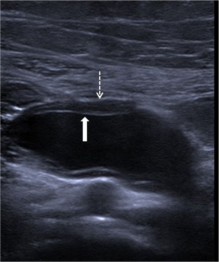
Karaosmanoglu et al. Insights into Imaging                (2020) 11:90                                                                                     Page 3 of 19

Table 1 Congenital and hereditary cystic diseases of the abdomen. Associated radiological findings. (Continued)
Organ                     Disease                                                   Typical imaging findings
Diaphragm                 Mesothelial cyst                                          - Homogeneous bilobulated cystic lesion located between posterolateral
*[29]                                                                                 aspect of the right liver lobe and the adjacent diaphragm
Prostate                  Prostatic utricle cyst                                    - Midline cyst communicating with the prostatic urethra and not extending
 *[30, 31]                                                                            above the base of the prostate
                          Mullerian duct cyst                                       - Teardrop-shaped midline cyst extending above the superior margin of the
                                                                                      prostate and not communicating with the prostatic urethra
Urachus                   Urachal cyst                                              - Homogeneous midline cyst along the trajectory of the urachus (between
*[32]                                                                                 the bladder dome and umbilicus)
                                                                                    - Inhomogeneous cyst content, cyst wall thickening, and inflammatory
                                                                                      stranding adjacent to the cyst indicate infection
Zinner’s syndrome         Seminal vesicle cysts                                     - Ipsilateral renal agenesis, seminal vesicle cysts, and ejaculatory duct obstruction
 *[33]                                                                              - In case of hemorrhage or infection the cyst content may appear as bright on
                                                                                      T1W MR images
*: References , (a) CT: Computed tomography,   (b)
                                                     T1W: T1-weighted,   (c)
                                                                               MR: Magnetic resonance, (d) US: Ultrasonography

Liver and biliary tract                                                                     Considering the LI-RADS v2018 (Liver Imaging Report-
Polycystic liver disease                                                                    ing and Data System Version 2018), non-complicated
Polycystic liver disease (PLD) is a part of the spectrum of                                 cysts are categorized as LR-1 lesions, which also include
fibropolycystic liver disease. It has an autosomal dominant                                 typical hemangiomas, vascular anomaly, confluent fibro-
inheritance pattern and may also be related to polycystic                                   sis, hepatic fat deposition or sparing, and focal scar [38].
kidney disease (PKD). This association with PKD is not                                      Hepatocyte-specific contrast agents are useful for dem-
rare and may be seen in around 50% of the patients [34].                                    onstrating the absence of any communication between
It is a rare disease, with an estimated incidence of < 0.01%,                               the biliary system and the cysts [2]. Although it is rare,
with a slight female preponderance [34, 35]. Genetic                                        the possibility of concomitant cholangiocarcinoma
mechanisms are the most important underlying cause                                          should be considered in patients with PLD and abnormal
which gives rise to the separation of ductal structures                                     liver function tests [39] (Fig. 4).
from the biliary tree, ultimately resulting in cyst formation
[36]. These disconnected bile ducts typically remain clinic-                                Caroli disease
ally silent until cysts begin to form in adulthood [36].                                    Caroli disease (CD) manifests with saccular, non-
These cysts are not distinct from simple hepatic cysts                                      obstructive, multisegmental dilation of the large intrahe-
from a histopathologic standpoint. Their walls are lined by                                 patic bile ducts [4]. The disease is mostly inherited in an
cuboidal biliary epithelium and contain serous fluid in                                     autosomal recessive fashion. In the so-called pure form
their cavities [1]. The cysts tend to emerge after puberty
and they generally remain asymptomatic. Cyst rupture,
hemorrhage, or infection may be counted among the po-
tential complications of these cysts. Malignant degener-
ation and liver failure are rare. Liver transplantation is
reserved for symptomatic relief [37].
   On cross-sectional imaging, the cysts are typically lo-
cated within the peripheral parenchyma (Fig. 1). They
highly vary in size, ranging from a few millimeters to 80
mm [1]. The peribiliary cysts, in the periportal distribu-
tion, may also be seen and they are typically small (< 10
mm) (Fig. 2). The cysts tend to increase both in size and
number with the advancing age. Infected or hemorrhagic
cysts may appear as hyperdense on CT and hyperintense
on T1-weighted (T1W) MR images [2] (Fig. 3). Fluid
level, cyst wall thickening, calcification, or endocavitary
air bubbles may be seen in infected cysts [3]. Non-                                           Fig. 1 A 60-year-old female patient with known long-standing PLD.
                                                                                              Axial plane T2W fat-saturated MR image shows multiple
complicated cysts have well-defined margins and the
                                                                                              parenchymal cysts in the liver (arrows)
cyst walls are smooth without any mural nodularity.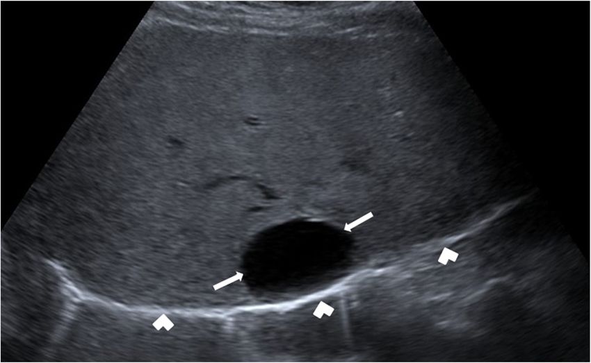
Karaosmanoglu et al. Insights into Imaging      (2020) 11:90                                                                      Page 4 of 19

 Fig. 2 A 55-year-old female patient with known ADPKD and PLD.
 Axial plane T2W fat-saturated MR image shows multiple bilateral         Fig. 4 A 60-year-old male patient with known PLD presenting with
 renal cysts with hemorrhagic cysts (arrows) and peribiliary cysts       recent onset jaundice. Axial plane T2W MR image demonstrates
 (arrowheads) in the liver                                               innumerable cysts scattered throughout the liver parenchyma. Also,
                                                                         note is made of dilation in the intrahepatic bile ducts (arrowheads).
                                                                         This dilation abruptly ends at the confluence of the right and left
of CD, there is no associating background parenchymal                    hepatic ducts (arrow). The patient underwent endoscopic retrograde
                                                                         cholangiopancreatography and endoscopic brush biopsy confirmed
liver abnormality. In the coexistence of CD and congeni-
                                                                         Klatskin tumor.
tal hepatic fibrosis, the disease process is called as the
“Caroli syndrome”, which is the more commonly en-
countered disease form. CD is also included in the                      the classic finding. Endoluminal stones or sludge may
Todani classification and classified as type V abnormality              be observed in these enlarged bile ducts. The detec-
[4]. The disease mostly manifests around 30 years of age;               tion of “central dot sign” is highly suggestive for CD
however, patients with Caroli syndrome may present                      (Fig. 5). This imaging finding refers to portal radicle
earlier.                                                                within these dilated bile ducts and they tend to show
   On imaging, cystic appearing enlarged intrahepatic bile              strong enhancement after contrast injection [4]. Mag-
ducts (in a saccular fashion) up to 5 cm in diameter is                 netic resonance cholangiopancreatography (MRCP) is
                                                                        helpful not only for detecting the presence of endo-
                                                                        luminal abnormalities but also for demonstrating the
                                                                        extent of the disease (Fig. 6). The extrahepatic bile
                                                                        ducts are typically not affected and disease may be

 Fig. 3 A 44-year-old female patient with known PLD presenting
 with right upper quadrant pain. On the US, a large hyperechoic
 mass was found in the right hepatic lobe (not shown). Axial plane
 precontrast T1W MR image shows subcentimeter cysts (arrowheads)
 and a cystic lesion with hyperintense content (arrows). There was no
 discernible enhancement on postcontrast series (not shown).
 Imaging findings were found to be consistent with the hemorrhagic
 cyst. Follow-up imaging studies demonstrated the decrease in the        Fig. 5 A 6-year-old male patient with known Caroli syndrome. Axial
 size of the lesion                                                      plane arterial phase CT image shows central dot sign (arrow)
Karaosmanoglu et al. Insights into Imaging (2020) 11:90 Page 5 of 19
Fig 6 A 30-year-old male patient with known Caroli syndrome presenting with jaundice. a Coronal plane MRCP MIP (maximum intensity
projection) image shows multifocal segmental dilation of the intrahepatic bile ducts (arrows). Choledoc is normal (arrowhead). b Axial plane T2W
fat-saturated MR image shows increased T2 signal in the periportal area (arrows) and surface irregularity of the liver. These findings are consistent
with congenital hepatic fibrosis
bilobar or limited to one lobe, mostly the left lobe. Type I: This group has been subclassified into three
Surgical resection of the affected liver lobe or seg- subgroups with type IC being the most common. Type
ment may be curative in limited disease. Hepatocyte- IA refers to diffuse cystic dilation of the extrahepatic bile
specific contrast agents may be helpful for demon- duct whereas type IB is seen as focal cystic dilation in
strating the communication between the parenchymal the extrahepatic bile duct. Type IC is characterized by
cysts and the biliary system. diffuse fusiform dilation of the entire extrahepatic bile
duct (Fig. 7).
Type II: This type is the least common one with focal
Choledochal cysts diverticular outpouching of the common bile duct (Fig. 8).
Choledochal cysts are rare congenital malformations of
the extrahepatic and intrahepatic biliary system. They
are more common in females and the incidence is higher
in Asian countries [5]. The most commonly accepted
classification is proposed by Todani et al. in 1977 [6].
Fig. 8 A 2-year-old female patient presenting with jaundice. On the
Fig. 7 A 48-year-old female patient presenting with jaundice. US, a saccular outpouching arising from the supraduodenal
Coronal plane MRCP MIP image shows fusiform dilation of the extrahepatic bile duct was found (not shown). Coronal plane MRCP
common bile duct (arrows). Also, note is made of mild dilation in MIP image shows a diverticulum (asterisk), arising from the
the intrahepatic bile ducts (arrowheads) supraduodenal extrahepatic bile duct (arrowhead)Karaosmanoglu et al. Insights into Imaging (2020) 11:90 Page 6 of 19
Fig 9 An 11-year-old female patient with newly diagnosed Hodgkin lymphoma underwent an initial US exam. a US image reveals
choledochocele (long arrows) that involves the intramural segment of the distal common bile duct (arrowheads). b Coronal plane MRCP MIP
image shows choledochocele (asterisk), common bile duct (arrowhead), and duodenum (arrow)
Type III: This group is also known as choledochoceles the disease. The risk of malignancy development is not
and refers to intramural dilation of the most distal por- rare (around 10–15% of the cases) with the extrahepatic
tion of the common bile duct (Fig. 9). biliary system and gallbladder being the most common
Type IV: This group has been subclassified into two [40, 41].
subgroups. Type IVA is characterized by combined
saccular-shaped dilations in the intrahepatic and extra- Biliary hamartomas
hepatic bile ducts. Type IVB refers to saccular dilations Biliary hamartomas, also known as the “von Meyenburg
restricted to extrahepatic bile ducts (Fig. 10). complex (VMC)” was first described in 1918 and is char-
Type V: This group is also known as Caroli disease. acterized by the presence of multiple bile duct hamarto-
Choledochal cysts typically manifest before the age of mas [7]. Histopathologically, they are composed of
10 years. The diagnosis is typically made after the clinical abnormally dilated intrahepatic bile ducts embedded in a
emergence of complications. Among these potential fibrous stroma. The prevalence of this abnormality is be-
complications, cholangitis and pancreatitis are the most tween 0.6% and 2.8% on autopsy studies [8]. This abnor-
common. Malignancy may also be seen in the course of mality is asymptomatic in the majority of the patients
and is incidentally detected [7]. The detection of biliary
hamartomas may cause diagnostic confusion in patients
having a history of cancer.
These lesions appear as focal hypodense lesions on CT
sometimes with irregular borders (Fig. 11). They typic-
ally do not enhance after contrast injection. T2-weighted
(T2W) MR images are very helpful for diagnosis as bil-
iary hamartomas are typically homogeneously hyperin-
tense (Fig. 11) whereas they are seen as hypointense on
T1W images with no apparent contrast enhancement on
dynamic T1W 3D gradient echo sequences. The cysts
are generally subcentimeter in diameter and
innumerable spread throughout the liver parenchyma
[7].
Ciliated hepatic foregut cyst
Ciliated hepatic foregut cysts (CHFC) are generally
asymptomatic and they are most commonly diagnosed
Fig. 10 A 23-year-old pregnant patient presenting with jaundice. incidentally on imaging or during surgery [42]. It has
Coronal plane MRCP MIP image shows fusiform dilation of the entire been proposed that CHFCs originate from intrahepati-
extrahepatic bile duct (long arrows) with extensive dilation of the cally entrapped detached hepatic diverticulum or abnor-
intrahepatic bile ducts in the left lobe (short arrows). Intrahepatic
mal tracheobronchiolar bud that may have migrated
bile ducts in the right lobe are normal (arrowheads)
caudally at the early stages of the embryonicKaraosmanoglu et al. Insights into Imaging (2020) 11:90 Page 7 of 19
Fig. 11 A 48-year-old female patient with known breast cancer underwent abdominal CT scanning for distant metastasis evaluation. a Axial plane
post-contrast CT image shows multiple hypodense lesions (arrows) in both liver lobes. b Axial plane fat-saturated T2W MR image shows multiple
cystic lesions (arrows) consistent with biliary hamartomas
development of the foregut [43]. It is a very rare clinical typical hyperintensity on T2W MR images. Enhance-
finding and histopathologically, they are composed of ment is typically not detected after contrast injection.
ciliated, pseudostratified columnar epithelium, a layer of Characteristic location is an important diagnostic clue
subepithelial connective tissue, a smooth muscle layer, for diagnosis as signal characteristics are not different
and outer capsule [44]. Management strategy is contro- from usual benign hepatic cysts.
versial but a more aggressive approach such as surgical
resection has been recommended as the malignant
transformation has been reported in few cases [9]. They Kidney
are more common in men and the medial segment of Autosomal dominant polycystic kidney disease
the left hepatic lobe (segments 4A and 4B) is the most Autosomal dominant polycystic kidney disease (ADPKD)
common location. is a relatively common disease and occurs in approxi-
On US and cross-sectional imaging, they typically ap- mately 1/1000 individuals. This hereditary condition is
pear as unilocular cystic lesion located in the subcapsu- largely inherited in autosomal dominant fashion. Most
lar area along the anterior surface of the liver (Fig. 12). patients become hemodialysis dependent around the 5th
The mean size is 3 cm, with a range of 1–12 cm [9]. to 7th decade of life [10].
They are seen as hypodense lesions on CT studies with a The cysts appear as typically hypoechoic on the US
and hypodense on non-contrast CT images (Fig. 13).
MRI findings are typical on advanced-stage disease with
a bright T2 signal. Despite the fact that most of the cysts
have typical imaging findings, some cysts may demon-
strate unusual signal characteristics due to proteinaceous
content. MRI studies with subtraction images may be
helpful in the mural and cavitary evaluation of these
cysts [10].
In the early stages of the disease, the kidneys may ap-
pear as either normal kidneys to single or multiple cysts
in one or both kidneys (Fig. 14). The cysts typically in-
crease in size and number into adulthood and during
the final stages of the disease the entire renal paren-
chyma may be replaced with the cysts. Epidemiological
and molecular biological data demonstrate that patients
with ADPKD bear an increased risk for renal cell cancer
Fig. 12 A 40-year-old female patient with known breast cancer. (RCC). But preoperative image-based diagnosis is often
Axial plane post-contrast CT image shows a unilocular cystic lesion challenging because of the distortion of the renal paren-
located in the subcapsular area of the segment 4 (arrows). The chyma [10]. (Fig. 15).
location was found to be typical for CHFC. The cyst was stable on 2-
year follow-up study (not shown)Karaosmanoglu et al. Insights into Imaging (2020) 11:90 Page 8 of 19
Fig. 13 A 63-year-old male patient with known ADPKD. a Axial plane post-contrast CT image shows enlarged kidneys with innumerable cysts
(arrows). b More cranial CT image demonstrates multiple hepatic (asterisks) and splenic cysts (arrowheads)
Autosomal recessive polycystic kidney disease may be seen in a certain subset of patients [13] (Fig. 16).
Autosomal recessive polycystic kidney disease (ARPKD) With the advancement of the age, the cysts typically en-
is much less common than ADPKD and occurs in ap- large and may replace the renal parenchyma. On US stud-
proximately one in 20.000 individuals [11]. The diagno- ies, findings suggestive of hepatic abnormalities should
sis is generally made in utero due to oligohydramnios also be sought after for early diagnosis and intervention.
from decreased fetal urine production. Severe renal dys- CT and MRI are rarely needed for diagnosis and IV con-
function immediately after birth is typical. Abdominal trast is not typically used due to limited renal reserve.
distension due to enlarged kidneys, bilateral flank Non-complicated cysts appear as hypodense on CT and
“masses”, and/or abdominal distension are common clin- T2 hyperintense on MR examinations (Fig. 17).
ical findings. Pulmonary hypoplasia and pneumothorax
are also common. Congenital hepatic fibrosis, Caroli dis-
ease, and bile duct ectasia may also be detected as asso- Multicystic dysplastic kidney disease
ciating abnormalities [12]. Multicystic dysplastic kidney (MCDK) is a congenital
The US is the most commonly used modality for diag- non-heritable cystic disease of the kidneys in the
nosis. Enlarged kidneys, with thickened parenchyma, is pediatric patient group. Renal cysts are formed in utero
the typical finding. Renal medulla appears as hyperechoic and may be observed on antenatal US examinations.
due to the presence of several ectatic renal tubules, also The disease is typically unilateral and the affected kidney
called as microcysts [11]. Larger cysts (larger than 1 cm) typically does not function [45].
Fig. 15 A 56-year-old female patient with known ADPKD. Axial
Fig. 14 A 34-year-old female patient with known hypertension and plane postcontrast CT image shows enlarged kidneys with
family history of ADPKD. Axial plane post-contrast CT image shows innumerable cysts and a solid mass in the right kidney (arrow).
bilateral renal cysts with different sizes (arrows). Normal renal Histopathological examination after surgery revealed papillary type
parenchyma can be seen. Genetic analysis confirmed the ADPKD renal cell carcinomaKaraosmanoglu et al. Insights into Imaging (2020) 11:90 Page 9 of 19
Fig. 16 A 3-year-old male patient known ARPKD. a Sagittal view US image shows an enlarged kidney with cortical and medullary
hyperechogenicity due to small cysts. There are also bigger cysts in the kidney (arrows). b Axial plane US image of the liver shows contour
irregularities and parenchymal heterogeneity (arrows) consistent with congenital hepatic fibrosis
The US is typically the imaging modality of choice in size, in medullary and corticomedullary locations may be
this patient group. The detection of renal cysts in a dis- detected [15, 16] (Fig. 20). Early in life, it may be pos-
organized pattern is the typical imaging finding and sible not to detect any cysts, the kidneys may appear
renal parenchyma is characteristically completely re- hyperechoic with the loss of corticomedullary differenti-
placed by these cysts [14] (Fig. 18). MRI demonstrates ation [47]. The cystic changes in the renal parenchyma
similar findings with the US exam (Fig. 19). are generally progressive and advance with age.
Nephronophthisis and medullary cystic kidney disease Von Hippel–Lindau disease
Medullary cystic kidney disease and nephronophthisis, Von Hippel–Lindau (VHL) syndrome is a phakomatosis
which are a common cause of end-stage renal disease inherited in an autosomal dominant fashion. It is a rare
during the first 3 decades of life, are inherited diseases disease which affects 17/36.000–53.000 individuals [48].
with similar renal morphology and histopathologic fea- This syndrome predisposes the affected individual to dif-
tures. The inheritance pattern is variable; nephro- ferent cancers.
nophthisis is autosomal recessive in inheritance and Renal manifestations are common and renal cysts may
medullary cystic kidney disease is autosomal dominant be detected in 59–63% of patients and bilateral involve-
[15]. Several other syndromes may be associated with ment is extremely common (around 75% of the cases)
nephronophthisis [46]. [17]. RCCs are also common and are seen in 24–45% of
On US studies, the kidneys, contrary to ARPKD, ap- the affected individuals [49]. Periodic screening of the
pear small due to parenchymal fibrosis. Cysts, of varying kidneys is mandatory as untreated RCCs carry a poor
Fig. 18 Newborn with prenatally detected left kidney cysts
underwent an US study on the first day of life. Sagittal view US
image shows multiple cysts in the left renal fossa with no
Fig. 17 A 5-year-old male patient with known ARPKD. Coronal plane discernible normal renal parenchyma. The right kidney was normal
T2W MR image shows enlarged kidneys with diffusely increased T2 (not shown). Imaging findings were found to be consistent
signal. Also note subcentimeter liver cysts (arrowheads) with MCDKKaraosmanoglu et al. Insights into Imaging (2020) 11:90 Page 10 of 19
Fig. 21 A 43-year-old male patient with known VHL underwent a
follow-up CT exam. Axial plane post-contrast CT image shows
several renal cysts (asterisks) in both kidneys. Also note is made of a
new focus of clear cell RCC (surgically confirmed) in the right kidney
(arrow). A pancreatic cyst in the uncinate process was also
detected (arrowheads)
Fig. 19 A 12-day-old newborn with prenatally diagnosed right kidney at the same time. Tumors may arise from precur-
kidney cysts underwent an abdominal MRI examination. Coronal sor cystic lesions or completely de novo [49]. Thus, con-
plane T2W MR image shows multiple cysts completely replacing the
tinuous imaging screening is fundamental for early
right kidney parenchyma (arrows). There was no discernible renal
parenchyma aside from the cysts. The left kidney was normal diagnosis and treatment to prevent metastatic disease.
Cysts may enlarge or regress with time leaving parenchy-
mal scars, and no relationship has been observed with
prognosis with a tendency to metastasize [18]. The his- the cyst size and number and the malignant potential
topathologic features of the renal cysts vary, ranging [17].
from simple and hyperplastic cysts to cystic clear cell The US may be helpful to differentiate the cysts from
carcinomas and to solid tumors [50]. All mentioned be- the solid masses but CT and MRI are more commonly
nign and malignant lesions may occur in the same used for both diagnosis and follow-up (Fig. 21). Mural
nodules within the cyst walls are suggestive for cancer
and they can be observed with relative ease on both CT
and MRI. Pure solid lesions may also be assessed with
these two modalities and renal vein involvement can also
be detected. CT and MRI are more sensitive for detect-
ing small lesions (2 cm) [49]. MRI might be preferable
for follow-up purposes, especially in young patients, due
to lack of radiation with this modality. Subtraction im-
ages may be helpful for differentiating proteinaceous
cysts from solid masses. Due to the progressive course
of VHL syndrome, partial nephrectomy and percutan-
eous ablative measures are common approaches for the
treatment of RCCs in these patients.
Tuberous sclerosis complex
Tuberous sclerosis complex (TSC) disease affects around
1/5.000–10.000 individuals [51]. TSC is typically inher-
Fig. 20 A 13-year-old female patient with known histopathologically
proven nephronophthisis now presenting with flank pain. The ited in an autosomal dominant fashion but the expres-
patient is on chronic hemodialysis. Coronal plane T2W MR image sion is variable. Around 66% of the cases are secondary
shows multiple cysts in both kidneys located at the corticomedullary to sporadic mutation. TSC is a multisystemic disease
junction. There was no discernible healthy renal parenchyma in the and may manifest with numerous mesodermal and ecto-
kidneys. Also note that both kidneys (arrows) are small in their
dermal abnormalities. Kidneys are also affected along
overall sizes in contrast to ARPKD
the course of the disease. Angiomyolipomas, withKaraosmanoglu et al. Insights into Imaging (2020) 11:90 Page 11 of 19 Fig. 22 A 9-year-old male patient with known TSC underwent follow-up US exam. US images show punctate hyperechoic foci consistent with subcentimeter angiomyolipomas (fine arrows). Also note is made of several parenchymal cysts (thick arrows) variable fat content, and renal cysts are the two most Pancreas common renal lesions in these patients [19]. Large Von Hippel-Lindau disease angiomyolipomas may spontaneously rupture in the As VHL is a multisystemic disease, the pancreas may course of the disease, renal cysts are almost always also be affected among the course of the disease. The asymptomatic [52]. Angiomyolipomas tend to be more pancreatic manifestations are either cysts, serous cysta- numerous and common compared to renal cysts [19]. denomas, or neuroendocrine tumors (NETs). Combined On imaging, renal cysts in TSC appear like simple lesion pattern, the presentation of solid and cystic mani- renal cysts (Fig. 22). Differential diagnosis from ADPKD festations, may be observed in 11.5% of patients with may be difficult as fewer cysts are typically seen at the VHL disease and in 7.6% of the cases pancreas may be early stage of this disease. The very common association the only affected organ [20]. of renal cysts with angiomyolipomas may serve as a reli- The pancreatic cysts follow the typical imaging charac- able imaging clue for correct diagnosis. The cysts tend teristics of cysts elsewhere in the body (Fig. 23). Pancre- not to be large in size and the average diameter of these atic NETs are mostly solid but cystic tumors were also cysts was reported to be around 20 mm which may be reported [21] (Fig. 24). These tumors, be it cystic or another helpful clue considering the large sizes of the solid, generally tend to have early arterial phase-contrast cysts in patients with ADPKD [19]. enhancement for detection and correct characterization. Fig. 23 Two different patients with VHL. a Coronal plane post-contrast CT image of a 47-year-old male patient shows innumerable pancreatic cysts (arrowheads). Also note is made of large-sized solid RCC (arrows) in the left kidney and bilateral renal cysts (asterisks). b Axial plane T2W MR image of a 40-year-old male patient demonstrates multiple cysts of the pancreas, almost completely replacing pancreatic parenchyma (arrows)
Karaosmanoglu et al. Insights into Imaging       (2020) 11:90                                                                    Page 12 of 19

 Fig. 24 A 21-year-old female patient with recently diagnosed (with
 genetic testing after a recent diagnosis of her elder sister) VHL        Fig. 26 A 13-year-old male patient with known CF and pancreatic
 underwent a baseline CT exam. Axial plane post-contrast pancreatic       insufficiency. Axial plane T2W MR image reveals innumerable cysts
 phase CT image shows a predominantly cystic lesion in the uncinate       of subcentimeter size scattered throughout the pancreas, almost
 process (arrows) with enhancing mildly thickened internal septation      completely replacing the parenchyma (arrows)
 (arrowhead). The lesion was found to be highly suspicious of a cystic
 NET. Surgical removal and histopathological examination confirmed
 the presence of cystic NET                                              characteristics of spontaneous NETs. Rarely, the tumors
                                                                         can be cystic [22] (Fig. 25).

Multiple endocrine neoplasia type I                                      Cystic fibrosis
MEN 1 is an inherited endocrine tumor syndrome in                        Cystic fibrosis (CF) is a common hereditary systemic dis-
autosomal dominant fashion. The pituitary gland, islet                   ease. Exocrine glands are commonly affected and pan-
cells of the pancreas, and parathyroid glands are the                    creatic insufficiency is a common manifestation of the
most common tumor sites. Imaging plays an important                      disease.
role in the diagnosis and management of the disease                         Four different imaging patterns were described in pa-
[53].                                                                    tients with CF: (1) partial fatty replacement of the pan-
  Most pancreatic NETs in MEN 1 are functional with                      creas, (2) complete fatty replacement of the pancreas, (3)
the gastrinoma being the most common [54]. The pan-                      atrophy of the pancreas, and (4) pancreatic cystosis.
creatic NETs seen in MEN 1 follow the typical imaging                    Among these four different patterns, pancreatic cystosis
                                                                         is the least common [55]. In pancreatic cystosis, the
                                                                         organ parenchyma is filled with macrocysts and the de-
                                                                         velopment of these cysts has been linked to bicarbonate
                                                                         transport [23].
                                                                            On the US, the cysts appear as hypoechoic structures
                                                                         with no associating mural wall thickening and nodular-
                                                                         ity. The size of the cysts is variable ranging from 0.5 cm
                                                                         to 1.2 cm in diameter. Vascular displacement due to the
                                                                         mass effect of these cysts are may be seen [56]. MRI is
                                                                         also a very helpful modality to detect the cysts and their
                                                                         anatomic relationship, due to its high soft-tissue reso-
                                                                         lution (Fig. 26). Malignant transformation of these cysts
                                                                         has not reported before [57]. Polycystic kidney disease
                                                                         and VHL may be considered in differential diagnosis but
                                                                         detection of normal kidneys and patient history are gen-
                                                                         erally diagnostic without any significant confusion.
 Fig. 25 A 31-year-old male patient with known MEN-1 syndrome
 underwent CT scanning to detect manifestations of the syndrome.         Gastrointestinal tract duplication cysts
 Axial plane post-contrast pancreatic phase CT image shows a             Gastrointestinal (GI) tract duplication cysts are rare con-
 predominantly cystic lesion in the pancreatic body (arrows). Focal      genital malformations which are typically detected in
 contrast-enhancing mural wall thickening can also be seen
 (arrowhead). Histopathological findings revealed cystic NET
                                                                         young patients and adults [58]. They may occur any-
                                                                         where along the GI tract but distal ileum is the most
Karaosmanoglu et al. Insights into Imaging (2020) 11:90 Page 13 of 19
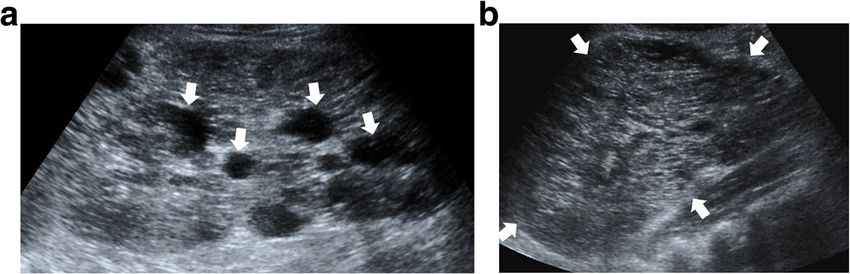
Fig. 27 An 8-year-old male patient presenting to the emergency
Fig. 28 A 7-year-old female patient presenting to ED with right
department (ED) with right lower quadrant pain. Axial view US
lower quadrant pain. Axial view US image demonstrates a purely
image shows a cystic mass with thick wall suggested for “double-
cystic mass (star) in the right lower quadrant. The wall of this lesion
wall” sign: The mucosa appears hyperechoic (arrow) whereas the
was in direct continuity (arrowhead) with the adjacent terminal
muscular layer hypoechoic (dashed-line arrow). Laparoscopic surgery
ileum segment (arrow)
and histopathologic examination confirmed the ileal duplication cyst
causing intestinal obstruction
commonly affected segment followed by the esophagus,
colon, jejunum, stomach, and the duodenum [59]. These
lesions may be contained within the wall of the affected
segments but may also be detected extrinsic to the bowel
segment and may appear as either spherical (80%) or
tubular cysts (20%) [60, 61]. This morphologic difference
may provide a clue regarding the possible communica-
tion between the enteric lumen and the duplication
cysts, as the spheric ones generally do not communicate
with the lumen, whereas, its tubular counterparts typic-
ally do.
Histopathologically, GI tract duplication cysts consist
of an epithelial lining containing the mucosa of the GI
tract and a surrounding smooth muscle. The cyst is also
closely attached to the enteric wall [24].
GI tract duplication cysts are most commonly diag-
nosed incidentally but complications may also occur, in-
cluding obstruction, volvulus, intussusception, bleeding,
Fig. 29 A 9-year-old female patient with palpable epigastric mass
perforation, and infection. Malignant transformation was found to have a large cystic mass in the epigastrium on US
from the mucosa is extremely rare and surgical resection examination (not shown). An abdominal CT study was planned for
is the preferred approach for treatment [59]. better assessment of the anatomic relationship of this lesion. Axial
The US is the most commonly used imaging modality. plane post-contrast CT image shows a huge cystic mass (arrows)
compressing and displacing the stomach (star). Surgery confirmed
The diagnosis is generally straightforward when the cyst
gastric duplication cyst
within the close proximity of the bowel segment. TheKaraosmanoglu et al. Insights into Imaging (2020) 11:90 Page 14 of 19
double-wall or muscular rim sign is the typical imaging
finding (Fig. 27). This imaging appearance is due to the
inner hyperechoic mucosa and the surrounding hypoe-
choic smooth muscle layer (muscularis propria) [24].
The GI tract duplication cysts share a common wall with
the adjacent gut segment. The so-called “Y configur-
ation” is a helpful diagnostic sign that is indicative of a
shared wall with the cyst and the neighboring bowel wall
(Fig. 28). This sign is caused by the splitting of the
shared muscularis propria with the cyst and bowel wall
[25, 26]. Due to the presence of smooth muscle content,
these cysts may change shape due to muscular contrac-
tions of the cyst wall, which is another useful finding for
diagnosis on real-time sonographic examination [58].
The cysts are generally homogenous on the sonographic
exam; however, internal septations or luminal debris
may also be observed in certain patients. Color Doppler
Fig. 31 A 39-year-old female patient with progressive abdominal
US exam may be helpful to detect wall inflammation in distension underwent an US exam which showed large volume
complicated duplication cysts [24]. ascites in the abdomen. The clinical and imaging findings were
CT and MRI are not typically used for diagnosis due concerning for peritoneal carcinomatosis. Axial plane CT image after
to ionizing radiation and increased patient cooperation, IV and oral contrast use reveals a large cystic lesion insinuating
between the mesenteric leaves. This cystic mass was separating
consecutively. However, these modalities may be used
apart the adjacent bowel loops (stars). Also noted were internal
for better assessing the anatomical relations of duplica- septations (arrows) within this large cystic mass. Surgical and
tion cysts (Fig. 29). They can also be used in patients subsequent histopathological examinations confirmed large
with questionable malignant degeneration within these mesenteric lymphangioma.
cysts.
Abdominal lymphatic malformations
Abdominal lymphatic malformations (LMs) are rare mesentery, followed by omentum, mesocolon, and retro-
congenital malformations of the lymphatic system. They peritoneum [62]. Abdominal LMs are rare as LMs are
are most commonly located in the small bowel most commonly located in the head-neck region [63].
LMs are generally small in size and most commonly di-
agnosed incidentally on imaging but symptoms may
occur, including abdominal pain or distension, particu-
larly in large size LMs. Asymptomatic LMs do not re-
quire treatment and can be followed with serial imaging.
The common approach for the treatment of symptom-
atic LMs is surgical resection [27, 64, 65].
US and CT are the two most commonly used mo-
dalities for diagnosis. The anatomic relations of these
cystic masses and their internal contents may be
assessed with high precision with both techniques
(Fig. 30). Ascites may be confused with these LMs
but well-circumscribed morphology, as opposed to
free-floating ascites, favors LMs over ascites. However,
LMs may also conform to omental anatomy and dif-
ferential diagnosis from loculated ascites may be diffi-
cult in certain cases (Fig. 31). The fluid in the LMs
Fig. 30 A 1-year-old male patient presented with progressively may contain fat which may be better appreciated on
enlarging abdominal girth. Axial plane CT image after IV and oral CT or MRI studies [28] (Fig. 32). Small LMs are fre-
contrast use shows a large purely cystic mass (star), displacing the quently mobile and, therefore, they can be observed
bowel loops to the left side of the abdomen. Histopathological in different locations on separate follow-up imaging
examination after surgical extirpation confirmed
studies [27].
omental lymphangiomaKaraosmanoglu et al. Insights into Imaging (2020) 11:90 Page 15 of 19 Fig. 32 A 41-year-old female patient with newly diagnosed breast cancer underwent a staging body CT examination. a Axial plane post-contrast CT image shows a well-circumscribed retroperitoneal mass (arrows) with a fat-fluid level (arrowhead). b, c Abdominal MRI exam of the same patient showed signal loss within the cyst content on out-of-phase image (c) compared to in-phase image (b). Imaging findings were found to be compatible with chylous fluid containing lymphangioma. Follow-up imaging studies confirmed the stability of this lesion Diaphragm the diaphragmatic origin of the cyst [67]. These cysts may Diaphragmatic mesothelial cyst be effectively treated with a percutaneous approach [29]. Diaphragmatic mesothelial cysts (DMC) are derived from coelomic remnants and are lined with mesothelial Prostate cells [66]. Mesothelial cysts may be found in several Prostatic utricle cyst—Mullerian duct cyst places including the diaphragm. Due to its close proxim- Unlike paramedian ejaculatory ductus cysts, prostatic ut- ity of DMCs to liver, lung, and pleura, it may be difficult ricle and Mullerian duct cysts are two different cystic to determine the diaphragm as the source organ. entities which are both located midline [68]. Prostatic On imaging, they have characteristic findings of an or- utricle cysts may be associated with several genitourinary dinary cyst located elsewhere in the body. The walls of abnormalities [30]. On the contrary, Mullerian duct cysts these cysts are thin with no associating solid component. are not expected to associate with any congenital genito- They may also appear as bilobulated on sonography [29]. urinary malformations. Both cysts may manifest with On the US, they appear as homogenously hypoechoic le- various symptoms, including difficulty urinating, dysuria, sions located between the posterolateral aspect of the right ejaculatory impairment, and hematospermia [31, 69]. In liver lobe and the adjacent diaphragm (Fig. 33). CT and these cystic lesions, the mechanism of hematospermia is MRI may also be used as confirmatory studies. In these considered to be due to ejaculatory duct obstruction studies, internal content of the lesion may be better appre- [68]. ciated. Bronchogenic cysts, hydatid cysts, or an ordinary Differentiation of these entities by imaging alone may liver cyst may be considered in differential diagnosis. Bilo- be difficult. Typically, prostatic utricle cysts do not bulated morphology of the cyst is an important clue for Fig. 33 A 23-year-old female patient with recently diagnosed hepatitis B infection underwent an initial liver US exam. Axial view US image shows a oval-shaped purely cystic lesion (arrows) in the posterolateral aspect of the right liver lobe in close proximity to the right hemidiaphragm (arrowheads). The lesion was found to be Fig. 34 A 17-year-old male patient with urinary incontinence. representing a diaphragmatic mesothelial cyst. Follow-up studies Coronal plane T2W MR image shows a midline prostatic cyst confirmed the stability of this lesion (asterisk) that does not extend above the prostate gland
Karaosmanoglu et al. Insights into Imaging       (2020) 11:90                                                                       Page 16 of 19

                                                                         Fig. 37 A 6-year-old female patient presenting to ED with
                                                                         suprapubic pain. Sagittal view US image shows a round-shaped
                                                                         cystic lesion (arrows) above the superior surface of the bladder (star).
                                                                         Due to the mixed echogenicity of the lesion, it was considered an
                                                                         infected urachal cyst. Surgical findings confirmed the diagnosis

 Fig. 35 A 2-year-old male patient with recurrent urinary tract         extend above the base of the prostate (Fig. 34), whereas
 infection underwent a US exam and a midline cystic lesion was seen     Mullerian duct cysts are characteristically observed as
 posterior of the bladder (not shown). Confirmatory MRI exam shows      teardrop-shaped midline cysts extending above the su-
 a tear-drop shaped midline cystic lesion (arrows) on the sagittal
 plane T2W image. The cystic lesion was extending above the
                                                                        perior margin of the prostate (Fig. 35). In terms of size,
 posterior superior margin of the prostate, highly suggestive for the   prostatic utricle cysts are typically smaller than Muller-
 Mullerian duct cyst                                                    ian duct cysts. From an anatomical standpoint, prostatic
                                                                        utricle cysts communicate with the prostatic urethra un-
                                                                        like Mullerian duct cyst [31, 68, 69]. Voiding cystoure-
                                                                        thrography may help differential diagnosis by
                                                                        demonstrating the connection between the prostatic ut-
                                                                        ricle cyst and the urethra [70] (Fig. 36).

                                                                        Urachal cyst
                                                                        The urachus is a ductal remnant that originates from
                                                                        the involution of the allantois and cloaca. It extends
                                                                        from the bladder dome to the umbilicus. In the late
                                                                        stages of gestation, it involutes and obliterates and ob-
                                                                        served as a median umbilical ligament in the postgesta-
                                                                        tional period. The failure of this mentioned involution
                                                                        may result in persistent of this canal after birth. The
                                                                        most common types of this failed involution are patent
                                                                        urachus and urachal cyst [71].
                                                                          Urachal cysts form when both the umbilical and blad-
                                                                        der ends of the urachus are obliterated with nonobliter-
                                                                        ated segment in between. These urachal cysts are
                                                                        typically small and asymptomatic; however, infection of
                                                                        the cysts may cause symptoms [32, 71].
                                                                          On imaging, the diagnosis of an uncomplicated cyst
                                                                        may be easily made by detecting the cyst in the midline
 Fig. 36 A 5-year-old male patient with recurrent urinary tract         along the trajectory of the urachus. The detection of in-
 infection. Voiding cystourethrogram demonstrates a contrast filling    homogeneous cyst content and inflammatory stranding
 prostatic utricle cyst (arrows)
                                                                        adjacent to the cyst may indicate infection [32] (Fig. 37).
Karaosmanoglu et al. Insights into Imaging (2020) 11:90 Page 17 of 19
relatively straightforward and clinical management may
be implemented appropriately.
Abbreviations
ADPKD: Autosomal dominant polycystic kidney disease; ARPKD: Autosomal
recessive polycystic kidney disease; CD: Caroli disease; CF: Cystic fibrosis;
CHFC: Ciliated hepatic foregut cyst; CT: Computed tomography;
DMC: Diaphragmatic mesothelial cyst; ED: Emergency department;
GI: Gastrointestinal; LI-RADS v2018: Liver Imaging Reporting and Data System
Version 2018; LM: Lymphatic malformation; MCDK: Multicystic dysplastic
kidney; MEN1: Multiple endocrine neoplasia type I; MR: Magnetic resonance;
MRCP: Magnetic resonance cholangiopancreatography; NET: Neuroendocrine
tumor; PKD: Polycystic kidney disease; PLD: Polycystic liver disease;
RCC: Renal cell cancer; T1W: T1-weighted; T2W: T2-weighted; TSC: Tuberous
sclerosis complex; US: Ultrasonography; VHL: von Hippel–Lindau; VMC: Von
Meyenburg complex
Authors’ contributions
ADK wrote the manuscript. SA collected the data and contributed to the
writing of the text. DA, MO, MH, and BO contributed to the writing of the
text and edited the text. The authors read and approved the final
manuscript. MK edited the text and first proposed the idea of writing this
manuscript.
Funding
Not applicable.
Availability of data and materials
Fig. 38 A 31-year-old male patient presenting with left groin pain. Data sharing is not applicable to this article as no datasets were generated
Coronal plane postcontrast CT image demonstrates left renal or analyzed during the current study.
agenesis and left seminal vesicle cysts (arrows)
Ethics approval and consent to participate
Not applicable.
Zinner’s syndrome
Consent for publication
Zinner’s syndrome is a rare developmental anomaly. It Not applicable.
refers to the triad of ipsilateral ejaculatory duct obstruc-
tion, seminal vesicle cysts, and renal agenesis. The pa- Competing interests
The authors declare that they have no competing interests.
tients usually admit to the hospital with genitourinary
symptoms in the 2nd–3rd decades of their life [72]. Received: 10 May 2020 Accepted: 16 July 2020
On imaging, the obstructed ejaculatory ducts are seen
as tubular structures in the pelvis and the ipsilateral kid-
References
ney is typically agenetic (Fig. 38). The content of ejacula- 1. Borhani AA, Wiant A, Heller MT (2014) Cystic hepatic lesions: a review and
tory ducts is characteristically anechoic on the US and an algorithmic approach. AJR Am J Roentgenol 203(6):1192–1204
homogenously hyperintense on T2W MR images. In the 2. Mamone G, Carollo V, Cortis K, Aquilina S, Liotta R, Miraglia R (2019)
Magnetic resonance imaging of fibropolycystic liver disease: the spectrum
case of hemorrhage or infection, the duct content may of ductal plate malformations. Abdom Radiol (NY) 44(6):2156–2171
appear as bright on T1W MR images. The detection of 3. Arnold HL, Harrison SA (2005) New advances in evaluation and
the tail-like connection between the cystic tubules and management of patients with polycystic liver disease. Am J Gastroenterol
100(11):2569–2582
the seminal vesicle may indicate that the seminal vesicle 4. Cannella R, Giambelluca D, Diamarco M et al (2020) Congenital cystic
is the site of origin [33]. lesions of the bile ducts: imaging-based diagnosis. Curr Probl Diagn Radiol
49(4):285–293
5. Santiago IS, Loureiro R, Curvo-Semedo L et al (2012) Congenital cystic
lesions of the biliary tree. AJR Am J Roentgenol 198(4):825–835
Conclusion 6. Todani T, Watanabe Y, Narusue M, Tabuchi K, Okajima K (1977) Congenital
Congenital and hereditary cystic lesions of the abdomen bile duct cysts: classification, operative procedures, and review of thirty-
seven cases including cancer arising from choledochal cyst. Am J Surg
are relatively rare. They may be diagnosed incidentally 134(2):263–269
or may give rise to symptoms which prompt their diag- 7. Maher MM, Dervan P, Keogh B, Murray JG (1999) Bile duct hamartomas (von
nosis. Correct diagnosis is critical as they may simulate Meyenburg complexes): value of MR imaging in diagnosis. Abdom Imaging
24(2):171–173
several other benign and malignant acquired diseases of 8. Lev-Toaff AS, Bach AM, Wechsler RJ, Hilpert PL, Gatalica Z, Rubin R (1995)
the abdomen, all of which have very different treatment The radiologic and pathologic spectrum of biliary hamartomas. AJR Am J
approaches and prognostic implications. With the cor- Roentgenol 165(2):309–313
9. Ansari-Gilani K, Modaresi EJ (2017) Ciliated hepatic foregut cyst: report of
rect and appropriate use of imaging, with relevant clin- three cases and review of imaging features. Gastroenterol Rep (Oxf) 5(1):75–
ical information and patient history, diagnosis may be 78Karaosmanoglu et al. Insights into Imaging (2020) 11:90 Page 18 of 19
10. Jilg CA, Drendel V, Bacher J et al (2013) Autosomal dominant polycystic 37. Brancatelli G, Federle MP, Vilgrain V, Vullierme MP, Marin D, Lagalla R (2005)
kidney disease: prevalence of renal neoplasias in surgical kidney specimens. Fibropolycystic liver disease: CT and MR imaging findings. Radiographics
Nephron Clin Pract 123(1-2):13–21 25(3):659–670
11. Dillman JR, Trout AT, Smith EA, Towbin AJ (2017) Hereditary renal cystic 38. Abdel Razek AAK, El-Serougy LG, Saleh GA, Shabana W, Abd E-WR (2020)
disorders: imaging of the kidneys and beyond. Radiographics 37(3):924–946 Liver Imaging Reporting and Data System Version 2018: what radiologists
12. Capisonda R, Phan V, Traubuci J, Daneman A, Balfe JW, Guay-Woodford LM need to know. J Comput Assist Tomogr 44(2):168–177
(2003) Autosomal recessive polycystic kidney disease: outcomes from a 39. Landais P, Grünfeld J-P, Droz D et al (1984) Cholangiocellular carcinoma in
single-center experience. Pediatr Nephrol 18(2):119–126 polycystic kidney and liver disease. Arch Intern Med 144(11):2274–2276
13. Avni FE, Guissard G, Hall M, Janssen F, DeMaertelaer V, Rypens F (2002) 40. Singham J, Yoshida EM, Scudamore CH (2009) Choledochal cysts: part 2 of
Hereditary polycystic kidney diseases in children: changing sonographic 3: Diagnosis. Can J Surg 52(6):506
patterns through childhood. Pediatr Radiol 32(3):169–174 41. Singham J, Yoshida EM, Scudamore CH (2010) Choledochal cysts: part 3 of
14. Gimpel C, Avni EF, Breysem L et al (2019) Imaging of kidney cysts and cystic 3: management. Can J Surg 53(1):51
kidney diseases in children: An international working group consensus 42. Sharma S, Dean AG, Corn A et al (2008) Ciliated hepatic foregut cyst: an
statement. Radiology 290(3):769–782 increasingly diagnosed condition. Hepatobiliary Pancreat Dis Int 7(6):581–589
15. Hildebrandt F, Waldherr R, Kutt R, Brandis M (1992) The nephronophthisis 43. Khoddami M, Kazemi Aghdam M, Alvandimanesh A (2013) Ciliated hepatic
complex: clinical and genetic aspects. Clin Investig 70(9):802–808 foregut cyst: two case reports in children and review of the literature. Case
16. Garel LA, Habib R, Pariente D, Broyer M, Sauvegrain J (1984) Juvenile Rep Med 2013:372017
nephronophthisis: sonographic appearance in children with severe uremia. 44. Bogner B, Hegedus G (2002) Ciliated hepatic foregut cyst. Pathol Oncol Res
Radiology 151(1):93–95 8(4):278–279
17. Leung RS, Biswas SV, Duncan M, Rankin S (2008) Imaging features of von 45. Lalli AF (1967) Multicystic kidney disease. Radiology 89(5):857–860
Hippel-Lindau disease. Radiographics 28(1):65–79 46. Hildebrandt F, Benzing T, Katsanis N (2011) Ciliopathies. N Engl J Med
18. Levine E, Lee KR, Weigel JW, Farber B (1979) Computed tomography in the 364(16):1533–1543
diagnosis of renal carcinoma complicating Hippel-Lindau syndrome. 47. Blowey DL, Querfeld U, Geary D, Warady BA, Alon U (1996) Ultrasound
Radiology 130(3):703–706 findings in juvenile nephronophthisis. Pediatr Nephrol 10(1):22–24
19. Casper KA, Donnelly LF, Chen B, Bissler JJ (2002) Tuberous sclerosis 48. Maher ER, Neumann HP, Richard S (2011) von Hippel-Lindau disease: a
complex: renal imaging findings. Radiology 225(2):451–456 clinical and scientific review. Eur J Hum Genet 19(6):617–623
20. Ayloo S, Molinari M (2016) Pancreatic manifestations in von Hippel-Lindau 49. Choyke PL, Glenn GM, Walther MM, Patronas NJ, Linehan WM, Zbar B (1995)
disease: a case report. Int J Surg Case Rep 21:70–72 von Hippel-Lindau disease: genetic, clinical, and imaging features. Radiology
21. Hammel PR, Vilgrain V, Terris B et al (2000) Pancreatic involvement in von 194(3):629–642
Hippel–Lindau disease. Gastroenterology 119(4):1087–1095 50. Choyke PL, Glenn GM, Walther MM et al (1992) The natural history of renal
22. Sheth S, Hruban RK, Fishman EK (2002) Helical CT of islet cell tumors of the lesions in von Hippel-Lindau disease: a serial CT study in 28 patients. AJR
pancreas: typical and atypical manifestations. AJR Am J Roentgenol 179(3):725– Am J Roentgenol 159(6):1229–1234
730 51. von Ranke FM, Zanetti G, e Silva JL et al (2015) Tuberous sclerosis complex:
23. Taylor CJ, Aswani N (2002) The pancreas in cystic fibrosis. Paediatr Respir state-of-the-art review with a focus on pulmonary involvement. Lung
Rev 3(1):77–81 193(5):619–627
24. Sanguesa Nebot C, Llorens Salvador R, Carazo Palacios E, Pico Aliaga S, 52. Ewalt DH, Sheffield E, Sparagana SP, Delgado MR, Roach ES (1998) Renal
Ibanez PV (2018) Enteric duplication cysts in children: varied presentations, lesion growth in children with tuberous sclerosis complex. J Urol 160(1):
varied imaging findings. Insights Imaging 9(6):1097–1106 141–145
25. Kumar D, Ramanathan S, Haider E, Khanna M, Otero C (2015) Education and 53. Scarsbrook AF, Thakker RV, Wass JA, Gleeson FV, Phillips RR (2006) Multiple
imaging. gastroenterology: revisiting the forgotten sign: five layered gut endocrine neoplasia: spectrum of radiologic appearances and discussion of
signature and Y configuration in enteric duplication cysts on high a multitechnique imaging approach. Radiographics 26(2):433–451
resolution ultrasound. J Gastroenterol Hepatol 30(7):1111 54. Pannett A, Thakker R (1999) Multiple endocrine neoplasia type 1. Endocr
26. Tritou I, Sfakianaki E, Prassopoulos P (2015) The sonographic multilaminar Relat Cancer 6(4):449–473
appearance is not enough for the diagnosis of enteric duplication cyst in 55. Feigelson J, Pecau Y, Poquet M et al (2000) Imaging changes in the
children. AJR Am J Roentgenol 204(2):W222–W223 pancreas in cystic fibrosis: a retrospective evaluation of 55 cases seen over a
27. McEwing R, Hayward C, Furness M (2003) Foetal cystic abdominal masses. period of 9 years. J Pediatr Gastroenterol Nutr 30(2):145–151
Australas Radiol 47(2):101–110 56. Monti L, Salerno T, Lucidi V et al (2001) Pancreatic cystosis in cystic fibrosis:
28. Yoo E, Kim MJ, Kim KW, Chung JJ, Kim SH, Choi JY (2006) A case of case report. Abdom Imaging 26(6):648–650
mesenteric cystic lymphangioma: fat saturation and chemical shift MR 57. la Denise JP a, Hubert D, Gaudric M, Scatton O, Soubrane O (2011)
imaging. J Magn Reson Imaging 23(1):77–80 Pancreatic mucinous cystadenoma in an adult with cystic fibrosis. Clin Res
29. Akinci D, Akhan O, Ozmen M, Ozkan OS, Karcaaltincaba M (2005) Hepatol Gastroenterol 35(11):759–761
Diaphragmatic mesothelial cysts in children: radiologic findings and 58. Liu R, Adler DG (2014) Duplication cysts: diagnosis, management, and the
percutaneous ethanol sclerotherapy. AJR Am J Roentgenol 185(4):873–877 role of endoscopic ultrasound. Endosc Ultrasound 3(3):152–160
30. Nghiem HT, Kellman GM, Sandberg SA, Craig BM (1990) Cystic lesions of the 59. Lee NK, Kim S, Jeon TY et al (2010) Complications of congenital and
prostate. Radiographics 10(4):635–650 developmental abnormalities of the gastrointestinal tract in adolescents and
31. Mittal PK, Camacho JC, Sahani DV et al (2016) Hematospermia evaluation at adults: evaluation with multimodality imaging. Radiographics 30(6):1489–
MR imaging. Radiographics 36(5):1373–1389 1507
32. Yu J-S, Kim KW, Lee H-J, Lee Y-J, Yoon C-S, Kim M-J (2001) Urachal remnant 60. Bhatia V, Tajika M, Rastogi A (2010) Upper gastrointestinal submucosal
diseases: spectrum of CT and US findings. Radiographics 21(2):451–461 lesions—clinical and endosonographic evaluation and management. Trop
33. Mehra S, Ranjan R, Garga UC (2016) Zinner syndrome—a rare Gastroenterol 31(1):5–29
developmental anomaly of the mesonephric duct diagnosed on magnetic 61. Domajnko B, Salloum RM (2009) Duplication cyst of the sigmoid colon.
resonance imaging. Radiol Case Rep 11(4):313–317 Gastroenterol Res Pract 2009:918401
34. Everson GT, Taylor MR, Doctor RB (2004) Polycystic disease of the liver. 62. Alqahtani A, Nguyen LT, Flageole H, Shaw K, Laberge JM (1999) 25 years'
Hepatology 40(4):774–782 experience with lymphangiomas in children. J Pediatr Surg 34(7):1164–1168
35. Qian Q (2010) Isolated polycystic liver disease. Adv Chronic Kidney Dis 17(2): 63. Mendez-Gallart R, Solar-Boga A, Gomez-Tellado M, Somoza-Argibay I (2009)
181–189 Giant mesenteric cystic lymphangioma in an infant presenting with acute
36. Cnossen WR, Drenth JP (2014) Polycystic liver disease: an overview of bowel obstruction. Can J Surg 52(3):E42–E43
pathogenesis, clinical manifestations and management. Orphanet J Rare Dis 64. O’Brien M, Winter D, Lee G, Fitzgerald E, O’Sullivan G (1999) Mesenteric
9:69 cysts—a series of six cases with a review of the literature. Ir J Med Sci
168(4):233You can also read