Recent advances on the mechanisms of kidney stone formation (Review)
←
→
Page content transcription
If your browser does not render page correctly, please read the page content below
INTERNATIONAL JOURNAL OF MOlecular medicine 48: 149, 2021

                              Recent advances on the mechanisms of
                                kidney stone formation (Review)
                  ZHU WANG1,2*, YING ZHANG1*, JIANWEN ZHANG1, QIONG DENG1,2 and HUI LIANG1

                          1
                          Department of Urology and 2Central Laboratory, People's Hospital of Longhua,
                            Southern Medical University, Shenzhen, Guangdong 518109, P.R. China

                                          Received April 4, 2021; Accepted May 31, 2021

                                                    DOI: 10.3892/ijmm.2021.4982

Abstract. Kidney stone disease is one of the oldest diseases          Contents
known to medicine; however, the mechanisms of stone
formation and development remain largely unclear. Over                1. Introduction
the past decades, a variety of theories and strategies have           2. Physicochemical mechanism of kidney stone formation
been developed and utilized in the surgical management of             3. Randall's plaque and calcium oxalate stone formation
kidney stones, as a result of recent technological advances.          4. Role of sex hormones in calcium oxalate nephrolithiasis
Observations from the authors and other research groups               5. Role of the microbiome in stone formation
suggest that there are five entirely different main mechanisms        6. Immune response to urinary crystals
for kidney stone formation. Urinary supersaturation and               7.	Conclusion and future perspectives
crystallization are the driving force for intrarenal crystal
precipitation. Randall's plaques are recognized as the origin
of calcium oxalate stone formation. Sex hormones may be key           1. Introduction
players in the development of nephrolithiasis and may thus
be potential targets for new drugs to suppress kidney stone           Kidney stone disease, also known as nephrolithiasis or
formation. The microbiome, including urease‑producing                 urolithiasis, is one of the oldest diseases known to medicine.
bacteria, nanobacteria and intestinal microbiota, is likely to        It is estimated that 1‑15% individuals suffer from kidney stone
have a profound effect on urological health, both positive and        formation at some point during their lifetime, and the prevalence
negative, owing to its metabolic output and other contributions.      and incidence of kidney stone is reported to be increasing
Lastly, the immune response, and particularly macrophage              worldwide (1,2). A recent study concluded that the prevalence
differentiation, play crucial roles in renal calcium oxalate          of kidney stones was 5.8% among Chinese adults (6.5% in
crystal formation. In the present study, the current knowledge        men and 5.1% in women), with about 1 in 17 adults currently
for each of these five aspects of kidney stone formation is           affected (3). Without proper treatment, kidney stones can cause
reviewed. This knowledge may be used to explore novel                 the blockage of the ureter, blood in the urine, frequent urinary
research opportunities and improve the understanding of the           tract infections, vomiting or painful urination, culminating
initiation and development of kidney stones for urologists,           in the permanent functional damage of the kidneys (4). The
nephrologists and primary care.                                       worldwide prevalence of urolithiasis has increased over the
                                                                      past decades. Urolithiasis is often a recurrent and lifelong
                                                                      disease with a recurrence rate of 50% within 5‑10 years and
                                                                      75% within 20 years (5). Some studies have indicated that
                                                                      an increase in kidney stone occurrence is expected, due to
                                                                      multiple environmental factors, including changes in lifestyle
                                                                      and dietary habits, as well as global warming (1,4,6). However,
                                                                      precise factors responsible for the upward prevalence and
Correspondence to: Dr Zhu Wang or Dr Hui Liang, Department of         recurrence of urolithiasis have not been identified yet. Due to its
Urology, People's Hospital of Longhua, Southern Medical University,
                                                                      high prevalence in adults of working age, kidney stone disease
38 Jinglong Jianshe Road, Shenzhen, Guangdong 518109, P.R. China
                                                                      has a substantial impact on the individual and society, and
E‑mail: wangzhu1223@hotmail.com
E‑mail: lianghui1976@163.com                                          has become a public health issue, particularly in populations
                                                                      residing in regions with a hot and dry climate (7,8).
*
    Contributed equally                                                    There are mainly five types of kidney stones according
                                                                      to the mineralogical composition, including calcium oxalate
Key words: kidney stone, urolithiasis, mechanism, nanobacteria,       (CaOx; 65.9%), carbapatite (15.6%), urate (12.4%), struvite
microbiome                                                            [(magnesium ammonium phosphate), 2.7%], brushite
                                                                      (1.7%) (9,10). Kidney stones can be broadly categorized into
                                                                      calcareous (calcium containing) stones and non‑calcareous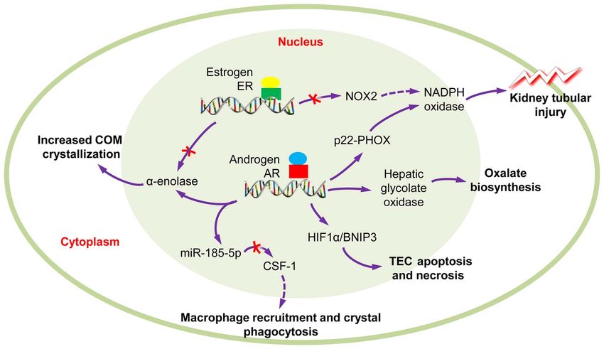
2 WANG et al: MECHANISM OF KIDNEY STONE FORMATION
stones. The most common types of human kidney stones are formation modulators, which has been thoroughly reviewed
CaOx and calcium phosphate (CaP), either alone or combined, previously (32). Several structures and molecular components
which are calcareous and radio‑opaque stones (9,11). Kidney also play the role of receptor in crystal attachments, including
stones form at a foundation of CaP termed Randall's plaques the phosphatidylserine component of the lipid bilayer and the
(RPs), which begins at the basement membranes of thin limbs acidic side chains of proteins (33). Calcium, oxalate, urate and
of the loop of Henle on the renal papillary surface (12). CaOx phosphate ions are the main promoters of crystal formation,
and urate stones exhibit a higher occurrence in males, whereas which can promote crystallization of stone constituents or their
higher percentages of carbapatite and struvite stones are aggregation through the activation of several mechanisms.
observed in females than in males (10,13). However, the role Ketha et al (34) demonstrated that the first time nephrolithiasis
of sex differences in the pathophysiological mechanisms of patients had increased serum calcium and 1,25(OH)2D levels
urinary stone disease are not yet fully understood. than the corresponding healthy individual serum calcium
Regardless of the type, kidney stone formation is a levels, suggesting that stone formation is a manifestation of
complex and multistep process that includes urinary super‑ altered calcium and vitamin D regulation. Higher serum
saturation, crystal nucleation, growth and aggregation (11,14). calcium concentration acts as a promoter in lithogenesis,
Kidney stone formation is associated with systemic disorders, which directly regulated by the calcium‑sensing receptor
including diabetes (15), obesity, cardiovascular diseases, (CaSR) through different pathways (35). Similarly, urate and
hypertension and metabolic syndrome (16,17). Conversely, phosphate ions have also been reported to promote hetero‑
nephrolithiasis patients [also known as kidney stone formers geneous nucleation and enhance the attachment of crystals
(KSF)] are at a risk of developing hypertension (18), chronic to epitheliums (36,37). Another important promoter of stone
kidney disease (CKD) (19) and progression to end‑stage renal formation is urine pH (38). Low pH urine may lead to CaOx
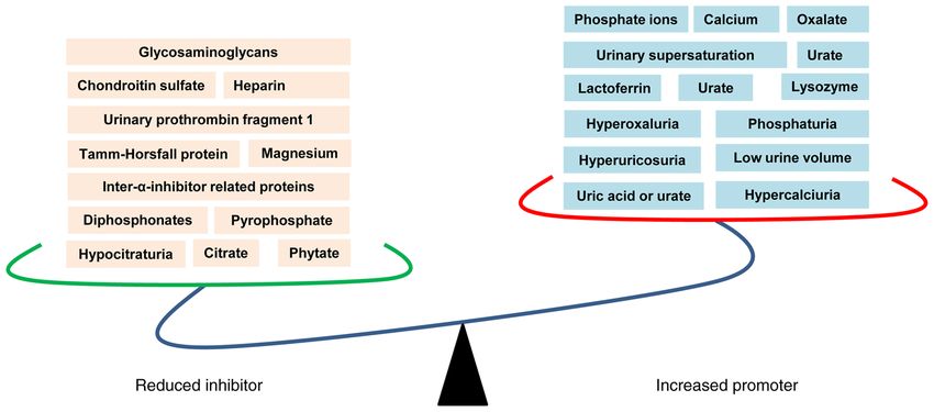
disease (ESRD) (20,21). Multiple promoting factors and inhib‑ crystallization and crystal precipitation (39). High‑alkaline
itors have been reported to play critical roles in kidney stone urine may also promote precipitation and nucleation of
formation. For example, hyperoxaluria, hyperuricosuria and CaOx crystals (40,41). Lysozyme and lactoferrin are two
phosphaturia are common promoting factors linked to kidney most recently identified proteins that promote COM growth
stone formation (22,23); inter‑α‑inhibitor (IαI), a member of through the acceleration of layer advancement rate on crystal
the protease inhibitor family, has been shown to inhibit CaOx surfaces (42).
crystallization in vitro (24).
Although details of human stone formation have Inhibitors of stone formation. Normal urine contains numerous
accumulated, kidney stone formation and growth mechanisms inhibitors that act both in competition and cooperation,
are far from being clarified. The present review provides an consequently decrease crystallization and inhibit crystals
update on the mechanisms of kidney stone formation, in order aggregation and/or adhesion to the tubular epithelial cells (43,44).
to improve the understanding of kidney stones for urologists, These inhibitors can be divided into three groups: Anions,
nephrologists and primary care givers. metallic cations and macromolecules. Anions such as citrate,
can inhibit crystal growth very efficiently, at concentrations
2. Physicochemical mechanism of kidney stone formation above 0.1 mM (45,46). A majority of nephrolithiasis patients
exhibited a decrease in citrate excretion. Alkali supplements
Urinary supersaturation and crystallization are the driving are widely used for hypocitraturic recurrent nephrolithiasis
force for intrarenal crystal precipitation and is mainly patients to restore citrate excretion (47,48). Hydroxycitrate
caused by inherited or acquired diseases associated is a structural analog of citrate, which has been reported to
with renal function impairment. Additionally, urinary show equivalent capacity in forming complexes with calcium,
supersaturation and crystallization are influenced by urine in order to inhibit crystallization (49,50). Metallic cations
pH and specific concentrations of substance excess, including such as magnesium, have been reported to inhibit crystal
CaOx, CaP, uric acids and urates, struvite, amino acids growth and aggregation, which is synergistic with citrate in
(cysteine), purines (2,8‑dihydroxyadenine and xanthine) acidic environments (51‑53). Macromolecules are the most
and drugs (e.g., atazanavir, sulfamethoxazole, amoxicillin, effective inhibitors of crystal growth. More specifically,
ceftriaxone) (25,26). Additionally, crystal formation and OPN, Tamm‑Horsfall protein (THP), urinary prothrombin
development are influenced by multiple modulator molecules, fragment 1 (UPTF‑1), nephrocalcin (NC) and some subunits
which are known as receptors, promoters and inhibitors. of the serum IαI are able to inhibit crystal growth, aggregation
and/or adhesion to the tubular cells (11,38,45).
Promoters of stone formation. A number of receptors or However, there is a competition between supersaturation
receptor‑like features have been reported to play critical and inhibitors of crystallization as mentioned above, which ulti‑
roles in crystal‑cell interaction, which is recognized as the mately determines the pattern of crystalluria in nephrolithiasis
most important process for crystal retention in kidney (8,27). patients and healthy individuals (54). As a consequence of the
Recently, protein alterations in a CaOx monohydrate (COM) increased promoters and reduced inhibitors, crystal formation
crystal‑cell interaction model were screened by the authors, and kidney stone occurrence have been observed (Fig. 1).
and 1,141 differentially expressed proteins (DEPs) were
identified in COM treated HK‑2 cells (28). Proteins and 3. Randall's plaque and calcium oxalate stone formation
glycosaminoglycan like CD44, nucleolin, hyaluronan (HA),
heat shock protein 90 (HSP90) (29), Αnnexin II (30) and RPs, first proposed by Alexander Randall in 1937 (55), are
osteopontin (OPN) (28,31), have been reported to act as stone regions of subepithelial mineralized tissue at the papillary tip,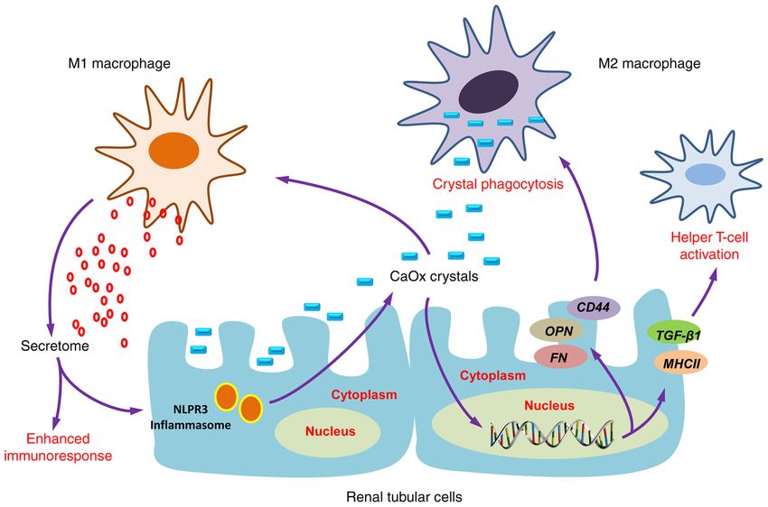
INTERNATIONAL JOURNAL OF MOlecular medicine 48: 149, 2021                                                          3

Figure 1. Physicochemical mechanisms of kidney stone formation. The reduced inhibitors (left panel) and increased promoters (right panel) are suggested to
play critical roles in kidney stone formation.

surrounding the openings of the ducts of Bellini containing                    4. Role of sex hormones in calcium oxalate nephrolithiasis
CaP (56). Scanning electron microscopy (SEM) examination
has shown that RP are made of a mixing of tubules with                         Statistical analyses have revealed that males have a higher
calcified walls and of tubules obstructed by CaP plugs (57).                   incidence of CaOx nephrolithiasis than females at a ratio
RP consists of CaP crystals mixed with an organic matrix                       of 2‑3:1 (4,65); however, the exact mechanism remain unclear.
that is rich in various proteins and lipids, and includes                      Previous studies have indicated that androgens increase
membrane‑bound vesicles or exosomes, collagen fibers,                          and estrogens decrease urinary oxalate excretion, plasma
as well as other components of the extracellular matrix (58).                  oxalate concentration and kidney CaOx crystal deposition.
An increasing number of studies have suggested that RPs are                    Additionally, enhanced androgen signaling may be responsible
the origin of renal stones (57‑60). Winfree et al (61) clarified               for the association between sex and kidney stone forma‑
that kidney stones develop as an overgrowth on RP, which                       tion (65‑68). Androgen receptor (AR) signaling can directly
contains unique organic composition (fibrillar collagen) that                  upregulate hepatic glycolate oxidase (69) and kidney epithe‑
can be differentiated from the stone overgrowth by specific                    lial nicotinamide adenine dinucleotide phosphate oxidase
autofluorescence signatures. Of note, a previous study using a                 (NAPDH ), subunit p22‑PHOX at the transcriptional level, so
murine mode of RP revealed that vitamin D supplementation                      as to increase oxalate biosynthesis, ultimately leading to kidney
and calcium intake could notably accelerate RP formation (60).                 stone formation (70). Peng et al (71) reported that testosterone
However, the precise mechanisms of RP formation remain                         contributes to nephrolithiasis development through the induc‑
unclear.                                                                       tion of renal tubular epithelial cells apoptosis and necrosis
    Recently, studies indicated that long non‑coding RNAs                      through HIF‑1α /BNIP3 pathway. Changtong et al (72)
(lcnRNAs) H19 and MALAT1 mediated osteogenic                                   revealed that testosterone could promote kidney stone disease
differentiation of human renal interstitial fibroblasts (hRIFs)                via the enhanced COM crystal‑cell adhesion by the increased
and participated in RP formation (62‑64). lcnRNA H19 has                       surface α‑enolase. Zhu et al (73) demonstrated that AR can
been shown to be significantly upregulated in RP, which can                    inhibit the recruitment of macrophages and suppress the COM
promote the osteogenic differentiation of hRIFs by activating                  crystals phagocytic ability of macrophages via the decrease
Wnt/β‑catenin signaling (63). lcnRNA H19 can also serve as                     of the colony‑stimulating factor 1 (CSF‑1) signals, through
a facilitator in the process of CaOx nephrocalcinosis‑induced                  miR‑185‑5p upregulation. These findings suggest that androgen
oxidative stress and renal tubular epithelial cell injury through              receptor signaling may be a key player in the development of
the interaction with miR‑216b and exerts its effect via the                    nephrolithiasis (Fig. 2).
HMGB1/TLR4/NF‑ κ B signaling pathway (64). lcnRNA                                  Theoretically, AR may be a new potential target and can be
MALAT1 can function as a competing endogenous RNA                              evaluated for novel therapeutics for the suppression of kidney
(ceRNA) that sponges miR‑320a‑5p, upregulates Runx2                            stone formation. The 5α‑reductase inhibitor, finasteride, has
expression and thus promotes the osteogenic phenotype of                       been reported to abolish the promoting effect of testosterone
hRIFs (62).                                                                    on COM crystallization (74). Another newly developed AR
    These studies provide novel insight into the pathogenesis                  degradation enhancer, dimethylcurcumin (ASC‑J9), has
of RP‑mediated kidney stone disease, while further studies                     been reported to suppress oxalate crystal formation via the
are urgently anticipated to explore the mechanisms of RP                       modulation of oxalate biosynthesis and reactive oxygen
formation, as well as additional roles of RP in the context of                 species (ROS)‑induced kidney tubular epithelial cell injury
stone formation.                                                               in a rat model (73). Reversely, estrogen may serve as a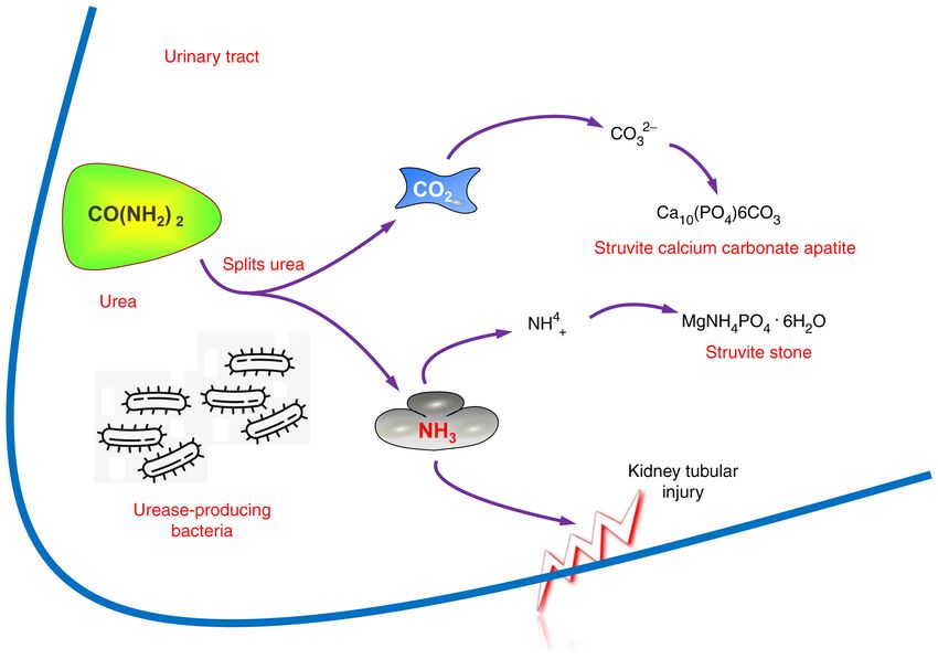
4                                          WANG et al: MECHANISM OF KIDNEY STONE FORMATION

Figure 2. Role of sex hormones in calcium oxalate nephrolithiasis. The AR signaling could induce TECs apoptosis and necrosis and kidney tubular injury,
promote COM crystallization and oxalate biosynthesis; however, macrophage recruitment and crystal phagocytosis are inhibited. Conversely, ER signaling
can reduce ROS‑mediated kidney tubular injury and COM crystallization. COM, calcium oxalate monohydrate; AR, androgen receptor; ER, estrogen receptor;
ROS, reactive oxygen species.

Figure 3. Role of urease‑producing bacteria in stone formation. The urease‑producing bacteria split urea and promote the formation of ammonia and carbon
dioxide, leading to kidney tubular injury and urine alkalinization and subsequent formation of phosphate salts.

protective factor against kidney stone formation. An in vitro                 oxidase subunit 2 (NOX2) through the direct binding to
study demonstrated that estrogen led to changes in the cellular               the estrogen response elements (EREs) on the NOX2 5'
proteome of [Madin darby canine kidney (MDCK)] renal                          promoter (76), which exerts protective effects on renal CaOx
tubular cells that led to the decreased CaOx crystal receptor                 crystal deposition.
surface expression (annexin A1 and α‑enolase), reduced                            All these findings may partly explain why a higher
intracellular ATP, and enhanced cell proliferation and renal                  incidence of nephrolithiasis is encounter in males than in
tubular cell tissue healing (75). There is evidence to suggest                females. Targeting AR may be developed as a potential therapy
that estrogen receptor β (ERβ) can suppress oxalate‑induced                   for CaOx crystal‑related kidney stone disease. However, these
oxidative stress via transcriptional suppression of the NADPH                 studies were performed in vitro and in vivo, using only cell
INTERNATIONAL JOURNAL OF MOlecular medicine 48: 149, 2021 5
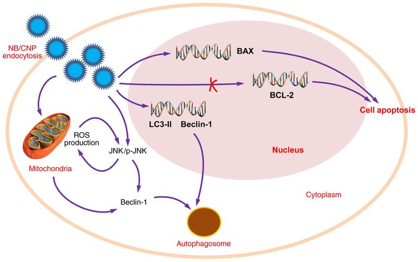
Figure 4. Role of nanobacteria in stone formation. The nanobacteria may induce ROS production through the JNK/p‑JNK signaling induction, may decrease
mitochondrial membrane potential and promote cell apoptosis through the downregulation of Bcl‑2 expression and the upregulation of Bax expression.
Additionally, nanobacteria may lead to autophagy through the upregulation of LC3‑II and Beclin‑1 expression. ROS, reactive oxygen species; LC3‑II,
microtubule‑associated proteins 1A/1B light chain 3B.
lines or animal models. Further validation and clinical studies
are required. Finasteride and ASC‑J9 have been demonstrated
to suppress a number of AR‑mediated diseases, including
prostate cancer (77,78), liver cancer and spinal and bulbar
muscular atrophy neuron disease (79). However, additional
future studies are necessary before the clinical application of
finasteride and ASC‑J9 in kidney stone prevention, considering
the side‑effects, including sexual dysfunction (80).
5. Role of the microbiome in stone formation
Emerging evidence has indicated that microorganisms
belonging to the human microbiome, including microorganisms
of the kidney and urinary tract, are likely to have a profound Figure 5. Role of oxalate‑degrading bacteria in stone formation.
Oxalate‑degrading bacteria use oxalate as a carbon energy source and thrive
effect on urological health, both positive and negative, due to in the presence of the oxalate anion, reduce urinary oxalate level and exhibit
their metabolic output and other contributions (81). growth inhibition in the calcium oxalate crystallization in the kidney.
Urease‑producing bacteria. Urease‑producing bacteria, such
as Proteus mirabilis, Klebsiella pneumonia, Staphylococcus
aureus, Pseudomonas aeruginosa, Providentia stuartii, Nanobacteria (NB). NB have been isolated from kidney stones
Serratia and Morganella morganii, are always associated for >30 years (87‑89); however, the nature and the mechanisms
with struvite stone formation and recurrence (82,83). The involved remain obscure. Ansari et al (90) demonstrated
bacterial urease degrades urea and promotes ammonia and that the size of cultured NB varies between 60 and 160 nm,
carbon dioxide formation, leading to urine alkalinization and and that they could infect patients with apatite kidney stone.
phosphate salt formation (Fig. 3). Kajander et al (91) indicated that NB can adapt to growing in
Urinary acidification and urease inhibitors have been plain DMEM or RPMI‑1640, through self‑proliferation. In the
proposed and implemented for the prevention and/or dissolution study by Ciftçioglu et al (92), it was demonstrated that 70 out
of struvite stones and encrustations in patients with infection of 72 (97.2%) kidney stones contained NB. The presence of
by urea‑degrading bacteria; however, their long term use is NB was independent of the stone type, although apatite‑based
limited due to their ineffectiveness and toxicity (84). Secondarily kidney stones presented the highest immunopositivity (91).
infected stones caused by non‑urease‑producing bacteria, NB are considered to play roles in calcium nucleation, as they
including Escherichia coli and Enterococcus spp., have also been can produce sufficient calcium apatite in their cell walls to
described (85,86). However, whether kidney stones form and initiate pathologic calcifications and stone formation (93‑95).
become secondarily infected or result from a nidus of infection This evidence is strongly in favor of the suggestion that NB are
that propagates stone formation remains largely unclear. living organisms.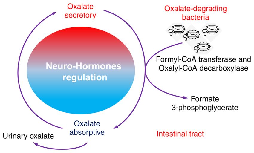
6                                          WANG et al: MECHANISM OF KIDNEY STONE FORMATION

Figure 6. Immune response to urinary crystals. Macrophage accumulation and macrophage‑related inflammation or anti‑inflammation is the main immune
response alteration observed as a result of kidney stone formation. M1 macrophages are important effectors of CaOx stone formation, while M2 macrophages
could prevent CaOx inflammatory damage through crystal phagocytosis. CaOx, calcium oxalate.

    However, an increasing number of studies have indicated                   well‑studied Gram‑negative anaerobic bacterium that degrades
that NB, also termed ‘Calcifying nanoparticles (CNPs)’,                       oxalate in the intestinal tract and has potential probiotic char‑
‘nanobacteria‑like particles’ or ‘Nanobes’, are merely mineral                acteristics for the prevention of CaOx kidney stone formation.
protein nanoparticles with biomimetic functions (88,89).                          In a pilot study, Stern et al (101) investigated the distinct
Although the definition and nature of these nanoparticles                     differences in the gut microbiome of nephrolithiasis patients,
remains controversial (96), their roles in kidney stone diseases              as compared with patients without kidney stone formation.
has been widely reported. CNPs have been identified in                        Their results demonstrated that the genus Bacteroides were
RPs and have been proven to be cytotoxic to 3T6 fibroblasts                   3.4‑fold more abundant in the kidney stone group, while the
and HK‑2 cells in vitro (89), which contributes to the renal                  genus Prevotella were 2.8‑fold more abundant in the non‑stone
tubular epithelial cell injury linked to kidney stone formation.              control group. A 24 h urine analysis revealed that the genus
Hong et al (97) demonstrated that catalase (CAT) and                          Eubacterium was inversely associated with oxalate levels
malonaldehyde (MDA) levels were significantly higher                          and the genus Escherichia trended to an inverse correlation
in CNP‑treated HK‑2 cells than the HK‑2 control group,                        with citrate level (101). However, the potential causative role
suggesting that CNPs may induce lipid peroxidation and result                 of pre‑existing dysbiosis of gut microbiome in kidney stone
in damaging HK‑2 cells. Wu et al (89) demonstrated that the                   disease is still unclear, and the association of urinary oxalate
CNPs may: Induce ROS production through JNK activation;                       excretion and oxalate‑degrading bacteria abundances remain
decrease mitochondrial membrane potential and promote cell                    limited (87,98,102,103).
apoptosis through the downregulation of Bcl‑2 expression and                      Both absorptive and secretory pathways for oxalate
the upregulation of Bax expression; lead to autophagy through                 have been identified in the proximal and distal segments
the upregulation of microtubule‑associated proteins 1A/1B                     of the colon, regulated by neuro‑hormones that direct net
light chain 3B (LC3‑II) and Beclin‑1 expression (Fig. 4).                     oxalate level. Thus, it has been suggested that intestinal tract
    According to currently available findings in the literature,              participates significantly in oxalate balance and subsequent
NB are localized in high concentrations in kidneys, excreted                  oxalate homeostasis (104‑106). The intestinal tract is also
in urine, are isolated from RPs and the majority of renal                     where oxalate‑degrading bacteria tend to reside, particularly
stones, and play the role of the initiator, by favoring nucleation            Oxalobacter formigenes, which requires a strict anaerobic
and crystal formation. Continued investigations are required,                 environment to survive. One hypothesis for the role of the
in order to solve the controversy of whether NB are living                    microbiome in the prevention of kidney stone has been that
or non‑living, as well as the mechanisms through which NB                     specific functional bacteria, such as the oxalate‑degrading
induce calcification and stone formation.                                     bacteria (such as Oxalobacter formigenes, Bifidobacterium
                                                                              sp. Porphyromonas gingivalis and Bacillus sp.) in human
Intestinal microbiota. The intestinal microbiome, which                       gut and intestinal tract, which use oxalate as their carbon
has been a recent area of wide interest, has been reported to                 energy source and thrive in the presence of the oxalate anion,
play a role in both the pathogenesis and prevention of kidney                 exhibit growth inhibition in the CaOx crystallization in the
stone disease (87,98‑100). Oxalobacter formigenes is the most                 kidney (102,107,108) (Fig. 5).
INTERNATIONAL JOURNAL OF MOlecular medicine 48: 149, 2021 7
The activity of oxalate‑degrading bacteria mediates 7. Conclusion and future perspectives
extra‑renal elimination of oxalate in the intestines and has
a significantly impact on the homeostatic levels of oxalate In the present review article, emerging concepts of mechanisms
in plasma and urine (109). This activity exhibits a strong contributing to stone formation were summarized, by reviewing
association with the occurrence of CaOx stone formation. novel insight into kidney stone disease related‑metabolic
risk factors, receptors, promoters and inhibitors, through the
6. Immune response to urinary crystals examination of the roles of immune‑response, microbiome
and sex hormones in stone formation and development. The
Macrophage accumulation and macrophage‑related pathophysiology of kidney stone disease cannot be completely
inflammation or anti‑inflammation is the main immune explained by crystallization processes alone. However, due to
response alteration observed in kidney stone disease, which current limitations in research, there are still some research
has been widely reported to play a crucial role in renal CaOx areas in kidney stone formation that remain poorly understood,
crystal formation (110). and were not been discussed herein. Future comprehensive
Firstly, the recruited macrophages could promote the studies are mandatory to further elucidate the mechanisms of the
development of COM crystals via the interaction of CD44 microbiome and immune response in kidney stone formation, in
with OPN and fibronectin (FN) (111), which are upregulated order to develop novel prophylactic and therapeutic approaches.
in renal tubular cells induced by crystals. Secondly,
macrophages have been evidenced to secrete various Acknowledgements
mediators via classical secretory pathways that cause renal
interstitial inflammation (112,113), particularly macrophage Not applicable.
inhibitory protein‑1, monocyte chemoattractant protein‑1 and
interleukin‑8 (IL‑8) (112). These chemokines consequently Funding
enhance the recruitment of various immune cells, including
monocytes, macrophages, neutrophils, dendritic cells and The present study was supported by the National Natural
T‑cells into the inflammatory locale (114,115). Several studies Science Foundation of China (grant no. 81802566), and
have demonstrated that macrophage‑derived exosomes Shenzhen Science and Technology Program (Basic Research
following COM exposure are involved in kidney stone Project, grant no. JCYJ20180228163919346).
pathogenesis (112,113,116). A set of proteins in COM‑treated
macrophage exosomes were previously identified as Availability of data and materials
proteins involved mainly in immune processes, including
T‑cell activation and homeostasis, Fc γ receptor‑mediated Not applicable.
phagocytosis, interferon‑ γ (IFN‑ γ) regulation and cell
migration (112). Additionally, infiltrated monocytes could Authors' contributions
differentiate into different macrophage subtypes with a wide
range of clinical manifestations, presentations and histological ZW and YZ prepared and drafted the manuscript. ZW
phenotypes (110,117), display protective or pathogenic obtained funding for the study, and drafted and revised the
activities in kidney stone development (110). manuscript. QD, JZ and HL assisted in obtaining data for the
Increasing evidence has revealed that M1/M2‑macrophage review article, drafted the manuscript and provided critical
differentiation plays an important role in renal CaOx revision of the manuscript for intellectual content. ZW and HL
crystal formation (111,115,118‑120). However, whether M1 confirm the authenticity of all the raw data. All authors have
macrophage‑mediated inflammation that contributes to stone read and approved the final manuscript.
formation will initiate stone promoters and reduce stone
inhibitors remains controversial. Khan et al (58) demonstrated Ethics approval and consent to participate
that M1 macrophages could cause acute tissue injury, which
was associated with crystal deposition and RP formation. Not applicable.
Conversely, Taguchi et al (121) concluded that there was no
association between renal dysfunction and increased crystal Patient consent for publication
deposition, based on their observation that no changes
were observed in urinary variables in lipopolysaccharide Not applicable.
(LPS)‑induced M1 macrophage‑mediated acute renal
injury. M2 anti‑inflammatory macrophages can phagocytize Competing interests
and degrade CaOx kidney stone fragments through a
clathrin‑dependent mechanism (110,113,115,120,121) (Fig. 6). The authors declare that they have no competing interests.
Given the critical role of immune‑response in CaOx crystal
formation and development, the immunotherapy approach has References
been proposed to prevent stone recurrences in certain individ‑
uals through the modulation of the immune response, in order 1. Romero V, Akpinar H and Assimos DG: Kidney stones: A global
to degrade CaOx crystals and thus prevent stones from devel‑ picture of prevalence, incidence, and associated risk factors. Rev
Urol 12: e86‑e96, 2010.
oping (122). However, investigations into immunotherapeutic 2. Morgan MS and Pearle MS: Medical management of renal
targets for kidney stone disease are urgently required. stones. BMJ 352: i52, 2016.8 WANG et al: MECHANISM OF KIDNEY STONE FORMATION
3. Zeng G, Mai Z, Xia S, Wang Z, Zhang K, Wang L, Long Y, 28. Wang Z, Li MX, Xu CZ, Zhang Y, Deng Q, Sun R, Hu QY,
Ma J, Li Y, Wan SP, et al: Prevalence of kidney stones in China: Zhang SP, Zhang JW and Liang H: Comprehensive study of
An ultrasonography based cross‑sectional study. BJU Int 120: altered proteomic landscape in proximal renal tubular epithelial
109‑116, 2017. cells in response to calcium oxalate monohydrate crystals. BMC
4. Ziemba JB and Matlaga BR: Epidemiology and economics of Urol 20: 136, 2020.
nephrolithiasis. Investig Clin Urol 58: 299‑306, 2017. 29. Fong‑Ngern K, Sueksakit K and Thongboonkerd V: Surface
5. Eisner BH and Goldfarb DS: A nomogram for the prediction of heat shock protein 90 serves as a potential receptor for calcium
kidney stone recurrence. J Am Soc Nephrol 25: 2685‑2687, 2014. oxalate crystal on apical membrane of renal tubular epithelial
6. Brikowski TH, Lotan Y and Pearle MS: Climate‑related increase cells. J Biol Inorg Chem 21: 463‑474, 2016.
in the prevalence of urolithiasis in the United States. Proc Natl 30. Kumar V, Farell G, Deganello S and Lieske JC: Annexin II
Acad Sci USA 105: 9841‑9846, 2008. is present on renal epithelial cells and binds calcium oxalate
7. Abeywickarama B, Ralapanawa U and Chandrajith R: monohydrate crystals. J Am Soc Nephrol 14: 289‑297, 2003.
Geoenvironmental factors related to high incidence of human 31. Anan G, Yoneyama T, Noro D, Tobisawa Y, Hatakeyama S,
urinary calculi (kidney stones) in Central Highlands of Sri Lanka. Sutoh Yoneyama M, Yamamoto H, Imai A, Iwamura H,
Environ Geochem Health 38: 1203‑1214, 2016. Kohada Y, et al: The impact of glycosylation of osteopontin on
8. Wang Z, Zhang JW, Zhang Y, Zhang SP, Hu QY and Liang H: urinary stone formation. Int J Mol Sci 21: 93, 2019.
Analyses of long non‑coding RNA and mRNA profiling using 32. Wiener SV, Ho SP and Stoller ML: Beginnings of nephrolithiasis:
RNA sequencing in calcium oxalate monohydrate‑stimulated Insights into the past, present and future of Randall's plaque forma‑
renal tubular epithelial cells. Urolithiasis 47: 225‑234, 2019. tion research. Curr Opin Nephrol Hypertens 27: 236‑242, 2018.
9. Parmar MS: Kidney stones. BMJ 328: 1420‑1424, 2004. 33. Sheng X, Ward MD and Wesson JA: Crystal surface adhesion
10. Ye Z, Zeng G, Yang H, Li J, Tang K, Wang G, Wang S, Yu Y, explains the pathological activity of calcium oxalate hydrates in
Wang Y, Zhang T, et al: The status and characteristics of urinary kidney stone formation. J Am Soc Nephrol 16: 1904‑1908, 2005.
stone composition in China. BJU Int 125: 801‑809, 2020. 34. Ketha H, Singh RJ, Grebe SK, Bergstralh EJ, Rule AD, Lieske JC
11. Aggarwal KP, Narula S, Kakkar M and Tandon C: Nephrolithiasis: and Kumar R: Altered calcium and vitamin D homeostasis in
Molecular mechanism of renal stone formation and the critical first‑time calcium kidney stone‑formers. PLoS One 10: e0137350,
role played by modulators. Biomed Res Int 2013: 292953, 2013. 2015.
12. Khan SR, Pearle MS, Robertson WG, Gambaro G, Canales BK, 35. Vezzoli G, Macrina L, Magni G and Arcidiacono T:
Doizi S, Traxer O and Tiselius HG: Kidney stones. Nat Rev Dis Calcium‑sensing receptor: Evidence and hypothesis for its role
Primers 2: 16008, 2016. in nephrolithiasis. Urolithiasis 47: 23‑33, 2019.
13. Sun X, Shen L, Cong X, Zhu H, He L and Lu J: Infrared spec‑ 36. Farell G, Huang E, Kim SY, Horstkorte R and Lieske JC:
troscopic analysis of 5,248 urinary stones from Chinese patients Modulation of proliferating renal epithelial cell affinity for
presenting with the first stone episode. Urol Res 39: 339‑343, calcium oxalate monohydrate crystals. J Am Soc Nephrol 15:
2011. 3052‑3062, 2004.
14. Hamamoto S, Taguchi K and Fujii Y: Molecular mechanism 37. Gao J, Xue JF, Xu M, Gui BS, Wang FX and Ouyang JM:
of renal stone formation. Clin Calcium 21: 1481‑1487, 2011 Nanouric acid or nanocalcium phosphate as central nidus to
(In Japanese). induce calcium oxalate stone formation: A high‑resolution trans‑
15. Pak CY, Sakhaee K, Moe O, Preminger GM, Poindexter JR, mission electron microscopy study on urinary nanocrystallites.
Peterson RD, Pietrow P and Ekeruo W: Biochemical profile Int J Nanomedicine 9: 4399‑4409, 2014.
of stone‑forming patients with diabetes mellitus. Urology 61: 38. Ratkalkar VN and Kleinman JG: Mechanisms of stone formation.
523‑527, 2003 Clin Rev Bone Miner Metab 9: 187‑197, 2011.
16. Carbone A, Al Salhi Y, Tasca A, Palleschi G, Fuschi A, 39. Moe OW, Abate N and Sakhaee K: Pathophysiology of uric acid
De Nunzio C, Bozzini G, Mazzaferro S and Pastore AL: Obesity nephrolithiasis. Endocrinol Metab Clin North Am 31: 895‑914,
and kidney stone disease: A systematic review. Minerva Urol 2002.
Nefrol 70: 393‑400, 2018. 40. Shekarriz B and Stoller ML: Uric acid nephrolithiasis: Current
17. Devarajan A: Cross‑talk between renal lithogenesis and concepts and controversies. J Urol 168: 1307‑1314, 2002.
atherosclerosis: An unveiled link between kidney stone formation 41. Song L and Maalouf NM: Nephrolithiasis. In: Endotext.
and cardiovascular diseases. Clin Sci (Lond) 132: 615‑626, 2018. Feingold KR, Anawalt B, Boyce A, Chrousos G, de Herder WW,
18. Kittanamongkolchai W, Mara KC, Mehta RA, Vaughan LE, Dungan K, Grossman A, Hershman JM, Hofland HJ, Kaltsas G,
Denic A, Knoedler JJ, Enders FT, Lieske JC and Rule AD: Risk et al (eds). MDText.com, Inc., South Dartmouth, MA, 2000.
of hypertension among first‑time symptomatic kidney stone 42. Farmanesh S, Chung J, Sosa RD, Kwak JH, Karande P and
formers. Clin J Am Soc Nephrol 12: 476‑482, 2017. Rimer JD: Natural promoters of calcium oxalate monohydrate
19. Rule AD, Bergstralh EJ, Melton LJ III, Li X, Weaver AL and crystallization. J Am Chem Soc 136: 12648‑12657, 2014.
Lieske JC: Kidney stones and the risk for chronic kidney disease. 43. Worcester EM: Urinary calcium oxalate crystal growth inhibitors.
Clin J Am Soc Nephrol 4: 804‑811, 2009. J Am Soc Nephrol 5 (Suppl 1): S46‑S53, 1994.
20. Keddis MT and Rule AD: Nephrolithiasis and loss of kidney 44. Schepers MS, van der Boom BG, Romijn JC, Schroder FH and
function. Curr Opin Nephrol Hypertens 22: 390‑396, 2013. Verkoelen CF: Urinary crystallization inhibitors do not prevent
21. Dhondup T, Kittanamongkolchai W, Vaughan LE, Mehta RA, crystal binding. J Urol 167: 1844‑1847, 2002.
Chhina JK, Enders FT, Hickson LJ, Lieske JC and Rule AD: Risk 45. Khan SR and Kok DJ: Modulators of urinary stone formation.
of ESRD and mortality in kidney and bladder stone formers. Am Front Biosci 9: 1450‑1482, 2004.
J Kidney Dis 72: 790‑797, 2018. 46. Hess B, Jordi S, Zipperle L, Ettinger E and Giovanoli R: Citrate
22. Voss S, Hesse A, Zimmermann DJ, Sauerbruch T and determines calcium oxalate crystallization kinetics and crystal
von Unruh GE: Intestinal oxalate absorption is higher in morphology‑studies in the presence of Tamm‑Horsfall protein of
idiopathic calcium oxalate stone formers than in healthy a healthy subject and a severely recurrent calcium stone former.
controls: Measurements with the [(13)C2]oxalate absorption test. Nephrol Dial Transplant 15: 366‑374, 2000.
J Urol 175: 1711‑1715, 2006. 47. Cicerello E, Ciaccia M, Cova G and Mangano M: The impact
23. Ha YS, Tchey DU, Kang HW, Kim YJ, Yun SJ, Lee SC and of potassium citrate therapy in the natural course of Medullary
Kim WJ: Phosphaturia as a promising predictor of recurrent Sponge Kidney with associated nephrolithiasis. Arch Ital Urol
stone formation in patients with urolithiasis. Korean J Urol 51: Androl 91: 102-106, 2019.
54‑59, 2010. 48. Siener R: Dietary treatment of metabolic acidosis in chronic
24. Dean C, Kanellos J, Pham H, Gomes M, Oates A, Grover P kidney disease. Nutrients 10: 512, 2018.
and Ryall R: Effects of inter‑alpha‑inhibitor and several of its 49. Kim D, Rimer JD and Asplin JR: Hydroxycitrate: A potential
derivatives on calcium oxalate crystallization in vitro. Clin Sci new therapy for calcium urolithiasis. Urolithiasis 47: 311‑320,
(Lond) 98: 471‑480, 2000. 2019.
25. Daudon M, Frochot V, Bazin D and Jungers P: Drug‑induced 50. Chung J, Granja I, Taylor MG, Mpourmpakis G, Asplin JR and
kidney stones and crystalline nephropathy: Pathophysiology, Rimer JD: Molecular modifiers reveal a mechanism of pathological
prevention and treatment. Drugs 78: 163‑201, 2018. crystal growth inhibition. Nature 536: 446‑450, 2016.
26. Rodgers AL: Physicochemical mechanisms of stone formation. 51. Ryall RL, Harnett RM and Marshall VR: The effect of urine,
Urolithiasis 45: 27‑32, 2017. pyrophosphate, citrate, magnesium and glycosaminoglycans on
27. Thongboonkerd V: Proteomics of crystal‑cell interactions: the growth and aggregation of calcium oxalate crystals in vitro.
A model for kidney stone research. Cells 8: 1076, 2019. Clin Chim Acta 112: 349‑356, 1981.INTERNATIONAL JOURNAL OF MOlecular medicine 48: 149, 2021 9
52. Riley JM, Kim H, Averch TD and Kim HJ: Effect of magnesium on 74. Sueksakit K and Thongboonkerd V: Protective effects of finaste‑
calcium and oxalate ion binding. J Endourol 27: 1487‑1492, 2013. ride against testosterone‑induced calcium oxalate crystallization
53. Grases F, Rodriguez A and Costa‑Bauza A: Efficacy of mixtures of and crystal‑cell adhesion. J Biol Inorg Chem 24: 973‑983, 2019.
magnesium, citrate and phytate as calcium oxalate crystallization 75. Peerapen P and Thongboonkerd V: Protective cellular mechanism
inhibitors in urine. J Urol 194: 812‑819, 2015. of estrogen against kidney stone formation: A proteomics approach
54. Robertson WG: Do ‘inhibitors of crystallisation’ play any role in and functional validation. Proteomics 19: e1900095, 2019.
the prevention of kidney stones? A critique. Urolithiasis 45: 43‑56, 76. Zhu W, Zhao Z, Chou FJ, Zuo L, Liu T, Bushinsky D, Chang C,
2017. Zeng G and Yeh S: The protective roles of estrogen receptor β
55. Randall A: The origin and growth of renal calculi. Ann Surg 105: in renal calcium oxalate crystal formation via reducing the liver
1009‑1027, 1937. oxalate biosynthesis and renal oxidative stress‑mediated cell
56. Wiener SV, Chen L, Shimotake AR, Kang M, Stoller ML and injury. Oxid Med Cell Longev 2019: 5305014, 2019.
Ho SP: Novel insights into renal mineralization and stone forma‑ 77. Loughlin KR: The clinical applications of five‑alpha reductase
tion through advanced imaging modalities. Connect Tissue inhibitors. Can J Urol 28: 10584‑10588, 2021.
Res 59: S102‑S110, 2018. 78. Tian H, Chou FJ, Tian J, Zhang Y, You B, Huang CP, Yeh S, Niu Y
57. Daudon M, Bazin D and Letavernier E: Randall's plaque as the and Chang C: ASC‑J9® suppresses prostate cancer cell prolifera‑
origin of calcium oxalate kidney stones. Urolithiasis 43 (Suppl 1): tion and invasion via altering the ATF3‑PTK2 signaling. J Exp
S5‑S11, 2015. Clin Cancer Res 40: 3, 2021.
58. Khan SR, Canales BK and Dominguez‑Gutierrez PR: Randall's 79. Hu H, Zhou H and Xu D: A review of the effects and molecular
plaque and calcium oxalate stone formation: Role for immunity mechanisms of dimethylcurcumin (ASC‑J9) on androgen
and inflammation. Nat Rev Nephrol 17: 417‑433, 2021. receptor‑related diseases. Chem Biol Drug Des 97: 821‑835, 2021.
59. Chung HJ: The role of Randall plaques on kidney stone forma‑ 80. Andy G, John M, Mirna S, Rachita D, Michael K, Maja K, Aseem S
tion. Transl Androl Urol 3: 251‑254, 2014. and Zeljana B: Controversies in the treatment of androgenetic
60. Bouderlique E, Tang E, Perez J, Coudert A, Bazin D, Verpont MC, alopecia: The history of finasteride. Dermatol Ther 32: e12647,
Duranton C, Rubera I, Haymann JP, Leftheriotis G, et al: Vitamin D 2019.
and calcium supplementation accelerates Randall's plaque forma‑ 81. Whiteside SA, Razvi H, Dave S, Reid G and Burton JP: The
tion in a murine model. Am J Pathol 189: 2171‑2180, 2019. microbiome of the urinary tract‑a role beyond infection. Nat Rev
61. Winfree S, Weiler C, Bledsoe SB, Gardner T, Sommer AJ, Urol 12: 81‑90, 2015.
Evan AP, Lingeman JE, Krambeck AE, Worcester EM, 82. Bichler KH, Eipper E, Naber K, Braun V, Zimmermann R and
El‑Achkar TM and Williams JC Jr: Multimodal imaging Lahme S: Urinary infection stones. Int J Antimicrob Agents 19:
reveals a unique autofluorescence signature of Randall's plaque. 488‑498, 2002.
Urolithiasis 49: 123‑135, 2021. 83. Espinosa‑Ortiz EJ, Eisner BH, Lange D and Gerlach R: Current
62. Zhu Z, Huang F, Xia W, Zeng H, Gao M, Li Y, Zeng F, He C, insights into the mechanisms and management of infection
Chen J, Chen Z, et al: Osteogenic differentiation of renal inter‑ stones. Nat Rev Urol 16: 35‑53, 2019.
stitial fibroblasts promoted by lncRNA MALAT1 may partially 84. Marien T and Miller NL: Treatment of the Infected Stone. Urol
contribute to Randall's plaque formation. Front Cell Dev Biol 8: Clin North Am 42: 459‑472, 2015.
596363, 2020. 85. de Cógáin MR, Lieske JC, Vrtiska TJ, Tosh PK and Krambeck AE:
63. Zhu Z, Cui Y, Huang F, Zeng H, Xia W, Zeng F, He C, Chen J, Secondarily infected nonstruvite urolithiasis: A prospective
Chen Z, Chen H and Li Y: Long non‑coding RNA H19 promotes evaluation. Urology 84: 1295‑1300, 2014.
osteogenic differentiation of renal interstitial fibroblasts through 86. Flannigan R, Choy WH, Chew B and Lange D: Renal struvite
Wnt‑beta‑catenin pathway. Mol Cell Biochem 470: 145‑155, 2020. stones‑pathogenesis, microbiology, and management strategies.
64. Liu H, Ye T, Yang X, Liu J, Jiang K, Lu H, Xia D, Peng E, Nat Rev Urol 11: 333‑341, 2014.
Chen Z, Sun F, et al: H19 promote calcium oxalate nephrocal‑ 87. Mehta M, Goldfarb DS and Nazzal L: The role of the microbiome
cinosis‑induced renal tubular epithelial cell injury via a ceRNA in kidney stone formation. Int J Surg 36: 607‑612, 2016.
pathway. EBioMedicine 50: 366‑378, 2019. 88. Martel J, Peng HH, Young D, Wu CY and Young JD: Of nanobac‑
65. Fan J, Chandhoke PS and Grampsas SA: Role of sex hormones teria, nanoparticles, biofilms and their role in health and disease:
in experimental calcium oxalate nephrolithiasis. J Am Soc Facts, fancy and future. Nanomedicine (Lond) 9: 483‑499, 2014.
Nephrol 10 (Suppl 14): S376‑S380, 1999. 89. Wu J, Tao Z, Deng Y, Liu Q, Liu Y, Guan X and Wang X:
66. Li JY, Zhou T, Gao X, Xu C, Sun Y, Peng Y, Chang Z, Zhang Y, Calcifying nanoparticles induce cytotoxicity mediated by
Jiang J, Wang L and Hou J: Testosterone and androgen receptor ROS‑JNK signaling pathways. Urolithiasis 47: 125‑135, 2019.
in human nephrolithiasis. J Urol 184: 2360‑2363, 2010. 90. Ansari H, Akhavan Sepahi A and Akhavan Sepahi M: Different
67. Gupta K, Gill GS and Mahajan R: Possible role of elevated serum approaches to detect ‘Nanobacteria’ in patients with kidney
testosterone in pathogenesis of renal stone formation. Int J Appl stones: An infectious cause or a subset of life? Urol J 14:
Basic Med Res 6: 241‑244, 2016. 5001‑5007, 2017.
68. Fuster DG, Morard GA, Schneider L, Mattmann C, Lüthi D, Vogt 91. Kajander EO, Ciftcioglu N, Aho K and Garcia‑Cuerpo E:
B and Dhayat NA: Association of urinary sex steroid hormones Characteristics of nanobacteria and their possible role in stone
with urinary calcium, oxalate and citrate excretion in kidney formation. Urol Res 31: 47‑54, 2003.
stone formers. Nephrol Dial Transplant: Dec 9, 2020 (Epub 92. Ciftçioglu N, Björklund M, Kuorikoski K, Bergström K and
ahead of print). Kajander EO: Nanobacteria: An infectious cause for kidney
69. Yoshihara H, Yamaguchi S and Yachiku S: Effect of sex stone formation. Kidney Int 56: 1893‑1898, 1999.
hormones on oxalate‑synthesizing enzymes in male and female 93. Khullar M, Sharma SK, Singh SK, Bajwa P, Shiekh FA, Relan V
rat livers. J Urol 161: 668‑673, 1999. and Sharma M: Morphological and immunological characteris‑
70. Liang L, Li L, Tian J, Lee SO, Dang Q, Huang CK, Yeh S, tics of nanobacteria from human renal stones of a north Indian
Erturk E, Bushinsky D, Chang LS, et al: Androgen receptor population. Urol Res 32: 190‑195, 2004.
enhances kidney stone‑CaOx crystal formation via modulation 94. Shiekh FA, Khullar M and Singh SK: Lithogenesis: Induction of
of oxalate biosynthesis & oxidative stress. Mol Endocrinol 28: renal calcifications by nanobacteria. Urol Res 34: 53‑57, 2006.
1291‑1303, 2014. 95. Kajander EO and Ciftçioglu N: Nanobacteria: An alternative
71. Peng Y, Fang Z, Liu M, Wang Z, Li L, Ming S, Lu C, Dong H, mechanism for pathogenic intra‑ and extracellular calcification
Zhang W, Wang Q, et al: Testosterone induces renal tubular and stone formation. Proc Natl Acad Sci USA 95: 8274‑8279, 1998.
epithelial cell death through the HIF‑1alpha/BNIP3 pathway. 96. Abrol N, Panda A, Kekre NS and Devasia A: Nanobacteria in the
J Transl Med 17: 62, 2019. pathogenesis of urolithiasis: Myth or reality? Indian J Urol 31:
72. Changtong C, Peerapen P, Khamchun S, Fong‑Ngern K, 3‑7, 2015.
Chutipongtanate S and Thongboonkerd V: In vitro evidence of 97. Hong X, Wang X, Wang T, Yu C and Li H: Role of nanobacteria
the promoting effect of testosterone in kidney stone disease: A in the pathogenesis of kidney stone formation. Am J Transl
proteomics approach and functional validation. J Proteomics 144: Res 8: 3227‑3234, 2016.
11‑22, 2016. 98. Sadaf H, Raza SI and Hassan SW: Role of gut microbiota against
73. Zhu W, Zhao Z, Chou F, Zuo L, Liu T, Yeh S, Bushinsky D, calcium oxalate. Microb Pathog 109: 287‑291, 2017.
Zeng G and Chang C: Loss of the androgen receptor suppresses 99. Ticinesi A, Nouvenne A, Chiussi G, Castaldo G, Guerra A and
intrarenal calcium oxalate crystals deposition via altering Meschi T: Calcium oxalate nephrolithiasis and gut microbiota:
macrophage recruitment/M2 polarization with change of the Not just a gut‑kidney axis. A nutritional perspective. Nutrients 12:
miR‑185‑5p/CSF‑1 signals. Cell Death Dis 10: 275, 2019. 548, 2020.10 WANG et al: MECHANISM OF KIDNEY STONE FORMATION
100. Ticinesi A, Milani C, Guerra A, Allegri F, Lauretani F, 112. Singhto N, Kanlaya R, Nilnumkhum A and Thongboonkerd V:
Nouvenne A, Mancabelli L, Lugli GA, Turroni F, Duranti S, et al: Roles of macrophage exosomes in immune response to calcium
Understanding the gut‑kidney axis in nephrolithiasis: An oxalate monohydrate crystals. Front Immunol 9: 316, 2018.
analysis of the gut microbiota composition and functionality of 113. Singhto N and Thongboonkerd V: Exosomes derived from
stone formers. Gut 67: 2097‑2106, 2018. calcium oxalate‑exposed macrophages enhance IL‑8 produc‑
101. Stern JM, Moazami S, Qiu Y, Kurland I, Chen Z, Agalliu I, tion from renal cells, neutrophil migration and crystal invasion
Burk R and Davies KP: Evidence for a distinct gut microbiome through extracellular matrix. J Proteomics 185: 64‑76, 2018.
in kidney stone formers compared to non‑stone formers. 114. Tamura M, Aizawa R, Hori M and Ozaki H: Progressive renal
Urolithiasis 44: 399‑407, 2016. dysfunction and macrophage infiltration in interstitial fibrosis
102. Falony G: Beyond Oxalobacter: The gut microbiota and kidney in an adenine‑induced tubulointerstitial nephritis mouse model.
stone formation. Gut 67: 2078‑2079, 2018. Histochem Cell Biol 131: 483‑490, 2009.
103. Miller AW and Dearing D: The metabolic and ecological inter‑ 115. Kusmartsev S, Dominguez‑Gutierrez PR, Canales BK, Bird VG,
actions of oxalate‑degrading bacteria in the Mammalian gut. Vieweg J and Khan SR: Calcium oxalate stone fragment and
Pathogens 2: 636‑652, 2013. crystal phagocytosis by human macrophages. J Urol 195:
104. Worcester EM, Fellner SK, Nakagawa Y and Coe FL: Effect 1143‑1151, 2016.
of renal transplantation on serum oxalate and urinary oxalate 116. Sintipr ungrat K, Singhto N and Thongboon kerd V:
excretion. Nephron 67: 414‑418, 1994. Characterization of calcium oxalate crystal‑induced changes
105. Hatch M, Freel RW and Vaziri ND: Mechanisms of oxalate in the secretome of U937 human monocytes. Mol Biosyst 12:
absorption and secretion across the rabbit distal colon. Pflugers 879‑889, 2016.
Arch 426: 101‑109, 1994. 117. Histiocytosis syndromes in children. Writing Group of the
106. Peck AB, Canales BK and Nguyen CQ: Oxalate‑degrading Histiocyte Society. Lancet 1: 208‑209, 1987.
microorganisms or oxalate‑degrading enzymes: Which is the 118. Okada A, Yasui T, Hamamoto S, Hirose M, Kubota Y, Itoh Y,
future therapy for enzymatic dissolution of calcium‑oxalate Tozawa K, Hayashi Y and Kohri K: Genome‑wide analysis
uroliths in recurrent stone disease? Urolithiasis 44: 45‑50, 2016. of genes related to kidney stone formation and elimination in
107. Knight J, Deora R, Assimos DG and Holmes RP: The genetic the calcium oxalate nephrolithiasis model mouse: Detection
composition of Oxalobacter formigenes and its relationship to of stone‑preventive factors and involvement of macrophage
colonization and calcium oxalate stone disease. Urolithiasis 41: activity. J Bone Miner Res 24: 908‑924, 2009.
187‑196, 2013. 119. Vervaet BA, Verhulst A, Dauwe SE, De Broe ME and
108. Batagello CA, Monga M and Miller AW: Calcium oxalate D'Haese PC: An active renal crystal clearance mechanism in rat
urolithiasis: A case of missing microbes? J Endourol 32: and man. Kidney Int 75: 41‑51, 2009.
995‑1005, 2018. 120. Dominguez‑Gutierrez PR, Kusmartsev S, Canales BK and
109. Cornelius JG and Peck AB: Colonization of the neonatal rat Khan SR: Calcium oxalate differentiates human monocytes into
intestinal tract from environmental exposure to the anaerobic inflammatory M1 macrophages. Front Immunol 9: 1863, 2018.
bacterium Oxalobacter formigenes. J Med Microbiol 53: 121. Taguchi K, Okada A, Hamamoto S, Unno R, Moritoki Y, Ando R,
249‑254, 2004. Mizuno K, Tozawa K, Kohri K and Yasui T: M1/M2‑macrophage
110. Nikolic‑Paterson DJ, Wang S and Lan HY: Macrophages phenotypes regulate renal calcium oxalate crystal development.
promote renal fibrosis through direct and indirect mechanisms. Sci Rep 6: 35167, 2016.
Kidney Int Suppl (2011) 4: 34‑38, 2014. 122. Dominguez‑Gutierrez PR, Kwenda EP, Khan SR and
111. Okada A, Yasui T, Fujii Y, Niimi K, Hamamoto S, Hirose M, Canales BK: Immunotherapy for stone disease. Curr Opin
Kojima Y, Itoh Y, Tozawa K, Hayashi Y and Kohri K: Renal Urol 30: 183‑189, 2020.
macrophage migration and crystal phagocytosis via inflam‑
matory‑related gene expression during kidney stone formation This work is licensed under a Creative Commons
and elimination in mice: Detection by association analysis of
Attribution-NonCommercial-NoDerivatives 4.0
stone‑related gene expression and microstructural observation.
J Bone Miner Res 25: 2701‑2711, 2010. International (CC BY-NC-ND 4.0) License.You can also read