Nanocarrier-enhanced intracellular delivery of benznidazole for treatment of Trypanosoma cruzi infection - JCI Insight
←
→
Page content transcription
If your browser does not render page correctly, please read the page content below
Nanocarrier-enhanced intracellular delivery of benznidazole for
treatment of Trypanosoma cruzi infection
Xiaomo Li, … , Evan A. Scott, David M. Engman
JCI Insight. 2021;6(9):e145523. https://doi.org/10.1172/jci.insight.145523.

 Research Article   Microbiology

Chagas disease is caused by infection with the protozoan parasite Trypanosoma cruzi (T. cruzi), an intracellular pathogen
that causes significant morbidity and death among millions in the Americas from Canada to Argentina. Current therapy
involves oral administration of the nitroimidazole benznidazole (BNZ), which has serious side effects that often
necessitate cessation of treatment. To both avoid off-target side effects and reduce the necessary dosage of BNZ, we
packaged the drug within poly(ethylene glycol)-block-poly(propylene sulfide) polymersomes (BNZ-PSs). We show that
these vesicular nanocarriers enhanced intracellular delivery to phagocytic cells and tested this formulation in a mouse
model of T. cruzi infection. BNZ-PS is not only nontoxic but also significantly more potent than free BNZ, effectively
reducing parasitemia, intracellular infection, and tissue parasitosis at a 466-fold lower dose of BNZ. We conclude that
BNZ-PS was superior to BNZ for treatment of T. cruzi infection in mice and that further modifications of this nanocarrier
formulation could lead to a wide range of custom controlled delivery applications for improved treatment of Chagas
disease in humans.

Find the latest version:
https://jci.me/145523/pdf
RESEARCH ARTICLE

                                          Nanocarrier-enhanced intracellular
                                          delivery of benznidazole for treatment of
                                          Trypanosoma cruzi infection
                                          Xiaomo Li,1,2 Sijia Yi,3 Débora B. Scariot,3 Santiago J. Martinez,1,4 Ben A. Falk,1 Cheryl L. Olson,2
                                          Patricia S. Romano,4 Evan A. Scott,3 David M. Engman1,2,5
                                          Department of Pathology and Laboratory Medicine, Cedars-Sinai Medical Center, Los Angeles, California, USA.
                                          1

                                          2
                                              Department of Pathology, Northwestern University, Chicago, Illinois, USA. 3Department of Biomedical Engineering,
                                          Chemistry of Life Processes Institute, and Simpson Querrey Institute, Northwestern University, Evanston and Chicago,
                                          Illinois, USA. 4Institute of Histology and Embryology, “Dr. Mario H. Burgos”, IHEM-CONICET, National University of Cuyo,
                                          Mendoza, Argentina. 5Department of Pathology and Laboratory Medicine, University of California, Los Angeles, Los
                                          Angeles, California, USA.

                                                     Chagas disease is caused by infection with the protozoan parasite Trypanosoma cruzi (T. cruzi), an
                                                     intracellular pathogen that causes significant morbidity and death among millions in the Americas
                                                     from Canada to Argentina. Current therapy involves oral administration of the nitroimidazole
                                                     benznidazole (BNZ), which has serious side effects that often necessitate cessation of treatment.
                                                     To both avoid off-target side effects and reduce the necessary dosage of BNZ, we packaged the
                                                     drug within poly(ethylene glycol)-block-poly(propylene sulfide) polymersomes (BNZ-PSs). We
                                                     show that these vesicular nanocarriers enhanced intracellular delivery to phagocytic cells and
                                                     tested this formulation in a mouse model of T. cruzi infection. BNZ-PS is not only nontoxic but also
                                                     significantly more potent than free BNZ, effectively reducing parasitemia, intracellular infection,
                                                     and tissue parasitosis at a 466-fold lower dose of BNZ. We conclude that BNZ-PS was superior to
                                                     BNZ for treatment of T. cruzi infection in mice and that further modifications of this nanocarrier
                                                     formulation could lead to a wide range of custom controlled delivery applications for improved
                                                     treatment of Chagas disease in humans.

                                          Introduction
                                          Chagas disease is an insect-transmitted parasitic infection first described in 1909 by the Brazilian physician
                                          Carlos Chagas (1). The protozoan Trypanosoma cruzi (T. cruzi) causes lifelong infection in humans and
                                          other vertebrates by infecting a wide variety of cells throughout the body, typically controlled by adap-
                                          tive immunity without causing any of the adverse sequelae known as Chagas disease. Indeed, most T.
Authorship note: XL and SY                cruzi–infected individuals are unaware of their infections, with approximately one-third developing either
contributed equally to this work.         a chronic cardiomyopathy or a megadisease of the esophagus or colon, often many years after infection
Conflict of interest: The authors have    (2). There are approximately 7 million infections and 14,000 deaths each year from T. cruzi and Chagas
declared that no conflict of interest     disease worldwide (3), and several hundred thousand infected people currently reside in the United States
exists.                                   (4). Although most T. cruzi–infected people in the United States are immigrants who were infected in their
Copyright: © 2021, Li et al. This is an   countries of origin, T. cruzi–infected triatomine bugs are found in most Southern states, and vector-borne
open access article published under       autochthonous transmission is now understood to be a small but significant risk to many Americans (5).
the terms of the Creative Commons         Blood transfusion and organ transplantation are additional modes of transmission, but these have been
Attribution 4.0 International License.    reduced significantly through global screening of blood and organs for T. cruzi (6).
Submitted: December 11, 2020                   Only 2 drugs, the hydrophobic nitroimidazoles benznidazole (BNZ) and nifurtimox (NFX), have been avail-
Accepted: March 31, 2021                  able to treat T. cruzi–infected individuals since 1970 (7). Despite their poor bioavailability and permeability (8),
Published: May 10, 2021                   both drugs are up to 80% effective when used during acute infection. Unfortunately, these agents have severe side
                                          effects, including neutropenia, nausea, vomiting and diarrhea, weight loss, and hypersensitivity skin reactions
Reference information: JCI Insight.
2021;6(9):e145523.                        and hives, which lead to treatment cessation in many individuals (9). In chronic disease, when cardiomyopathy
https://doi.org/10.1172/jci.              and megadisease typically develop, the decision to treat has been a matter of debate for many years. The very
insight.145523.                           large BENEFIT trial tested the effect of treatment on those with established cardiomyopathy and concluded

                                                                                                                                                                     1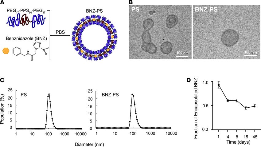
RESEARCH ARTICLE
that treatment does not reduce progression to a major cardiac event or death, despite reducing parasitemia (10).
However, results from a more recent BNZ clinical trial showed that treatment of indeterminate individuals hav-
ing no clinical disease reduces progression to cardiomyopathy (11), which many consider equally important.
Regardless, there is no treatment that reverses Chagas cardiomyopathy once established (12). Chagas is consid-
ered a neglected tropical disease and is still overlooked by pharmaceutical companies due to it primarily being
found within low-income countries (13, 14).
T. cruzi is an obligate intracellular pathogen that can infect any nucleated cell. The tropism to myocytes
has been extensively studied because the presence of parasite in muscles cells is intrinsically related to
cardiac dysfunction, especially in the acute stage (2). Therefore, trypanocidal drugs must have the ability
to cross the host cell plasma membrane to effectively kill intracellular parasites (14). It is possible that the
toxicity observed with BNZ and NFX treatment is due to the high concentration necessary to achieve
an effective intracellular killing concentration. The development of drug nanocarriers has revolutionized
therapeutics by permitting drug transport to specific organs, cells, and intracellular targets, thus reducing
toxicity and enhancing potency (15, 16). Whereas the development of these delivery platforms has focused
mainly on the treatment of cancer (17, 18), several studies have presented promising results after the deliv-
ery of BNZ and NFX via nanocarriers (13, 19, 20).
BNZ nanocrystals produced by a nanoprecipitation process have been shown to improve the BNZ
solubility and permeability, potentiating BNZ trypanocidal activity in acute Chagas disease despite the
nanocrystal instability (21). Mesoporous silica nanoparticles combined with chitosan facilitates cellular
uptake, which enhances in vitro trypanocidal activity, but with significant cytotoxicity (22). Complexes
of BNZ and cyclodextrin have been reported as a strategy to increase the hydrophilicity of BNZ, which
improves the absorption, permeability, and bioavailability of BNZ oral formulations. Modifying these
pharmacokinetic properties reduces the toxicity of BNZ, possibly by reducing lipophilicity of the drug (23)
without inhibiting trypanocidal activity (24). Nevertheless, the BNZ-cyclodextrin complex was not superior
to free BNZ in trypanocidal activity during murine T. cruzi infection (25). BNZ liposomes have also been
investigated due to their biocompatibility and biodegradability. However, their rapid hepatic clearance and
low BNZ encapsulation efficiency are likely responsible for suboptimal in vivo efficacy (25, 26). Finally,
incorporation of BNZ into a self-emulsifying delivery system has been successfully used in children (27).
Mice represent an excellent animal model of human Chagas disease, with different parasite-mouse and
strain-strain combinations being able to represent diverse outcomes of infection, ranging from no disease to
acute fulminating disease and death. In the present study, we developed BNZ-loaded vesicular nanocarriers
by packaging BNZ within poly(ethylene glycol)-block-poly(propylene sulfide) (PEG-b-PPS) polymersomes
(BNZ-PSs), which we compared with free BNZ in a mouse model of T. cruzi infection. PEG-b-PPS nano-
carriers have been employed for enhanced delivery and efficacy with lower toxicity for a wide range of
therapeutic and diagnostic agents (28–30).
Results
Production and characterization of BNZ-PSs. BNZ-PSs were prepared using the thin-film hydration meth-
od (Figure 1A). BNZ-PS and unloaded PS have similar structures and sizes, as determined by cryogenic
transmission electron microscopy (Figure 1B) and dynamic light scattering (Figure 1C), respectively. The
hydrodynamic size of BNZ-PS was approximately 115 nm, which is comparable to unloaded PS (Table 1).
Although PEG-b-PPS PS is effectively neutral in surface charge (zeta potential), that of BNZ-PS in PBS is
slightly positive. The encapsulation efficiency and loading efficiency of BNZ-PS were approximately 31%
and approximately 1%, respectively (Table 1), and stable for well over a month at room temperature (Figure
1D). LC-MS was used to characterize BNZ loading within PEG-b-PPS PS (Figure 2).
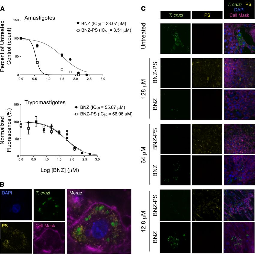
Effective killing of human life cycle stages of T. cruzi by BNZ-PSs. The trypanocidal effectiveness of BNZ-
PSs was evaluated in vitro against 2 human life cycle stages, the amastigote form, which replicates in the
host cell cytoplasm, and the trypomastigote form, which arises from the amastigote through differentia-
tion and is liberated from the host cell as it lyses to infect adjacent cells or distant cells after hematogenous
spread. The half-inhibitory concentrations (IC50) of free BNZ and BNZ-PSs against amastigotes were
33.07 ± 8.17 μM and 3.51 ± 0.79 μM, respectively, and against trypomastigotes were 55.87 ± 11.39 μM
and 56.06 ± 12.21 μM, respectively (Figure 3A). Therefore, although the 2 drug preparations were simi-
larly effective against free trypomastigotes, BNZ-PSs were effective at one-tenth the concentration of free
BNZ against amastigotes. The spatial relationships among the host cell, intracellular T. cruzi amastigotes,
JCI Insight 2021;6(9):e145523 https://doi.org/10.1172/jci.insight.145523 2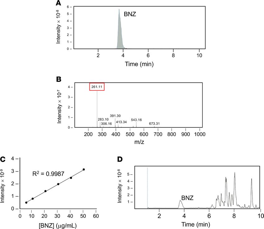
RESEARCH ARTICLE

Figure 1. BNZ-PSs are 100 nm particles with good in vitro stability. (A) BNZ was loaded into PSs by the thin-film hydration method. (B) Representative cryo–
transmission electron microscopy images of PS and BNZ-PS. Scale bar: 100 nm. (C) The diameters of PS and BNZ-PS are approximately 100 nm as determined
by dynamic light scattering analysis. (D) The stability of BNZ-PS was determined by incubating the particles in PBS at 4°C and quantifying the BNZ in BNZ-PS
over time by LC-MS. Results expressed as mean ± SEM for 3 independent experiments. LC-MS, liquid chromatography–coupled mass spectrometry.

                                   and Alexa Fluor 630–loaded PSs are easily seen (Figure 3B), indicating a successful uptake of PSs by host
                                   cells. The effects of including different concentrations of BNZ in the PSs on the levels of T. cruzi are also
                                   clearly visualized (Figure 3C). As the concentration of BNZ/BNZ-PSs was decreased, a clear increase in
                                   T. cruzi was observed in the BNZ-treated cells but not in the BNZ-PS–treated cells. These results clearly
                                   demonstrate the superiority of BNZ-PSs to free BNZ at each drug concentration.
                                        BNZ-PSs are superior to free BNZ in treating T. cruzi–infected mice. We infected BALB/c mice with the
                                   Y strain of T. cruzi, which leads to acute myocarditis and eventual development of chronic cardiomy-
                                   opathy. We began drug administration at 7 days postinfection (d.p.i.) when parasitemia approached 2 ×
                                   105 trypomastigotes per milliliter. Mice were administered no drug, empty PSs, free BNZ at 100 mg/kg,
                                   or 3 concentrations of BNZ-PS (Figure 4A). Parasitemia dropped in all groups by 11 d.p.i., as expected
                                   in this model of T. cruzi infection, then rebounded by 11 d.p.i. in the untreated and PS groups. Parasite-
                                   mias in the treatment groups generally paralleled the BNZ concentrations employed. The development
                                   of adaptive immunity caused a drop in parasitemia in all groups by 15 d.p.i., as expected, but by 18
                                   d.p.i. significant differences were observed among the groups, with the BNZ 100 mg/kg and PS 1.5
                                   and PS 0.15 mg/kg groups all having substantially lower parasitemia than the untreated and empty PS
                                   controls. The group receiving the extremely low dose of 0.03 mg/kg BNZ-PSs showed a 50% reduction
                                   in parasitemia, but the difference from untreated controls did not reach statistical significance. Interest-
                                   ingly, the only treatment that significantly reduced cardiac parasitosis, the number of parasites in the
                                   heart, was BNZ-PSs at 1.5 mg/kg (Figure 4B). Even BNZ at 100 mg/kg did not reduce parasitosis any
                                   better than the other BNZ-PS formulations. The medium-dose BNZ-PS (0.15 mg/kg) also significantly
                                   reduced myocarditis (Figure 4C). Representative cardiac histology is shown (Figure 4E), with hearts
                                   from untreated, PS-treated, BNZ 100 mg/kg–treated, and BNZ-PS 0.03 mg/kg–treated mice showing
                                   focal to diffuse interstitial inflammation and hearts from BNZ-PS 1.5 and 0.15 mg/kg–treated mice dis-
                                   playing normal cardiac histology (Figure 4D).
                                        BNZ-PS preparations are not toxic to mice. To test whether effective doses of BNZ-PSs would also
                                   have the benefit of reduced toxicity, groups of mice were given no treatment or were administered
                                   PS or doses of BNZ or BNZ-PSs previously determined as effective. Mice treated with BNZ experi-
                                   enced significant weight loss, whereas all other mice gained weight during the 21-day course of the

JCI Insight 2021;6(9):e145523 https://doi.org/10.1172/jci.insight.145523                                                                                   3
RESEARCH ARTICLE
Table 1. Characterization of PSs and BNZ-loaded PSs in PBS pH 7.4
Name of sample Average diameter Polydispersity index Zeta potential Encapsulation efficiency Loading efficiency
(nm) (mV) (%) (% w/w)
PSs 120 ± 6.2 0.06 ± 0.01 –1.97 ± 0.46 N/A N/A
BNZ-loaded PSs (BNZ- 114.3 ± 4.1 0.11 ± 0.02 4.92 ± 1.93 ~31 ~1
PSs)
experiment (Figure 5A). BNZ caused a doubling of the serum alanine aminotransferase concentration
relative to all other mice (Figure 5B).
Discussion
A large variety of nanocarriers have been developed for drug delivery that differ in their physicochemical
properties, including surface chemistry, shape, size, charge, stiffness, and stability (31, 32). Each of these
properties, as well as the molecular payload incorporated, affects biodistribution, cell membrane interac-
tions, mechanisms of cell uptake, and blood protein interactions (30, 33). Predicting how specific nanocar-
riers influence drug efficacy remains an active area of research, with thousands of possible combinations of
physicochemical properties that can be customized for a variety of applications (34). In this regard, PEG-b-
PPS nanocarriers display high loading efficiency for a wide array of small molecules, increase the effective
water solubility of these agents, and enhance intracellular delivery (30, 35–37). They are noninflammatory
and nontoxic in mice (38, 39) and nonhuman primates (40). Vesicular nanocarriers like PS display a supe-
rior capacity to target antigen-presenting cells, such as DCs and macrophages.
In this study, PEG-b-PPS was engineered to assemble vesicles loaded with BNZ, a hydrophobic drug, as
the lipophilic PPS block ensures stable BNZ incorporation into the vesicle membrane. The combination of
PPS hydrophobicity and PEG hydrophilicity creates a highly stable macroamphiphile copolymer assembly
(41). BNZ-PSs are similar in structure and stability to prior PEG-b-PPS PS formulations (28–30). We found
that incorporation of BNZ into PEG-b-PPS PSs reduced by 9-fold the IC50 for intracellular T. cruzi amastig-
otes, the predominant life cycle stage of the parasite during human infection, while having the same IC50 as
free-form BNZ for trypomastigotes. The high trypanocidal efficiency of BNZ-PSs for amastigotes is most
likely due to the fact that the host cell effectively concentrates BNZ through nanoparticle uptake. Cellular
uptake of nanocarriers smaller than 500 nm, such as PEG-b-PPS PSs, occurs primarily by endocytosis (42),
with cell entry via both macropinocytosis and receptor-mediated endocytosis (30, 37, 39). Once inside the
cell, the nanocarrier becomes unstable in the cellular microenvironment (43) with oxidative enzymes inside
the PS-containing endolysosomal vacuoles promoting the oxidation of sulfide moieties in the PPS block that
modify the hydrophilic-lipophilic balance of the PEG-b-PPS copolymer (37). This promotes disassembly of
the nanostructure and consequent drug release during the initial stages of oxidation. The vesicle disassembly
also liberates amphiphilic copolymers that insert into the endosomal membrane, promoting membrane per-
meabilization and escape of PEG-b-PPS vesicle drug payloads into the cell cytoplasm (36). During cell inva-
sion, T. cruzi trypomastigotes first enter a parasitophorous vacuole and then escape from the vacuole into the
cytoplasm after vacuolar acidification by lysosomal fusion. Based on prior work with PEG-b-PPS nanocarri-
ers, the enhanced therapeutic effect of BNZ-PS is likely due to protection of BNZ from the acidic pH of endo-
lysosomal vacuoles and the ability of PEG-b-PPS nanocarriers to improve cytoplasmic delivery via nontoxic
disruption of endosomal lipid bilayer membranes (37, 44). Thus, the cell invasion and uptake pathways of T.
cruzi and PEG-b-PPS are very similar, which may also increase the efficacy of treatment. Two other features
of PEG-b-PPS are important. First, the copolymer is physiologically inert, releasing drug only after cellular
internalization, which may also contribute to the reduced toxicity (45–47). Second, PEG-b-PPS enhances the
solubility of highly hydrophobic agents like BNZ via entrapment within a lipophilic PPS membrane, allowing
more facile and stable transport in the blood, and enhances cellular uptake (36, 38, 39).
The most important finding from our study is that BNZ-PS was extremely potent in treating T.
cruzi–infected mice, with no detectable hepatotoxicity. The total amount of BNZ delivered by 2 injec-
tions of BNZ-PS is 466-fold lower than that delivered by daily dosing of free BNZ, leading to similar
decreases in parasitemia with notable, if not statistically significant, decreases at even a 23,000-fold
JCI Insight 2021;6(9):e145523 https://doi.org/10.1172/jci.insight.145523 4RESEARCH ARTICLE
Figure 2. Analysis of BNZ loading within BNZ-PSs. (A) Representative chromatogram and (B) positive ion mass spec-
trum of BNZ standards. The BNZ peak is indicated at m/z of approximately 261. (C) Calibration curve of BNZ standards
using ordinary least square. (D) Representative chromatogram of BNZ-PS displaying a BNZ peak at the same elution
time as BNZ standards in A.
lower dose (the cumulative amount over the entire experiment). Although parasitemia may be a useful
measure of infection and drug efficacy, the major serious sequela of infection is myocarditis/cardio-
myopathy. We assessed the efficacy of the various drug preparations to prevent cardiomyopathy in 2
ways: cardiac parasitosis and cardiac inflammation, with both metrics frequently tracking together.
Importantly, BNZ-PSs at 1.5 mg/kg significantly reduced myocarditis, whereas free-form BNZ at 100
mg/kg/d did not. Even at one-tenth the dose (0.15 mg/kg), BNZ-PS reduced cardiac inflammation,
although it did not significantly reduce cardiac parasitemia. This finding suggests that BNZ-PSs had
the additional benefit of ameliorating cardiac inflammation, even in the presence of a substantial par-
asite tissue burden. The potential immunomodulatory effect of BNZ-PSs needs further exploration.
Although our study was of short duration, possibly representative of acute human infection, the anti-
inflammatory effect observed here suggests that BNZ-PSs might be effective in chronic Chagas disease,
particularly because the same mechanisms of inflammation, immunity, and intracellular parasite killing
by drugs are similar in chronic infection.
BNZ is widely used for the treatment of T. cruzi infection. However, therapeutic efficacy requires the
administration of high daily doses of the drug, which is frequently accompanied by toxicity and additional
adverse side effects (48, 49). The low BNZ serum half-life in mice (t1/2 = 2 h) results from the mouse’s high
metabolic rate, including high first-pass metabolism after oral administration with resultant low bioavail-
ability. This is why high BNZ doses of 100 mg/kg/d are required (8). In humans, BNZ displays a longer
serum half-life (t1/2 = 12 hours) (19) and a lower metabolic rate. Therefore, a lower effective dose is needed
but daily doses are still necessary (43). Although daily administration of BNZ is important to achieve the
trypanocidal effect, it also increases the concentrations of toxic BNZ metabolites, the major drawback of
current BNZ therapy (19). Because BNZ is practically insoluble in water (50), its slow release from the PSs
to the aqueous environment of the cytoplasm was likely responsible for the successful antiparasitic effect
JCI Insight 2021;6(9):e145523 https://doi.org/10.1172/jci.insight.145523 5RESEARCH ARTICLE
Figure 3. BNZ-PSs are more potent than free BNZ against T. cruzi in vitro. (A) In vitro killing of purified T. cruzi Y strain trypomastigotes by BNZ and
BNZ-PS. Trypomastigotes were purified from infected H9C2 cell cultures and tested in a 24-hour resazurin cell viability assay using increasing doses of
BNZ or BNZ-PS. Results from 3 independent experiments are expressed as mean ± SEM. (B) PS are readily taken up by T. cruzi–infected H9C2 cells. H9C2
cells were infected with T. cruzi Tulahuen strain trypomastigotes expressing Luc-mNeonGreen (green) for 24 hours and Alexa Fluor 630–labeled PS (yellow)
were added and cultures incubated for an additional 24 hours. Cells were imaged after staining with DAPI (blue) and Cell Mask Deep Red Dye (purple)
(original magnification, ×1000). (C) BNZ-PSs are significantly more potent against intracellular T. cruzi than free BNZ. Cells were cultured and treated as in
B, but with different concentrations of BNZ or BNZ-PS, and imaged after DAPI and Cell Mask staining. The key images for comparison are the left (T. cruzi)
BNZ and BNZ-PS panels at each drug concentration (original magnification, ×400).
of only 2 BNZ-PS administrations during the 14 days of treatment. An equally important finding from our
study is that the low effective dose of BNZ achieved through the BNZ-PS formulation completely abrogat-
ed the weight loss and hepatotoxicity observed with the effective dose of free-form BNZ.
In conclusion, we have developed an advanced formulation of BNZ based on incorporation into a
PEG-b-PPS nanocarrier that increased the potency and decreased the toxicity of this effective trypanocidal
drug. The likely mechanisms were enhanced cytoplasmic delivery via well-established uptake mechanisms
and reduced exposure of BNZ and its metabolites due to molecular sequestration within PS nanocarriers.
The most clinically relevant consequence of BNZ-PS was the reduction of cardiac parasitosis and inflam-
mation, which strongly supports further development of PEG-b-PPS nanocarriers for Chagas drugs and
drug combinations. Ultimately, a sustained-release BNZ formulation may further advance therapy. PEG-
b-PPS nanocarriers have been employed for sustained intracellular drug release for up to 12 days through
the modification of the PPS chain to achieve slow release by partitioning the drug from the hydrophobic
JCI Insight 2021;6(9):e145523 https://doi.org/10.1172/jci.insight.145523 6RESEARCH ARTICLE
Figure 4. BNZ-PSs are more potent than free BNZ against T. cruzi in vivo. (A) Mice (n = 5 per group) were infect-
ed with T. cruzi Y strain trypomastigotes on day 0 and treated with 2 i.v. doses of BNZ-PS and PS after parasite-
mia had reached approximately 2 × 10⁵/mL on day 7. Standard oral treatment with BNZ was given daily for 14
days (7–21 d.p.i.). Parasitemia was monitored every few days through the end of the experiment on day 25, when
mice were sacrificed and organs were collected for further analysis. (B) Effective suppression of parasitemia by
BNZ and BNZ-PS. *P ≤ 0.05 for BNZ 100 mg/kg and BNZ-PS 1.5 and 0.15 mg/kg vs. untreated and PS. (C) Cardiac
parasitosis was quantitated by quantitative PCR. *P ≤ 0.01 vs. untreated and PS. (D) Cardiac inflammation was
quantitated in heart sections 2 ways — by total cellularity (top) and by the percentage of cellular area occupied by
nuclei (bottom). *P ≤ 0.05 vs. untreated, PS, and BNZ. (E) Representative cardiac histology from the experiment
in C. Scale bar: 100 μM. All error bars reflect mean ± SEM. One-way ANOVA with Tukey’s post hoc test was used
for multiple comparisons.
JCI Insight 2021;6(9):e145523 https://doi.org/10.1172/jci.insight.145523 7RESEARCH ARTICLE
Figure 5. BNZ-PSs are significantly less toxic than free BNZ. Healthy mice (n = 5 per group) were treated with BNZ (100
mg/kg) or BNZ-PS (1.5 or 0.15 mg/kg), plus controls. (A) Mice treated with BNZ, but not BNZ-PS, lost weight during
2 weeks of treatment. Weights were determined every few days. *P ≤ 0.05 vs. untreated. (B) Mice treated with BNZ
show hepatotoxicity as reflected by increased serum ALT on day 21. Serum ALT was also measured and did not show
significant elevation in any mouse. *P ≤ 0.0001. All error bars reflect mean ± SEM. One-way ANOVA with Tukey’s post
hoc test was used for multiple comparisons. ALT, alanine aminotransferase.
PPS phase into the aqueous phase (35, 51, 52) and PS can also be delivered by routes other than i.v. (53,
54). Furthermore, PEG-b-PPS nanostructures can be surface-engineered for cell selective receptor-mediat-
ed uptake (29, 30, 33). These approaches could also be of use in treating other intracellular parasitic, viral,
bacterial, and fungal infections, which are often treated with highly toxic drugs.
Methods
Synthesis of PEG-b-PPS copolymers and loading of BNZ into PSs. PSs were prepared by the controlled self-as-
sembly of PEG-b-PPS block copolymers with the 25%–45% molecular weight of hydrophilic PEG fraction
in the total block copolymer. PEG-b-PPS block copolymers were synthesized as previously described (30).
Briefly, the anionic ring-opening polymerization of propylene sulfide was initiated by PEG thioacetate and
end-capped with PEG mesylate. The obtained block copolymers (PEG17-PPS60-PEG17) were purified by
precipitation in methanol and then characterized by NMR spectroscopy and gel permeation chromatogra-
phy (Thermo Fisher Scientific). The loading of BNZ (MilliporeSigma) into PS to make BNZ-PS was per-
formed by the thin-film rehydration method in PBS as described previously (30, 39). Briefly, 30 mg of the
copolymer (PEG17-PPS60-PEG17) with or without 1.5 mg of BNZ was dissolved in 150 μL tetrahydrofuran
within 1.8 mL clear glass vials (Thermo Fisher Scientific) and placed under vacuum to remove the solvent.
The resulting thin films were dehydrated in sterile PBS (1 mL) under shaking at 1500 rpm for 48 hours.
The BNZ-PSs were purified to remove free BNZ by Zeba Spin Desalting Columns (7K MWCO, Thermo
Fisher Scientific).
Characterization of PSs. LC-MS was performed using a Bruker AmaZon-X instrument. Samples were
chromatographed on a Hypersil BDS C18 column (2.4 mM particle size, 2.1 × 50 mM) (Thermo Fisher
Scientific) at 40°C. The separation was achieved by a gradient of water with 0.1% formic acid (eluent A)
and acetonitrile with 0.1% formic acid (eluent B) with a flow rate of 0.3 mL/min. Detection was performed
at 324 nm and the injection volume was 2 μL. The gradient was started at 40% B for 1 minute, increased
to 100% B over 4 minutes, held at 100% B for 5 minutes, decreased to 90% B over 0.1 minute, and held at
90% B for 1.9 minutes. The standard calibration solution of BNZ was prepared in acetonitrile/water (95:5
v/v), ranging from 3.125 to 100 mg/mL. BNZ-PS samples were dissolved in acetonitrile/water (95:5 v/v)
and then filtered through a 0.2 mM membrane (Thermo Fisher Scientific). The loading efficiency of the
BNZ-PS was determined by the percentage of the loaded weight of BNZ of the total weight of BNZ-PS.
The encapsulation efficiency of the BNZ-PS was calculated by the percentage of BNZ weight loaded into
the PS of the initial BNZ weight used. The size distribution and zeta potential of PS and BNZ-PS (1 mg/
mL) were characterized by Zetasizer Nano-ZS using a 4 mW He-Ne 633 laser (Malvern Instruments). The
morphology of PS and BNZ-PS was determined by cryo–transmission electron microscopy as described
previously (55). In brief, 200 mesh Cu grids with a lacey carbon membrane (catalog LC200-CU-100, EMS)
JCI Insight 2021;6(9):e145523 https://doi.org/10.1172/jci.insight.145523 8RESEARCH ARTICLE
were glow discharged in a Pelco easiGlow glow discharger (Ted Pella Inc.) using an atmosphere plasma
generated at 15 mA for 15 seconds with a pressure of 0.24 mbar. PS and BNZ-PS samples (4 μL, 10 mg/
mL in PBS) were pipetted onto the grid and blotted for 5 seconds with a blot offset of +0.5 mM, followed
by immediate plunging into liquid ethane within an FEI Vitrobot Mark III plunge-freezing instrument
(Thermo Fisher Scientific). The plunge-frozen grids were kept vitreous at –180°C in a Gatan Cryo Trans-
fer Holder model 626.6 (Gatan Inc.) and viewed in a JEOL JEM1230 LaB6 emission transmission elec-
tron microscope (JEOL USA, Inc.) at 100 kiloelectron volts. Image data were collected by a Gatan Orius
SC1000 CCD camera model 831 (Gatan Inc.). The images were processed and analyzed using NIH ImageJ
software. To test the stability of BNZ-PSs upon storage, a suspension of BNZ-PSs (30 mg/mL in PBS) was
kept in sealed tubes and maintained at 4°C. At different time points (0, 1, 4, 8, 15, and 45 days), the released
or unloaded BNZ was removed by using Zeba Spin Desalting Columns (7K MWCO, Thermo Fisher Scien-
tific). The percentage of loaded BNZ in PS at different time points (compared with day 0) was determined
by LC-MS by methods mentioned above.
Cells, cell culture, and parasite purification. The Y strain of T. cruzi was used for all experiments oth-
er than the nanocarrier uptake/cell imaging experiment, which employed Tulahuen strain expressing the
dual reporter mNeonGreen fused to red-shifted luciferase (Luc-mNeonGreen) (56). Epimastigotes were
cultured using standard methods. H9C2 rat myoblasts (ATCC) were inoculated with small numbers of
cultured log-phase epimastigotes, which contain small numbers of metacyclic trypomastigotes capable of
initiating mammalian cell infection. After several days of culture in H9C2 cells, amastigotes and trypomas-
tigotes are produced, and trypomastigotes can be purified from culture supernatants using standard meth-
ods. Amastigotes can be easily studied in situ as intracellular parasites (e.g., IC50 determination) or purified
from host cells for additional studies.
Intracellular amastigotes. H9C2 rat myoblast cells, 50,000 per well, were seeded in Chamber Slides (Nunc
Lab-Tek II, Thermo Fisher Scientific) and incubated in RPMI medium (RPMI 1640, 5% FBS, 100 IU/mL
penicillin, 100 μg/mL streptomycin, and 2 mM l-glutamine) at 37°C in 5% CO2. After 24 hours, cells were
infected by the addition of 250,000 T. cruzi Y strain trypomastigotes per well (5:1 MOI), incubated for 24
hours, and washed to remove trypomastigotes in the medium. Different concentrations of BNZ and BNZ-
PS in RPMI were added to each well. The medium (with drug) was replaced after 24 hours, and the slides
were fixed and stained with Giemsa after a further 24-hour incubation. The number of amastigotes per 100
H9C2 cells was calculated for each chamber. The Resazurin method was used to evaluate the activity of
BNZ and BNZ-PS against trypomastigotes. Purified trypomastigotes (1 × 10⁷ cells/mL) were incubated in
96-well plates containing serial dilutions of BNZ or BNZ-PS in RPMI medium. After incubation for 24
hours, 10 μL of alamarBlue cell viability reagent (Thermo Fisher Scientific) was added to each well, and
plates were incubated for an additional 4 hours. Fluorescence was measured on a microplate reader (BMG
Labtech) at 560/590 nm to evaluate trypomastigote viability. A log concentration versus response curve
was generated, and the BNZ IC50 was calculated using GraphPad Prism 7.0.
Nanocarrier uptake. H9C2 cells were seeded in Chamber Slides for 24 hours and then infected with
Luc-mNeonGreen–expressing T. cruzi trypomastigotes at a 5:1 MOI for 24 hours. Free trypomastigotes
were washed away and the T. cruzi–infected H9C2 cells were incubated with Alexa Fluor 630–labeled PS
for 24 hours. The slides were washed 5 times with PBS and stained with CellMask Deep Red Plasma Mem-
brane Stain (Invitrogen, Thermo Fisher Scientific) for 5 minutes and DAPI for 20 minutes. Leica confocal
microscopy was used to image the slides.
Mouse infections. Female BALB/c mice (4–6 weeks of age) were purchased from The Jackson Labo-
ratory and housed in pathogen-free conditions on a 12-hour dark/12-hour light cycle at 22 ± 3°C with
access to food and water ad libitum. H9C2-derived T. cruzi Y strain tissue culture trypomastigotes to be
used for infections of BALB/c mice were first passaged through female SCID mice (The Jackson Labo-
ratory) bloodstream trypomastigotes. SCID mice were infected by i.p. injection of 2 × 10³ tissue culture
trypomastigotes in 0.2 mL PBS and peripheral blood parasitemia was monitored by analysis of saphenous
venous blood (3 μL). When SCID mice parasitemia reached 1 × 10⁸ trypomastigotes/mL, infected blood
from SCID mice was harvested and adjusted to 2 × 10⁴ bloodstream trypomastigotes/mL by the addition
of PBS. BALB/c mice were then infected with 2 × 10³ bloodstream trypomastigotes by i.p. injection. Par-
asitemia of Y strain–infected mice was determined and animals were euthanized after the final time point.
BNZ and BNZ-PS therapy. BNZ-PS was prepared as described above, and BNZ (MilliporeSigma) was
prepared from powder at 22 mg/mL in 5% methylcellulose. After confirming establishment of infection by
JCI Insight 2021;6(9):e145523 https://doi.org/10.1172/jci.insight.145523 9RESEARCH ARTICLE
peripheral blood analysis 7 d.p.i., 30 mice were randomized into 6 groups of 5 animals each, as follows:
(a) untreated control mice, (b) mice administered 0.3 mg/mL PS by i.v. injection, (c) mice treated with 100
mg/kg BNZ daily orally (po) for 14 days (7–21 d.p.i.), (d) mice treated with BNZ-PS i.v. at a BNZ dose of
1.5 mg/kg 7 and 14 d.p.i. (2 doses), (e) mice treated with BNZ-PS i.v. at a BNZ dose of 0.15 mg/kg 7 and
14 d.p.i. (2 doses), and (f) mice treated with BNZ-PS at a BNZ dose of 0.03 mg/kg 7 and 14 d.p.i. (2 doses).
All animals were euthanized 25 d.p.i. and blood and organs were procured for further analysis.
Tissue parasitosis. Organs were harvested and snap-frozen on dry ice. DNA was extracted using QIAamp
DNA Mini Kit (QIAGEN) following the manufacturer’s instructions. To make the standard for parasite bur-
den quantification, 25 mg of heart of noninfected mouse was mixed with 1 × 10⁷ T. cruzi trypomastigotes.
Total DNA was extracted, and the DNA concentration was adjusted to 50 ng/μL. The standard curve was
established from serial dilutions of the sample, ranging from 1 × 10⁷ to 1 × 10⁻¹ parasite equivalents. Real-
time PCR reactions were carried out using a QuantStudio 5 instrument (Thermo Fisher Scientific). PCR
reactions contained 50 ng of DNA, 0.5 μL of primers TCZ-F 5′-GCTCTTGCCCACAMGGGTGC-3′ and
TCZ-R 5′-CCAAGCAGCGGATAGTTCAGG-3′, and 10 L of EXPRESS SYBR GreenER qPCR Super-
mix (Invitrogen, Thermo Fisher Scientific) in a final volume of 20 L. Reactions were run in triplicate using
the following cycling parameters: 50°C for 2 minutes followed by 40 cycles of 95°C for 10 seconds, 55°C for
15 seconds, 72°C for 5 seconds. Mouse-specific GADPH forward and reverse were used as internal controls.
Histopathology and image analysis. Hearts were obtained from all mice, fixed in 10% buffered formalin
for 15 hours, and embedded in paraffin. Five-micrometer sections were stained with H&E and Masson’s tri-
chrome stain. All slides were scanned using an Aperio Scanscope AT slide scanner, and images were taken
with ImageScope software and analyzed using NIH ImageJ software by a scientist blinded to the groups.
The inflammatory index was derived by quantifying the total number of nuclei present in 10 randomly
selected microscopic fields of each H&E-stained section. Tissue cellularity and percentage of cellular area
occupied by nuclei were determined as separate but related histologic indicators of inflammation.
Assessment of the toxicity of BNZ and BNZ-PS in vivo. Twenty-five healthy BALB/c mice were randomized
into 5 groups of 5 animals each, as follows: (a) untreated control mice, (b) mice administered 0.3 mg/mL PS
i.v., (c) mice treated with 100 mg/kg BNZ daily po for 14 days (days 7–21), (d) mice treated with BNZ-PS
i.v. at a BNZ dose of 1.5 mg/kg on days 7 and 14 (2 doses), and (e) mice treated with BNZ-PS i.v. at a BNZ
dose of 0.15 mg/kg on days 7 and 14 (2 doses). Mouse weights were recorded during the course of the exper-
iment. On day 15, mice were euthanized and serum and liver tissue were collected from each animal. Serum
alanine aminotransferase (ALT/SGPT), was determined at a reference laboratory (IDEXX BioResearch).
Statistics. Numerical results are expressed as mean ± SEM. Individual animals were used as the unit
of analysis for in vivo and ex vivo experiments. Animal group size was determined empirically. One-way
ANOVA and Tukey’s multiple comparisons test were used in GraphPad Prism v.7 to evaluate differences
between groups. P values of less than 0.05 were considered statistically significant.
Study approval. All animal protocols were reviewed and approved by the Institutional Animal Care and
Use Committee of Cedars-Sinai Medical Center (protocol 7053).
Author contributions
XL, SY, EAS, and DME conceived and designed the research. XL, SY, SJM, BAF, and CLO carried out
all the experiments and analyzed the data. XL, SY, DBS, PSR, EAS, and DME wrote the manuscript with
feedback from all the authors. XL is listed before co–first author SY because she did more than half of the
work on this project.
Acknowledgments
We thank Conrad Epting and Robert Brown for helpful discussions. This work was supported by NIH
grants R21-AI144529 (to DME and EAS) and DP2-HL132390 (to EAS) and by a Catalyzer Award from
the Northwestern University Institute for Global Health.
Address correspondence to: Evan A. Scott, Northwestern University, 2145 Sheridan Road, Evanston, Illi-
nois, 60208, USA. Phone: 847.467.6719; Email: evan.scott@northwestern.edu. Or to: David M. Engman,
University of California, Los Angeles, 609 Charles E. Young Drive E, Los Angeles, California, 90024,
USA. Phone: 312.953.0709; Email: dengman@mednet.ucla.edu.
JCI Insight 2021;6(9):e145523 https://doi.org/10.1172/jci.insight.145523 10RESEARCH ARTICLE
1. Chagas C. Nova tripanozomiaze humana: estudos sobre a morfolojia e o ciclo evolutivo do Schizotrypanum cruzi n. gen., n.
sp., ajente etiolojico de nova entidade morbida do homem. Mem Inst Oswaldo Cruz. 1909;1(2):159–218.
2. Bonney KM, et al. Pathology and pathogenesis of Chagas heart disease. Annu Rev Pathol. 2019;14:421–447.
3. WHO. Chagas disease (American trypanosomiasis). https://www.who.int/health-topics/chagas-disease#tab=tab_1. Accessed
April 5, 2021.
4. Montgomery SP, et al. What do we know about Chagas disease in the United States? Am J Trop Med Hyg. 2016;95(6):1225–1227.
5. Lynn M, et al. Contemporary autochthonous human Chagas disease in the USA. Acta Trop. 2020;205:105361.
6. Whitman JD, et al. Chagas disease serological test performance in U.S. blood donor specimens. J Clin Microbiol.
2019;57(12):e01217–19.
7. Bern C. Antitrypanosomal therapy for chronic Chagas’ disease. N Engl J Med. 2011;364(26):2527–2534.
8. Perin L, et al. Pharmacokinetics and tissue distribution of benznidazole after oral administration in mice. Antimicrob Agents
Chemother. 2017;61(4):e02410–16.
9. Andrade DV, et al. Acute Chagas disease: new global challenges for an old neglected disease. PLoS Negl Trop Dis.
2014;8(7):e3010.
10. Morillo CA, et al. Randomized trial of benznidazole for chronic Chagas’ cardiomyopathy. N Engl J Med. 2015;373(14):1295–1306.
11. Hasslocher-Moreno AM, et al. Benznidazole decreases the risk of chronic Chagas disease progression and cardiovascular
events: a long-term follow up study. EClinicalMedicine. 2021;31:100694.
12. Nunes MCP, et al. Chagas cardiomyopathy: an update of current clinical knowledge and management: a scientific statement
from the American Heart Association. Circulation. 2018;138(12):e169–e209.
13. Arrua EC, et al. Nanocarriers for effective delivery of benznidazole and nifurtimox in the treatment of Chagas disease: a review.
Acta Trop. 2019;198:105080.
14. Clayton J. Chagas disease 101. Nature. 2010;465(7301):S4–S5.
15. Kreuter J. Nanoparticles--a historical perspective. Int J Pharm. 2007;331(1):1–10.
16. Ventola CL. Progress in nanomedicine: approved and investigational nanodrugs. P T. 2017;42(12):742–755.
17. Barenholz Y. Doxil(R)--the first FDA-approved nano-drug: lessons learned. J Control Release. 2012;160(2):117–134.
18. van der Meel R, et al. Smart cancer nanomedicine. Nat Nanotechnol. 2019;14(11):1007–1017.
19. Morilla MJ, Romero EL. Nanomedicines against Chagas disease: an update on therapeutics, prophylaxis and diagnosis. Nano-
medicine (Lond). 2015;10(3):465–481.
20. Quijia Quezada C, et al. Advances in nanocarriers as drug delivery systems in Chagas disease. Int J Nanomedicine.
2019;14:6407–6424.
21. Rial MS, et al. Elucidating the impact of low doses of nano-formulated benznidazole in acute experimental Chagas disease.
PLoS Negl Trop Dis. 2017;11(12):e0006119.
22. Nhavene EPF, et al. Chitosan grafted into mesoporous silica nanoparticles as benznidazol carrier for Chagas diseases treatment.
Microporous Mesoporous Mater. 2018;272:265–275.
23. Lyra MAM, et al. Study of benznidazole–cyclodextrin inclusion complexes, cytotoxicity and trypanocidal activity. J Incl Phenom
Macrocycl Chem. 2012;73(1–4):397–404.
24. Leonardi D, et al. Effects of benznidazole:cyclodextrin complexes on the drug bioavailability upon oral administration to rats.
Int J Biol Macromol. 2013;62:543–548.
25. Vinuesa T, et al. Benznidazole nanoformulates: a chance to improve therapeutics for Chagas disease. Am J Trop Med Hyg.
2017;97(5):1469–1476.
26. Morilla MJ, et al. Intravenous liposomal benznidazole as trypanocidal agent: increasing drug delivery to liver is not enough. Int
J Pharm. 2004;278(2):311–318.
27. Mazzeti AL, et al. Benznidazole self-emulsifying delivery system: A novel alternative dosage form for Chagas disease treatment.
Eur J Pharm Sci. 2020;145:105234.
28. Karabin NB, et al. Sustained micellar delivery via inducible transitions in nanostructure morphology. Nat Commun.
2018;9(1):624.
29. Stack T, et al. Targeted delivery of cell softening micelles to Schlemm’s canal endothelial cells for treatment of glaucoma. Small.
2020;16(43):e2004205.
30. Yi S, et al. Surface engineered polymersomes for enhanced modulation of dendritic cells during cardiovascular immunotherapy.
Adv Funct Mater. 2019;29(42):1904399.
31. Frey M, et al. Influences of nanocarrier morphology on therapeutic immunomodulation. Nanomedicine (Lond).
2018;13(14):1795–1811.
32. Scott EA, et al. Overcoming immune dysregulation with immunoengineered nanobiomaterials. Annu Rev Biomed Eng.
2017;19:57–84.
33. Vincent MP, et al. Surface chemistry-mediated modulation of adsorbed albumin folding state specifies nanocarrier clearance by
distinct macrophage subsets. Nat Commun. 2021;12(1):1–18.
34. Le TC, Winkler DA. Discovery and optimization of materials using evolutionary approaches. Chem Rev. 2016;116(10):6107–6132.
35. Allen SD, et al. Celastrol-loaded PEG-b-PPS nanocarriers as an anti-inflammatory treatment for atherosclerosis. Biomater Sci.
2019;7(2):657–668.
36. Scott EA, et al. Dendritic cell activation and T cell priming with adjuvant- and antigen-loaded oxidation-sensitive polymer-
somes. Biomaterials. 2012;33(26):6211–6219.
37. Vasdekis AE, et al. Precision intracellular delivery based on optofluidic polymersome rupture. ACS Nano. 2012;6(9):7850–7857.
38. Dowling DJ, et al. Toll-like receptor 8 agonist nanoparticles mimic immunomodulating effects of the live BCG vaccine and
enhance neonatal innate and adaptive immune responses. J Allergy Clin Immunol. 2017;140(5):1339–1350.
39. Yi S, et al. Tailoring nanostructure morphology for enhanced targeting of dendritic cells in atherosclerosis. ACS Nano.
2016;10(12):11290–11303.
40. Allen SD, et al. Polymersomes scalably fabricated via flash nanoprecipitation are non-toxic in non-human primates and associ-
ate with leukocytes in the spleen and kidney following intravenous administration. Nano Res. 2018;11(10):5689–5703.
JCI Insight 2021;6(9):e145523 https://doi.org/10.1172/jci.insight.145523 11RESEARCH ARTICLE
41. Napoli A, et al. Oxidation-responsive polymeric vesicles. Nat Mater. 2004;3(3):183–189.
42. Zhao J, Stenzel MH. Entry of nanoparticles into cells: the importance of nanoparticle properties. Polym Chem. 2018;9(3):259–272.
43. Romero EL, Morilla MJ. Nanotechnological approaches against Chagas disease. Adv Drug Deliv Rev. 2010;62(4):576–588.
44. Smith SA, et al. The endosomal escape of nanoparticles: toward more efficient cellular delivery. Bioconjug Chem.
2019;30(2):263–272.
45. Cooper DL, et al. Nanoparticles in drug delivery: mechanism of action, formulation and clinical application towards reduction
in drug-associated nephrotoxicity. Expert Opin Drug Deliv. 2014;11(10):1661–1680.
46. Ezrahi S, et al. Basic principles of drug delivery systems — the case of paclitaxel. Adv Colloid Interface Sci. 2019;263:95–130.
47. Ladavière C, Gref R. Toward an optimized treatment of intracellular bacterial infections: input of nanoparticulate drug delivery
systems. Nanomedicine (Lond). 2015;10(19):3033–3055.
48. Castro JA, et al. Toxic side effects of drugs used to treat Chagas’ disease (American trypanosomiasis). Hum Exp Toxicol.
2006;25(8):471–479.
49. Molina I, et al. Randomized trial of posaconazole and benznidazole for chronic Chagas’ disease. N Engl J Med.
2014;370(20):1899–1908.
50. World Health Organization. WHO Drug Information 2018. Vol. 32. https://www.who.int/medicines/publications/druginfor-
mation/issues/DrugInformation2018_Vol32-2/en/. 2018. Accessed April 5, 2021.
51. Velluto D, et al. PEG-b-PPS diblock copolymer aggregates for hydrophobic drug solubilization and release: cyclosporin A as an
example. Mol Pharm. 2008;5(4):632–642.
52. Bobbala S, et al. Employing bicontinuous-to-micellar transitions in nanostructure morphology for on-demand photo-oxidation
responsive cytosolic delivery and off-on cytotoxicity. Nanoscale. 2020;12(9):5332–5340.
53. Karabin NB, et al. Sustained micellar delivery via inducible transitions in nanostructure morphology. Nat Commun. 2018;9(1):1–13.
54. Shang S, et al. Induction of mycobacterium tuberculosis lipid-specific T cell responses by pulmonary delivery of mycolic
acid-loaded polymeric micellar nanocarriers. Front Immunol. 2018;9:2709.
55. Yi S, et al. An injectable hydrogel platform for sustained delivery of anti-inflammatory nanocarriers and induction of regulatory
T cells in atherosclerosis. Front Bioeng Biotechnol. 2020;8:542.
56. Taylor MC, et al. Exploiting genetically modified dual-reporter strains to monitor experimental trypanosoma cruzi infections
and host-parasite interactions. Methods Mol Biol. 2019;1955:147–163.
JCI Insight 2021;6(9):e145523 https://doi.org/10.1172/jci.insight.145523 12You can also read