Cytoplasmic DNA accumulation preferentially triggers cell death of myeloid leukemia cells by interacting with intracellular DNA sensing pathway
←
→
Page content transcription
If your browser does not render page correctly, please read the page content below
Baba et al. Cell Death and Disease (2021)12:322
                                    https://doi.org/10.1038/s41419-021-03587-x                                                                                                             Cell Death & Disease

                                     ARTICLE                                                                                                                                                    Open Access

                                    Cytoplasmic DNA accumulation preferentially
                                    triggers cell death of myeloid leukemia cells by
                                    interacting with intracellular DNA sensing pathway
                                    Tomohisa Baba 1, Takeshi Yoshida2,3, Yamato Tanabe1, Tatsunori Nishimura4, Soji Morishita5, Noriko Gotoh                                                                                4
                                                                                                                                                                                                                             ,
                                    Atsushi Hirao3,6, Rikinari Hanayama2,3 and Naofumi Mukaida 1

                                      Abstract
                                      Accumulating evidence indicates the presence of cytoplasmic DNAs in various types of malignant cells, and its
                                      involvement in anti-cancer drug- or radiotherapy-mediated DNA damage response and replication stress. However,
                                      the pathophysiological roles of cytoplasmic DNAs in leukemias remain largely unknown. We observed that during
                                      hematopoietic stem cell transplantation (HSCT) in mouse myeloid leukemia models, double-stranded (ds)DNAs were
                                      constitutively secreted in the form of extracellular vesicles (EVs) from myeloid leukemia cells and were transferred to
                                      the donor cells to dampen their hematopoietic capabilities. Subsequent analysis of cytoplasmic DNA dynamics in
                                      leukemia cells revealed that autophagy regulated cytoplasmic dsDNA accumulation and subsequent redistribution
                                      into EVs. Moreover, accumulated cytoplasmic dsDNAs activated STING pathway, thereby reducing leukemia cell
                                      viability through reactive oxygen species (ROS) generation. Pharmaceutical inhibition of autophagosome formation
                                      induced cytoplasmic DNA accumulation, eventually triggering cytoplasmic DNA sensing pathways to exert
                  1234567890():,;
1234567890():,;
                  1234567890():,;
1234567890():,;

                                      cytotoxicity, preferentially in leukemia cells. Thus, manipulation of cytoplasmic dsDNA dynamics can be a novel and
                                      potent therapeutic strategy for myeloid leukemias.

                                    Introduction                                                                                        generating LICs and the presence of heteroclonal LICs
                                      Leukemia arises from the neoplastic transformation of                                             hinder the development of a molecular targeted therapy,
                                    hematopoietic stem cells (HSCs) or lineage-committed                                                which can be effective to treat various types of leukemia.
                                    progenitor cells. The transformed cells are called leuke-                                           Therefore, the establishment of a more effective and
                                    mia initiating cells (LICs), which expand in the bone                                               universal therapeutic strategy is urgently required to treat
                                    marrow and eventually spread systemically. Various types                                            leukemia, besides molecular targeting therapy.
                                    of oncogenic driver mutations have been identified in                                                  We have previously examined the cellular and mole-
                                    LICs even in leukemias which are morphologically clas-                                              cular mechanisms underlying the competitive interaction
                                    sified as the same disease entity. Despite the monoclonal                                            between normal hematopoietic cells and leukemia cells. In
                                    origin of LICs in most cases, some leukemia patients                                                the initiation step of chronic myeloid leukemia (CML)
                                    contain heteroclonal LICs simultaneously, thereby pro-                                              development, we demonstrated that CML LIC-derived
                                    moting leukemia progression1. The diversity of oncogenes                                            basophil-like leukemia cells constitutively secrete an
                                                                                                                                        inflammatory chemokine, CCL3, which has a potent
                                                                                                                                        capacity to inhibit normal hematopoiesis, thereby indir-
                                    Correspondence: Tomohisa Baba (sergenti@staff.kanazawa-u.ac.jp)
                                    1
                                     Division of Molecular Bioregulation, Cancer Research Institute, Kanazawa                           ectly promoting leukemia-tropic hematopoiesis in the
                                    University, Kanazawa, Japan
                                    2
                                                                                                                                        limited bone marrow (BM) space in CML-bearing mice2,3.
                                     Department of Immunology, Graduate School of Medical Sciences, Kanazawa
                                                                                                                                        Accordingly, the inhibition of CCL3-mediated signal by
                                    University, Kanazawa, Japan
                                    Full list of author information is available at the end of the article                              the treatment with an allosteric inhibitor of CCL3
                                    Edited by H.-U. Simon

                                    © The Author(s) 2021
                                                        Open Access This article is licensed under a Creative Commons Attribution 4.0 International License, which permits use, sharing, adaptation, distribution and reproduction
                                                        in any medium or format, as long as you give appropriate credit to the original author(s) and the source, provide a link to the Creative Commons license, and indicate if
                                    changes were made. The images or other third party material in this article are included in the article’s Creative Commons license, unless indicated otherwise in a credit line to the material. If
                                    material is not included in the article’s Creative Commons license and your intended use is not permitted by statutory regulation or exceeds the permitted use, you will need to obtain
                                    permission directly from the copyright holder. To view a copy of this license, visit http://creativecommons.org/licenses/by/4.0/.

                                    Official journal of the Cell Death Differentiation Association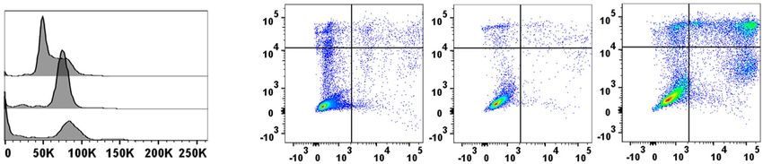
Baba et al. Cell Death and Disease (2021)12:322                                                               Page 2 of 14

receptor, maraviroc, efficiently prevented the develop-          TONBO Biosciences), anti-CD45R/B220 (RA3-6B2; 35-
ment of CML but failed to eradicate CML LICs3.                  0452, TONBO Biosciences), anti-CD81 (Rat-2; 104901,
  Hematopoietic stem cell transplantation (HSCT) is             Biolegend), anti-CD117/c-kit (ACK2; 20-1172, TONBO
widely used as a curative therapy for various hematologic       Biosciences), anti-GAPDH (3H12; M171-3, MEDICAL &
malignancies due to its ability to eradicate LICs. To           BIOLOGICAL LABORATORIES, Nagoya, Japan), anti-
examine the intercellular interaction between residual          γH2A.X (2F3; 613408, Biolegend), anti-IRF7 (MNGPKL;
LICs and donor-derived normal hematopoietic cells after         12-5829-82, eBioscience, San Diego, CA, USA), anti-Ly-
HSCT, we conducted BM transplantation (BMT) in CML              6A/E/Sca-1 (D7; 12-5981, eBioscience), anti-Ly-6G/Gr-1
mice. We observed that BMT mostly suppressed the                (RB6-8C5; 35-5931, TONBO Biosciences), and anti-TER-
recurrence of residual leukemia cells in the BM, spleen         119 (TER-119; 35-5921, TONBO Biosciences). Mouse
(SP), and peripheral blood (PB), probably due to the            lineage Ab cocktail with isotype control set, was pur-
elimination of residual leukemia cells by irradiation but       chased from BD Biosciences (561317, San Jose, CA, USA).
induced fatal pancytopenia with a long period of latency        Isotype-matched control IgGs for individual rat mAbs and
in most animals. Thus, donor cell-derived normal hema-          mouse mAb were purchased from BD Biosciences. Rabbit
topoiesis was disturbed fatally in mice subjected to BMT        anti-actin Ab (A5060, Sigma-Aldrich, St. Louis, MO,
after transient hematopoietic reconstitution.                   USA) and anti-human AIM2 (D5X7K; 12948), cGAS
  Here, we further revealed that CML cells, as well as          (D1D3G; 15102), and STING (D2P2F; 13647) were pur-
acute myeloid leukemia (AML) cells, could incorporate           chased from Cell Signaling (Danvers, MA, USA) and were
extranuclear DNA fragments into extracellular vesicles          used for western blot analysis. HRP-conjugated anti-rat
(EVs), which are taken up by normal hematopoietic stem/         IgG (405405, BioLegend), anti-rabbit IgG (7074, Cell
progenitor cells (HSPCs) and dampen their hematopoietic         Signaling), anti-hamster IgG (127-035-099, Jackson
capacity. Moreover, cytoplasmic DNA can trigger DNA             ImmunoResearch, West Grove, PA, USA), and anti-
sensing pathway-mediated cytotoxicity in leukemia cells         mouse IgG (405306, BioLegend) Abs were used as the
upon its accumulation. This accumulation arises from the        secondary Abs for western blot analysis.
inhibition of its autophagy-mediated degradation and
removal, as revealed by our present observations. Thus,         Culture of cell lines and cytotoxic assay
the manipulation of cytoplasmic dsDNA dynamics can be             Kasumi-14 (JCRB1003), KG-15 (JCRB0065), and BALL-16
a novel and potent therapeutic strategy for myeloid             (JCRB0071) cell lines were obtained from JCRB Cell Bank
leukemias.                                                      (Osaka, Japan). HL-60 (human AML cell line), THP-1
                                                                (human monocytic leukemia cell line), Kasumi-1 (human
Methods and Materials                                           AML cell line), KG-1 (human AML cell line), K562
Mice                                                            (human CML cell line), and BALL-1 (human B lympho-
  Specific pathogen-free 6 to 8-week-old male BALB/c             blastic leukemia cell line) cells were cultured in 10% FBS
and C57BL/6J mice were purchased from Charles River             RPMI-1640 medium. In the cytotoxic assay of autophagy
Japan (Yokohama, Japan) and designated as WT mice.              inhibitors, cell viability was determined using a Cell
CD45.1 congenic mice of the BALB/c strain and STING−/−          Counting kit-8 (DOJINDO, Kumamoto, Japan) or by
mice of the C57BL/6J strain were obtained from Jackson          counting absolute cell numbers 48 h after treatment with
Laboratories (Bar Harbor, ME, USA). All mice were kept          each inhibitor. The cells were not tested for mycoplasma
under specific pathogen-free conditions and all the animal       contamination.
experiments in this study complied with the Guidelines
for the Care and Use of Laboratory Animals of Kanazawa          Establishment of knock-down (KD) cell lines
University (AP-163699). Mouse experiments were not                 HL-60 and THP-1 cells were infected with MISSION
randomized. The investigators were not blinded when             lentivirus carrying pLKO.1 puro with shRNA constructs
separating mice into each experimental group.                   (STING-1: TRCN0000164628; STING2: TRCN0000135555;
                                                                AIM2-1: TRCN0000107501; AIM2-2: TRCN0000107502;
Antibodies (Abs)                                                and cGAS: TRCN0000149984), which were purchased from
  The following hamster, mouse, or rat anti-mouse               Sigma-Aldrich. Each cell line infected with MISSION len-
monoclonal Abs (mAbs) were used; anti-Alix (3A9;                tivirus carrying pLKO.1 puro and with a non-mammalian
634502, Biolegend, San Diego, CA, USA), anti-CD4                shRNA control construct was used as a shControl. Stably
(RM4-5; 35-0042, TONBO Biosciences, San Diego, CA,              transfected cells were selected in culture medium supple-
USA), anti-CD8 (53-6.7; 35-0081, TONBO Biosciences),            mented with 1 μg/ml puromycin for more than 2 weeks. For
anti-CD9 (MZ3; 124802, Biolegend), anti-CD11b (M1/70;           the transient KD of ATG5 gene expression, HL-60 cells were
35-0112, TONBO Biosciences), anti-CD45.1 (A20; 25-              transfected with ATG5 siRNA (Santa Cruz Biotechnology,
0453, TONBO Biosciences), anti-CD45.2 (104; 50-0454,            Santa Cruz, CA, USA) using GENOMONE-Si transfection

Official journal of the Cell Death Differentiation Association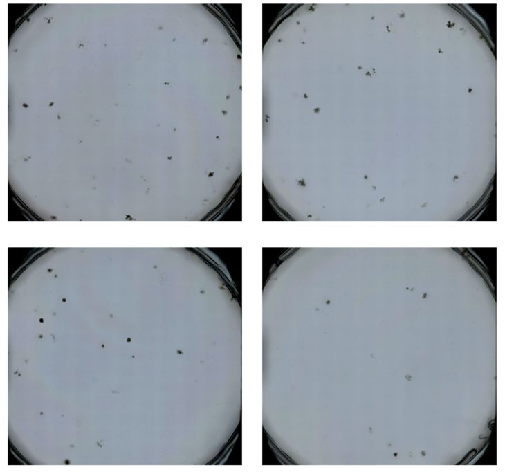
Baba et al. Cell Death and Disease (2021)12:322                                                                   Page 3 of 14

reagent (Ishihara Sangyo, Osaka, Japan). As a siControl, cells   transplanted intravenously into sublethally irradiated
were transfected with control siRNA-A (Santa Cruz Bio-           recipient mice (BALB/c strain, 5.5 Gy; C57BL/6J strain,
technology). The KD efficiency of each gene was confirmed          7 Gy) along with normal BM cells (BCR-ABL, 1 × 10⁶;
using quantitative real-time PCR (qRT-PCR) and western           MLL-AF9, 3 × 10⁵) in a 200 μl volume. When AML-like
blotting analyses.                                               splenomegaly was observed around 100 days after trans-
                                                                 plantation, BM cells were harvested from the primary
Cell preparation from BM and SP                                  AML mice and stored in liquid nitrogen. A frozen stock of
  Total BM cells from the femoral, tibial, and humeral           primary AML cells was used for the colony formation
bones were flushed using cold magnetic activated cell             assay. To prepare a mouse AML model, 2 × 10⁵ colony-
separation (MACS) buffer (PBS supplemented with 2 mM             forming cells were transplanted intravenously into sub-
EDTA and 3% FBS). Subsequently, erythrocytes were                lethally irradiated secondary recipient mice, along with
removed by density gradient centrifugation using                 1 × 10⁶ normal BM cells.
Histopaque-1083 reagent (Sigma-Aldrich) or by hemo-
lysis using ammonium chloride lysing buffer containing           BMT therapy
0.826% NH4Cl, 0.1% KHCO3, and 0.004% EDTA-4Na.                     CML-bearing mice were transplanted with 2 × 10⁶
Lineage marker (CD4, CD8, CD11b, Gr-1, B220, and                 healthy WT or STING−/− BM cells following x-
TER-119)−c-kit+Sca-1+ and lineage marker−c-kit+ cells            irradiation pretreatment (BALB/c strain, 5 Gy; C57BL/6J
were sorted using FACSAria Cell Sorter (BD Biosciences)          strain, 6 Gy) 10 days after the transplantation of leuke-
and were used as LKS+ and LK+ cells, respectively. In            mogenic cells. To discriminate the donor cells from the
some experiments, total BM cells were biopsied from live         recipient ones, including residual leukemia cells, CD45.1+
mice and were subjected to kinetics examination of resi-         congenic mouse-derived BM cells were used as donor
dual leukemia cells in the BM. Total SP cells were isolated      cells in some experiments.
via mechanical digestion from SP, and were subsequently
treated with ammonium chloride lysing buffer to remove           In vivo administration of autophagy inhibitors
the erythrocytes.                                                  SBI-0206965 (Sigma-Aldrich) and MRT-68921 (Sigma-
                                                                 Aldrich) were dissolved in DMSO and distilled water,
Retroviral preparation                                           respectively, to prepare the 10 mg/ml stock solution.
  MSCV-MLL-AF9-ires-GFP vector was gifted from                   AML-bearing mice were intraperitoneally injected with a
Akihiko Yokoyama, National Cancer Center, Tsuruoka               dose of 20 mg/kg SBI-0206965 diluted in PBS containing
Metabolomics Laboratory, Japan. MSCV-BCR-ABL-ires-               5% polyethylene glycol 400 (Sigma-Aldrich) and 5%
GFP and MSCV-MLL-AF9-ires-GFP plasmids were pre-                 Tween 80 (Sigma-Aldrich) or MRT-68921 diluted in PBS
pared as described previously7,8. Retroviral packaging cells     according to the schedule (Fig. 5a).
(Phoenix 293T) were transiently transfected with each
plasmid using jetPRIME transfection reagent (Polyplus-           Isolation of EVs from SP tissues
transfection, New York, NY, USA) to produce retrovirus             CML mice were irradiated with 5 Gy, 14 days after LIC
carrying MSCV-BCR-ABL-ires-GFP or MSCV-MLL-                      transplantation. The SP tissues were harvested from
AF9-ires-GFP in the culture supernatant, which were              normal mice, untreated leukemia mice (CML and AML),
subjected to the infection into LKS+ cells.                      or CML mice irradiated 7 days before sacrifice. Then, they
                                                                 were mechanically digested in 2 mM EDTA in PBS. The
Generation of CML and AML model                                  cells and their debris were removed using centrifugation
  LKS+ cells purified from BM of WT or STING−/− mice              for 5 min at 300 × g and filtration using a 1.2 μm filter
were cultured in serum-free S-Clone SF-03 medium                 (Minisart 17593-K; Sartorius Stedim Biotech, Goettingen,
(Sanko Junyaku, Tokyo, Japan) supplemented with 1%               Germany). After centrifugation for 30 min at 10,000 × g,
BSA, 100 ng/ml stem cell factor (WAKO, Osaka, Japan),            EVs were subsequently isolated from the supernatant via
100 ng/ml thrombopoietin, 25 ng/ml fms-like tyrosine             centrifugation for 2 h at 100,000 × g. EVs were further
kinase-3 ligand (R & D systems, Minneapolis, MN, USA),           purified using a 0.22 μm PVDF filter (Ultrafree; Merck
10 ng/ml IL-6 (PeproTech Cranbury, NJ, USA), and                 Millipore, Billerica, MA, USA) after they were resus-
10 ng/ml IL-3 (PeproTech) for 1 day. Cultured LKS+ cells         pended in HBSS buffer (Sigma-Aldrich). Isolated EVs
were infected with the retrovirus carrying MSCV-BCR-             were stored at −80 °C until use.
ABL-ires-GFP or MSCV-MLL-AF9-ires-GFP using Vir-
oMag R/L kit (OZ Bioscience, San Diego, CA, USA) to              Isolation of EVs from leukemia cells
obtain LICs. The resultant LICs (BCR-ABL, 200–300                  LKS+ cells were isolated from WT or STING−/− mice
GFP+ cells included in 15,000 LKS+ cells; MLL-AF9,               and were infected with a retrovirus carrying MSCV-BCR-
50–100 GFP+ cells included in 30,000 LKS+ cells) were            ABL-ires-GFP or MSCV-MLL-AF9-ires-GFP. Two days

Official journal of the Cell Death Differentiation Association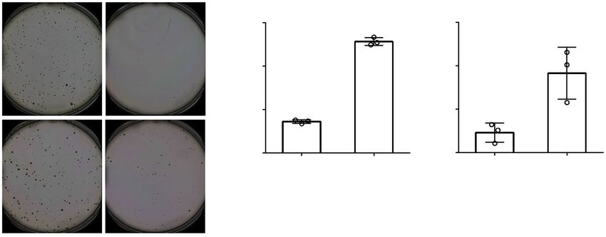
Baba et al. Cell Death and Disease (2021)12:322                                                                  Page 4 of 14

after the infection, 150 GFP+ or − cells were sorted using      primer sets for mouse Gapdh gene (sense: 5′-GCG GCA
FACSAria Cell Sorter and were subsequently cultured in          CGT CAG ATC CA-3′; antisense: 5′-CAT GGC CTT
1.1 ml of Methocult GF M3434. MLL-AF9+ colony-                  CCG TGT TTC CTA-3′), mouse Isg20 gene (sense: 5′-
forming cells were harvested at day 7 and were re-              ACA TCC AGA ACA ACT GGC GG-3′; antisense: 5′-
plated to obtain the secondary culture, and the procedure       TGA GGA GTG GCA GCT TCT AAC-3′), mouse Rsad2
was repeated at least two times to determine leukemic           gene (sense: 5′-TGC CTG AAT CTA ACC AGA AGA
transformation. EVs were isolated from the supernatant of       TGA A-3′; antisense: 5′-TTC TTC CAC GCC AAC ATC
each 3.5 mm dish for their characterization at days 7, 10,      CA-3′), mouse Ifit3 gene (sense: 5′-CCA TCA TGA GTG
or 12 of the colony formation assay.                            AGG TCA ACC G-3′; antisense: 5′-CAT TGT TGC CTT
                                                                CTC CTC AGA GT-3′), mouse Irf7 gene (sense: 5′-GTG
Co-culture of the primitive BM cells with EVs                   TAC GAA CTT AGC CGG GA-3′; antisense: 5′-GGT
  LKS+ cells isolated from WT mice were cultured and            TTG GAG CCC AGC ATT TT-3′), mouse Cxcl10 gene
propagated in serum-free S-Clone SF-03 medium sup-              (sense: 5′-CCA CGT GTT GAG ATC ATT GCC-3′;
plemented with 1% BSA, 100 ng/ml stem cell factor,              antisense: 5′-GAG GCT CTC TGC TGT CCA TC-3′),
10 ng/ml IL-6, and 10 ng/ml IL-3 for 2 days. After pro-         human AIM2 gene (sense: 5′- AAG AAG GCA AGC
pagated LKS+ cells were subsequently co-cultured with           AGG AGA TG-3′; antisense: 5′-TCA GCG GGA CAT
150 μg/ml SP EVs derived from control or irradiated CML         TAA CCT TT-3′), human cGAS gene (sense: 5′-GGC
mice for 24 h, they were subjected to the microarray            GGT TTT GGA GAA GTT G-3′; antisense: 5′-TCA TAG
analysis. Freshly isolated LKS+ cells and LK+ cells were        TAG CTC CCG GTG TTC-3′), human GAPDH gene
co-cultured with EVs in serum-free S-Clone SF-03 med-           (sense: 5′-GCC AAA AGG GTC ATC TC-3′; antisense:
ium supplemented with 1 % BSA and 100 ng/ml stem cell           5′-TGA GTC CTT CCA CGA TAC CA-3′), human IL-12
factor for 6 and 18 h, for the qRT-PCR analysis and the         gene (sense: 5′-GGT ATC ACC TGG ACC TTG GA-3′;
flow cytometric analysis of IRF7 expression, respectively.       antisense: 5′-GCT TAG AAC CTC GCC TCC TT-3′),
                                                                human ISG54 gene (sense: 5′-TGC GTG AAG AAG GTG
Extraction and measurement of double-stranded DNAs              AAG AG-3′; antisense: 5′-GCA GGT AGG CAT TGT
(dsDNAs) in EVs and cytoplasmic fraction                        TTG GT-3′), human ISG56 gene (sense: 5′-GCC CAG
   The total protein concentration of EVs isolated from SP      ACT TAC CTG GAC AA-3′; antisense: 5′-TTT CCT
tissues and culture supernatants was measured using a BCA       CCA CAC TTC AGC AA-3′), human MCP-1 gene (sense:
Protein Assay kit (Thermo Fisher Scientific, Waltham, MA,        5′-ATA GCA GCC ACC TTC ATT CC-3′; antisense: 5′-
USA). For the measurement of dsDNA concentration, total         GCT TCT TTG GGA CAC TTG CT-3′), human STING
DNA was purified using NucleoSpin Tissue kit                     gene (sense: 5′-CAT CGG ATA TCT GCG GCT GA-3′;
(MACHEREY-NAGEL, Duren, Germany) following the                  antisense: 5′-TCC AGG AAG CGA ATG TTG GG-3′),
digestion of vesicular-free DNAs using 27 Kunitz U/ml           and human TNF-α gene (sense: 5′-GGC GTG GAG CTG
DNase I (QIAGEN, Chatsworth, CA, USA) for 30 min at             AGA GAT AA-3′; antisense: 5′-GAT GGC AGA GAG
37 °C and its ensuing inactivation using 10 mM EDTA for         GAG GTT GA-3′). The relative expression of each gene
5 min at 65 °C. Cytoplasmic DNAs were extracted using a         was analyzed using the ΔΔCt method relative to the Ct
Cell Fractionation kit (Abcam, Cambridge, MA, USA), and         value of the Gapdh gene.
were purified using NucleoSpin Tissue kit. The con-
centration of dsDNA was measured using Qubit 4 (Thermo          Flow cytometry
Fisher Scientific) and Qubit 1X dsDNA HS Assay kit                 Isolated hematopoietic cells and leukemia cells were
(Thermo Fisher Scientific). dsDNA concentration in EVs           stained with various combinations of fluorescent dye-
was normalized with the determined protein concentration.       conjugated Abs. Intracellular IRF7 and intranuclear
In some experiments, BCR-ABL gene was detected using            γH2A.X were stained with PE-conjugated anti-IRF7 mAb
genomic PCR using a specific primer set (sense: 5′-TTC           and APC-conjugated anti-γH2A.X mAb using the Foxp3/
AGA AGC TTC TCC CTG ACA T-3′; antisense: 5′-TGT                 Transcription Factor Buffer Set (eBioscience). For the
TGA CTG GCG TGA TGT AGT TGC TTG G-3′)9.                         detection of apoptosis, cell cycle status, and total reactive
                                                                oxygen species (ROS) in AML cells, cells were stained
RNA isolation, cDNA synthesis, and qRT-PCR                      using Annexin V-FITC Apop kit (Thermo Fisher Scien-
  Total RNA was isolated from cells using an RNeasy             tific), Vybrant DyeCycle Green (Thermo Fisher Scientific),
Mini Kit (QIAGEN) and then reverse-transcribed using            and Total ROS/Superoxide Detection kit (Enzo Life Sci-
the SuperScript IV VILO (Thermo Fisher Scientific). qRT-         ences, Farmingdale, NY, USA) respectively. The expres-
PCR was performed using StepOne Real-time PCR system            sion of each molecule was determined using a
(Thermo Fisher Scientific), using the Fast SYBR Green            FACSCantoII (BD Biosciences) and analyzed using FlowJo
Master Mix (Thermo Fisher Scientific), and specific               software (Tree Star, Ashland, OR, USA).

Official journal of the Cell Death Differentiation Association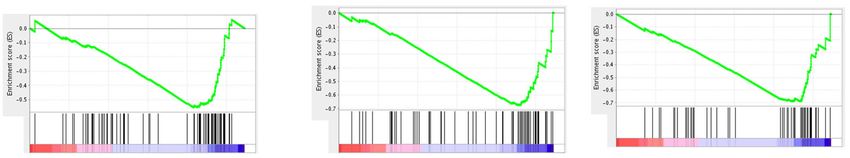
Baba et al. Cell Death and Disease (2021)12:322                                                                   Page 5 of 14

Characterization of EVs                                         Santa Clara, CA, USA). RNA Integrity Number (RIN) of
  Spleen tissues were lysed in RIPA buffer supplemented         all samples was 10.0, and the samples were subjected to
with a protease inhibitor cocktail (WAKO) on ice. After         microarray analysis according to the manufacturer’s
centrifugation at 14,000 × g for 30 min, the supernatants       instructions. In brief, 100 ng RNA samples were labeled
were used as a SP tissue lysate. The protein concentration      with cyanine 3-CTP using the Low Input Quick Amp
of EVs and SP lysate were determined using a BCA Pro-           Labeling Kit, one color (Agilent Technologies). Hybridi-
tein Assay kit (Thermo Fisher Scientific). A total of 2 μg of    zation was performed using the Gene Expression Hybri-
EVs and SP lysate were lysed in 20 µL of 2× SDS sample          dization Kit (Agilent Technologies). cRNA samples
buffer (4% SDS, 20% glycerol, 0.02% bromophenol blue,           (600 ng) were subjected to fragmentation (30 min at
100 mM Tris-HCl (pH 6.8)) at room temperature for               60 °C), and then hybridized on SurePrint G3 Mouse Gene
30 min, and were electrophoresed in a 5–20% poly-               Expression v2 8x60K Microarray Kit (G4852B, design ID:
acrylamide gel at 200 V. The protein on the gel was             074809, Agilent Technologies) in a rotary oven (10 rpm at
transferred onto a polyvinylidene fluoride membrane              65 °C for 17 h). Slides were washed in Agilent Gene
(Bio-Rad Laboratories, Hercules, CA, USA). The mem-             Expression Wash Buffers 1 and 2 (Agilent Technologies)
brane was incubated in PBS supplemented with 5% skim            and scanned using an Agilent DNA Microarray Scanner
milk and 0.05% Tween 20 for 1 h, each primary Ab for 2 h,       (G2600D, Agilent Technologies). The obtained data were
and each HRP-conjugated secondary Ab for 1 h. The               normalized by adjusting the differences in the probe
protein expression was detected using SuperSignal West          intensity distribution across different arrays as follows: the
Pico Chemiluminescent Substrate (Thermo Fisher Sci-             control probes were removed and the scaling factor for
entific) and ImageQuant LAS4000min (GE Healthcare,               each array was calculated by multiplying the expression
Chicago, IL, USA). For nano tracking analysis (NTA), EVs        values by the scaling factor of the array where the probes
were diluted at 300 ng/mL with PBS, and their size dis-         were. The scaling factor was 2500 divided by the average
tribution and particle concentration were measured using        value of probes in addition to the upper and lower 2% of
the NanoSight™ LM10 nanoparticle tracking system                the array. When there were multiple probes for a gene, the
(Malvern Panalytical, Malvern, UK).                             probe for which the value of the CML sample was the
                                                                highest was exploited for subsequent analysis. Gene set
Colony formation assay                                          enrichment analysis (GSEA) was performed using Gene-
  Two hundred LKS+ cells were isolated from WT or               Pattern server10–12. As the metric for ranking genes,
STING−/− mice and were cultured with or without 50 μg/          log2_ratio_of_means was exploited, and the gene set size
ml SP-derived EVs in 1.1 ml of Methocult GF M3434               was set to 20 to 200. The database gene sets used in this
(STEMCELL technologies, Vancouver, BC, Canada). The             study were hallmark and c2.cgp.v6.0.symbols.gmt.
number of colonies and total cells was determined 7 and
10 days after incubation, respectively. In some experi-         Statistical analysis
ments, 200 LKS+ cells or 1,250 mouse AML cells were               We did not use specific sample size calculation meth-
cultured with or without 5 μM SBI-0206965 in 1.1 ml of          ods. Any technically validated data were not excluded.
Methocult GF M3534 (STEMCELL technologies). The                 Data were analyzed statistically using GraphPad Prism
number of colonies and total cells was determined 7 days        software (Ver. 6; La Jolla, CA, USA) using the methods
after incubation. A wide-area continuous image of colony        indicated in the legend of each figure. Two-sided Stu-
formation in a 35 mm dish was obtained using BZ-X700            dent’s t-test and one-way ANOVA followed by Tukey-
microscope (Keyence, Osaka, Japan) and was analyzed             Kramer post-hoc test were used to compare the data
using Keyence Analysis Software.                                among two and more than two groups, respectively. Log-
                                                                rank test was used to evaluate the survival curve data. A
Blood test                                                      p < 0.05 was considered statistically significant.
  Whole blood samples were collected using K2 EDTA-
coated micro-hematocrit capillaries (VITREX, Herlev,            Results
Denmark). The number of WBCs, red blood cells (RBCs),           Leukemia SP-derived EVs inhibit the hematopoietic
platelets (PLTs), hemoglobin (HGB) concentration, and           capacity of normal HSPCs
hematocrit (HCT) values were determined using an                  CML-bearing mice underwent BMT after irradiation
automatic hematology analyzer (Celltacα MEK-6358;               (5 Gy) (Supplementary Fig. 1a), and donor cell-derived
NIHON KOHDEN CORPORATION, Tokyo, Japan).                        hematopoietic reconstitution, but not leukemia relapse,
                                                                was observed by 8 weeks after BMT (Supplementary Fig.
Microarray analysis                                             1b). However, most of them eventually died later than
  Total RNA was extracted and its quality was confirmed          10 weeks after BMT (Supplementary Fig. 1c) without
using an Agilent 2200 TapeStation (Agilent Technologies,        leukemia relapse, as evidenced by few BCR-ABL+ cells in

Official journal of the Cell Death Differentiation Association
Baba et al. Cell Death and Disease (2021)12:322 Page 6 of 14
the BM, SP, and PB (Supplementary Fig. 1d). However, CML EVs significantly enhanced IRF7 protein expression
marked reductions in RBCs, HGB concentration, HCT, in WT HSPCs, but not in STING−/− cells (Fig. 3c).
and PLTs were observed in the PB (Supplementary Fig. Moreover, the suppressive effect of CML-derived EVs on
1e). These observations suggest that donor cell-derived colony formation of normal HSPCs was mostly abrogated
hematopoiesis was fatally disturbed in CML mice treated by STING deficiency (Fig. 3d). Upon BMT of CML-bearing
with BMT. Moreover, a substantial proportion of mice, WT but not STING−/− donor cells displayed an
CD45.1+ donor cells in BM and SP expressed a recipient enhanced expression of a marker for DNA damage, γH2A.
marker, CD45.2, 4 weeks after BMT (Fig. 1a), together X, in the HSPCs, after 2 weeks (Fig. 3e). Furthermore, fatal
with the punctate fluorescent signals on their cell surface anemia development was significantly attenuated when
(Fig. 1a), suggesting the adhesion of EVs. Indeed, CML SP STING−/− cells were used as donor cells (Fig. 3f). Thus,
contained a larger amount of EVs than control SP, and dsDNA fragments contained in leukemia cell-derived EVs
their contents were further increased by irradiation damaged normal HSPCs in a cytosolic DNA-sensor sig-
(Fig. 1b). Furthermore, all isolated EVs exhibited EV naling pathway-dependent manner both in vivo and
marker proteins Alix, CD81, and CD9 (Fig. 1c), and their in vitro.
sizes ranged from 50 to 300 nm (Fig. 1d). Either irradiated
or non-irradiated CML-derived EVs reduced the colony Pharmaceutical inhibition of autophagosome formation
and whole cell numbers in the colony formation assays induces an accumulation of cytoplasmic dsDNA and
using normal LKS+ HSPCs, compared with control EVs cytotoxicity in leukemia cells
(Fig. 2a, b). Likewise, AML SP-derived EVs inhibited the We next examined the extranuclear DNA dynamics in
colony formation of normal HSPCs (Supplementary Fig. leukemia cells. BCR-ABL+ cells secreted EVs containing
2), similar to CML-derived EVs. Thus, leukemia EVs dsDNA at 10 days but not at 7 days of culture, when
reduced the hematopoietic capabilities of normal HSPCs. cytoplasmic DNA speckle formation was discerned
Additional comprehensive gene expression analysis and (Supplementary Fig. 5a, b). Additionally, dsDNA con-
subsequent GSEA identified only three differentially centration in EVs was reduced by rapamycin, which can
regulated gene sets in HSPCs treated with CML EVs, and induce the digestion of the cytoplasmic DNA by autop-
two of them were type I IFN-responsive gene sets hagic induction14, while the intravesicular dsDNA con-
(Fig. 2c). qRT-PCR validated the enhanced expression of tents were reciprocally increased by SBI-0206965, an
Rsad2, Irf7, and Cxcl10, among the top five upregulated autophagosome formation inhibitor (Supplementary Fig.
genes, which are common to two type I IFN-related gene 5c). These observations indicate that autophagy can reg-
sets in HSPCs (Fig. 2d), suggesting type I IFN signaling ulate cytoplasmic dsDNA accumulation and the sub-
pathway activation by CML EVs. sequent redistribution into EVs. It was previously
reported that cytoplasmic DNA activates DNA-sensor
Horizontal transfer of leukemia cell-derived DNA via EVs signaling in autophagy-deficient cells14. As leukemia EV-
disturbed normal hematopoiesis by activating cytosolic derived cytoplasmic DNA damages normal HSPCs (Fig. 3),
DNA-sensor signaling we assessed whether autophagy blockade can induce
As the cytoplasmic double-stranded (ds)DNA fragments cytotoxicity in leukemia cells by increasing the levels of
can robustly activate type I IFN signaling by acting on cytoplasmic DNA. Indeed, cytoplasmic dsDNA was sig-
several DNA sensors13, we determined dsDNA content in nificantly increased in HL-60 cells, which were defective
EVs. Indeed, irradiated and non-irradiated CML SP- in autophagy arising from the transfection with siRNA
derived EVs contained more dsDNA fragments than against ATG5 gene, a gene essentially involved in autop-
control SP-derived ones (Fig. 3a) in the presence of BCR- hagy (Fig. 4a, b), in line with the previous study14.
ABL gene (Supplementary Fig. 3). Moreover, BCR-ABL- Moreover, HL-60 cells exhibited cytoplasmic DNA
transformed cell-derived EVs contained a larger amount accumulation when they were treated with SBI-0206965
of dsDNA fragments than GFP-transduced cell-derived and MRT-68921, autophagosome formation inhibitors,
EVs (Fig. 3b). Likewise, the EVs present in the SP or in the whereas an autolysosome formation inhibitor, hydroxy-
culture supernatants of MLL-AF9-transformed cells chloroquine, did not induce cytoplasmic DNA accumu-
contained abundant dsDNAs (Fig. 3a, b). Moreover, lation, together with an increased autophagosome
dsDNA was detected in EVs derived from AML mouse formation (Fig. 4c, d, and Supplementary Fig. 6). Fur-
serum, but not in those derived from control-treated mouse thermore, SBI-0206965 or MRT-68921 efficiently induced
serum (Supplementary Fig. 4). As multiple DNA sensors cell death in various types of human leukemia cell lines
can recognize cytoplasmic DNAs to converge on a single (Fig. 4e and Supplementary Fig. 7a) with sequential
adaptor molecule, STING, thereby activating type I IFN induction of cell cycle arrest (Fig. 4f) and apoptotic cell
signaling13, we next examined the roles of STING pathways death (Fig. 4g), probably due to the cytotoxic activity of
in this process using STING−/− cells. The treatment with cytoplasmic dsDNA. On the contrary, cytotoxicity was
Official journal of the Cell Death Differentiation AssociationBaba et al. Cell Death and Disease (2021)12:322 Page 7 of 14
4
Fig. 1 Characterization of CML SP-derived EVs. (a) CD45.1 and CD45.2 expression on the BM cells and splenocytes in CML-bearing mice was
determined 4 weeks after BMT therapy and is shown in the left panels. As a control, cells derived from CD45.1+ and CD45.2+ mice were mixed
immediately before staining with molecule-specific Abs. Representative immunofluorescence images for CD45.2 on the sorted CD45.1+CD45.2+ cells
(inside square gates) from three independent experiments are shown in the right panels. Expression pattern of CD45.2 on the cells derived from
CD45.2+ mice are shown as a control. (b) Protein concentration of SP-derived EVs. Data represent the mean ± SD from three independent
experiments. **P < 0.01 using Tukey-Kramer test. (c) EV surface marker proteins (CD9 and CD81), an EV cytosolic marker protein (Alix), and a non-EV
protein (GAPDH) were detected using western blot. Each lane represents an individual sample (n = 3). (d) Size distribution of control EVs, non-rad
CML EVs, and rad CML EVs were analyzed using NTA. Representative results from three independent experiments are shown here.
Official journal of the Cell Death Differentiation AssociationBaba et al. Cell Death and Disease (2021)12:322 Page 8 of 14
Fig. 2 CML mouse-derived EVs disturb normal hematopoiesis. a Colony formation of LKS+ cells when they were co-cultured with or without EVs.
Representative results from four independent experiments are shown here. b The number of colonies and total cells are shown in the left and right
panels, respectively. Data represent mean ± SD from four independent experiments. c Gene expression was determined in LKS+ cells co-cultured
with EVs and differentially regulated gene sets (false discovery rate (FDR) < 0.25) were identified with GSEA using hallmark and C2CGP gene sets. FDR
and normalized enrichment score (NES) values were calculated from a single-sample experiment. d Relative mRNA expression of the indicated genes
in LKS+ cells co-cultured with or without (N.T.) EVs. Data represent mean ± SD from three independent experiments. **P < 0.01; *P < 0.05 using Tukey-
Kramer test.
not marked when leukemia cells were treated with lines (Supplementary Fig. 8a). Both SBI-0206965 and
hydroxychloroquine (Supplementary Fig. 7b). MRT-68921 efficiently reduced cell viability in control
shRNA transduced HL-60 cells, but the reduction was
Autophagosome formation inhibitor induces STING significantly reversed in STING KD cell lines (Fig. 5a and
pathway-mediated cytotoxicity in leukemia cells Supplementary Fig. 9a). STING shRNA treatment also
As STING pathway mainly contributed to cytoplasmic reduced SBI-0206965-mediated cytotoxicity in another
DNA-mediated damage of normal HSPCs, we examined leukemia cell line, THP-1 (Supplementary Fig. 9b).
its involvement in autophagy inhibitor-mediated cyto- Moreover, SBI-0206965-mediated cytotoxicity was sig-
toxicity in leukemia cells, using STING KD HL-60 cell nificantly reduced by shRNA treatment against cyclic
Official journal of the Cell Death Differentiation AssociationBaba et al. Cell Death and Disease (2021)12:322 Page 9 of 14
N.S
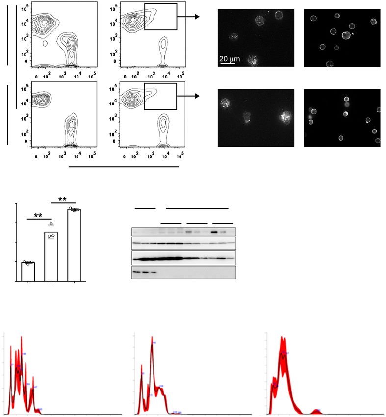
Fig. 3 STING pathway sensing leukemia cell-derived dsDNA damages normal HSPCs. a, b dsDNA concentration in the SP-derived EVs (a) and in
the 12-day-cultured cell-derived EVs (b). Data represent mean ± SD from three independent experiments. c Protein expression of IRF7 in LK+ cells
when they were co-cultured with or without EVs. Mean fluorescence intensity (MFI) of cells stained with PE-conjugated anti-IRF7 mAb is shown. Data
represent mean ± SD from three independent experiments. d Colony formation of LKS+ cells when they were co-cultured with or without EVs. Data
represent mean ± SD from four independent experiments. e Expression of γH2A.X in GFP−LKS+ cells (R1 region in the right panel) 2 weeks after BMT
in CML-bearing mice. As a control, normal mice were subjected to BMT. Representative results from 4 to 5 independent animals are shown in the
middle panels. Gray-filled and open histograms in the left panels show control and CML-bearing mice, respectively. Each symbol represents an
individual mouse (control, n = 4; CML, n = 5). f Survival rates within 120 days after BMT (WT, n = 10; STING−/−, n = 12). **P < 0.01; *P < 0.05; N.S., no
significant difference using Tukey-Kramer test or log-rank test.
GMP-AMP synthase (cGAS), a major DNA sensor in inhibitor-mediated cytotoxicity in leukemia cells. SBI-
STING pathway (Supplementary Fig. 10), but not against 0206965 reduced the colony formation ability of WT
another cytosolic DNA sensor, AIM2 (Fig. 5b and Sup- mouse-derived AML cells to a larger extent than nor-
plementary Fig. 8b). Furthermore, among the genes mal HSPCs (Fig. 5f and Supplementary Fig. 11),
known to be expressed in the STING pathway, the whereas the reduction was attenuated in STING−/−
expression of TNF-α, IL-12, and MCP-1 genes was mouse-derived AML cells (Fig. 5f, g, h).
enhanced by SBI-0206965 in the control but not in the
STING KD cells (Fig. 5c), further supporting the Autophagosome formation inhibitors can suppress the
assumption that the STING pathway was activated by progression of AML
cytoplasmic dsDNA accumulation induced by autop- Inspired by the observation that mouse normal HSPCs
hagosome formation inhibition. Moreover, consistent were more resistant than mouse AML cells to SBI-
with the previous report that STING pathway activa- 0206965, we examined its therapeutic effect. AML-
tion can induce ROS production, and eventually induce bearing mice were administered the agent every other
cellular senescence by activating the DNA damage day starting from 14 days after AML cell transplantation
response15, SBI-0206965 treatment increased intracel- (Fig. 6a) when leukemia cells could be apparently dis-
lular ROS levels in the control but not in STING KD cerned in PB. SBI-0206965 was only partially effective to
cells (Fig. 5d). Additionally, N-acetyl-L-cysteine, an prevent the increases in total WBCs and leukemia cells in
ROS inhibitor, efficiently attenuated SBI-0206965- the PB (Fig. 6b, c), but significantly improved the survival
induced cytotoxicity in a dose-dependent manner rate (Fig. 6d). Moreover, when AML-bearing mice were
(Fig. 5e), suggesting the involvement of STING treated with MRT-68921, with a lower IC50 against its
pathway-induced ROS in autophagosome formation molecular target, ULK-1, than SBI-0206965, both total
Official journal of the Cell Death Differentiation AssociationBaba et al. Cell Death and Disease (2021)12:322 Page 10 of 14 Fig. 4 Inhibitors of autophagosome formation induce the accumulation of cytoplasmic dsDNA and cytotoxicity in leukemia cells. a Relative mRNA expression of ATG5 in HL-60 cells 48 h after transfection of control or ATG5 siRNA. Data represent mean ± SD from three independent experiments. b Cytoplasmic dsDNA in HL-60 cells at 48 h after transfection of control or ATG5 siRNA. Fold change was calculated by dividing with the mean value of control. Data represent mean ± SD from three independent experiments. **P < 0.01; *P < 0.05 using two-sided Student’s t-test. c Cytoplasmic dsDNA in HL-60 cells 18 h after treatment with 20 μM SBI-0206965 (SBI), 2.5 μM MRT-68921 (MRT), and 20 μM hydroxychloroquine (HCQ, Sigma-Aldrich). Cells treated with DMSO were used as a control. Fold change was calculated by dividing with mean value of control. Data represent mean ± SD from three independent experiments. **P < 0.01 using Tukey-Kramer test. d Cytoplamic DNA was stained with Vybrant DyeCycle Green 18 h after treatment with SBI-0206965. e Cytotoxic assay of SBI-0206965 against leukemia cell lines (n = 4). f, g Cell cycle status (f) and apoptosis induction (g) was determined on HL-60 cells 18 and 42 h after treatment with SBI-0206965. Percentages of Annexin V+PI− and Annexin V+PI+ cells in SP are shown here. Representative results from three independent experiments are shown here. Official journal of the Cell Death Differentiation Association
Baba et al. Cell Death and Disease (2021)12:322                                                                                        Page 11 of 14

  Fig. 5 STING pathway contributes to the cytotoxicity arising from the inhibition of autophagosome formation. a Determination of
  cytotoxicity of SBI-0206965 against shRNA control or STING KD HL-60 cells (n = 4). b Determination of cytotoxicity of SBI-0206965 against shRNA
  control or AIM2 KD HL-60 cells (n = 4). c Relative mRNA expression of the indicated genes in control or STING shRNA-transfected HL-60 cells at 18 h
  after treatment with 5 μM SBI-0206965. As a control, cells were treated with DMSO. Data represent mean ± SD from three independent experiments.
  d Total ROS induction in control or STING shRNA-transfected HL-60 cells at 20 h after treatment with 5 μM SBI-0206965. Cells treated with DMSO were
  used as a control. MFI of cells stained using a total ROS/superoxide detection kit is shown. Data represent mean ± SD from three independent
  experiments. e Cytotoxic assay when HL-60 cells were co-cultured with 5 μM SBI-0206965 in the presence or absence of N-acetyl-L-cysteine (NAC,
  Sigma-Aldrich) (n = 4). f Colony formation of WT mice or STING−/− mice AML cells when they were co-cultured with 5 μM SBI-0206965. Cells treated
  with DMSO were used as a control. g, h Suppression of colony formation of mouse AML cells by SBI-0206965 was calculated in terms of colony (g)
  and total cell number (h) by normalization with DMSO control for each. Data represent mean ± SD from three independent experiments. **P < 0.01;
  *P < 0.05; N.S., no significant difference using Tukey–Kramer test or two-sided Student’s t-test.

WBCs and leukemia cells were efficiently reduced in the                       Discussion
PB (Fig. 6e, f) with an improved survival rate (Fig. 6g).                      We revealed that the DNA fragments contained in EVs
Thus, autophagosome inhibitors can be potent ther-                           are horizontally transferred to normal hematopoietic cells
apeutic agents against AML.                                                  to reduce their hematopoietic capabilities through the

Official journal of the Cell Death Differentiation Association
Baba et al. Cell Death and Disease (2021)12:322                                                                                             Page 12 of 14

                                                                                                                                               50d

                                                                                                                                               50d

  Fig. 6 In vivo administration of autophagy inhibitors retarded the progression of AML. a Schematic representation of the experimental
  procedure of autophagy inhibitor treatment. As a control, mice were treated with each vehicle. b, e Total WBC count in mice treated with SBI-
  0206965 (b) or MRT-68921 (e) at the indicated time points. Each symbol represents an individual mouse (n = 6 or 7). c, f Percentages of GFP+CD11b+
  AML cells in the PB of mice treated with SBI-0206965 (c) or MRT-68921 (f) at the indicated time points. Each symbol represents an individual mouse
  (n = 7). d, g Alteration of the survival rates of mice treated with SBI-0206965 (d) or MRT-68921 (g) (n = 7). **P < 0.01; *P < 0.05; N.S., no significant
  difference using Mann-Whitney u test or log-rank test.

activation of cytosolic DNA-sensor pathway after BMT                            to investigate the effects of extranuclear DNA on leu-
therapy for CML. We assumed that irradiation at BMT                             kemic hematopoiesis, which shares many features with
could enhance DNA fragmentation and subsequent EV                               normal ones, and we demonstrated that extranuclear
formation. In contrast with our hypothesis, non-irradiated                      DNA accumulated in the cytoplasm of leukemia cells and
CML SP contained EVs, which inhibited hematopoietic                             suppressed leukemic hematopoiesis.
colony formation to a similar extent as irradiated ones did.                      STING was originally identified as a central adaptor
Hence, CML cell-derived EVs may be able to promote                              protein to sense cytoplasmic DNA. Several lines of evi-
CML development by counteracting normal hematopoi-                              dence indicate that STING activation in HSCs can
esis. These effects may be concealed by the rapid division                      depress their hematopoietic capacity16,17. Consistently, we
of primary leukemia cells but may emerge when leukemia                          observed that a single exposure of leukemia cell-derived
cell growth is severely suppressed by irradiation. We                           EVs to normal HSPCs reduced their in vitro colony for-
further uncovered that AML cells abundantly secreted                            mation in a STING-dependent manner. Moreover, we
EVs containing DNA fragments, with a capacity to inhibit                        revealed that BMT caused less DNA damage in donor
normal hematopoiesis. Thus, it is likely that leukemia cell-                    cells when STING-deficient HSPCs were used as donor
derived extranuclear DNA contributes to the suppression                         cells in CML-bearing mice, compared with the use of WT
of normal hematopoiesis. This assumption prompted us                            HSPCs. Consequently, fatal anemia development was

Official journal of the Cell Death Differentiation Association
Baba et al. Cell Death and Disease (2021)12:322 Page 13 of 14
significantly delayed. Thus, it is plausible that leukemia We demonstrated that autophagosome inhibitors
cell-derived DNAs, which were transferred to the cyto- improved the survival of AML-bearing mice, suggesting
plasm of normal hematopoietic cells, can disturb normal the potential of these agents for AML treatment. More-
hematopoiesis in a STING pathway-dependent manner. over, their therapeutic effects can be ascribed to their
The potential cytotoxicity of extranuclear DNA18 capacity to reduce AML cell proliferation by the action of
prompted Takahashi et al. to elucidate the dynamics of ROS, which were generated through cytoplasmic DNA-
extranuclear DNA, which were released from the nuclei of mediated STING pathway activation. Cytoplasmic DNA
senescent fibroblasts19. They demonstrated that DNA accumulation is observed in various types of cancer
accumulated in the cytoplasm of senescent fibroblasts was cells25,26, and can be ascribed mainly to nuclear DNA
discarded after being incorporated into exosomes to release into the cytoplasm upon replication stress or DNA
maintain cellular homeostasis. Moreover, the repression damage27,28. Thus, autophagosome formation inhibitor
of cytoplasmic DNases accelerated the senescence status may be a potent therapeutic drug against various types of
and nuclear DNA damage, eventually inducing the accu- leukemias and other malignancies, which grow rapidly
mulation of cytoplasmic DNA, which activated cytosolic and are under replication stress. Moreover, as anti-cancer
DNA-sensor pathways15. Another study demonstrated drugs and irradiation can cause DNA damage in cancer
that the prevention of autophagy-mediated degradation of cells, these inhibitors may be able to supplement these
cytoplasmic DNA activated DNA-sensor signaling, even- therapies.
tually enhancing DNA damage14. These observations SBI-0206965 and MRT-68921 are inhibitors of a serine/
suggest that autophagy can reduce the cytoplasmic DNA threonine kinase, ULK-1, which can prevent autophago-
levels in addition to cytoplasmic DNases. Consistent with some formation, and can inhibit several additional kina-
these findings, we revealed that inhibition of autophago- ses29. Chen et al. reported that in addition to ULK1,
some formation efficiently induced cytoplasmic DNA MRT-68921 can inhibit NUAK family SNF1-like kinase 1
accumulation, and subsequently, STING pathway- (NUAK1), an anti-oxidant molecule, and eventually can
mediated cytotoxicity in leukemia cells. Given the mod- exert effective cytotoxicity in various solid cancer cell
erate effects of an autolysosome formation inhibitor on lines30. Moreover, Hwang et al. recently reported that
cytotoxicity, autophagosomes may be mainly utilized to both SBI-0206965 and MRT-68921 could induce degra-
sequester cytoplasmic DNA, thereby concealing them dation of internal tandem duplication mutations in the
from DNA sensing machinery to prevent cytotoxicity in FLT3 tyrosine kinase receptor (FLT3-ITD) protein,
leukemia cells. thereby exerting cytotoxicity in FLT-ITD AML cells31.
We observed that STING pathway activation in leu- Thus, SBI-0206965- or MRT-68921-mediated inhibition
kemia cells resulted in the generation of ROS, which of other kinases may also contribute to their cytotoxicity
could be a main executor of the cytotoxicity induced by in leukemia cells. Nevertheless, a more precise study on
autophagosome inhibitors. STING pathway activation the pharmacology of these agents is warranted to develop
can stimulate IκB kinase and TANK-binding kinase 1 to a novel strategy to treat leukemia.
activate two distinct transcription factors, NF-κB and
IRF3, respectively, and both these factors can induce the Acknowledgements
expression of several antimicrobial and inflammation- This work was supported in part by KAKENHI, grant number 17K19681 (Japan
Society for the Promotion of Science).
related molecules, including ROS20–22. We observed
that an autophagosome inhibitor enhanced the gene
Author details
expression of TNF-α, IL-12, and MCP-1, in a STING 1
Division of Molecular Bioregulation, Cancer Research Institute, Kanazawa
pathway-dependent manner. In contrast, the gene University, Kanazawa, Japan. 2Department of Immunology, Graduate School of
expression of ISG54 and ISG56, was augmented by an Medical Sciences, Kanazawa University, Kanazawa, Japan. 3Nano Life Science
Institute, Kanazawa University, Kanazawa, Japan. 4Division of Cancer Cell
autophagosome inhibitor, even in the absence of STING Biology, Cancer Research Institute, Kanazawa University, Kanazawa, Japan.
pathway activation. Several lines of evidence indicate 5
Department of Transfusion Medicine and Stem Cell Regulation, Juntendo
that NF-κB activation can regulate the gene expression University School of Medicine, Tokyo, Japan. 6Division of Molecular Genetics,
Cancer Research Institute, Kanazawa University, Kanazawa, Japan
of TNF-α, IL-12, and MCP-123 and that the gene
expression of ISG54 and ISG56 can be regulated by
Author contributions
IRF-324. Thus, autophagosome inhibitor-mediated T.B. designed, performed, and analyzed the experiments in their entirety. Y.T.
STING pathway activation might induce ROS produc- and S.M. established and analyzed the mouse leukemia models. T.Y. supervised
tion mainly through the NF-κB pathway. However, as the extracellular vesicle study. T.N. analyzed the microarray data. R.H., N.G., A.H.,
and N.M. supervised the entire study. T.B. prepared the manuscript.
the loss of STING did not completely abrogate autop-
hagy inhibitor-mediated cytotoxicity in leukemia cells,
Data availability
the contribution of STING-independent activation of Raw and processed microarray data have been deposited in the GEO
IRF3 cannot be excluded. repository. The accession number is G1 (GEO reviewer link).
Official journal of the Cell Death Differentiation AssociationBaba et al. Cell Death and Disease (2021)12:322 Page 14 of 14
Conflict of interest 12. Subramanian, A. et al. Gene set enrichment analysis: a knowledge-based
The authors declare no competing interests. approach for interpreting genome-wide expression profiles. Proc. Natl Acad.
Sci. USA 102, 15545–15550 (2005).
Ethics statement 13. Paludan, S. R. & Bowie, A. G. Immune sensing of DNA. Immunity 38, 870–880
All the animal experiments were approved by the Kanazawa University Ethics (2013).
Committee (AP-163699). 14. Lan, Y. Y., Londono, D., Bouley, R., Rooney, M. S. & Hacohen, N. Dnase2a
deficiency uncovers lysosomal clearance of damaged nuclear DNA via
autophagy. Cell Rep. 9, 180–192 (2014).
Publisher’s note
15. Takahashi, A. et al. Downregulation of cytoplasmic DNases is implicated in
Springer Nature remains neutral with regard to jurisdictional claims in
cytoplasmic DNA accumulation and SASP in senescent cells. Nat. Commun. 9,
published maps and institutional affiliations.
1249 (2018).
16. Kobayashi, H. et al. Bacterial c-di-GMP affects hematopoietic stem/progenitors
Supplementary information The online version contains supplementary
and their niches through STING. Cell Rep. 11, 71–84 (2015).
material available at https://doi.org/10.1038/s41419-021-03587-x.
17. Xia, P. et al. A circular RNA protects dormant hematopoietic stem cells from
DNA sensor cGAS-mediated exhaustion. Immunity 48, 688–701 e687 (2018).
Received: 26 October 2020 Revised: 2 March 2021 Accepted: 4 March 2021 18. Vanpouille-Box, C., Demaria, S., Formenti, S. C., Galluzzi, L. & Cytosolic, D. N. A.
Sensing in organismal tumor control. Cancer Cell 34, 361–378 (2018).
19. Takahashi, A. et al. Exosomes maintain cellular homeostasis by excreting
harmful DNA from cells. Nat. Commun. 8, 15287 (2017).
20. Bjorn, M. E. & Hasselbalch, H. C. The role of reactive oxygen species in mye-
References lofibrosis and related neoplasms. Mediators Inflamm. 2015, 648090 (2015).
1. Saunders, N. A. et al. Role of intratumoural heterogeneity in cancer drug 21. Song, J. H. et al. Manassantin B shows antiviral activity against coxsackievirus
resistance: molecular and clinical perspectives. EMBO Mol. Med. 4, 675–684 B3 infection by activation of the STING/TBK-1/IRF3 signalling pathway. Sci. Rep.
(2012). 9, 9413 (2019).
2. Baba, T. et al. MIP-1alpha/CCL3-mediated maintenance of leukemia-initiating 22. Zheng, J. et al. Comprehensive elaboration of the cGAS-STING signaling axis in
cells in the initiation process of chronic myeloid leukemia. J. Exp. Med. 210, cancer development and immunotherapy. Mol. Cancer 19, 133 (2020).
2661–2673 (2013). 23. Blaauboer, S. M., Gabrielle, V. D. & Jin, L. MPYS/STING-mediated TNF-alpha, not
3. Baba, T. et al. MIP-1alpha/CCL3-expressing basophil-lineage cells drive the type I IFN, is essential for the mucosal adjuvant activity of (3′-5′)-cyclic-di-
leukemic hematopoiesis of chronic myeloid leukemia in mice. Blood 127, guanosine-monophosphate in vivo. J. Immunol. 192, 492–502 (2014).
2607–2617 (2016). 24. Grandvaux, N. et al. Transcriptional profiling of interferon regulatory factor 3
4. Asou, H. et al. Establishment of a human acute myeloid leukemia cell line target genes: direct involvement in the regulation of interferon-stimulated
(Kasumi-1) with 8;21 chromosome translocation. Blood 77, 2031–2036 genes. J. Virol. 76, 5532–5539 (2002).
(1991). 25. Lam, A. R. et al. RAE1 ligands for the NKG2D receptor are regulated by STING-
5. Koeffler, H. P. & Golde, D. W. Acute myelogenous leukemia: a human cell line dependent DNA sensor pathways in lymphoma. Cancer Res. 74, 2193–2203
responsive to colony-stimulating activity. Science 200, 1153–1154 (1978). (2014).
6. Miyoshi, I. et al. Human B cell, T cell and null cell leukaemic cell lines derived 26. Hong C., Tijhuis A. E., Foijer F. The cGAS paradox: contrasting roles for cGAS-
from acute lymphoblastic leukaemias. Nature 267, 843–844 (1977). STING pathway in chromosomal instability. Cells 8, 1228 (2019).
7. Hoshii, T. et al. mTORC1 is essential for leukemia propagation but not stem cell 27. Ahn, J. et al. Inflammation-driven carcinogenesis is mediated through STING.
self-renewal. J. Clin. Invest. 122, 2114–2129 (2012). Nat. Commun. 5, 5166 (2014).
8. Naka, K. et al. TGF-beta-FOXO signalling maintains leukaemia-initiating cells in 28. Yang, Y. G., Lindahl, T. & Barnes, D. E. Trex1 exonuclease degrades ssDNA to
chronic myeloid leukaemia. Nature 463, 676–680 (2010). prevent chronic checkpoint activation and autoimmune disease. Cell 131,
9. Melo, J. V., Gordon, D. E., Cross, N. C. & Goldman, J. M. The ABL-BCR fusion 873–886 (2007).
gene is expressed in chronic myeloid leukemia. Blood 81, 158–165 29. Martin, K. R. et al. A potent and selective ULK1 inhibitor suppresses autophagy
(1993). and sensitizes cancer cells to nutrient. Stress. iScience 8, 74–84 (2018).
10. Mootha, V. K. et al. PGC-1alpha-responsive genes involved in oxidative 30. Chen, Y. et al. Dual targeting of NUAK1 and ULK1 using the multitargeted inhibitor
phosphorylation are coordinately downregulated in human diabetes. Nat. MRT68921 exerts potent antitumor activities. Cell Death Dis. 11, 712 (2020).
Genet. 34, 267–273 (2003). 31. Hwang, D. Y. et al. ULK1 inhibition as a targeted therapeutic strategy for FLT3-
11. Reich, M. et al. GenePattern 2.0. Nat. Genet. 38, 500–501 (2006). ITD-mutated acute myeloid leukemia. J. Exp. Clin. Cancer Res. 39, 85 (2020).
Official journal of the Cell Death Differentiation AssociationYou can also read