Domain: Role in Nucleotide Binding, Oligomerization
←
→
Page content transcription
If your browser does not render page correctly, please read the page content below
JOURNAL OF BACTERIOLOGY, Feb. 2009, p. 1169–1179                                                                                   Vol. 191, No. 4
0021-9193/09/$08.00⫹0 doi:10.1128/JB.01145-08
Copyright © 2009, American Society for Microbiology. All Rights Reserved.

    Functional Analysis of the Streptomyces coelicolor NrdR ATP-Cone
      Domain: Role in Nucleotide Binding, Oligomerization, and
                            DNA Interactions䌤†
                 Inna Grinberg,1 Tatyana Shteinberg,1 A. Quamrul Hassan,2 Yair Aharonowitz,1
                                     Ilya Borovok,1* and Gerald Cohen1*
 Department of Molecular Microbiology and Biotechnology, George S. Wise Faculty of Life Sciences, Tel Aviv University, Tel Aviv,
            Israel,1 and Department of Chemistry, Massachusetts Institute of Technology, Cambridge, Massachusetts2
                                             Received 14 August 2008/Accepted 24 November 2008

             Ribonucleotide reductases (RNRs) are essential enzymes in all living cells, providing the only known de novo
          pathway for the biosynthesis of deoxyribonucleotides (dNTPs), the immediate precursors of DNA synthesis and
          repair. RNRs catalyze the controlled reduction of all four ribonucleotides to maintain a balanced pool of
          dNTPs during the cell cycle. Streptomyces species contain genes, nrdAB and nrdJ, coding for oxygen-dependent
          class I and oxygen-independent class II RNRs, either of which is sufficient for vegetative growth. Both sets of

                                                                                                                                                     Downloaded from jb.asm.org at TEL AVIV UNIV on August 5, 2009
          genes are transcriptionally repressed by NrdR. NrdR contains a zinc ribbon DNA-binding domain and an
          ATP-cone domain similar to that present in the allosteric activity site of many class I and class III RNRs.
          Purified NrdR contains up to 1 mol of tightly bound ATP or dATP per mol of protein and binds to tandem 16-bp
          sequences, termed NrdR-boxes, present in the upstream regulatory regions of bacterial RNR operons. Previ-
          ously, we showed that the ATP-cone domain alone determines nucleotide binding and that an NrdR mutant
          defective in nucleotide binding was unable to bind to DNA probes containing NrdR-boxes. These observations
          led us to propose that when NrdR binds ATP/dATP it undergoes a conformational change that affects DNA
          binding and hence RNR gene expression. In this study, we analyzed a collection of ATP-cone mutant proteins
          containing changes in residues inferred to be implicated in nucleotide binding and show that they result in
          pleiotrophic effects on ATP/dATP binding, on protein oligomerization, and on DNA binding. A model is
          proposed to integrate these observations.

   Ribonucleotide reductases (RNRs) provide the only known                       replication anomalies, mutations, and genome instability (10,
de novo pathway for the biosynthesis of deoxyribonucleotides                     17, 35, 41, 49).
(dNTPs) for DNA synthesis and repair (37). RNRs catalyze                            Three major classes of RNRs have been characterized (36).
the controlled reduction of all four ribonucleotides (NTPs) to                   Class I RNRs are oxygen-dependent enzymes that occur in
maintain a balanced pool of dNTPs during the cell cycle and                      eubacteria, eukaryotes and some viruses, class II RNRs are
may constitute a rate-limiting step in chromosomal replication                   oxygen-independent enzymes confined to bacteria, archaea,
initiation (20). In prokaryotes RNR activity is controlled at two                and a few unicellular eukaryotes, and class III RNRs are oxy-
main levels. Nucleoside and deoxynucleoside triphosphate ef-                     gen-sensitive enzymes present in anaerobes. Despite signifi-
fector molecules allosterically regulate enzyme activity and                     cant differences in their structures and in cofactor require-
specificity (36), while equally important, though less well un-                  ments, all three classes of RNRs share similar catalytic
derstood, is genetic regulation of enzyme activity (42). These                   mechanisms (13, 26, 36, 37). In prokaryotes class I reductases
processes enable the cell to rapidly adapt to changes in the                     comprise two main subgroups. Class Ia RNRs are encoded in
intracellular replication machinery, to ensure faithful DNA                      operons containing nrdA and nrdB genes that specify the NrdA
replication and repair, and to respond to changes brought                        (R1) subunit, possessing catalytic and allosteric regulatory
about by environmental factors, such as oxygen tension and                       functions, and the NrdB (R2) subunit, possessing radical-gen-
oxidative stress agents (16, 17). Strict control of RNR activity                 erating activity. Class Ib RNRs are encoded in operons con-
and dNTP pool sizes is important since pool imbalances cause                     taining nrdE and nrdF genes that code for the corresponding
                                                                                 subunits NrdE and NrdF, respectively. Class Ia and class Ib
                                                                                 RNRs share many biochemical features, although their protein
                                                                                 subunits have limited sequence identity. Both require oxygen
   * Corresponding authors. Mailing address for G. Cohen: Depart-                for generation of a tyrosyl radical stabilized by an iron center,
ment of Molecular Microbiology and Biotechnology, George S. Wise                 which transfers the radical to an active-site cysteine of NrdA or
Faculty of Life Sciences, Tel Aviv University, Tel Aviv, Israel. Phone:
972 3 6409649. Fax: 972 3 6422245. E-mail: coheng@post.tau.ac.il.                NrdE. They differ in that class Ia RNRs, but not class Ib RNRs,
Mailing address for I. Borovok: Department of Molecular Microbiology             contain in the N-terminal part of NrdA an effector activity site
and Biotechnology, George S. Wise Faculty of Life Sciences, Tel Aviv             that enables allosteric regulation by ATP/dATP (13, 26,
University, Tel Aviv, Israel. Phone: 972 3 6407505. Fax: 972 3 6422245.          36, 37).
E-mail: IlyaBo@tauex.tau.ac.il.
   † Supplemental material for this article may be found at http://jb
                                                                                    Class II RNRs are encoded by the nrdJ gene and use coen-
.asm.org/.                                                                       zyme B12 (adenosylcobalamin) to generate a transient 5⬘-
   䌤
     Published ahead of print on 1 December 2008.                                deoxyadenosyl radical. The cofactor fulfills the function of the

                                                                          1169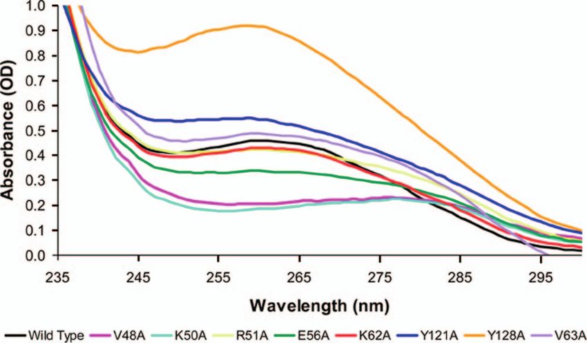
1170 GRINBERG ET AL. J. BACTERIOL.
radical generating subunit in class I enzymes. NrdJ consists of NrdR is a 146- to 200-amino-acid C4-type zinc ribbon/ATP-
a single polypeptide and is considered to be the simplest of the cone protein that is present in a very broad group of eubacteria
RNRs. Class III RNRs are encoded by nrdD, which occurs in (HAMAP: MF_00440 [http://tw.expasy.org/unirules/MF_00440],
an operon containing nrdG, coding for a specific activase that or COG1327 [http://www.ncbi.nlm.nih.gov/COG/grace/wiew.cgi
uses S-adenosylmethionine to create a stable oxygen-sensitive ?COG1327]). Computer analysis of NrdR (5) reveals that the
glycyl radical close to the active site of NrdD. N-terminal an ⬃45-amino-acid sequence defines a zinc ribbon
Allosteric regulation of RNRs is mediated by the binding of motif belonging to the family of zinc finger spatial structures that
nucleoside and deoxynucleoside triphosphate effectors to two typically function as interaction modules with nucleic acids,
distinct sites, a specificity site that regulates substrate specific- proteins, and small molecules (30). Immediately following, an
ity and an activity site that regulates overall enzyme activity ⬃90-amino-acid sequence is predicted to form an ATP-cone
(36). Effectors induce specific conformational changes in pro- domain similar to that present in the overall effector activity site
tein structure that modulate enzyme activity, however the mo- of E. coli NrdA (5). We previously showed that an intact zinc
lecular mechanisms are not well understood. Bacterial class Ia ribbon domain is necessary for binding of NrdR to conserved
RNRs (and some class III RNRs) typically contain an effector tandem 16-bp sequences, termed NrdR-boxes, located in the
binding site for regulating overall activity. Binding of ATP to upstream regulatory regions of both S. coelicolor RNR operons
that domain stimulates activity, whereas binding of dATP in- (18). Rodionov and Gelfand (38) subsequently used phylogenetic
hibits activity. In contrast, class Ib RNRs (and many class II profiling to show that the location of NrdR-boxes is almost
RNRs) lack the activity site. The crystal structure of E. coli invariably correlated with that of RNR operons. S. coelicolor
NrdA complexed with a nonhydrolyzable ATP analogue NrdR contains up to one mole of tightly bound ATP or dATP
Downloaded from jb.asm.org at TEL AVIV UNIV on August 5, 2009
[AMPPNP adenosine 5⬘-(-␥-imido)-triphosphate] established per mole protein. The ATP-cone domain alone determines
that the activity site lies in a sequence of approximately 100 nucleotide binding since a truncated protein that contains only
amino acids, located at the N-terminal portion of the molecule, that domain binds ATP/dATP (18). Moreover, a NrdR ATP-cone
which forms a cleft with a four-helix bundle covered by a mutant that is defective in nucleotide binding was found to be
three-stranded mixed -sheet (14, 48). Aravind et al. first unable to bind short DNA probes containing NrdR-boxes (18).
coined the term ATP-cone to describe the nucleotide binding These observations led us to propose that when NrdR binds
domain present in the N-terminal region of class Ia and class ATP/dATP it undergoes a conformational change that facilitates
III RNRs (1). The ATP-cone consensus sequence (http://pfam binding to its cognate DNA recognition sequences to repress
.sanger.ac.uk/family?acc⫽PF03477) contains the signature se- RNR gene expression. In the present study we show that
quence VXKRDG. In some bacteria the class Ia NrdA pro- mutations in the NrdR ATP-cone domain can have profound
teins contain more than one ATP-cone domain. Pseudomonas effects on ATP/dATP binding, on protein oligomerization, and on
aeruginosa, Legionella pneumophila, and Azotobacter vinelandii DNA binding. A model is proposed that attempts to integrate
all possess in the N-terminal region two ATP-cone domains. In these observations.
P. aeruginosa only the proximal N-terminal ATP-cone is func-
tional (47). Chlamydiaceae species are predicted to possess an MATERIALS AND METHODS
NrdA with three ATP-cones (1). In some other prokaryotes,
Bacterial strains and plasmids. S. coelicolor strain M145 is referred to as the
including Streptomyces, certain AT-rich Firmicutes and halo- wild type (29). E. coli strain XL1-Blue was used for plasmid constructions, and E.
philic Archaea, the class I RNR large subunit is distinguished coli strain BL21(DE3) was used for protein overexpression.
from the canonical class Ia NrdA subunit in that it lacks an Culture medium and DNA manipulations. E. coli strains were grown in Luria-
ATP-cone domain. Bertani (LB) medium and supplemented with kanamycin or ampicillin (50 or 100
g/ml, respectively) when appropriate. Plasmid DNA was isolated by using a
Relatively little is known about how bacteria control RNR High Pure plasmid isolation kit (Roche, Mannheim, Germany). DNA restriction
activity at the gene level (2, 17, 20, 23, 24, 42). Studies of digestions and ligations were carried out according to the manufacturer’s instruc-
Streptomyces have shown that RNRs are regulated at the tran- tions. DNA linear fragments were isolated from a 0.9% agarose (Sigma) gel by
scriptional level. Streptomyces spp. are gram-positive aerobic using a QIAquick gel extraction kit (Qiagen). DNA manipulations were as
described by Sambrook et al. (40). Electroporation was performed with a Gene
bacteria that produce a remarkable variety of metabolites and
Pulsar II apparatus (Bio-Rad Laboratories) according to the manufacturer’s
possess a complex life cycle (11). They and other members of instructions.
the high G⫹C branch of the actinomycetes contain class I and Construction of NrdR mutant proteins. Single point mutations in the ATP-
class II RNRs (6). The class I NrdAB reductase, encoded by cone domain—Val483Ala, Lys503Ala, Arg513Ala, Glu563Ala, Lys623Ala,
the nrdAB genes, is ordinarily very weakly expressed in vege- Val633Ala, Tyr1213Ala, and Tyr1283Ala—were created by an overlap PCR
procedure as described previously (18). Two PCR fragments were amplified from
tative growth, whereas the class II NrdJ RNR, encoded by nrdJ, M145 genomic DNA by use of two nonmutagenic external oligonucleotides, the
is highly expressed. Either RNR is sufficient for vegetative forward primer IG-1 (5⬘-ATATCATATGCACTGCCCCTTTGC-3⬘) contains an
growth (5, 6). We identified in Streptomyces coelicolor a tran- NdeI restriction site and the reverse primer IG-2 (5⬘-TCTCAAGCTTGTCGG
scriptional regulator, NrdR, that controls expression of both CGGCGCCTGCGG-3⬘) contains a HindIII restriction site (restriction sites are
underlined), as well as two complementary mutagenic internal oligonucleotides
sets of RNR genes (5). NrdR, encoded by nrdR, is coexpressed
(Table 1). The PCR products of the reaction were purified by using a High Pure
with NrdJ. Deletion of nrdR causes a dramatic increase in PCR product purification kit and treated with Klenow fragment to remove
transcription of class I and class II RNR genes (5). An analo- protruding 3⬘ A nucleotides, and the DNA fragments containing the mutations
gous situation occurs in Escherichia coli which contains class Ia were gel purified. The PCR fragments were mixed, denatured, reannealed, and
NrdAB and class Ib NrdEF RNRs (46). Normally, only the E. amplified with the two external primers. The mutant DNA fragments were
digested with NdeI and HindIII and ligated into the NdeI- and HindIII-digested
coli class Ia RNR functions during aerobic growth (25). When pET30a(⫹) vector. The resulting recombinant plasmids were introduced into E.
the nrdR gene was deleted transcription of the class Ib RNR coli BL21(DE3) by electroporation. DNA inserts were sequenced to verify the
genes was greatly elevated. presence of the expected mutation.VOL. 191, 2009 FUNCTIONAL ANALYSIS OF THE NrdR ATP-CONE DOMAIN 1171
TABLE 1. Primers and oligonucleotide sequences used for the troscopic determination of eluted nucleotides was performed as described
construction of S. coelicolor NrdR mutants previously (18).
Western blotting. S. coelicolor cell extracts were chromatographed on Su-
Primer Sequence (5⬘–3⬘)a
perdex 200. Eluted fractions were loaded on 12% sodium dodecyl sulfate-
NrdR_V48A_FOR.........................GCTCGCTCATGGCTGTGAAG polyacrylamide gel electrophoresis (SDS-PAGE) gels (33) and transferred
CGGTC onto a nitrocellulose membrane by electroblotting for 1 h at 350 mA. The
NrdR_V48A_REV.........................GACCGCTTCACAGCCATGAG unoccupied sites on the membrane were blocked with Tris-buffered saline
CGAGC (pH 7.5) containing 0.1% Tween 20 and 5% (wt/vol) nonfat milk powder. The
NrdR_K50A_FOR.........................CATGGTGGTGGCTCGGTCCG membrane was incubated with rabbit serum anti-NrdR (Sigma) at 1:500 in
GGGTC blocking solution at room temperature for 1 h, followed by five washes with
NrdR_K50A_REV.........................GACCCCGGACCGAGCCACCA Tris-buffered saline–Tween 20. The membrane was incubated for 1 h with
CCATG goat anti-rabbit immunoglobulin G–horseradish peroxidase conjugate (Jack-
NrdR_R51A_FOR.........................ATGGTGGTGAAGGCGTCCGG son) 1:10,000 in blocking solution, washed as described above, and developed
GGTCA using a SuperSignal kit (Pierce) according to the manufacturer’s instructions.
NrdR_R51A_REV.........................TGACCCGGACGCCTTCACCA Spectroscopic methods. UV absorption spectra were recorded with an Ultro-
CCAT spec 2100 pro UV/visible spectrophotometer (Amersham Biosciences) using
NrdR_E56A_FOR .........................CGGGGTCACCGCTCCGTTCAG 1-cm quartz cuvettes. Circular dichroism spectroscopy was carried out as previ-
CCGC ously described (18).
NrdR_E56A_REV.........................GCGGCTGAACGGAGCGGTGA Electrophoretic gel mobility shift assays. The following S. coelicolor DNA
CCCCG sense and antisense oligonucleotides containing 51 and 52 bp that span the
NrdR_K62A_FOR.........................TCAGCCGCACCGCTGTGATCA
tandem nrdAB and nrdRJ promoter NrdR-box sequences, respectively, were
ACGG
ordered from Syntezza, Israel: 5⬘-GGACACAACATCTGGGGGTGCTCGCG
NrdR_K62A_REV.........................CCGTTGATCACAGCGGTGCG
Downloaded from jb.asm.org at TEL AVIV UNIV on August 5, 2009
TCCCCCGGCACAAGATGTATGCTCA-3⬘ and 5⬘-AATCCCCACATCTAGT
GCTGA
GGTTGGATAGCGTGAGCAGCCCACAAGTTGTGGTCC-3⬘. (NrdR boxes
NrdR_Y121A_FOR.......................CCTCGTCGCCGCTCTGCGATT
are indicated by underlining.)
CGCC
Annealing was performed by dissolving the oligonucleotides in 0.1 M NaOH,
NrdR_Y121A_REV.......................GGCGAATCGCAGAGCGGCGA
CGAGG mixing and incubating complementary oligodeoxyribonucleotide at a ratio 1:1 in
NrdR_Y128A_FOR.......................TTCGCCTCCGTCGCCCGGGCG a final volume of ⬃100 l at room temperature for 25 min and then dialyzing the
TTCG samples against TEN buffer. The fragments were labeled at the 3⬘ end with
NrdR_Y128A_REV.......................CGAACGCCCGGGCGACGGAG digoxigenin-labeled ddUTP by the use of a terminal transferase kit (Roche).
GCGAA Binding assays were carried out as described previously (18). The final reaction
NrdR_V63A_FOR.........................TTCAGCCGCACCAAGGCCATC volume of 20 l contained labeled DNA (50 fmol), binding buffer (20 mM
AACGGTGTGCG Tris-HCl [pH 8.5], 5% [vol/vol] glycerol, 1 mM MgCl2, 40 mM KCl, 1 mM DTT),
NrdR_V63A_REV.........................CGCACACCGTTGATGGCCTTG purified wild-type or mutant recombinant NrdR (1 to 10 g of protein), 1 g of
GTGCGGCTGAAC poly(dI-dC), and 0.1 g of bovine serum albumin. After a 30-min incubation at
30°C, the reaction products were separated on a native 6% polyacrylamide gel in
a
Underlining indicates mutant nucleotides. 0.5⫻ TB (Tris-borate buffer [pH 8.5]). The gel was contact blotted onto a
Hybond-N⫹ membrane (Amersham Biosciences). Chemiluminescence detection
was performed according to the manufacturer’s instructions (Roche). The mem-
brane was exposed to X-ray film (Fuji) for 15 to 40 min at 37°C.
Protein overexpression. Overnight cultures of E. coli BL21(DE3)/pET30a(⫹) Bioinformatics and protein sequence analysis. Domain and motif analyses of
bearing wild-type or mutant nrdR genes were diluted to an absorbance at 600 nm protein sequences were performed using the following databases of protein
of 0.1 in LB containing kanamycin (50 g/ml) and shaken vigorously at 37°C. At families, domains, and functional sites: HAMAP (http://www.expasy.org/sprot
an absorbance at 600 nm of 0.6, IPTG (isopropyl--D-thiogalactopyranoside; /hamap/), Pfam (http://pfam.sanger.ac.uk/), ProDom (http://prodom.prabi.fr
Sigma) was added to a final concentration of 0.4 mM. The cells were incubated /prodom/current/html/home.php), ProSite (http://www.expasy.org/prosite/),
for 3 h at 37°C with shaking and harvested by centrifugation at 4,000 ⫻ g for 20 InterPro (http://www.ebi.ac.uk/interpro/), TIGRFAMs (http://www.tigr.org
min at 4°C. The supernatant was discarded, and the cell pellet was stored at /TIGRFAMs/), and BLOCKs (http://blocks.fhcrc.org/). Protein secondary-
⫺70°C. structure prediction was performed by using the GOR method (15) available
Protein purification. Frozen cells were thawed and suspended in sonication via the GORIV server (http://npsa-pbil.ibcp.fr/cgi-bin/npsa_automat.pl?page
buffer (50 mM Tris-HCl, [pH 8.5], 300 mM NaCl, 5 mM imidazole, 1 mM ⫽npsa_gor4.html) and the PredictProtein Server (http://www.predictprotein
dithiothreitol [DTT]). Phenylmethylsulfonyl fluoride was added to 1 mM to .org/). Pairwise and multiple amino acid sequence alignments were prepared
the cell suspension, and the mixture was sonicated in an ultrasonic processor
by using CLUSTAL W programs (21) improving the sensitivity of progressive
(Misonics) until clear. The sonicate was centrifuged at 10,000 ⫻ g for 45 min
multiple sequence alignment through sequence weighting, position-specific gap
at 4°C. The supernatant was loaded on a His-Trap HP column (GE Amer-
penalties, and weight matrix choice (34, 45), using either the EMBL CLUSTAL
sham Biosciences) using the AKTA prime system equilibrated in buffer (50
W2 server (http://www.ebi.ac.uk/Tools/clustalw2/index.html) or the PBIL-IBCP
mM Tris-HCl [pH 8], 300 mM NaCl, 15 mM imidazole, 1 mM DTT). After
Lyon-Gerland server at the Institute of Biology and Chemistry of Proteins (http:
loading the supernatant, the column was washed with 160 ml of the same
//npsa-pbil.ibcp.fr/cgi-bin/npsa_automat.pl?page⫽/NPSA/npsa_clustalw.html).
buffer containing increasing concentrations of imidazole. Bound NrdR was
Phylogenetic trees were generated by using multiple sequence alignments per-
eluted with buffer containing 250 mM imidazole. Protein samples after Ni2⫹
affinity purification were dialyzed against buffer containing 50 mM Tris-HCl formed with the EMBL CLUSTAL W2 server (see above) and Molecular Evo-
(pH 8.0), 300 mM NaCl, 5 mM DTT, 50 M ZnCl2, and 20% glycerol when lutionary Genetics Analysis software (MEGA, v.4) (32).
assayed for DNA binding and dialyzed against 50 mM Tris-HCl (pH 8)–300 Other methods. The protein concentration was determined by the method of
mM NaCl for spectroscopic analyses. DTT (5 mM) was added to the same Bradford (8) with bovine serum albumin as the standard. SDS-PAGE was per-
buffer for gel filtration chromatography. Recovery of recombinant protein formed as described by Laemmli (33). Nucleotide sequencing was determined by
was monitored by the Bradford assay (8). Purified recombinant proteins were using an AB1 Prism 3100 genetic analyzer (Applied Biosystems) and a BigDye
stored at ⫺70°C. terminator cycle sequencing kit (Applied Biosystems) as recommended by the
Chromatography. Gel filtration chromatography was carried out on a Su- manufacturer, except that 5% dimethyl sulfoxide was added to each reaction
perdex 200HR 10/30 column (Pharmacia) connected to a Pharmacia fast- mixture. Perchloric acid precipitation of NrdR was performed by adding per-
protein liquid chromatography system in 50 mM Tris-HCl (pH 8)–300 mM chloric acid to the protein solution to a final concentration of 1 M; the protein
NaCl–5 mM DTT. MW-GF-200 molecular weight markers (Sigma) were run was then left on ice for 1 h, and the solution was centrifuged for 30 min at 4°C
in parallel (see Fig. S5 in the supplemental material). Fractions (0.5 ml) were at 14,000 rpm. The supernatant was adjusted to neutral pH and used for spec-
collected and analyzed. Affi-Gel boronate affinity chromatography and spec- troscopic analysis and Affi-Gel boronate chromatography.1172 GRINBERG ET AL. J. BACTERIOL.
Downloaded from jb.asm.org at TEL AVIV UNIV on August 5, 2009
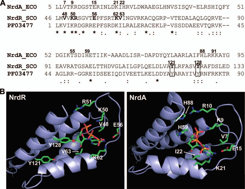
FIG. 1. (A) Alignment of deduced amino acid sequences of the E. coli NrdA and S. coelicolor NrdR ATP-cone domains. E. coli (ECO) NrdA
numbered residues are those known from the crystal structure to interact with an ATP analog. The corresponding S. coelicolor (SCO) NrdR
numbered residues that were modified in the present study are indicated in boldface. Two S. coelicolor tyrosines that are fully conserved in NrdRs,
but absent in NrdA, are boxed. Asterisks indicate fully conserved residues, colons indicate strongly similar residues, and dots indicate weakly similar
residues. PF03477 represents a 90-amino-acid consensus sequence of the ATP-cone (http://pfam.sanger.ac.uk/family?acc⫽PF03477). (B) Homol-
ogy modeling of the S. coelicolor NrdR ATP-cone domain. The right panel shows the E. coli NrdA ATP-cone domain. The left panel shows a
homology model of the S. coelicolor NrdR ATP-cone domain based on the crystal structure of E. coli NrdA (14, 48). S. coelicolor NrdR residues
changed in the present study, and the corresponding E. coli NrdA residues, are indicated.
Comparative protein modeling. Comparative homology modeling of the S. supplemental material) and the ATP-cone domains of NrdR,
coelicolor NrdR ATP-cone and E. coli NrdA ATP-cone was performed using the NrdA, NrdB, NrdD, and NrdJ (see Fig. S1B in the supplemen-
SWISS-MODEL (19).
tal material) shows that each of the above mentioned residues
(with the exception of Tyr121) are well conserved in the dif-
RESULTS ferent RNR protein subunits. They are also well conserved in
The NrdR ATP-cone domains form a well-defined group ATP-cones that were, unexpectedly, identified in several class
that is separated from ATP-cone domains belonging to class Ia RNR NrdB proteins. The phylogeny of 75 NrdR, NrdA,
Ia, II, and III RNR proteins. Fig. 1A shows a comparison of NrdB, NrdD, and NrdJ ATP-cone domains can be found in
the amino acid sequences of the S. coelicolor NrdR and E. coli Fig. S2 in the supplemental material.
NrdA ATP-cone domains. The S. coelicolor NrdR ATP-cone Currently, there is no crystal structure of the NrdR ATP-
consists of residues 46 to 133; the E. coli NrdA N-terminal cone domain. The crystal structure of E. coli NrdA with an
ATP-cone domain consists of residues 5 to 95 and contains the ATP analog (AMPPNP) bound has been solved (14, 48). The
allosteric overall activity site. The ATP-cone consensus motif N-terminal residues, 5 to 95 (PDB: 1RLR), form the allosteric
(PF03477 [http://pfam.sanger.ac.uk/family?entry ⫽ PF03477&type activity site cleft consisting of a four-helix bundle covered by a
⫽ Family]) is shown below the alignment. E. coli NrdA resi- three-stranded mixed -sheet (Fig. 1B). The ATP analog is
dues that are inferred from the crystal structure of the NrdA- deep in the cleft. The adenine base is hydrogen bonded to the
ATP analog complex to interact with the analog nucleotide mainchain residues 17 to 19 and stacked upon Val7 on one side
(14) are shown in bold numbers. The corresponding S. coeli- and upon Ile22 and Ile58 on the other. At the entrance of the
color NrdR residues are Val48, Lys50, Arg51, Lys62 and Val63. cleft four positively charged residues—Lys9, Arg10, Lys21, and
The highly conserved Glu56 was also mutated since the corre- Lys91—are positioned around the phosphates. The ribose
sponding NrdA Glu15 lies close to Val 7 which interacts with sugar 3⬘-OH is close to His59, while the 2⬘-OH is H-bonded to
the adenine base. These residues are highly conserved in bac- NH of the main chain and surrounded by hydrophobic resi-
terial NrdR proteins and were modified in this work. Two dues, including Ile22 and Val25. Thr55 binds to the 5⬘ oxygen.
highly conserved NrdR tyrosines, Tyr121 and Tyr128 were also Homology modeling of the S. coelicolor NrdR ATP-cone,
changed. Alignment of NrdR proteins (see Fig. S1A in the using the NrdA crystal structure as a template, shows that theVOL. 191, 2009 FUNCTIONAL ANALYSIS OF THE NrdR ATP-CONE DOMAIN 1173
groups based on the mole amount of bound nucleotide per
mole protein. Group 1 mutants bind a similar amount of nu-
cleotide as wild type (R51A, K62A, Val63A, and Y121A) or a
greater amount (Y128A); group 2 mutants bind significantly
less nucleotide than wild type, 30 to 70% less (V48A, K50A,
and E56A). The values reported in Table 2 for moles of nu-
cleotide bound/moles of protein are the averages of two inde-
pendent preparations of proteins. The absolute values are sub-
ject to possible error since protein concentrations were
determined by the method of Bradford, using bovine serum
albumin as a standard, and may depend on the amino acid
composition of the protein. However, the relative values of the
amounts of moles of nucleotide bound/moles of protein be-
FIG. 2. UV absorption spectra of NrdR wild-type and mutant pro- tween wild type and the different mutants do reflect the effects
teins. The UV absorption spectra of 0.04 mM solutions of wild-type of the mutations. Similar relative values for the moles of nu-
and mutant proteins in 50 mM Tris-HCl (pH 8.0)–300 mM NaCl were cleotide bound/moles of protein, 0.8 to 1.2 for group 1 mutants
determined. and 0.2 to 0.4 for group 2 mutants, were obtained after treat-
ment of proteins with perchloric acid, which partly releases
protein-bound nucleotide while precipitating the protein (18)
Downloaded from jb.asm.org at TEL AVIV UNIV on August 5, 2009
two ATP-cone domains may possess similar structures (Fig. (Table 2). The absolute values of nucleotide/protein in the
1B). It confirms that residues Lys50, Arg51, and Lys62 (corre- PCA method were two- to threefold less than those based on
sponding to E. coli NrdA Lys9, Arg10, and Lys21) are posi- the protein spectra, presumably due to incomplete release of
tioned close to the phosphates and that Val63 (corresponding nucleotide, but the relative values were the same for all of the
to E. coli NrdA Ile22) and Thr96 (probably corresponding to mutant proteins.
E. coli NrdA Thr55) are close to the sugar moiety (see Fig. 1B). To assess the relative amounts of ATP and dATP in wild-
S. coelicolor NrdR Val48 and Glu56 (corresponding to in E. type and mutant proteins, supernatants from proteins treated
coli NrdA Val7 and Glu15) lie close to the adenine ring. The with perchloric acid were analyzed by Affi-Gel boronate chro-
UV circular dichroism spectra (see Fig. S3 and S4 in the matography as previously described (18). Figure 3 shows that
supplemental material) of native S. coelicolor NrdR at different
temperatures and that of a NrdR mutant protein (18) contain-
ing only the ATP-cone domain (residues 42 to 150) indicates
TABLE 2. Properties of S. coelicolor wild-type and mutant
that the ATP-cone domain is a largely alpha-helical and stable NrdR proteins
structure consistent with the three-dimensional structure of the
DNA
NrdA ATP-cone domain (14, 48). Some differences are, how- Mol of ATP⫹dATP/ % dATP/
Major
bindingd
NrdR protein oligomeric
ever, evident in the two ATP-cone domains. Comparison of the mol of proteina dATP⫹ATPb
statec
amino acid sequences surrounding the ligand binding site re- L H
veals that NrdR contains two tyrosine residues, Tyr121 and Group 1
Tyr128, both of which are absent in NrdA. Tyr128 is predicted Wild type 0.71 (0.35) 38 L ⫹ –
to contact the sugar 3⬘-OH, while Tyr121 is distal to the ligand R51A 0.76 (0.26) 96 L ⫹ –
Y121A 0.73 (0.36) 96 L ⫹ –
binding site. Another difference is that NrdR lacks two histi- K62A 0.58 (0.34) 57 L ⫺ –
dines present in NrdA, His59, and His88. In NrdA, His59 is V63A 0.83 (0.35) 92 H ⫹/– –
close to the sugar 3⬘-OH and to His88 (which in alignments Y128A 1.56 (0.44) 100 L ⫹ –
appear close to Tyr128 [Fig. 1A]), which is positioned in the
cleft between His59 and Lys91. His59 and His88 were pro- Group 2
V48A 0.19 (0.06) 82 H ⫹ –
posed to be H bonded and to facilitate interaction between the K50A 0.27 (0.09) 91 H ⫹ –
NrdA and NrdB subunits (4). One other difference is the E56A 0.37 (0.13) 100 H ⫹ –
presence in NrdR of Val63 in place of the more bulky Ile22 in ATP- 0.70 (0.33) 25 Dimer NA NA
NrdA. The effect of these and other changes may cause a cone
narrowing of the cleft. a
The amounts of bound ATP and dATP per mole of wild-type and mutant
Functional analysis of NrdR ATP-cone mutant proteins: NrdR, determined by protein absorption spectra and nucleotide absorption spec-
tra after PCA treatment (in parentheses), are the average values of two mea-
effect of mutations on nucleotide content, DNA binding, and surements made with two independent protein preparations obtained after Ni2⫹
oligomeric state. Table 2 lists the S. coelicolor NrdR mutant affinity chromatography.
b
proteins made in the present study. Val48, Lys50, Arg51, The percent dATP of bound nucleotide, determined by Affi-Gel boronate
affinity chromatography, is the average value of two to three measurements with
Glu56, Lys62, Val 63, Tyr121, and Tyr128 were all changed to the same protein preparations. Individual values differed from the mean by no
alanines. UV spectroscopy was used to assess the amount of more than 6%.
c
The oligomeric state of the wild type and mutant is that of the major protein
bound nucleotide, ATP and dATP, present in wild-type and fraction obtained after Superdex 200 gel filtration chromatography (see Fig. 5),
mutant proteins as described previously (18) (Fig. 2). Previ- which is denoted as “L” for low-molecular-weight material and “H” for high-
ously, we showed, by using truncated S. coelicolor NrdR pro- molecular-weight aggregated material.
d
DNA binding refers to the ability of the low- and high-molecular-weight
teins, that the ATP-cone domain alone determines NrdR nu- fractions to bind to the probes nrdAB and nrdRJ. ⫹, binding; ⫹/⫺, weak
cleotide content (18). We divided the NrdR mutants into two binding; –, lack of binding. NA, not applicable.1174 GRINBERG ET AL. J. BACTERIOL.
FIG. 4. Gel filtration profiles of S. coelicolor wild-type NrdR.
FIG. 3. Differential binding of ATP and dATP by NrdR wild-type Crude cell extracts of S. coelicolor M145 were chromatographed on
and mutant proteins. Affi-Gel boronate chromatography of the super- Superdex 200, and fractions were subjected to Western analysis.
natant obtained after perchloric acid treatment of the wild-type and
mutant proteins was performed. Nucleotides were eluted, in succes-
sion, with buffer A to elute bound dATP and with buffer B to elute
bound ATP. The results are the average of two sets of measurements We next analyzed the effect of mutations in the NrdR ATP-
Downloaded from jb.asm.org at TEL AVIV UNIV on August 5, 2009
with independent protein preparations. Values differed by no more cone domain on oligomerization. Figure 5 shows the Superdex
than 6% from the mean. 200 gel filtration chromatography results for wild-type NrdR
and mutant proteins. The gel filtration profiles of the wild type
and group 1 mutants—R51A, K62A, Y121A, and Y128A—
in wild-type NrdR ca. 60% of the released nucleotides is ATP were similar. Each contained a major and a minor fraction.
and 40% dATP. In contrast, all of the mutant proteins were The major fraction consists of molecules with a mass of ⬃180
found to contain significantly more dATP than ATP. Compar- kDa; the minor fraction consists of heterogeneous molecules
ison of the UV circular dichroism spectra of wild-type and with a mass greater than 400 kDa that elutes in the void
group 2 mutant proteins failed to show any significant differ- volume (Fig. 5). In contrast, gel filtration chromatography of
ences in their secondary structure (see Fig. S3 in the supple- the group 2 V48A, K50A, and E56A mutants and the group 1
mental material). This does not preclude possible changes in V63A mutant revealed anomalous profiles in which the major
overall folding of the proteins. protein fraction occurred in an aggregated state with a size
Effect of ATP-cone mutations on protein oligomeric state. greater than 400 kDa. All contained a minor fraction with a
Previous studies showed that S. coelicolor NrdR when ex- size of about 80 to 120 kDa (Fig. 5). These observations show
pressed as a C-terminal His6 tag protein in E. coli and purified that alterations in the ATP-cone can have profound effects on
by Ni2⫹ affinity column chromatography is an oligomeric pro- protein oligomerization. To determine whether the ATP-cone
tein (18). In a low salt concentration (0.3 M NaCl) the molec- domain alone forms oligomers, gel filtration chromatography
ular mass of the recombinant protein, determined by Superdex was performed with a truncated NrdR protein consisting of
200 gel filtration, was ⬃180 kDa, corresponding to an oligomer NrdR residues 42 to 150 (18). Figure 5 shows that the ATP-
containing eight monomers of 21.2 kDa. At a high salt con- cone protein has a molecular mass corresponding to it con-
centration (1.5 M NaCl) its estimated size was ⬃85 kDa, cor- taining two subunits, while the native NrdR protein has a mass
responding to a tetramer. Under the former conditions two corresponding to about eight subunits.
peaks are observed, the main peak consists of a protein with a Effect of ATP-cone mutations on DNA binding. To assess
molecular mass of ⬃180 kDa and a minor peak consisting of binding of NrdR to DNA, electrophoretic gel mobility shift
protein with a much larger mass, ⬎400 kDa, which eluted in assays were performed with wild-type and mutant proteins
the void volume. We refer to this latter material as the high- using the 51- and 52-bp digoxigenin-labeled DNA probes
molecular-mass (H) or aggregated NrdR and the former as nrdAB and nrdRJ, respectively. The probes contain two tan-
low-molecular-mass (L) material. When the two peak fractions dem centrally located, NrdR-box sequence motifs. As previ-
were rechromatographed, each gave the same elution profile, ously reported, the amounts of NrdR proteins used in binding
indicating that the two forms do not readily interconvert (data reactions to obtain DNA gel shifts were relatively high, sug-
not shown). gesting that NrdR exists in a multimeric state (18) or may in
To determine the molecular size of native NrdR, cultures of part be due to low binding affinity. We cannot rule out that the
S. coelicolor were grown in YEME medium (29) and harvested, protein may be partially denatured or that additional factors
and cell extracts were chromatographed on Superdex 200. may affect its DNA-binding activity. Binding reactions were
Western analysis, with anti-NrdR antibodies, showed that na- carried out with wild-type NrdR and with the low (L)- and high
tive NrdR eluted in a single peak with a molecular mass of (H)-molecular-weight fractions of mutant proteins obtained by
⬃170 kDa (Fig. 4) corresponding to an oligomer of eight size chromatography (see Fig. 5). The low-molecular-mass
monomers of 19.7 kDa, similar to that of recombinant His tag (⬃180-kDa) protein fractions of the group 1 R51A, Y121A,
NrdR in a low salt concentration. Assuming this material con- and Y128A mutant proteins formed DNA-protein complexes
tains only NrdR, the finding confirms that native NrdR is a like that of wild type with both nrdAB and nrdRJ DNA probes
putative octomeric protein. (Fig. 6, upper and lower panels). No discernible DNA bindingVOL. 191, 2009 FUNCTIONAL ANALYSIS OF THE NrdR ATP-CONE DOMAIN 1175
Downloaded from jb.asm.org at TEL AVIV UNIV on August 5, 2009
FIG. 5. Superdex 200 gel filtration chromatography of NrdR wild-type and mutant proteins. Fractions were analyzed for protein by method of
Bradford (8) and by SDS-PAGE (33).
occurred with the high-molecular-mass aggregated protein NrdR species. However, the minor low-molecular-mass pro-
fractions (⬎400 kDa) with either probe. In contrast, the group tein fractions of V48A (85 kDa), K50A (120 kDa), E56A (120
1 K62A low- and high-molecular-mass protein fractions failed kDa), and V63A (120 kDa) showed partial binding to both
to bind to either DNA probe (Fig. 6). In the group 2 mutants DNA probes to form complexes that migrated like that of
V48A, K50A, and E56A and the group 1 mutant V63A the wild-type NrdR. Table 2 summarizes the properties of the S.
high-molecular-mass major protein fractions (⬎400 kDa) all coelicolor NrdR mutant proteins.
failed to form normal DNA protein complexes with the nrdAB
and nrdRJ probes, as judged by their mobility (Fig. 7). The DISCUSSION
K50A H fraction binds very weakly to the nrdAB probe but not
to the nrdRJ probe. The H fractions form higher-order com- We recently proposed that Streptomyces NrdR functions to
plexes which occur in diffuse irregular bands that most likely universally control bacterial RNR activity by acting as an ATP/
arise from nonspecific DNA interactions with the aggregated dATP-dependent regulator of RNR gene expression (18). In1176 GRINBERG ET AL. J. BACTERIOL.
Downloaded from jb.asm.org at TEL AVIV UNIV on August 5, 2009
FIG. 7. Binding of wild-type NrdR and group 2 mutant proteins
to DNA probes. Totals of 50 fmol of the nrdAB and nrdRJ probes
FIG. 6. Binding of wild-type NrdR and group 1 mutant proteins to were incubated with the indicated amounts (in g) of NrdR (1 g
DNA probes. Totals of 50 fmol of the nrdAB and nrdRJ probes were corresponds to 47 pmol of hexahistidyl-tagged NrdR protein) wild-
incubated with the indicated amounts (in g) of NrdR (1 g corre- type and mutant low (L)- and high (H)-molecular-weight protein
sponds to 47 pmol of hexahistidyl-tagged NrdR protein) wild-type and fractions obtained from Superdex 200 gel filtration chromatography
mutant low (L)- and high (H)-molecular-weight protein fractions ob- (see Fig. 3). (A) nrdAB probe; (B) nrdRJ probe. Lanes 1 to 4, NrdR
tained from Superdex 200 gel filtration chromatography (see Fig. 3). wild type (185 kDa); lanes 5 to 7, V48A (85 kDa); lanes 8 to 10,
(A) nrdAB probe; (B) nrdRJ probe. Lanes: 1 to 4, wild-type NrdR (185 V48A (⬎400 kDa); lanes 11 to 13, K50A (120 kDa); lanes 14 to 16,
kDa); 5 and 6, Y121A (⬎400 kDa); 7 and 8, Y121A (170 kDa); lane 9, K50A (⬎400 kDa); lanes 17 to 19, E56A (120 kDa); lanes 20 to 22,
Y128A (⬎400 kDa); lanes 10 to 12, Y128A (190 kDa); lane 13, R51A E56A (⬎400 kDa); lanes 23 to 24, V63A (⬎400 kDa); lanes 25 to
(⬎400 kDa); lanes 14 to 16, R51A (185 kDa); lanes 17 to 19, K62A 26, E56A (⬎400 kDa).
(⬎400 kDa); lanes 20 to 22, K62A (180 kDa).
this model NrdR binds ATP/dATP via its ATP-cone domain, held deep within the ATP-cone cleft and contacted by numer-
eliciting a conformational change that modulates its binding to ous residues (14, 48). We therefore adopted an alternative
tandem 16-bp NrdR-box sequences located in the promoter approach in which the NrdR ATP-cone residues that are con-
regions of class I and class II RNR operons. Differential ex- served in E. coli NrdA and are inferred from the crystal struc-
pression of the RNR operons arises, in part, from the respec- ture to be involved in ligand binding were modified. Three
tive positions of the NrdR-boxes which overlap with, or are NrdR positively charged residues, Lys50, Arg51, and Lys62
proximal to, their promoter elements, and to differences in (which in NrdA interact with phosphates), and the two hydro-
NrdR-box sequences. A similar role was proposed for the E. phobic residues, Val48 and Val63 (which in NrdA lie above
coli NrdR homolog (formerly named YbaD) for differential and below the adenine base), were changed to alanines. Also,
transcription of its class Ia, class Ib, and class III RNR operons we changed two highly conserved NrdR tyrosines, Tyr121 and
(46). The studies described here were aimed at further analyz- Tyr128, that are absent in the NrdA ATP-cone. The latter
ing the role of the NrdR ATP-cone domain in binding to DNA tyrosine can be aligned with E. coli NrdA His88, which is
probes containing NrdR-boxes. Initially, our goal was to obtain reported to play a role with His59 in allosteric regulation
NrdR with no nucleotides bound so that nucleotide binding and in the interaction between the E. coli NrdA and NrdB
could be quantitatively analyzed and the effects on DNA bind- subunits (4).
ing assessed. Several procedures were tried to remove bound The NrdR ATP-cone mutants described in the present study
nucleotide, including extensive chromatography and dena- exhibit pleiotrophic effects in terms of the absolute and relative
turation in urea; however, we could not release all of the amounts of bound ATP and dATP, as well as their oligomeric
nucleotide. We attempted to address this issue by perform- state and ability to bind DNA. The mutants were divided into
ing prolonged incubation of wild-type NrdR with different two groups based on the amount of bound ATP/dATP. Group
concentrations of ATP and dATP to see whether we could 1 mutants (R51A, Y121A, K62A, and V63A) were similar to
influence NrdR binding to DNA, but we failed to discern any the wild type in terms of nucleotide content; one mutant
significant effects on the binding profiles. These findings indi- (Y128A) bound significantly more nucleotide. The major, low-
cate that the nucleotides are tightly bound. Indeed, the struc- molecular-weight, protein fractions of R51A, Y121A, and
ture of the NrdA ATP-analog complex shows that the ligand is Y128A correspond in size to that of wild type, and all were ableVOL. 191, 2009 FUNCTIONAL ANALYSIS OF THE NrdR ATP-CONE DOMAIN 1177
to bind to nrdAB and nrdRJ DNA probes. In the case of gion and in communicating allosteric inhibition (4). The over-
K62A, the corresponding fraction did not bind to either probe; all activity site occupied by the ligand is open to the solvent and
in V63A the major fraction, the high-molecular-weight frac- favorably disposed to allow subunit interactions. In NrdR the
tion, also failed to bind to the probes, while the low-molecular- N-terminal DNA-binding domain consists of about 45 to 48
weight fraction bound very weakly. We suppose that the defect amino acid residues, of which residues 3 to 34 form the zinc
in binding of the V63A mutant arises from a change in its ribbon C4 motif. The remaining 11 to 14 residues form a linker
oligomeric state (as described for group 2 mutants [see be- that joins the DNA-binding domain to the ATP-cone domain.
low]). The inability of the K62A mutant to bind DNA is We propose that native NrdR is an oligomer, probably con-
puzzling since it possesses the same oligomeric state as the wild- sisting of eight subunits, each of which can bind ATP or dATP,
type NrdR. Lys62, unlike Arg51, is buried deep in the ATP- in which the zinc ribbon domain is free to bind to its DNA
cone cleft, suggesting that the K62A mutation perturbs the target sites to repress RNR gene expression. When the level of
ATP-cone structure which leads to a change in the DNA- cellular dNTPs is low, NrdR is depleted of dATP, and the
binding domain. Group 2 mutants (V48A, K50A, and E56A) ATP-cone undergoes a conformational change which induces,
all contained lesser bound nucleotide than did the wild type, via the linker, a change in its oligomeric state (or some higher
and all had abnormal gel filtration profiles, indicating that most order quaternary structure). Consequently, the disposition of
of the protein was in a highly aggregated state; the minor the zinc ribbon domain is changed, and DNA binding is abro-
low-molecular-weight fractions exhibited partial binding to gated. Hence, NrdR appears to function as an ATP/dATP-
both probes to form complexes that migrated like wild type, dependent regulatory switch that controls the expression of
whereas the major high-molecular-weight fractions all failed to RNR genes in response to cellular nucleotide needs. To estab-
Downloaded from jb.asm.org at TEL AVIV UNIV on August 5, 2009
bind (or bound very weakly) to one or both DNA probes. lish the validity of this model, it will be necessary to obtain apo
These observations agree with those of a previous study which NrdR and show that it binds ATP and dATP with different
showed that the K50NR51G double mutant was significantly affinities. Attempts to obtain the apo protein were unsuccessful
impaired in both nucleotide binding and DNA binding (18). and resulted in partial release of nucleotide; similarly, attempts
How these mutations, which presumably weaken interaction of to exchange the bound ATP/dATP by incubating NrdR with
the ATP-cone with its nucleotide ligand, effect changes in the high concentrations of nucleotides were apparently unsuccess-
protein oligomeric state and DNA binding is unclear. Remark- ful, as judged by the lack of effect on DNA binding, a finding
ably, all of the group 1 and group 2 mutants showed a marked consistent with the nucleotides being tightly bound. For these
preference for binding dATP, whereas wild-type NrdR binds reasons, we performed structure-function studies modifying
about twofold more ATP than dATP. However, an NrdR trun- residues predicted to be involved in nucleotide binding. One
cated protein containing only the ATP-cone domain exhibited possible reason for our failure to obtain apo NrdR is that NrdR
a marked preference for ATP (Table 2). A possible explana- may exist in vivo in a complex with other proteins that affect
tion for these observations is that wild-type NrdR binds either binding of ATP and dATP. Thioredoxin, for instance, was
ATP or dATP, depending on their relative abundance and shown in E. coli to be associated with NrdR (31). Moreover, in
their affinity, while it is being synthesized and then remains in E. coli several proteins, including Fis, DnaA, and IcA, which
that form, and that the mutant proteins have a lower affinity are known to regulate class Ia RNR gene expression, poten-
for ATP. tially interact with NrdR since they bind to sites near to the
Our working model for a mechanism for ATP/dATP-depen- NrdR boxes and coordinate DNA replication and RNR syn-
dent NrdR regulation of RNR gene expression is based pri- thesis during the cell cycle (2, 17, 20, 23, 24). Likewise, the
marily on two observations. First, native NrdR is an oligomeric regulatory region of the E. coli class III RNR genes contain
ATP/dATP-containing protein, and each subunit is assumed to sites for binding NrdR, DnaA, and FNR. Further studies
be able to bind one ATP/dATP molecule (18). Second, in the should clarify whether NrdR is associated in vivo with these or
nucleotide-bound state, the NrdR N-terminal zinc ribbon do- other proteins and whether they play a role in determining
main is free to bind to its DNA substrates. At present, we do nucleotide binding affinity (7, 43, 46).
not understand the nature of the putative NrdR structural Transcription regulators often function as oligomers, and a
changes triggered by binding ATP/dATP, whether ATP and few examples have been described for octameric regulatory
dATP have similar or opposing effects, and how these changes proteins (3, 12). The NrdR target binding sites in RNR oper-
affect aggregation and DNA binding. Indeed, the structural ons are tandem imperfect palindromic 16-bp NrdR-boxes sep-
basis of ATP/dATP allosteric regulation of activity is still not arated by 15 bp. An NrdR octamer composed of four dimers
resolved. In the case of E. coli NrdA (R1), the only ATP-cone could form the active regulator with one dimer binding to each
protein for which a crystal structure exists, it was not possible of the two palindromic half-sites of the tandem NrdR-boxes.
to determine whether binding of an ATP analog alters its Preliminary studies show that NrdR binds to either NrdR box
conformation (14, 48) However, it is noteworthy that prokary- though with different affinities. Interestingly, the gel filtration
otic and eukaryotic R1 ATP-cone proteins exist in different profile of a NrdR mutant containing only the ATP-cone do-
oligomeric states and that binding of ATP/dATP modulates main shows it to be most likely a dimer, suggesting that part of
oligomerization (9, 22, 27, 28, 39, 44, 47). The crystal structure the information necessary to form the larger oligomer resides
of the E. coli NrdA dimer with bound nucleotide shows that in regions outside the ATP-cone.
the ATP-cone domain is located at the N-terminal tip, at the NrdR ATP-cones contain two highly conserved tyrosines
surface of the molecule (14, 48). Two H-bonded histidines, that are missing in E. coli NrdA. Tyr128 (S. coelicolor number-
His59 and His88, located at and near the nucleotide binding ing) is highly conserved in certain bacterial NrdA, NrdJ, and
site, respectively, participate in forming the dimer contact re- NrdD ATP-cones and in five identified cases of bacterial R21178 GRINBERG ET AL. J. BACTERIOL.
ATP-cones (see Fig. S1 in the supplemental material). In E. Sjoberg, and H. Eklund. 1997. Binding of allosteric effectors to ribonucle-
otide reductase protein R1: reduction of active-site cysteines promotes sub-
coli NrdA, the corresponding position is occupied by Phe87 or strate binding. Structure 5:1077–1092.
His88. In contrast, Tyr121 is found uniquely in NrdR ATP- 15. Garnier, J., J. F. Gibrat, and B. Robson. 1996. GOR method for predicting
cones. Inspection of NrdR sequences in public databases shows protein secondary structure from amino acid sequence. Methods Enzymol.
266:540–553.
that the two tyrosines occur in the well-conserved decapeptide 16. Gon, S., and J. Beckwith. 2006. Ribonucleotide reductases: influence of
AY(L/V/I)RFASVY(R/K). Homology modeling of the S. environment on synthesis and activity. Antioxid. Redox Signal. 8:773–780.
coelicolor NrdR revealed that Tyr128 lies near to the nucleo- 17. Gon, S., J. E. Camara, H. K. Klungsoyr, E. Crooke, K. Skarstad, and J.
Beckwith. 2006. A novel regulatory mechanism couples deoxyribonucle-
tide binding site while Tyr121 is distal (Fig. 1B). The role of the otide synthesis and DNA replication in Escherichia coli. EMBO J. 25:
two tyrosines is unknown. Alignment of S. coelicolor NrdR with 1137–1147.
18. Grinberg, I., T. Shteinberg, B. Gorovitz, Y. Aharonowitz, G. Cohen, and I.
E. coli NrdA indicates that Tyr128 appears to occupy the po- Borovok. 2006. The Streptomyces NrdR transcriptional regulator is a Zn
sition of His88 (Fig. 1), which is thought to contribute to the ribbon/ATP cone protein that binds to the promoter regions of class Ia and
NrdA dimer interface (4) and might therefore play a role in class II ribonucleotide reductase operons. J. Bacteriol. 188:7635–7644.
19. Guex, N., and M. C. Peitsch. 1997. SWISS-MODEL and the Swiss-Pdb-
oligomerization. In fact, the S. coelicolor Y128A mutant pos- Viewer: an environment for comparative protein modeling. Electrophoresis
sesses normal DNA binding properties and oligomerization 18:2714–2723.
though, unexpectedly, it binds more nucleotide than wild type, 20. Herrick, J., and B. Sclavi. 2007. Ribonucleotide reductase and the regulation
of DNA replication: an old story and an ancient heritage. Mol. Microbiol.
predominantly dATP. By comparison, the Y121A mutant be- 63:22–34.
haved in all respects like the wild type. Structural studies of the 21. Higgins, D. G., J. D. Thompson, and T. J. Gibson. 1996. Using CLUSTAL
wild-type and mutant apo NrdR proteins, with or without ATP/ for multiple sequence alignments. Methods Enzymol. 266:383–402.
22. Ingemarson, R., and L. Thelander. 1996. A kinetic study on the influence of
dATP, in complex with DNA, should help uncover the molec- nucleoside triphosphate effectors on subunit interaction in mouse ribonucle-
Downloaded from jb.asm.org at TEL AVIV UNIV on August 5, 2009
ular mechanism of NrdR regulation. otide reductase. Biochemistry 35:8603–8609.
23. Jacobson, B. A., and J. A. Fuchs. 1998. A 45-bp inverted repeat is required
for cell cycle regulation of the Escherichia coli nrd operon. Mol. Microbiol.
ACKNOWLEDGMENTS 28:1307–1314.
24. Jacobson, B. A., and J. A. Fuchs. 1998. Multiple cis-acting sites positively
This research was funded in part by a grant from the Israel Science regulate Escherichia coli nrd expression. Mol. Microbiol. 28:1315–1322.
Foundation (1189/04). I.G. was supported by a fellowship from the 25. Jordan, A., I. Gibert, and J. Barbe. 1995. Two different operons for the same
NoE EuroPathoGenomics (EPG) program. Q.H. is an Anna Fuller function: comparison of the Salmonella typhimurium nrdAB and nrdEF
fellow funded by David H. Koch Institute for Integrative Cancer Re- genes. Gene 167:75–79.
search at MIT and was supported by grant GM-29595 from the Na- 26. Jordan, A., and P. Reichard. 1998. Ribonucleotide reductases. Annu. Rev.
tional Institutes of Health, Bethesda, MD. Biochem. 67:71–98.
27. Kashlan, O. B., and B. S. Cooperman. 2003. Comprehensive model for
We thank JoAnne Stubbe for helpful discussions.
allosteric regulation of mammalian ribonucleotide reductase: refinements
and consequences. Biochemistry 42:1696–1706.
REFERENCES 28. Kashlan, O. B., C. P. Scott, J. D. Lear, and B. S. Cooperman. 2002. A
1. Aravind, L., Y. I. Wolf, and E. V. Koonin. 2000. The ATP-cone: an evolu- comprehensive model for the allosteric regulation of mammalian ribonucle-
tionarily mobile, ATP-binding regulatory domain. J. Mol. Microbiol. Bio- otide reductase. Functional consequences of ATP- and dATP-induced oligo-
technol. 2:191–194. merization of the large subunit. Biochemistry 41:462–474.
2. Augustin, L. B., B. A. Jacobson, and J. A. Fuchs. 1994. Escherichia coli Fis 29. Kieser, T., M. J. Bibb, M. J. Buttner, K. F. Chater, and D. A. Hopwood. 2000.
and DnaA proteins bind specifically to the nrd promoter region and affect Practical Streptomyces genetics. John Innes Foundation, Norwich, United
expression of an nrd-lac fusion. J. Bacteriol. 176:378–387. Kingdom.
3. Beloin, C., S. McKenna, and C. J. Dorman. 2002. Molecular dissection of 30. Krishna, S. S., I. Majumdar, and N. V. Grishin. 2003. Structural classifica-
VirB, a key regulator of the virulence cascade of Shigella flexneri. J. Biol. tion of zinc fingers: survey and summary. Nucleic Acids Res. 31:532–550.
Chem. 277:15333–15344. 31. Kumar, J. K., S. Tabor, and C. C. Richardson. 2004. Proteomic analysis of
4. Birgander, P. L., A. Kasrayan, and B. M. Sjoberg. 2004. Mutant R1 proteins thioredoxin-targeted proteins in Escherichia coli. Proc. Natl. Acad. Sci. USA
from Escherichia coli class Ia ribonucleotide reductase with altered responses 101:375937–375964.
to dATP inhibition. J. Biol. Chem. 279:14496–14501. 32. Kumar, S., K. Tamura, and M. Nei. 2004. MEGA3: Integrated software for
5. Borovok, I., B. Gorovitz, M. Yanku, R. Schreiber, B. Gust, K. Chater, Y. molecular evolutionary genetics analysis and sequence alignment. Brief
Aharonowitz, and G. Cohen. 2004. Alternative oxygen-dependent and oxy- Bioinform. 5:150–163.
gen-independent ribonucleotide reductases in Streptomyces: cross-regulation 33. Laemmli, U. K. 1970. Cleavage of structural proteins during the assembly of
and physiological role in response to oxygen limitation. Mol. Microbiol. the head of bacteriophage T4. Nature 227:680–685.
54:1022–1035. 34. Larkin, M. A., G. Blackshields, N. P. Brown, R. Chenna, P. A. McGettigan,
6. Borovok, I., R. Kreisberg-Zakarin, M. Yanko, R. Schreiber, M. Myslovati, F. H. McWilliam, F. Valentin, I. M. Wallace, A. Wilm, R. Lopez, J. D. Thomp-
Aslund, A. Holmgren, G. Cohen, and Y. Aharonowitz. 2002. Streptomyces son, T. J. Gibson, and D. G. Higgins. 2007. CLUSTAL W and CLUSTAL X
spp. contain class Ia and class II ribonucleotide reductases: expression anal- version 2.0. Bioinformatics 23:2947–2948.
ysis of the genes in vegetative growth. Microbiology 148:391–404. 35. Mathews, C. K. 2006. DNA precursor metabolism and genomic stability.
7. Boston, T., and T. Atlung. 2003. FNR-mediated oxygen-responsive regula- FASEB J. 20:1300–1314.
tion of the nrdDG operon of Escherichia coli. J. Bacteriol. 185:5310–5313. 36. Nordlund, P., and P. Reichard. 2006. Ribonucleotide reductases. Annu. Rev.
8. Bradford, M. M. 1976. A rapid and sensitive method for the quantitation of Biochem. 75:681–706.
microgram quantities of protein utilizing the principle of protein-dye bind- 37. Reichard, P. 1993. From RNA to DNA, why so many ribonucleotide reduc-
ing. Anal. Biochem. 72:248–254. tases? Science 260:1773–1777.
9. Brown, N. C., and P. Reichard. 1969. Ribonucleoside diphosphate reductase: 38. Rodionov, D. A., and M. S. Gelfand. 2005. Identification of a bacterial
formation of active and inactive complexes of proteins B1 and B2. J. Mol. regulatory system for ribonucleotide reductases by phylogenetic profiling.
Biol. 46:25–38. Trends Genet. 21:385–389.
10. Chabes, A., and B. Stillman. 2007. Constitutively high dNTP concentration 39. Rofougaran, R., M. Vodnala, and A. Hofer. 2006. Enzymatically active mam-
inhibits cell cycle progression and the DNA damage checkpoint in yeast malian ribonucleotide reductase exists primarily as an ␣62 octamer. J. Biol.
Saccharomyces cerevisiae. Proc. Natl. Acad. Sci. USA 104:1183–1188. Chem. 281:27705–27711.
11. Chater, K. F. 1993. Genetics of differentiation in Streptomyces. Annu. Rev. 40. Sambrook, J., E. F. Fritsch, and T. Maniatis. 1989. Molecular cloning: a
Microbiol. 47:685–713. laboratory manual. Cold Spring Harbor Laboratory Press, Cold Spring Har-
12. de los Rios, S., and J. J. Perona. 2007. Structure of the Escherichia coli bor, NY.
leucine-responsive regulatory protein Lrp reveals a novel octameric assem- 41. Stubbe, J. 2000. Ribonucleotide reductases: the link between an RNA and a
bly. J. Mol. Biol. 366:1589–1602. DNA world? Curr. Opin. Struct. Biol. 10:731–736.
13. Eklund, H., U. Uhlin, M. Farnegardh, D. T. Logan, and P. Nordlund. 2001. 42. Sun, L., and J. A. Fuchs. 1992. Escherichia coli ribonucleotide reductase
Structure and function of the radical enzyme ribonucleotide reductase. Prog. expression is cell cycle regulated. Mol. Biol. Cell 3:1095–1105.
Biophys. Mol. Biol. 77:177–268. 43. Sun, X., J. Harder, M. Krook, H. Jornvall, B. M. Sjoberg, and P. Reichard.
14. Eriksson, M., U. Uhlin, S. Ramaswamy, M. Ekberg, K. Regnstrom, B. M. 1993. A possible glycine radical in anaerobic ribonucleotide reductase fromYou can also read