Mol Cell Proteomics Papers in Press. Published on March 27, 2019 as Manuscript RA119.001378
←
→
Page content transcription
If your browser does not render page correctly, please read the page content below
Mol Cell Proteomics Papers in Press. Published on March 27, 2019 as Manuscript RA119.001378

      Quantitative early auxin root proteomics identifies GAUT10, a
      galacturonosyltransferase, as a novel regulator of root meristem maintenance

      Yunting Pu1, Justin W. Walley3, Zhouxin Shen2, Michelle G. Lang1, Steven P. Briggs2,
      Mark Estelle2, Dior R. Kelley1*
      1Iowa  State University, Department of Genetics, Development and Cell Biology, Ames,
      IA, USA
      2University of California, San Diego, Section of Cell and Developmental Biology, La

      Jolla, CA
      3Iowa State University, Department of Plant Pathology and Microbiology, Ames, IA,

      USA

      *Correspondence
      Corresponding Author

                                                                                                     Downloaded from http://www.mcponline.org/ by guest on April 30, 2019
      Dior Kelley dkelley@iastate.edu

      Keywords: auxin, proteomics, apical meristem, cell wall, pectin, root development,
      hormone signaling, Arabidopsis

      Abstract

      Auxin induces rapid gene expression changes throughout root development. How auxin
      induced transcriptional responses relate to changes in protein abundance is not well
      characterized. This report identifies early auxin responsive proteins in roots at 30 minutes
      and 2 hours after hormone treatment using a quantitative proteomics approach in which
      3,514 proteins were reliably quantified. A comparison of the >100 differentially
      expressed proteins at each the time point showed limited overlap suggesting a dynamic
      and transient response to exogenous auxin. Several proteins with established roles in
      auxin mediated root development exhibited altered abundance, providing support for this
      approach. While novel targeted proteomics assays demonstrate that all six auxin receptors
      remain stable in response to hormone. Additionally, 15 of the top responsive proteins
      display root and/or auxin response phenotypes, demonstrating the validity of these
      differentially expressed proteins. Auxin signaling in roots dictates proteome
      reprogramming of proteins enriched for several gene ontology terms, including
      transcription, translation, protein localization, thigmatropism, and cell wall modification.
      In addition, we identified auxin-regulated proteins that had not previously been
      implicated in auxin response. For example, genetic studies of the auxin responsive
      protein GALACTURONOSYLTRANSFERASE 10 demonstrate that this enzyme plays a
      key role in root development. Altogether these data complement and extend our
      understanding of auxin response beyond that provided by transcriptome studies and can
      be used to uncover novel proteins that may mediate root developmental programs.

      Introduction
Auxin responsive root proteomes

Auxin is a major phytohormone involved in regulating many aspects of seedling
development, including cotyledon formation, hypocotyl cell elongation, meristem
maintenance and root morphogenesis (1). In land plants, the response to auxin is
controlled by co-receptors comprised of TIR1/AUXIN F-BOX (TIR1/AFB) and
Aux/IAA transcriptional regulators. In Arabidopsis there are 6 TIR1/AFB proteins and 29
Aux/IAA proteins (2). The Aux/IAA proteins actively repress transcription by interacting
with transcription factors called AUXIN RESPONSE FACTORS (ARFs) and recruiting a
co-repressor protein called TOPLESS (TPL). Auxin acts by promoting the degradation of
the Aux/IAAs leading to tightly regulated changes in gene expression that have been well
documented (3–6).

One of the outstanding questions in the field is how these auxin-mediated transcriptional
changes collectively influence proteome composition. Arabidopsis roots are an excellent
model for proteomic profiling because they exhibit tissue-specific auxin responses and
provide sufficient quantities of plant material for sampling. Additionally, transcriptional

                                                                                              Downloaded from http://www.mcponline.org/ by guest on April 30, 2019
changes in Arabidopsis roots have been well documented (3, 7–9). Initial characterization
of auxin responsive proteomes in seedlings and roots identified proteins that are
responsive 6-24 hours after auxin treatment (Slade et al., 2017) and protein
phosphorylation events associated with auxin mediated lateral root formation (12).
However, further studies of the auxin-regulated proteome are needed to generate a more
comprehensive view of auxin mediated gene expression (13).

In this study, we characterized early auxin-regulated proteomes in Arabidopsis roots
following exposure to a naturally occurring auxin, indole-3-acetic acid (IAA), for 30 and
120 minutes. These data provide a proteomic description of how auxin influences early
gene expression events in roots that has not been previously captured. Comparisons
between differentially expressed proteins at both time points showed limited overlap,
suggesting that regulation of protein abundance by auxin is dynamic. Additionally, the
identification of novel auxin regulated proteins provides the opportunity to uncover new
regulators of root development. Towards this goal we have characterized loss of function
alleles of one auxin responsive protein, GALACTURONOSYLTRANSFERASE10
(GAUT10), for auxin related root phenotypes. GAUT10 mutants have short roots which
are enhanced in the absence of sucrose and smaller root apical meristems. In addition,
gaut10 roots are auxin responsive in the presence of sucrose, suggesting that this protein
may act downstream or independent of TIR1/AFB-Aux/IAA co-receptor action. Sugar
signaling has been show to affect root growth through interactions with auxin as part of
root developmental plasticity to environmental conditions. Given the potential role of
GAUT10 to directly modify pectin composition, we propose that this
galacturonosyltransferase provides a novel link between cell wall modification and auxin
signaling that is required for cell expansion within the developing primary root.

Experimental Procedures

Plant Material
Arabidopsis thaliana plants used in this study were Columbia (Col-0) ecotype.
SALK_029319 (gaut10-1) is a knock-out allele and SALK_082273C (gaut10-2) is a

                                                                                          2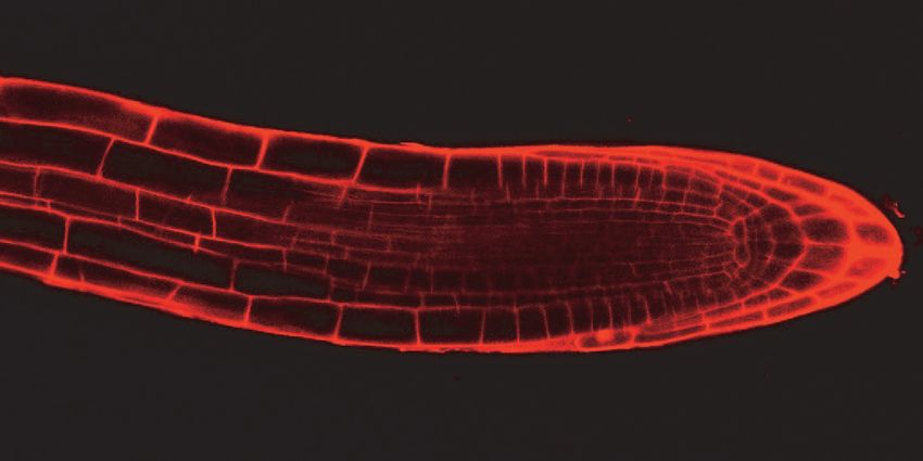
Auxin responsive root proteomes
knock-down allele that were previously characterized (14). SALK_092577C (gaut10-3)
is a null allele characterized in this study. Other alleles used in this study have been
previously published as null/knock-out mutants: vps35b-1 (SALK_014345) (15); bdx-2
(SALK_142260) (16); camta2 (SALK_007027) (17); gapcp2.2 (SALK_008979) (18);
atrh8 (SALK_016830) (19); smp2 (SALK_127730) (20); pme17-2 (SALK_059908) (21);
SALK_063023 (22); nadp-me4 (SALK_064163) (23); rhip1-1 (SALK_091518) (24);
SALK_111575 (25); cka2-1 (SALK_129331) (26); SALK_145341 (27); SALK_151595
(28); impl2-3 (SAIL_35_A08) (29). For proteomics profiling Col-0 seeds were surfaced
sterilized using 50% bleach and 0.01% Triton X-100 for 10 minutes and then washed five
times with sterile water. Seeds were then imbibed in sterile water for two days at 4C and
then transferred to 0.5X Murashige-Skoog (MS) medium plates overlaid with sterile
nylon mesh squares to facilitate tissue harvesting. Seedlings were grown under long day
photoperiods (16 h light/8 h dark) at 23C. Five day-old seedlings were treated with 1 μM
indole-3-acetic acid (IAA) (“auxin”) or an equivalent volume of 95% dimethyl sulfoxide
(DMSO) (“mock”) for 30 minutes or 2 hours by transferring the seedlings on mesh
Downloaded from http://www.mcponline.org/ by guest on April 30, 2019
squares to square petri dishes containing 10 mL of fresh 0.5X MS supplemented with
IAA or solvent. Following treatments, the roots were then hand dissected at the root-
hypocotyl junction with a sterile scalpel, pooled to reach 1 g of tissue per biological
replicate per treatment, and immediately frozen in liquid nitrogen. Four independent
biological replicates were generated for each treatment and time point. For phenotyping
assays seeds were surfaced sterilized using 50% bleach and 0.01% Triton X-100 for 10
minutes and then washed five times with sterile water. Seeds were then imbibed in sterile
water for two days at 4C and then transferred to 0.5X MS medium plates. Seedlings
were grown under long day photoperiods (16 h light/8 h dark) at 23C. For auxin
response assays, five day-old seedlings were transferred to 0.5X MS plates supplemented
with DMSO or 1 μM indole-3-acetic acid (IAA) (“auxin”) and grown for another two
days.
Root phenotyping
Five and seven day-old seedlings were photographed and images were saved as JPEG
files. Measurements of primary root length were calculated using the FIJI software
program (https://fiji.sc/). For lateral root measurements on seven day-old seedlings, all
visible emerged lateral roots were counted. For phenotypic assays, at least 10 biological
replicates were analyzed per genotype and phenotype. The root phenotyping assays
(primary root length, response to auxin treatment and lateral root formation) were
repeated twice. Mutants that were significantly different in each phenotype or treatment,
relative to Col-0 or mock treatment, were determined using t-tests (two-sample
heteroscedastic); p-value of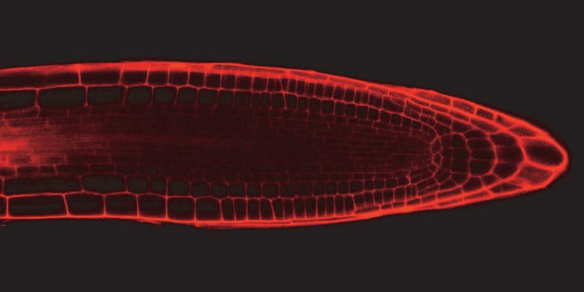
Auxin responsive root proteomes

using t-tests (two-sample heteroscedastic). Proteins with a p-value of
Auxin responsive root proteomes

custom pressure cell. A new set of columns was used for each LC-MS/MS analysis.
Peptides were first eluted from the RP1 column to the SCX column using a 0 to 80%
acetonitrile gradient for 60 minutes. The peptides were then fractionated by the SCX
column using a series of 27 salt steps for non-modified iTRAQ profiling (20, 40, 50 55,
60, 62.5, 65, 67.5, 70, 72.5, 75, 77.5, 80, 82.5, 85, 87.5, 90, 92.5, 95, 97.5, 100, 120, 150,
180, 200, 500, 1000 mM ammonium acetate) followed by high-resolution reverse phase
separation using an acetonitrile gradient of 0 to 80% for 150 minutes.

Spectra were acquired using an LTQ Velos linear ion trap tandem mass spectrometer
(Thermo Electron Corporation, San Jose, CA) employing automated, data-dependent
acquisition. The mass spectrometer was operated in positive ion mode with a source
temperature of 250 °C. The full MS scan range of 400-2000 m/z was divided into 3
smaller scan ranges (400-800, 800-1050, 1050-2000) to improve the dynamic range (32–
34). Both CID (Collision Induced Dissociation) and PQD (Pulsed-Q Dissociation) scans
of the same parent ion were collected for protein identification and quantitation. Each MS

                                                                                                 Downloaded from http://www.mcponline.org/ by guest on April 30, 2019
scan was followed by 4 pairs of CID-PQD MS/MS scans of the most intense ions from
the parent MS scan. A dynamic exclusion of 1 minute was used to improve the duty cycle
of MS/MS scans.

The raw data were extracted and searched using Spectrum Mill v3.03 (Agilent). The CID
and PQD scans from the same parent ion were merged together. MS/MS spectra with a
sequence tag length of 1 or less were considered to be poor spectra and were discarded.
The remaining MS/MS spectra were searched against the Arabidopsis TAIR10 database,
which contains 70,800 protein sequences in the database. The enzyme parameter was
limited to fully tryptic peptides with a maximum miscleavage of 1. All other search
parameters were set to default settings of Spectrum Mill (carbamidomethylation of
cysteines and iTRAQ modification). Carbamidomethylation of cysteines and iTRAQ
modifications as the fixed modifications, Ox-Met and n-term pyro-Gln as the variable
modifications. Mass tolerances were +/- 2.5 Da for precursor ions, +/- 0.7 Da for
fragment ions. A concatenated forward-reverse database was constructed to calculate the
in-situ false discovery rate (FDR). All datasets were summarized together to maintain
FDR across the datasets. Cutoff scores were dynamically assigned resulting in FDR of
0.05%, 0.12%, and 0.66% at the spectrum, peptide, and protein level, respectively.
Proteins that share common peptides were grouped to address the database redundancy
issue. The proteins within the same group shared the same set or subset of unique
peptides.

iTRAQ intensities were calculated by summing the peptide iTRAQ intensities from each
protein group. Peptides shared among different protein groups were removed before
quantitation using custom Perl scripts implemented in Spectrum Mill v3.03 (Agilent).
Isotope impurities of iTRAQ reagents were corrected using correction factors provided
by the manufacturer (Applied Biosystems). Median normalization was performed to
normalize the protein iTRAQ reporter intensities in which the log ratios between different
iTRAQ tags (115/114, 116/114, 117/114) are adjusted globally such that the median log
ratio is zero. Protein ratios between the mock and each treatment were calculated by

                                                                                             5
Auxin responsive root proteomes

taking the ratios of the total iTRAQ intensities from the corresponding iTRAQ reporter.
Protein ratios were then log2 converted. Data analysis was performed using Perseus.

Development and Analytical Validation Targeted MS Assays/Measurements
“Tier 2” targeted MS assays were developed to detect and quantify seven endogenous
proteins (six auxin receptor proteins and one ACTIN control protein) from Arabidopsis
root tissues. These MRM assays were designed and performed according to (32) with the
following modifications. Reference peptides were designed for ACTIN and TIR1/AFB
proteins using Skyline (Table S5). The ACTIN peptide (VAPEEHPVLLTEAPLNPK)
was used for normalization of sample loading and therefore designed to detect multiple
ACTIN proteins (AT2G42170, AT5G59370, AT3G46520, AT3G12110, AT5G09810). A
synthetic plasmid (pDK185) containing these peptide sequences separated by lysine
residues (Supplemental Figure 3) was generated from Genewiz by insertion of the
chimeric DNA sequence into a pUC57 backbone. This chimeric sequence was then
cloned into pT7CFE1-CGST-HA-His (Thermo Scientific) using BamHI and SalI sites

                                                                                              Downloaded from http://www.mcponline.org/ by guest on April 30, 2019
generating plasmid pDK186. Heavy labeled reference peptides were then generated by in
vitro transcription translation using the 1-Step Heavy Protein IVT kit (Thermo
Scientific). Peptides were purified using His-tag Dynabeads (Invitrogen) and subjected to
on-bead digestion with trypsin to recover heavy labeled reference peptides; 2 uL of IVT
peptides were spiked into samples. Method optimization was performed to determine the
optimal transitions and collision energies for each protein. Three to four biological
replicates were assayed for each treatment/timepoint and 2-3 technical replicates were
run for each biological replicate (21 samples in total). Digested peptides were injected by
Agilent 1200 autosampler and subjected to a 40-min reverse phase separation. The
intensities from the best transitions were used to quantify relative abundance of TIR1,
AFB1, AFB2, AFB3, AFB4 and AFB5 in auxin treated samples compared to mock
treated samples (Table S5; Figure S3). Interference free transitions with high intensities
were selected for quantification in Skyline (Figure S3).

Gene Ontology (GO) enrichment
GO enrichment analyses were performed using PANTHER 13.1 using Arabidopsis
thaliana as the organism (http://pantherdb.org/). A heatmap of enriched GO biological
processes was generated following hierarchical clustering (HCL) in MeV.

Confocal imaging and propidium iodide staining
Confocal imaging of roots was performed using a Leica SP5 X MP confocal/multiphoton
microscope system at the Iowa State University Microscopy and Nanoimaging Facility.
Prior to confocal imaging, seedlings were stained with 10 mg/ml propidium iodide for 1-
3 minutes, rinsed in water, and then imaged under 40x/1.25 oil immersion objective with
excitation wavelength of 488 nm and emission wavelength of 500-550 nm.

Results

Quantitative proteomic analysis of roots following auxin treatments identifies
dynamic changes

                                                                                          6
Auxin responsive root proteomes
The transcriptional responses to exogenous auxin in Arabidopsis roots have been well
characterized (3, 8, 36). We sought to extend these studies by examining early proteome
changes in Arabidopsis roots associated with auxin using mass spectrometry. Thus, we
selected 30 min and 2 hours as “early” time points to profile auxin regulated proteins
(Figure 1A). A total of 5,312 proteins were detected and 3,514 proteins were reliably
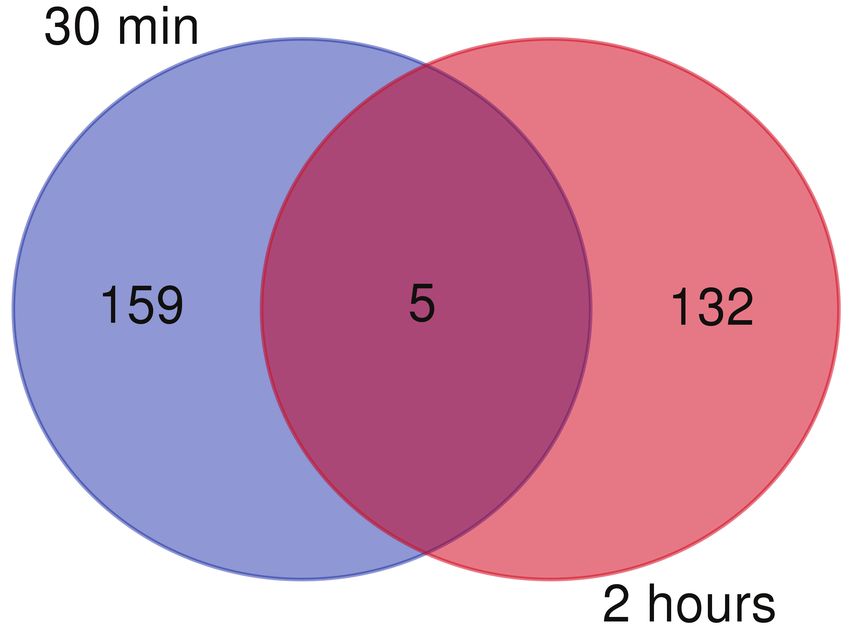
quantified (i.e. detected in at least 3 out of 4 biological replicates) (Table S1).
At 30 min, 164 proteins are differentially expressed relative to the mock control while
137 proteins are differentially expressed at 2 hours (p-value 0.05) (Figure 1B). In
general, most of the significantly differentially expressed proteins exhibited modest fold
change (FC) values (Figure 1C, D; Table S1), which is in line with the observed ratio
compression associated with isobaric tags for relative and quantitative abundance
(iTRAQ) methodology (37–41). Specifically, the observed fold changes in iTRAQ data
are often compressed and may lead to underestimation of relative protein levels. Notably,
a number of the top responsive proteins (log2 FC 0.58 or 0.58) have known roles in
Downloaded from http://www.mcponline.org/ by guest on April 30, 2019
auxin-mediated pathways, thus their modest change in auxin driven protein abundance is
likely sufficient for driving phenotypic changes (Figure 2).
While 3,419 proteins were detected in at least 3 out of the 4 replicates at both time points
(Table S1), only five proteins are differentially expressed at both time points (Figure 1D).
This may indicate that auxin-mediated changes in root proteomes are rapid and transient.
Proteins common to both time points include NITRILASE 1 (NIT1) (At3g44310),
MUCILAGE-MODIFIED 2 (At5g63800), a putative eukaryotic elongation factor 1A
(eEF1A), a methyltransferase (At1g66680), eukaryotic translation initiation factor
isoform 4G1 (At5g57870), and an unknown protein (At3g03150). NIT1 regulates root
growth and development through modulation of auxin metabolism (42) and was
previously shown to be differentially expressed in roots at later timepoints following
auxin treatment using iTRAQ (11). These other common auxin responsive proteins do
not yet have established roles in auxin signaling.
Key auxin responsive proteins are dynamically regulated
We examined the differentially expressed proteins in more detail in order to identify
particular proteins that may play known roles in auxin biology. Proteins with altered
abundance levels in auxin-treated roots have been previously linked to auxin pathways,
providing support for these profiling data (Figure 2 and Table S1). For example, this
group includes several proteins associated with auxin transport in roots, such as
SORTING NEXIN 1 (SNX1), TIME FOR COFFEE (TIC), MAP KINASE 6 (MAPK6),
PROTEIN PHOSPHATASE 2A A3 (PP2AA3), TOUCH3 and exocyst subunit exo70
family protein A1 (EXO70A1) (Figure 2). SNX1 has been previously reported to increase
in both abundance and phosphorylation in roots following auxin treatment (12), which is
consistent with the modest increase in abundance observed in our root dataset. SNX1 is a
key component of the retromer complex that acts to retrieve the PIN-FORMED (PIN)
family of auxin transporters from late/pre-vacuolar compartment back to recycling
pathways; fine tunes auxin responses during gravitropism (12, 43–46). TIC has a role in
controlling root meristem size, reduced PIN expression and acropetal auxin transport in
7Auxin responsive root proteomes
tic-2 mutants (47). MAPK6 has been shown to regulate post-embryonic root development
and auxin levels (48), while MAPK6 activity is correlated with repression of primary root
growth and auxin signaling induces MAPK6 activity (49). In our datasets, we only
observed unmodified (i.e. unphosphorylated) levels of MAPK6 and observed
downregulation at 120 min (Figure 2B). PP2AA3 regulates auxin distribution and stem
cell function at the root apex through interaction with PIN proteins (50, 51). TOUCH3
(Benjamins et. al., Plant Phys 2003) interacts physically with PINOID (52) and TOUCH3
expression was speculated to be under the influence of auxin (53). In these data TOUCH3
levels increase 120 min following auxin treatment in roots (Figure 2) which is consistent
with these published reports. Finally, EXO70A1 is of interest because the exocyst is
involved in PIN1 and PIN2 recycling and thus contributes to polar auxin transport
regulation (54). Modest upregulation of EXO70A1 occurs at 30 min after auxin treatment
(Figure 2A) which is in line with timing observed for PIN1 and PIN2 recycling. Finally,
ATP-BINDING CASSETTE G37/PLEIOTROPIC DRUG RESISTANCE 9/POLAR
AUXIN TRANSPORT INSENSITIVE 1 (ABCG37/PDR9/PIS1) regulates auxin
Downloaded from http://www.mcponline.org/ by guest on April 30, 2019
distribution and homeostasis in roots by excluding indole butyric acid (IBA) from the
root apex (55–57) and was observed to be downregulated in roots 30 min after exogenous
auxin exposure suggesting rapid feedback on auxin homeostasis pathways (Figure 2).
Auxin receptors are stable in response to hormone
Auxin perception by the TIR1/AFB-Aux/IAA families of co-receptor complexes is
central to ARF action. However, none of these proteins were among the DE protein lists.
This could be to lack of detection via MS or other reasons. Further examination of the
detected proteins revealed that only AFB1 (At4g03190) was detected in these datasets
and was not found to be auxin responsive. In order to verify these results we developed a
multiplexed targeted proteomics assay to simultaneously quantify all six auxin receptor
proteins and an actin control protein using heavy labeled synthetic proteotypic peptides
(Supplemental Figures S2 and S3). From these assays we were able to confirm our
iTRAQ results for AFB1 (Figure S2). Additionally, all of the six auxin receptors appear
to be stable in the presence of 1 uM IAA at 30 min and 120 min.
Mutant validation of top responsive proteins
In order to further verify the biological relevance of the identified differentially expressed
proteins we performed phenotypic assays. We examined 17 of the top responsive proteins
that have not been previously phenotyped for auxin-mediated root growth for analysis
(Table 2). All alleles used for these assays have been previously published as null/strong
alleles (see Experimental Procedures for references). We examined three phenotypes: (1)
primary root length in 5 day-old seedlings, (2) primary root inhibition following auxin
treatment, and (3) lateral root formation in 7 day-old seedlings both in the absence and
presence of auxin. In five day-old seedlings, 9/17 lines exhibited shorter root lengths that
were statistically different compared to wild-type Col-0 (Table 2, “Root length” column),
which equates to 53% of the proteins tested as displaying primary root phenotypes. In
young Arabidopsis seedlings auxin treatment can inhibit primary root growth. To test the
auxin responsiveness of these mutant lines we grew them on 0.5X MS for five days and
8Auxin responsive root proteomes
then transferred them to either 0.5X MS control plates or plates supplemented with 1 M
IAA for two more days; root length was measured before and after the treatments.
Relative root growth on auxin was calculated as percentage relative to untreated
seedlings. Col-0 has a 78% reduction in root growth in response to auxin (Table 2, “%
Root growth” column). Notably, one mutant, gapcp2.2 (SALK_008979) exhibited auxin
insensitivity while all the other mutants tested had a normal response to auxin with
respect to primary root inhibition. Finally, auxin is a positive regulator of lateral root
formation. Subsequently we also examined lateral root formation phenotypes in response
to auxin treatment in these same seedlings. In order to account for differences in root
length we calculated the number of lateral roots per mm of root length. Col-0 produces
0.23 +/- 0.01 lateral roots/mm of root length following two days of auxin treatment. In
comparison, 13/17 mutants exhibited reduced lateral root formation; 2/17 mutants failed
to form any lateral roots and were thus auxin insensitive in this assay. Altogether 76% of
the top responsive proteins tested had auxin mediated lateral root defects. Overall,
identification of >50% root and/or auxin regulated phenotypes via reverse genetics is a
Downloaded from http://www.mcponline.org/ by guest on April 30, 2019
significant validation of these data.
Auxin responsive proteins fall into diverse functional categories
Most of the auxin-regulated proteins were distinct between the two time points (Figure
1), leading us to hypothesize that they may have different biological functions. In order to
test this idea we performed gene ontology (GO) enrichment analysis on the differentially
expressed proteins (Figure 3, Figure S3, Table S2). Several enriched GO categories are
common to both time points, which suggests that while the individual proteins may vary,
the overall biological processes that are impacted by auxin signaling are retained (Figure
3, Figure S3, Table S2). Such categories include ‘translation (GO:0006412)’, ‘cellular
amino acid metabolic process (GO:0006520)’, ‘response to cadmium ion (GO:0046686)’,
and ‘response to heavy metal ion (GO:0010038)’ (Figure 3). The interaction between
auxin homeostasis and heavy metal ion toxicity (including cadmium) is of interest given
the widespread nature of this environmental stress. This result is in line with published
studies indicating auxin metabolism and polar transport pathways can be modulated by
heavy metal stimuli (58–61).
Additionally, a couple notable GO biological processes are enriched temporally in these
data. For example, after 30 minutes of auxin treatment, GO categories related to
transcription, protein localization and microtubule dynamics are enriched (Figure 3;
Figure S3) which is consistent with current models for early downstream auxin signaling
events involving active regulation of transcription and organization of cellular
transporters and actin (reviewed in (62)). Whereas cell wall modification (GO:0042545),
growth (GO:0040007), and thigmotropism are enriched after 120 min of auxin treatment
(Figure 3; Figure S3), which would fit well with the timing related to these processes.
A galacturonosyltransferase protein, GAUT10, is auxin regulated and required for
root development
9Auxin responsive root proteomes
We wanted to explore these datasets to uncover novel proteins downstream of auxin co-
receptor action that may mediate root developmental programs. One of the top auxin
responsive proteins is GAUT10 (Table 1; ranked #5 in down-regulated proteins after 120
min). GAUT10 has been implicated in pectin biosynthesis as gaut10 alleles have reduced
altered glycosyl residue compositions compared to wild type, including reduced levels of
galacturonic acid in seedlings (14). Auxin-mediated cell expansion has long been linked
to cell wall mechanics and thus we hypothesized that downregulation of GAUT10 levels
may be involved in such a process. Additionally, ‘cell wall modification’ was one of the
GO enriched biological process terms (Figure 3, Figure S3) adding further support for
testing this candidate protein.
In order to test this idea, we performed functional characterization of gaut10 mutants
with respect to root development and auxin response. In addition to the previously
published gaut10 alleles, gaut10-1 and gaut10-2 (14), we characterized an additional T-
DNA insertional allele, SALK_092577C as a null and designated it gaut10-3 (Figure 4A,
Downloaded from http://www.mcponline.org/ by guest on April 30, 2019
B). During the process of growing these mutant alleles we observed a sucrose dependent
short root phenotype (Figure 4C-4G). While light grown wild-type Arabidopsis roots
have somewhat impaired growth on MS media lacking sucrose due to arrest of the root
apical meristem (RAM) (63, 64), gaut10 mutants are hypersensitive to sucrose deficient
media compared to wild-type.
To test whether gaut10 mutants remain normal auxin response, we performed auxin
response assays on wild-type and gaut10 seedlings. For these experiments, seedlings
were grown on 0.5X MS without sucrose or with 1% sucrose media for 5 days and
imaged (Figure 4H, left panels). Seedlings were then transferred to fresh 0.5X MS
without sucrose or with 1% sucrose media plates supplemented with DMSO or 1 M
IAA (Figure 4H, right panels). After two additional days of growth, seedlings were re-
imaged and primary root length was measured again. The ratio of primary root length
post-treatment to pre-treatment of auxin was calculated from 3 independent replicate
experiments with at least 9 seedlings measured per genotype each condition (Figure 4I).
When grown on sucrose or without sucrose, the ratio of root elongation in gaut10-3 are
similar to WT, indicating inhibition of primary root growth following 2 days of
exogenous auxin treatment (Figure 4I), suggesting gaut10 roots exhibit normal response
to auxin. Additionally, exogenous auxin treatment cannot overcome the short root
phenotype of gaut10 seedlings when grown without sucrose.
Short root phenotypes can manifest due to RAM arrest, lack of cell elongation and/or a
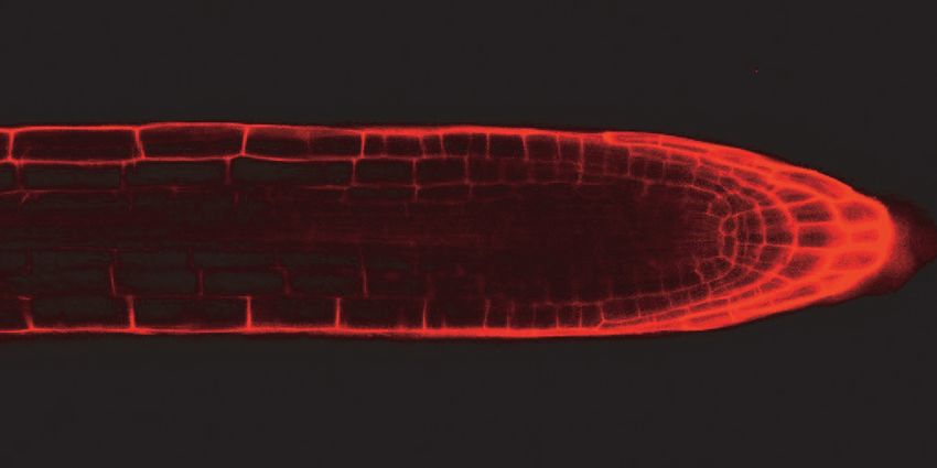
reduction in cell number. Through confocal imaging of five day-old seedlings, we
observed that both gaut10 alleles, gaut10-2 and gaut10-3, appeared to have a shorter
RAM in roots compared to wild-type roots when grown in the absence of sucrose (Figure
5A). The distance between the quiescent center (QC) and elongation zone appeared to be
shorter in gaut10 roots than wild-type (Figure 5A, B). We also counted the number of
visible epidermal cells along one side of each root from the QC to the first elongated
epidermal cell (Figure 5C), which indicated that gaut10 RAM contains fewer cells in the
absence of sucrose. Thus, loss of gaut10 leads to shorter RAM which underlies the short
root phenotype in the absence of sucrose.
10Auxin responsive root proteomes
Discussion
The effects of auxin on gene regulation have been well appreciated at the transcriptional
level. In this study, we describe rapid and quantitative auxin-dependent proteome
changes that occur in Arabidopsis roots using quantitative proteomics. These datasets
show that auxin regulated proteins belong to diverse functional categories such as amino
acid metabolism, RNA and protein regulation and cell wall modification which is
consistent with the conventional wisdom that auxin signaling impacts many aspects of
plant growth and development. Additionally, auxin responsive proteins exhibit a degree
of temporal specificity as very few auxin-responsive proteins in roots are found in
common between 30 min and 120 min following exogenous auxin treatment. This is
consistent with the long-standing notion that auxin drives dynamic developmental
outcomes within primary roots (3, 8, 65–67), and we propose that early root
Downloaded from http://www.mcponline.org/ by guest on April 30, 2019
morphogenesis events are shaped by distinct cellular proteomes.
Previous proteome studies based on auxin responses in seedlings and roots involved older
seedlings and later time points compared to this study (10, 11). In Slade et. al., Proteomes
2017 the authors examined auxin mediated proteome changes in young seedlings at 8, 12
and 24 h after exogenous auxin treatment and thus captured auxin regulated proteins
associated with root differentiation. Because these are later time points than what we
sampled here, it is difficult to directly compare the results between these studies.
However, we did examine the overlap between these studies and found several proteins in
common that are differentially regulated in the root following auxin treatment (Table S3).
Altogether these proteins may represent a set of auxin biomarkers that are rapidly and
stably expressed following auxin treatment and are reproducibly detected via peptide
mass spectrometry. They include proteins such as SORTING NEXIN1 and
NITRILASE1; collectively these proteins play important functional roles in various
aspects of auxin transport, signaling and biosynthesis (12, 42–46).
In addition to global characterization of auxin regulated proteins we developed targeted
proteomics assays to simultaneously quantify all six endogenous auxin receptors. These
targeted assays both validated and extended our iTRAQ studies. All six auxin receptors
(TIR1 and AFB1-5) appear to have stable protein levels at both 30 and 120 min following
auxin treatment. Additionally, these targeted proteomics assays provide a novel method
for quantification of auxin receptors from in vivo tissues which could be applied to
several biological questions related to auxin signaling, including natural variation and
parameterization of existing mathematical models.
This study sought to describe how early auxin signaling events influences cellular
proteomes in organ specific context. Hundreds of proteins change rapidly in response to
auxin in roots, including ‘cell wall modification’ enzymes. We examined the role of one
such protein, GAUT10, in auxin mediated root development using genetic analyses. In
our proteomic profiling, GAUT10 is down regulated at both 30 and 120 min following
exogenous auxin treatment in roots. Loss of function alleles of gaut10 have short roots
11Auxin responsive root proteomes
which are exacerbated when grown without sucrose. Auxin response assays demonstrated
that gaut10 roots can respond normally to auxin when grown in the presence of sucrose.
The short root phenotype is attributed to a reduction in root apical meristem size. Because
GAUT10 is a glucuronosyltransferase, we hypothesize that gaut10 mutants are
hypersensitive to low sucrose conditions due to the function of this enzyme to modify
pectin composition via attachment of sugar moieties. Notably, several other root mutants
involved in auxin signaling also exhibit a similar sugar dependent phenotype (63, 68–70).
This includes gin2 (Hexokinase) and MEDIATOR med 12 med13 double mutants (71,
72). Additionally, sugar signaling has been shown to positively affect root growth via
auxin (72) and transcriptional studies indicate that glucose can affect the expression of
auxin biosynthesis genes, PIN transporter proteins and several genes involved in auxin
signaling (72). Additionally, glucose has been proposed to control non transcriptional
processes such as protein stability. Auxin and glucose act agonistically to activate TOR
kinase activity (73, 74) while a large portion of auxin regulated genes are antagonistically
regulated by glucose (72), indicating that there is much to still understand related to these
Downloaded from http://www.mcponline.org/ by guest on April 30, 2019
complex signaling pathways. Further genetic and molecular studies will be required in
order to examine possible molecular links between GAUT10 activity, nutrient sensing
and auxin signaling.
Altogether these datasets provide a rich resource for mining novel protein function. In
particular, numerous proteins show significant altered abundance levels in a temporal
fashion which makes these excellent candidates for future functional studies.
Additionally, these datasets can inform new hypotheses of what biological processes may
govern rapid auxin responses downstream of perception, including complex levels of
gene regulation and rapid alteration of metabolic states.
Figure Legends
Figure 1. Quantitative proteomic analysis of early auxin responsive proteomes in
Arabidopsis roots identifies ~300 differentially expressed proteins. (A) Schematic of the
experimental workflow. Five day-old wild-type seedlings were treated with 1 uM indole-
3-acetic acid (“auxin”) or an equivalent volume of solvent control (“mock”) for 30 min
and 120 min and dissected roots were processed for proteome profiling using 4-plex
iTRAQ labeling as diagramed. This was repeated three more times for a total of four
multiplexed 2D-LC-MS/MS runs (B) Only five proteins are differentially expressed at
both time points (P-value 0.05). 164 differentially expressed proteins (P-value 0.05)
detected at 30 min (C) and 137 proteins at 120 min (D).
Figure 2. Several well-characterized proteins involved in various auxin pathways exhibit
modest but significant differential expression in roots (P-value 0.05) following 30 min
(A) and 120 min (B) of exogenous auxin treatment. This includes NITRILASE1 (NIT1),
CALMODULIN-BINDING TRANSCRIPTION ACTIVATOR 2 (CAMTA2),
hydroxycinnamoyl-CoA shikimate/quinate hydroxycinnamoyl transferase (HCT),
SORTING NEXIN1 (SNX1), exocyst subunit exo70 family protein A1 (EXO70A1),
STOMATAL CYTOKINESIS DEFECTIVE 2 (SCD2), GroES-like zinc-binding
dehydrogenase family protein (GSNOR), Protein phosphatase 2A-4 (PP2A-4), p23-1
12Auxin responsive root proteomes
(HSP20-like chaperones superfamily protein), NAKED PINS IN YUC MUTANTS 5
(NPY5), ATP-BINDING CASSETTE G37/PLEIOTROPIC DRUG RESISTANCE
9/POLAR AUXIN TRANSPORT INSENSITIVE 1 (ABCG37/PDR9/PIS1), an auxin-
responsive GH3 family protein, TIME FOR COFFEE (TIC), TRYPTOPHAN
BIOSYNTHESIS 1 (TRP1), HSP70, MAP KINASE 6 (MAPK6), PROTEIN
PHOSPHATASE 2A subunit A3 (PP2AA3), TOUCH3, and PECTIN
METHYLESTERASE 1 (PME1). Heatmap indicates the log2 fold change of auxin/mock
for all four biological replicates; increased abundance is indicated in yellow while
reduced protein abundance is indicated in blue.
Figure 3. Auxin regulated proteins are enriched in several gene ontology (GO) biological
process categories; log10 p-values as indicated in the color scale (grey boxes indicate not
significantly enriched) (A) Hierarchical clustering of GO functional categories enriched
in differentially expressed (DE) proteins following 30 min and 120 min of auxin
treatment in roots. Categories in common to both time points include amino acid
Downloaded from http://www.mcponline.org/ by guest on April 30, 2019
metabolism, response to metal ions, and translation. Categories unique to the 30 min DE
proteins include protein localization and transcription while cell wall metabolism and
growth are enriched in the 120 min data set.
Figure 4. Loss of function gaut10 allele gaut10-3 have short roots in the absence of
sucrose but retain auxin responsiveness. (A) SALK T-DNA insertion alleles of GAUT10.
gaut10-1 (SALK_029319) and gaut10-2 (SALK_082273C) were previously
characterized as knock-out and knock-down alleles, respectively (Caffall et. al., 2009).
(B) RT-PCR analysis of gaut10-3 (SALK_092577C) indicates it is a null allele of
GAUT10. (C-G) Five day-old seedlings of wild-type and gaut10-3 grown with or without
sucrose. (H) gaut10-3 roots are shorter than wild-type in the absence of sucrose but can
still respond normally to exogenous auxin (IAA) treatment when grown on sucrose as
indicated by lack of primary growth past the black mark and induced lateral root
formation. (I) Quantification of inhibition of primary root growth by exogenous auxin.
Scale bars shown in C-F is 5 mm.
Figure 5. gaut10 mutant roots have a shorter root apical meristem compared to wildtype.
(A) Confocal images of propidium iodide stained five day-old roots of WT and gaut10
alleles grown on media without sucrose. The gaut10 alleles have smaller root apical
meristem size based on the distance between the quiescent center (QC) and the beginning
of the elongation zone (indicated by a white bar in each root). Scale bar = 50 μm. All
images were acquired at the same magnification. (B-C) Quantification of (A), the smaller
root apical meristems of gaut10 alleles are due to (B) a shorter meristem length and (C)
fewer cells in the meristem and transition zone. Asterisk indicates statistical significance
as determined by the P value for each comparison as determined by t tests.
AUTHOR CONTRIBUTIONS:
Y.P., D.R.K, Z.S., S.P.B., and M.E. designed the research. Y.P., D.R.K, Z.S. and M.G.L.
performed the research. D.R.K, J.W.W., Y.P, and Z.S. analyzed data. D.R.K., M.E. and
S.P.B. supported the research. D.R.K. wrote the article with input from the other authors.
13Auxin responsive root proteomes
Funding
This work was supported by grants from NIH (GM43644 to ME), the Gordon and Betty
Moore Foundation (ME) and the Howard Hughes Medical Institute (ME).
Acknowledgments
We wish to thank Tracey Stewart and Margie Carter at the ISU Microscopy and
Nanoimaging Facility and Dr. Katie (Katayoon) Dehesh for providing camta2 seeds.
Data Availability
All supporting data have been deposited to the MassIVE repository developed by the
NIH-funded UCSD Center for Computational Mass Spectrometry. The data obtained are
available at MassIVE (MassIVE ID MSV000079857):
https://massive.ucsd.edu/ProteoSAFe/static/massive.jsp.
Downloaded from http://www.mcponline.org/ by guest on April 30, 2019
References
1. Finet, C., and Jaillais, Y. (2012) AUXOLOGY: When auxin meets plant evo-devo.
Dev. Biol. 369, 19–31
2. Strader, L. C., and Zhao, Y. (2016) Auxin perception and downstream events.
Curr. Opin. Plant Biol. 33, 8–14
3. Bargmann, B. O. R., Vanneste, S., Krouk, G., Nawy, T., Efroni, I., Shani, E.,
Choe, G., Friml, J., Bergmann, D. C., Estelle, M., and Birnbaum, K. D. (2014) A
map of cell type-specific auxin responses. Mol. Syst. Biol. 9, 688–688
4. Chapman, E. J., Greenham, K., Castillejo, C., Sartor, R., Bialy, A., Sun, T. ping,
and Estelle, M. (2012) Hypocotyl transcriptome reveals auxin regulation of
growth-promoting genes through GA-dependent and -independent pathways. PLoS
One 7,
5. Overvoorde, P. J., Okushima, Y., Alonso, J. M., Chan, A., Chang, C., Ecker, J. R.,
Hughes, B., Liu, A., Onodera, C., Quach, H., Smith, A., Yu, G., and Theologis, A.
(2005) Functional genomic analysis of the AUXIN/INDOLE-3-ACETIC ACID
gene family members in Arabidopsis thaliana. Plant Cell 17, 3282–3300
6. Weijers, D., and Wagner, D. (2016) Transcriptional Responses to the Auxin
Hormone. Annu. Rev. Plant Biol. 67, 539–574
7. Laskowski, M., Biller, S., Stanley, K., Kajstura, T., and Prusty, R. (2006)
Expression profiling of auxin-treated Arabidopsis roots: Toward a molecular
analysis of lateral root emergence. Plant Cell Physiol. 47, 788–792
8. Lewis, D. R., Olex, A. L., Lundy, S. R., Turkett, W. H., Fetrow, J. S., and Muday,
G. K. (2013) A Kinetic Analysis of the Auxin Transcriptome Reveals Cell Wall
Remodeling Proteins That Modulate Lateral Root Development in Arabidopsis.
Plant Cell 25, 3329–3346
9. Stepanova, A. N., Yun, J., Likhacheva, A. V., and Alonso, J. M. (2007) Multilevel
Interactions between Ethylene and Auxin in Arabidopsis Roots. PLANT CELL
ONLINE 19, 2169–2185
14Auxin responsive root proteomes
10. Xing, M., and Xue, H. (2012) A proteomics study of auxin effects in Arabidopsis
thaliana. Acta Biochim. Biophys. Sin. (Shanghai). 44, 783–796
11. Slade, W., Ray, W., Hildreth, S., Winkel, B., and Helm, R. (2017) Exogenous
Auxin Elicits Changes in the Arabidopsis thaliana Root Proteome in a Time-
Dependent Manner. Proteomes 5, 16
12. Zhang, H., Zhou, H., Berke, L., Heck, A. J. R., Mohammed, S., Scheres, B., and
Menke, F. L. H. (2013) Quantitative Phosphoproteomics after Auxin-stimulated
Lateral Root Induction Identifies an SNX1 Protein Phosphorylation Site Required
for Growth. Mol. Cell. Proteomics 12, 1158–1169
13. Mattei, B., Sabatini, S., and Schininà, M. E. (2013) Proteomics in deciphering the
auxin commitment in the Arabidopsis thaliana root growth. J. Proteome Res. 12,
4685–4701
14. Caffall, K. H., Pattathil, S., Phillips, S. E., Hahn, M. G., and Mohnen, D. (2009)
Arabidopsis thaliana T-DNA mutants implicate GAUT genes in the biosynthesis of
pectin and xylan in cell walls and seed testa. Mol. Plant 2, 1000–1014
Downloaded from http://www.mcponline.org/ by guest on April 30, 2019
15. Munch, D., Teh, O.-K., Malinovsky, F. G., Liu, Q., Vetukuri, R. R., El Kasmi, F.,
Brodersen, P., Hara-Nishimura, I., Dangl, J. L., Petersen, M., Mundy, J., and
Hofius, D. (2015) Retromer Contributes to Immunity-Associated Cell Death in
Arabidopsis. Plant Cell Online,
16. Zúñiga-Sánchez, E., Soriano, D., Martínez-Barajas, E., Orozco-Segovia, A., and
Gamboa-deBuen, A. (2014) BIIDXI, the At4g32460 DUF642 gene, is involved in
pectin methyl esterase regulation during Arabidopsis thaliana seed germination
and plant development. BMC Plant Biol. 14,
17. Benn, G., Wang, C. Q., Hicks, D. R., Stein, J., Guthrie, C., and Dehesh, K. (2014)
A key general stress response motif is regulated non-uniformly by CAMTA
transcription factors. Plant J.,
18. Munoz-Bertomeu, J., Cascales-Minana, B., Mulet, J. M., Baroja-Fernandez, E.,
Pozueta-Romero, J., Kuhn, J. M., Segura, J., and Ros, R. (2009) Plastidial
Glyceraldehyde-3-Phosphate Dehydrogenase Deficiency Leads to Altered Root
Development and Affects the Sugar and Amino Acid Balance in Arabidopsis.
PLANT Physiol.,
19. Huang, T.-S., Wei, T., Laliberte, J.-F., and Wang, A. (2010) A Host RNA
Helicase-Like Protein, AtRH8, Interacts with the Potyviral Genome-Linked
Protein, VPg, Associates with the Virus Accumulation Complex, and Is Essential
for Infection. PLANT Physiol.,
20. Clay, N. K., and Nelson, T. (2005) The Recessive Epigenetic Mutation Affects the
Expression of Two Step II Splicing Factors Required for the Transcription of the
Cell Proliferation Gene STRUWWELPETER and for the Timing of Cell Cycle
Arrest in the Arabidopsis Leaf. Plant Cell 17, 1994 LP-2008
21. Sénéchal, F., L’Enfant, M., Domon, J. M., Rosiau, E., Crépeau, M. J., Surcouf, O.,
Esquivel-Rodriguez, J., Marcelo, P., Mareck, A., Guérineau, F., Kim, H. R.,
Mravec, J., Bonnin, E., Jamet, E., Kihara, D., Lerouge, P., Ralet, M. C., Pelloux,
J., and Rayon, C. (2015) Tuning of pectin methylesterification: Pectin
methylesterase inhibitor 7 modulates the processive activity of co-expressed pectin
methylesterase 3 in a pH-dependentmanner. J. Biol. Chem.,
22. Luhua, S., Hegie, A., Suzuki, N., Shulaev, E., Luo, X., Cenariu, D., Ma, V., Kao,
15Auxin responsive root proteomes
S., Lim, J., Gunay, M. B., Oosumi, T., Lee, S. C., Harper, J., Cushman, J., Gollery,
M., Girke, T., Bailey-Serres, J., Stevenson, R. A., Zhu, J. K., and Mittler, R.
(2013) Linking genes of unknown function with abiotic stress responses by high-
throughput phenotype screening. Physiol. Plant. 148, 322–333
23. Wheeler, M. C. G., Tronconi, M. A., Drincovich, M. F., Andreo, C. S., Flügge, U.-
I., and Maurino, V. G. (2005) A comprehensive analysis of the NADP-malic
enzyme gene family of Arabidopsis. Plant Physiol.,
24. Huang, J.-P., Tunc-Ozdemir, M., Chang, Y., and Jones, A. M. (2015) Cooperative
control between AtRGS1 and AtHXK1 in a WD40-repeat protein pathway in
Arabidopsis thaliana. Front. Plant Sci.,
25. Lee, U., Rioflorido, I., Hong, S. W., Larkindale, J., Waters, E. R., and Vierling, E.
(2007) The Arabidopsis ClpB/Hsp100 family of proteins: Chaperones for stress
and chloroplast development. Plant J. 49, 115–127
26. Lu, S. X., Liu, H., Knowles, S. M., Li, J., Ma, L., Tobin, E. M., and Lin, C. (2011)
A Role for Protein Kinase Casein Kinase2 -Subunits in the Arabidopsis Circadian
Downloaded from http://www.mcponline.org/ by guest on April 30, 2019
Clock. PLANT Physiol.,
27. Su, M., Huang, G., Zhang, Q., Wang, X., Li, C., Tao, Y., Zhang, S., Lai, J., Yang,
C., and Wang, Y. (2016) The LEA protein, ABR, is regulated by ABI5 and
involved in dark-induced leaf senescence in Arabidopsis thaliana. Plant Sci.,
28. Silverblatt-Buser, E. W., Frick, M. A., Rabeler, C., and Kaplinsky, N. J. (2018)
Genetic Interactions Between BOB1 and Multiple 26S Proteasome Subunits
Suggest a Role for Proteostasis in Regulating Arabidopsis Development.
G3: Genes|Genomes|Genetics,
29. Nourbakhsh, A., Collakova, E., and Gillaspy, G. E. (2015) Characterization of the
inositol monophosphatase gene family in Arabidopsis. Front. Plant Sci.,
30. Walley, J., Xiao, Y., Wang, J.-Z., Baidoo, E. E., Keasling, J. D., Shen, Z., Briggs,
S. P., and Dehesh, K. (2015) Plastid-produced interorgannellar stress signal
MEcPP potentiates induction of the unfolded protein response in endoplasmic
reticulum. Proc. Natl. Acad. Sci. U. S. A. 112, 6212–6217
31. Zhang, X., Facette, M., Humphries, J. A., Shen, Z., Park, Y., Sutimantanapi, D.,
Sylvester, A. W., Briggs, S. P., and Smith, L. G. (2012) Identification of PAN2 by
Quantitative Proteomics as a Leucine-Rich Repeat-Receptor-Like Kinase Acting
Upstream of PAN1 to Polarize Cell Division in Maize. Plant Cell,
32. O’Brien, R. N., Shen, Z., Tachikawa, K., Lee, P. A., and Briggs, S. P. (2010)
Quantitative Proteome Analysis of Pluripotent Cells by iTRAQ Mass Tagging
Reveals Post-transcriptional Regulation of Proteins Required for ES Cell Self-
renewal. Mol. Cell. Proteomics 9, 2238–2251
33. Zhang, X., Facette, M., Humphries, J. A., Shen, Z., Park, Y., Sutimantanapi, D.,
Sylvester, A. W., Briggs, S. P., and Smith, L. G. (2012) Identification of PAN2 by
quantitative proteomics as a leucine-rich repeat-receptor-like kinase acting
upstream of PAN1 to polarize cell division in maize. Plant Cell 24, 4577–89
34. Walley, J. W., Shen, Z., McReynolds, M. R., Schmelz, E. A., and Briggs, S. P.
(2018) Fungal-induced protein hyperacetylation in maize identified by acetylome
profiling. Proc. Natl. Acad. Sci. 115, 210–215
35. Czechowski, T. (2005) Genome-Wide Identification and Testing of Superior
Reference Genes for Transcript Normalization in Arabidopsis. PLANT Physiol.,
16Auxin responsive root proteomes
36. Nemhauser, J. L., Hong, F., and Chory, J. (2006) Different Plant Hormones
Regulate Similar Processes through Largely Nonoverlapping Transcriptional
Responses. Cell 126, 467–475
37. Mahoney, D. W., Therneau, T. M., Heppelmann, C. J., Higgins, L., Benson, L. M.,
Zenka, R. M., Jagtap, P., Nelsestuen, G. L., Bergen, H. R., and Oberg, A. L.
(2011) Relative quantification: Characterization of bias, variability and fold
changes in mass spectrometry data from iTRAQ-labeled peptides. J. Proteome
Res. 10, 4325–4333
38. Karp, N. A., Huber, W., Sadowski, P. G., Charles, P. D., Hester, S. V, and Lilley,
K. S. (2010) Addressing accuracy and precision issues in iTRAQ quantitation.
Mol. Cell. Proteomics 9, 1885–97
39. Wang, H., Alvarez, S., and Hicks, L. M. (2011) Comprehensive Comparison of
iTRAQ and Label-free LC-Based Quantitative Proteomics Approaches Using Two
Chlamydomonas reinhardtii Strains of Interest for Biofuels Engineering. J.
Proteome Res. 11, 487–501
Downloaded from http://www.mcponline.org/ by guest on April 30, 2019
40. Ow, S. Y., Salim, M., Noirel, J., Evans, C., Rehman, I., and Wright, P. C. (2009)
iTRAQ Underestimation in Simple and Complex Mixtures: “The Good, the Bad
and the Ugly.” J. Proteome Res. 8, 5347–5355
41. Hultin-Rosenberg, L., Forshed, J., Branca, R. M. M., Lehtiö, J., and Johansson, H.
J. (2013) Defining, comparing, and improving iTRAQ quantification in mass
spectrometry proteomics data. Mol. Cell. Proteomics 12, 2021–31
42. Lehmann, T., Janowitz, T., Sánchez-Parra, B., Alonso, M.-M. P., Trompetter, I.,
Piotrowski, M., and Pollmann, S. (2017) Arabidopsis NITRILASE 1 Contributes
to the Regulation of Root Growth and Development through Modulation of Auxin
Biosynthesis in Seedlings. Front. Plant Sci. 8,
43. Hanzawa, T., Shibasaki, K., Numata, T., Kawamura, Y., Gaude, T., and Rahman,
A. (2013) Cellular Auxin Homeostasis under High Temperature Is Regulated
through a SORTING NEXIN1-Dependent Endosomal Trafficking Pathway. Plant
Cell 25, 3424–3433
44. Ambrose, C., Ruan, Y., Gardiner, J., Tamblyn, L. M., Catching, A., Kirik, V.,
Marc, J., Overall, R., and Wasteneys, G. O. (2013) CLASP Interacts with Sorting
Nexin 1 to Link Microtubules and Auxin Transport via PIN2 Recycling in
Arabidopsis thaliana. Dev. Cell 24, 649–659
45. Kleine-Vehn, J., Leitner, J., Zwiewka, M., Sauer, M., Abas, L., Luschnig, C., and
Friml, J. (2008) Differential degradation of PIN2 auxin efflux carrier by retromer-
dependent vacuolar targeting. Proc. Natl. Acad. Sci. 105, 17812–17817
46. Jaillais, Y., Fobis-Loisy, I., Miège, C., Rollin, C., and Gaude, T. (2006) AtSNX1
defines an endosome for auxin-carrier trafficking in Arabidopsis. Nature 443, 106–
109
47. Hong, L. W., Yan, D. W., Liu, W. C., Chen, H. G., and Lu, Y. T. (2014) TIME for
COFFEE controls root meristem size by changes in auxin accumulation in
Arabidopsis. J. Exp. Bot. 65, 275–286
48. Smékalová, V., Luptovčiak, I., Komis, G., Šamajová, O., Ovečka, M.,
Doskočilová, A., Takáč, T., Vadovič, P., Novák, O., Pechan, T., Ziemann, A.,
Košútová, P., and Šamaj, J. (2014) Involvement of YODA and mitogen activated
protein kinase 6 in Arabidopsis post-embryogenic root development through auxin
17Auxin responsive root proteomes
up-regulation and cell division plane orientation. New Phytol. 203, 1175–1193
49. Contreras-Cornejo, H. A., Lopez-Bucio, J. S., Mendez-Bravo, A., Macias-
Rodriguez, L., Ramos-Vega, M., Guevara-Garcia, A. A., and Lopez-Bucio, J.
(2015) Mitogen-Activated Protein Kinase 6 and Ethylene and Auxin Signaling
Pathways Are Involved in Arabidopsis Root-System Architecture Alterations by
Trichoderma atroviride. Mol. Plant. Microbe. Interact. 28, MPMI01150005R
50. Dai, M., Zhang, C., Kania, U., Chen, F., Xue, Q., Mccray, T., Li, G., Qin, G.,
Wakeley, M., Terzaghi, W., Wan, J., Zhao, Y., Xu, J., Friml, J., Deng, X. W., and
Wang, H. (2012) A PP6-Type Phosphatase Holoenzyme Directly Regulates PIN
Phosphorylation and Auxin Efflux in Arabidopsis. Plant Cell 24, 2497–2514
51. Blakeslee, J. J., Zhou, H.-W., Heath, J. T., Skottke, K. R., Barrios, J. A. R., Liu,
S.-Y., and DeLong, A. (2007) Specificity of RCN1-Mediated Protein Phosphatase
2A Regulation in Meristem Organization and Stress Response in Roots. PLANT
Physiol. 146, 539–553
52. Benjamins, R. (2003) PINOID-Mediated Signaling Involves Calcium-Binding
Downloaded from http://www.mcponline.org/ by guest on April 30, 2019
Proteins. PLANT Physiol. 132, 1623–1630
53. Antosiewicz, D. M., Polisensky, D. H., and Braam, J. (1995) Cellular localization
of the Ca2+ binding TCH3 protein of Arabidopsis. Plant J. 8, 623–636
54. Drdová, E. J., Synek, L., Pečenková, T., Hála, M., Kulich, I., Fowler, J. E.,
Murphy, A. S., and Žárský, V. (2013) The exocyst complex contributes to PIN
auxin efflux carrier recycling and polar auxin transport in Arabidopsis. Plant J. 73,
709–719
55. Fujita, H., and Syono, K. (1997) PIS1, a negative regulator of the action of auxin
transport inhibitors in Arabidopsis thaliana. Plant J.,
56. Ruzicka, K., Strader, L. C., Bailly, A., Yang, H., Blakeslee, J., Langowski, L.,
Nejedla, E., Fujita, H., Itoh, H., Syono, K., Hejatko, J., Gray, W. M., Martinoia, E.,
Geisler, M., Bartel, B., Murphy, A. S., and Friml, J. (2010) Arabidopsis PIS1
encodes the ABCG37 transporter of auxinic compounds including the auxin
precursor indole-3-butyric acid. Proc. Natl. Acad. Sci.,
57. Ito, H. (2006) A Gain-of-Function Mutation in the Arabidopsis Pleiotropic Drug
Resistance Transporter PDR9 Confers Resistance to Auxinic Herbicides. PLANT
Physiol.,
58. Wang, R., Wang, J., Zhao, L., Yang, S., and Song, Y. (2015) Impact of heavy
metal stresses on the growth and auxin homeostasis of Arabidopsis seedlings.
BioMetals 28, 123–132
59. Yuan, H. M., Xu, H. H., Liu, W. C., and Lu, Y. T. (2013) Copper regulates
primary root elongation through PIN1-mediated auxin redistribution. Plant Cell
Physiol. 54, 766–778
60. Camacho-Cristóbal, J. J., Martín-Rejano, E. M., Herrera-Rodríguez, M. B.,
Navarro-Gochicoa, M. T., Rexach, J., and González-Fontes, A. (2015) Boron
deficiency inhibits root cell elongation via an ethylene/auxin/ROS-dependent
pathway in Arabidopsis seedlings. J. Exp. Bot. 66, 3831–3840
61. Bücker-Neto, L., Paiva, A. L. S., Machado, R. D., Arenhart, R. A., and Margis-
Pinheiro, M. (2017) Interactions between plant hormones and heavy metals
responses. Genet. Mol. Biol. 40, 373–386
62. Leyser, O. (2018) Auxin Signaling. Plant Physiol. 176, 465–479
18Auxin responsive root proteomes
63. Xiong, Y., McCormack, M., Li, L., Hall, Q., Xiang, C., and Sheen, J. (2013)
Glucose-TOR signalling reprograms the transcriptome and activates meristems.
Nature 496, 181–186
64. Moore, B., Zhou, L., Rolland, F., Hall, Q., Cheng, W. H., Liu, Y. X., Hwang, I.,
Jones, T., and Sheen, J. (2003) Role of the Arabidopsis glucose sensor HXK1 in
nutrient, light, and hormonal signaling. Science (80-. ).,
65. Leyser, O. (2006) Dynamic Integration of Auxin Transport and Signalling. Curr.
Biol.,
66. Möller, B. K., Xuan, W., and Beeckman, T. (2017) Dynamic control of lateral root
positioning. Curr. Opin. Plant Biol.,
67. Du, Y., and Scheres, B. (2018) Lateral root formation and the multiple roles of
auxin. J. Exp. Bot.,
68. Li, X., Cai, W., Liu, Y., Li, H., Fu, L., Liu, Z., Xu, L., Liu, H., Xu, T., and Xiong,
Y. (2017) Differential TOR activation and cell proliferation in Arabidopsis root
and shoot apexes. Proc. Natl. Acad. Sci.,
Downloaded from http://www.mcponline.org/ by guest on April 30, 2019
69. Raya-González, J., López-Bucio, J. S., Prado-Rodríguez, J. C., Ruiz-Herrera, L. F.,
Guevara-García, Á. A., and López-Bucio, J. (2017) The MEDIATOR genes
MED12 and MED13 control Arabidopsis root system configuration influencing
sugar and auxin responses. Plant Mol. Biol.,
70. Racolta, A., Bryan, A. C., and Tax, F. E. (2014) The receptor-like kinases GSO1
and GSO2 together regulate root growth in arabidopsis through control of cell
division and cell fate specification. Dev. Dyn.,
71. Gillmor, C. S., Park, M. Y., Smith, M. R., Pepitone, R., Kerstetter, R. A., and
Poethig, R. S. (2010) The MED12-MED13 module of Mediator regulates the
timing of embryo patterning in Arabidopsis. Development 137, 113–122
72. Mishra, B. S., Singh, M., Aggrawal, P., and Laxmi, A. (2009) Glucose and auxin
signaling interaction in controlling arabidopsis thaliana seedlings root growth and
development. PLoS One,
73. Sheen, J. (2014) Master regulators in plant glucose signaling networks. J. Plant
Biol.,
74. Pu, Y., Luo, X., and Bassham, D. C. (2017) TOR-Dependent and -Independent
Pathways Regulate Autophagy in Arabidopsis thaliana. Front. Plant Sci.,
19You can also read