Describing variability in pig genes involved in coronavirus infections for a One Health perspective in conservation of animal genetic resources ...
←
→
Page content transcription
If your browser does not render page correctly, please read the page content below
www.nature.com/scientificreports

                OPEN             Describing variability in pig genes
                                 involved in coronavirus infections
                                 for a One Health perspective
                                 in conservation of animal genetic
                                 resources
                                 Samuele Bovo1, Giuseppina Schiavo1, Anisa Ribani1, Valerio J. Utzeri1, Valeria Taurisano1,
                                 Mohamad Ballan1, Maria Muñoz2, Estefania Alves2, Jose P. Araujo3, Riccardo Bozzi4,
                                 Rui Charneca5, Federica Di Palma6, Ivona Djurkin Kušec7, Graham Etherington8,
                                 Ana I. Fernandez2, Fabián García2, Juan García‑Casco2, Danijel Karolyi9, Maurizio Gallo10,
                                 José Manuel Martins5, Marie‑José Mercat11, Yolanda Núñez2, Raquel Quintanilla12,
                                 Čedomir Radović13, Violeta Razmaite14, Juliette Riquet15, Radomir Savić16, Martin Škrlep17,
                                 Graziano Usai18, Christoph Zimmer19, Cristina Ovilo2 & Luca Fontanesi1*

                                 Coronaviruses silently circulate in human and animal populations, causing mild to severe diseases.
                                 Therefore, livestock are important components of a “One Health” perspective aimed to control
                                 these viral infections. However, at present there is no example that considers pig genetic resources
                                 in this context. In this study, we investigated the variability of four genes (ACE2, ANPEP and DPP4
                                 encoding for host receptors of the viral spike proteins and TMPRSS2 encoding for a host proteinase)
                                 in 23 European (19 autochthonous and three commercial breeds and one wild boar population) and
                                 two Asian Sus scrofa populations. A total of 2229 variants were identified in the four candidate genes:
                                 26% of them were not previously described; 29 variants affected the protein sequence and might
                                 potentially interact with the infection mechanisms. The results coming from this work are a first
                                 step towards a “One Health” perspective that should consider conservation programs of pig genetic
                                 resources with twofold objectives: (i) genetic resources could be reservoirs of host gene variability
                                 useful to design selection programs to increase resistance to coronaviruses; (ii) the described

                                 1
                                  Department of Agricultural and Food Sciences, Division of Animal Sciences, University of Bologna, Viale
                                 Fanin 46, 40127 Bologna, Italy. 2Departamento Mejora Genética Animal, Instituto Nacional de Investigación
                                 y Tecnología Agraria yAlimentaria (INIA), Crta. de la Coruña, km. 7, 5, 28040 Madrid, Spain. 3Centro de
                                 Investigação de Montanha (CIMO), Instituto Politécnico de Viana do Castelo, Escola Superior Agrária, Refóios
                                 do Lima, 4990‑706 Ponte de Lima, Portugal. 4DAGRI – Animal Science Section, University of Florence, Via delle
                                 Cascine 5, 50144 Florence, Italy. 5MED – Mediterranean Institute for Agriculture, Environment and Development,
                                 Universidade de Évora, Pólo da Mitra, Apartado 94, 7006‑554 Évora, Portugal. 6Biodiversity School of Biological
                                 Sciences, University of East Anglia, Norwich Research Park, Norwich, Norfolk NR47UH, UK. 7Faculty of
                                 Agrobiotechnical Sciences Osijek, Josip Juraj Strossmayer University of Osijek, Vladimira Preloga 1, 31000 Osijek,
                                 Croatia. 8Earlham Institute, Norwich Research Park, Colney Lane, Norwich, Norfolk NR47UZ, UK. 9Department
                                 of Animal Science, Faculty of Agriculture, University of Zagreb, Svetošimunska c. 25, 10000 Zagreb,
                                 Croatia. 10Associazione Nazionale Allevatori Suini (ANAS), Via Nizza 53, 00198 Rome, Italy. 11IFIP Institut du porc,
                                 La Motte au Vicomte, BP 35104, 35651 Le Rheu Cedex, France. 12Programa de Genética y Mejora Animal, Institute
                                 for Research and Technology in Food and Agriculture (IRTA), Torre Marimon, 08140 Caldes de Montbui, Barcelona,
                                 Spain. 13Department of Pig Breeding and Genetics, Institute for Animal Husbandry, 11080 Belgrade‑Zemun,
                                 Serbia. 14Animal Science Institute, Lithuanian University of Health Sciences, Baisogala, Lithuania. 15Génétique
                                 Physiologie et Systèmes d’Elevage (GenPhySE), Université de Toulouse, INRA, Chemin de Borde‑Rouge 24,
                                 Auzeville Tolosane, 31326 Castanet Tolosan, France. 16Faculty of Agriculture, University of Belgrade, Nemanjina 6,
                                 11080 Belgrade‐Zemun, Serbia. 17Kmetijski Inštitut Slovenije, Hacquetova 17, 1000 Ljubljana, Slovenia. 18AGRIS
                                 SARDEGNA, Loc. Bonassai, 07100 Sassari, Italy. 19Bäuerliche Erzeugergemeinschaft Schwäbisch Hall, Schwäbisch
                                 Hall, Germany. *email: luca.fontanesi@unibo.it

Scientific Reports |   (2021) 11:3359                | https://doi.org/10.1038/s41598-021-82956-0                                                  1

                                                                                                                                              Vol.:(0123456789)
www.nature.com/scientificreports/
variability in genes involved in coronavirus infections across many different pig populations might be
part of a risk assessment including pig genetic resources.
Coronaviruses (CoVs) are enveloped single-stranded, positive-strand RNA viruses belonging to the Corona-
viridae family, which includes four genera (Alphacoronavirus, Betacoronavirus, Gammacoronavirus, and Del-
tacoronavirus). Several viruses of this family constantly and silently circulate or emerge and re-emerge in the
human and animal populations causing, in many cases, mild to severe diseases1–8. The most recent dramatic
example of a novel human coronavirus is the severe acute respiratory syndrome-coronavirus 2 (SARS-CoV-2),
detected in the city of Wuhan, China, in December 2019, and that caused the severe pandemic of Coronavirus
Disease 2019 (COVID-19) in this Asian country and then worldwide, critically threatening the public health at
the global level9–13.
Several animal species can act as reservoirs of coronaviruses and different mechanisms have been suggested
for host cell and cross-species transmission of coronaviruses i nfections14–19.
Viral entry, that starts from the receptor recognition, is an essential step determining host range and cross-
species infection. Coronaviruses encode a spike (S) glycoprotein, which recognizes and binds to the host receptor
on the cell s urface20. The region of the spike protein that mediates the interaction with the host-cell receptor
is called receptor-binding domain (RBD). This domain is constituted by the ectodomain subunit S1 which, in
turn, has two main domains: the N-terminal domain (S1-NTD) and the C-terminal domain (S1-CTD;21). The
S1-NTDs are usually responsible for binding sugar components of the receptors22–25 whereas the S1-CTDs are
responsible for recognizing protein receptors26–31. Subsequently, nearby host proteases cleave the spike glyco-
protein, which releases the spike fusion peptide S2. The cleaved S2 peptide allows fusion of viral and cellular
membranes facilitating virus entry into the host c ell20. The infection process has two critical and general issues
that should be considered: (i) the diversity of the host receptor usage from different coronaviruses and (ii) the
different level of sequence similarity of the S1 subunit of the spike from different genera, whereas those from the
same genus have significant sequence similarity of this subunit20.
A few host receptors, that could be specific or less specific for different coronavirus groups, have been identi-
fied: (i) angiotensin-converting enzyme 2 (ACE2) is specific for the alphacoronavirus HcoV-NL63 and the beta-
coronaviruses SARS-CoV and SARS-CoV-232–36, (ii) aminopeptidase N (APN or ANPEP), described to be the
receptor of the human coronavirus NL63 (HcoV-NL63) and other alphacoronaviruses, like the porcine epidemic
diarrhea virus or PEDV, the porcine respiratory coronavirus or PRCV and the transmissible gastroenteritis virus
or TGEV25,37,38 and (iii) dipeptidyl peptidase-4 (DPP4), the receptor of the Middle-East respiratory syndrome
coronavirus (MERS-CoV) and a possible receptor for MERS-like bat coronaviruses including the Tylonycteris
bat coronavirus HKU4 (Bat-CoV HKU4)39,40. All these coronavirus receptors also play their own additional
physiological functions in the host other than their role in the viral surface recognition step. The most studied
host protease for S protein priming is the transmembrane serine protease 2 (TMPRSS2) which is mainly involved
in SARS-CoV and SARS-CoV-2 i nfections36,41–43.
Crystal structures resolved for a number of S1 domains of different coronaviruses complexed with their
respective receptor, along with functional studies and in silico comparative analyses of receptor sequences across
host species, have identified several critical receptor domains and structures that are relevant for the interactions
between the host and the infecting v iruses44,45. These studies also suggested the utilizing capability of receptors
from different animal species by coronaviruses, indicating potential cross-species transmission according to the
structural compatibility between the spike domains and the host receptors46,47.
Structural variations and different expression levels of the receptors and S protein priming proteases could
potentially affect the spike/receptor interactions and subsequent spike cleavage efficiency which might cause
differences of susceptibility of the host for the coronavirus infection capability and disease progression. A few
studies in humans that investigated the ACE2 and TMPRSS2 genes reported variants segregating in different
cohorts that might confer resistance against SARS-CoV-2 infection or modulate COVID-19 s everity48–53.
Several coronaviruses (PEDV, PDCV, SADS-CoV and TGEV), that originated from interspecies transmission,
infect the pig (Sus scrofa) and cause acute gastroenteritis in neonatal piglets and death of the animals, leading
to economically relevant problems to the pig industry7,54. Genetic resistance to the infection of these corona-
viruses might be present within and among pig populations and breeds55. Only few studies have evaluated if
pigs can become infected with other coronaviruses causing human diseases, such as SARS-CoV or MERS-CoV.
These studies challenged the pigs with the two viruses and the obtained results indicated that a small fraction
of the challenged animals were SARS-CoV or MERS-CoV antibody positives without any clinical signs or
lesions, indicating that, even if remote, transmission of these viruses to the pigs and other animals cannot be
excluded56–58. Shi et al.59 reported that SARS-CoV-2 replicates poorly in pigs but other animals such as ferrets
and cats are permissive to infection. Still, Zhou et al.12 reported that SARS-CoV-2 could use ACE2 from four
animal species including the porcine ACE2 as the receptor to enter the cell in vitro, suggesting that pigs might
be potentially susceptible to SARS-CoV-2 infection and could be a potential intermediate host. In other studies,
however, pigs did not result to have developed antibodies against SARS-Cov-2 and were negative for viral RNA
after intranasal infection60,61.
Epidemiological, biological and virological characteristics of coronaviruses, including their demonstrated
ability to easily cross species barriers, suggest that pets and livestock should be considered as part of a global
control and of a “One Health” approach to evaluate if animals that are close to human contacts could represent
a risk source of infections for humans and vice versa62,63. Based on the mentioned preliminary evidences on the
potential relationships between SARS-CoV-2 and pigs (even if contrasting) and considering (i) the relevance
of the pig production systems for meat supply, (ii) that several other coronaviruses circulate in pigs and cause
Scientific Reports | (2021) 11:3359 | https://doi.org/10.1038/s41598-021-82956-0 2
Vol:.(1234567890)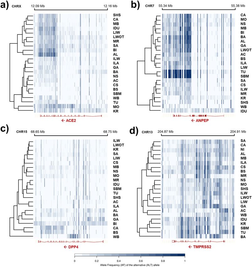
www.nature.com/scientificreports/
diseases in this livestock species7,8,40, (iii) that receptor variants may confer different susceptibility to infections
within species48–53, iv) that coronaviruses may jump the species barriers easily5,18,46,57 and (v) that variability of
the RBD region of the spike protein might determine a quite large host spectrum for every c oronaviruses45,64, as
part of a “One Health” a pproach63, it is needed to evaluate the genetic variability segregating in pig populations
potentially conferring differences of sensitivity to coronavirus-related diseases.
In this study, we investigated the variability in several pig genes (ACE2, ANPEP, DPP4 and TMPRSS2) that
can serve as receptors or protease for priming the infection of coronaviruses. We also evaluated their relevance
in conferring potential differences in susceptibility to coronavirus diseases, also considering a comparative
analysis between the corresponding human genes and the information available in other species. Analysis of
variability included a total of 22 European pig breeds and wild boars and two Asian pig populations using next
generation sequencing data (NGS). This dataset covered a broad number of pig genetic resources raised in
Europe65,66 in comparison with a few Asian populations. The obtained results could be useful (i) to establish a
risk evaluation system in a “One Health” approach, including information on the diversity of pig populations,
(ii) to define cross species evolutionary analyses of genes involved in coronavirus infections and (iii) to identify
natural genetic variability within the Sus scrofa species that could help to design genetic improvement strate-
gies to increase genetic resistance in commercial and autochthonous pig populations against emerging and
re-emerging coronavirus diseases.
Methods
Identification of polymorphisms by next generation sequencing in different pig popula‑
tions. Animals and whole genome sequencing in DNA pools. Blood samples from pigs were obtained by spe-
cialized professionals following standard breeding procedures and health monitoring practices and guidelines at
farm or at slaughter. No treatments or other procedures with animals were performed that would demand ethical
protocols according to Directive 2010/63/EU (2010) and in compliance with the ARRIVE guidelines. Collected
DNA or samples from previous projects were also re-used in this study. This work took advantage from a study
design developed within the Horizon 2020 TREASURE project65–68. Animals included in the study were 30 or 35
from each of the 22 pig breeds that were investigated. These breeds are raised in nine European countries (from
West to East and then North): Portugal (Alentejana and Bísara); Spain (Majorcan Black); France (Basque and
Gascon); Italy (autochthonous: Apulo-Calabrese, Casertana, Cinta Senese, Mora Romagnola, Nero Siciliano and
Sarda; and commercial breeds: Italian Large White, Italian Landrace and Italian Duroc); Slovenia (Krškopolje
pig, hereafter indicated as Krškopolje); Croatia (Black Slavonian and Turopolje); Serbia (Moravka and Swallow-
Bellied Mangalitsa); Germany (Schwäbisch-Hällisches Schwein); and Lithuania (Lithuanian indigenous wattle
and Lithuanian White old type). Selection of individuals for sampling was performed by avoiding highly related
animals (no full- or half-sibs), balancing between sexes, and prioritizing adult individuals or at least animals
with adult morphology. All animals were registered to their respective Herd Books. In addition, 35 Italian wild
boars, previously genotyped for the absence of introgressed domestic alleles at major loci69, were used in this
study. Details on the analysed animals and investigated breeds and wild boars, including geographical distribu-
tion, are reported in Supplementary Table S1.
For each pig, genomic DNA was extracted from 8–15 mL of peripheral blood (collected in Vacutainer tubes
containing 10% 0.5 M EDTA) using either a standardized phenol–chloroform70 or the NucleoSpin Tissue com-
mercial kit (Macherey–Nagel, Düren, Germany). A total of 22 DNA pools were constructed from the European
pig breeds and one DNA pool was constructed from European wild boars, including in each pool 30 or 35
individual DNA samples pooled at equimolar concentration (Supplementary Table S1). For the 22 DNA pools
of the pig breeds, a sequencing library was generated for each DNA pool by using the Truseq Nano DNA HT
Sample preparation Kit (Illumina, CA, USA), following the manufacturer’s recommendations. Briefly, DNA was
randomly sheared to obtain 350 bp fragments which were end polished, A-tailed, and ligated with the full-length
adapter for Illumina sequencing with further PCR amplification. PCR products were purified (AMPure XP sys-
tem) and libraries were analysed for size distribution by Agilent 2100 Bioanalyzer and quantified using real-time
PCR. The qualified libraries were then fed into an Illumina HiSeq X Ten sequencer for paired-end sequencing,
obtaining 150 bp length reads. The wild boar DNA pool was sequenced from 250 bp fragment libraries, with
100 bp long paired-end reads, on the BGISeq 500 platform, following the provider’s procedures.
Quality controls, sequence alignment and variant detection from sequencing data. Reads that were obtained
from the sequenced libraries were cleaned by removing adapter sequences and filtering out sequences presenting
more than 10% unknown bases (N) and/or containing low quality bases (Q ≤ 5) over 50% of the total sequenced
bases. These procedures on FASTQ files were sub-sequentially carried out using FASTQC v.0.11.7 (https://www.
bioinformatics.babraham.ac.uk/projects/fastqc/). Then, filtered high quality reads were mapped on the latest
version of the Sus scrofa reference genome (Sscrofa11.1) using the BWA-MEM algorithm v.0.7.1771 and the
parameters for paired-end data. Picard v.2.1.1 (https://broadinstitute.github.io/picard/) was used to remove
duplicated reads. A summary of whole genome sequencing data statistics is reported in Supplementary Table S2.
Detection of variants on aligned reads was carried out using CRISP v.12271372. CRISP parameters were
tuned to maximize the discovery of variations (–ctpval -0.6 –minc 1 –EM 0). A three-step filtering procedure
was adopted to retain high quality variants:
• first step: (i) retention of only bi-allelic variants, (ii) a minimum read depth (RDmin) in each pool equal to
ten, (iii) a minimum number of alternative reads, over DNA pools, equal to three, (iv) a maximum read
depth (RDmax), in each pool, equal to 68 (computed as proposed by L i73; RDmax = RDmean + 4√RDmean, where
RDmean = 42), and (v) removal of variants mapping in low-quality regions or suffering of strand-bias;
Scientific Reports | (2021) 11:3359 | https://doi.org/10.1038/s41598-021-82956-0 3
Vol.:(0123456789)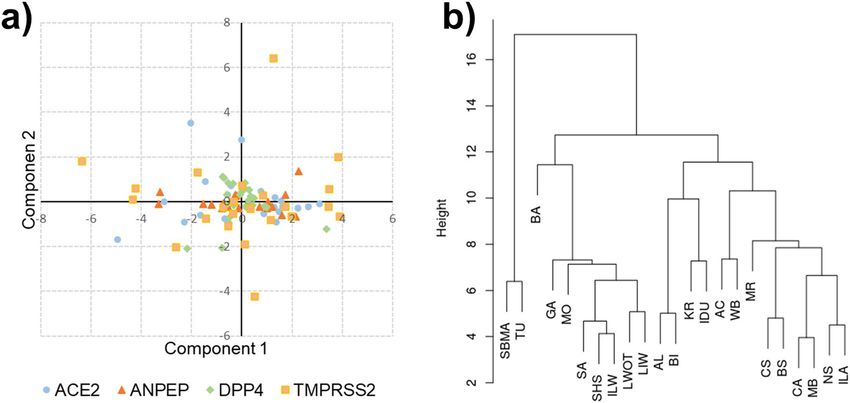
www.nature.com/scientificreports/
Pig Human
Gene name Gene symbol SSC location1 Gene2 Transcript3 Protein-Length4 Protein5
Angiotensin I converting
ACE2 X:12099853-12151275:-1 ENSSSCG00000012138 ENSSSCT00000034032.2 K7GLM4-805 Q9BYF1
enzyme 2
Alanyl aminopeptidase,
ANPEP 7:55351083-55373881:-1 ENSSSCG00000001849 ENSSSCT00000086218.1 A0A5G2QI26(P15145*)-1017 P15144
membrane
Dipeptidyl peptidase 4 DPP4 15:68660849-68743818:-1 ENSSSCG00000015894 ENSSSCT00000067722.1 A0A5G2Q7G7(P27487*)-833 P27487
Transmembrane serine protease
TMPRSS2 13:204876561-204902561:-1 ENSSSCG00000024336 ENSSSCT00000041631.2 A0A287AFA0-526 O15393
2
Table 1. Candidate genes investigated in the present study. 1 Porcine chromosome, starting position, ending
position, gene orientation. Coordinates are based on the Sscrofa11.1 reference genome; 2 Ensembl gene
identifier; 3 Ensembl canonical transcript identifier (it is defined as the longest CCDS translation with no stop
codons); 4 UniProtKB accession number related to the Ensembl canonical transcript. The number of residues
of the protein is reported; 5 UniProtKB accession number. *Alternative reviewed entry (Swiss-Prot).
• second step: implementation of the quality filter procedures described by Anand et al.74. Despite the low false
RISP72, these procedures allow the filtering out of other possible false variants. In this step,
positive rate of C
we made use of dbSNP v.150 (75; no. of variants equal to 64,535,988). Briefly, variants were initially annotated
as reported in dbSNP (“in.dbSNP” class) or not (“novel” class). These two classes were then subdivided in
“rare” and “common” variants. Rare variants were defined as variants presenting a minor allele frequency
(MAF) lower than 0.0143. This number represents the “ideal” lower limit of detection (i.e. 1/70), since pools
were in general composed by 35 diploid individuals (Supplementary Table S1). This is an approximated
estimation that did not take into account the average sequencing depth. Then, considering the “rare” class,
the Kolmogorov–Smirnov (KS) test was used to compare the distributions of the quality score of the variant
of the sub-classes “in.dbSNP” and “novel”. The KS test measures the similarity of the two distributions in a
quantitative way via the D-statistics (a metric ranging from 0 to 1). Lower values of D indicate more similar
distributions. Different cut-off values, in the range 0–50 with steps of 1, were tested. The CRISP quality score
(QCRISP) minimizing the D value was selected as the best score;
• third step: to globally evaluate the quality of our dataset, the transition-to-transversion ratio (Ts/Tv) was used
as quality indicator (1000 Genomes Project Consortium).
Variant detection in the wild boar DNA pool was carried out with Samtools v.1.776 considering a R
Dmin equal
to 3.
Polymorphisms were detected in four porcine candidate genes (ACE2, ANPEP, DPP4 and TMPRSS2) involved
in coronavirus infections considering a region spanning 5 kbp upstream and 5 kbp downstream the correspond-
ing gene coordinates as reported in Ensembl database (http://www.ensembl.org/). Information on the annotated
features of these genes in the Sscrofa11.1 genome version as retrieved in Ensembl database (release 100, April
2020) are reported in Table 1. Variants were annotated using the Variant Effect Predictor (VEP) v.95.077, by
predicting with SIFT v.5.2.278 their impact to the protein function. Variants that affected the protein coding
regions were manually checked. Pipelines were developed either in Python v.2.7.12 or in R v.3.4.479; the Kol-
mogorov–Smirnov test was carried out with the function “ks.test”. SNP allele frequencies (AF) were estimated
by counting the number of reads covering the SNP position.
Mining sequence data from other whole genome resequencing datasets in public data‑
bases. As European and Asian pigs derives from independent domestication routes (e.g.80), for comparative
analyses with information obtained from European pig breeds, sequence data of five Chinese Meishan pigs
and two Asian wild boars were retrieved from the EMBL-EBI European Nucleotide Archive (ENA) repository
(http://www.ebi.ac.uk/ena), project PRJEB9922. Reads were aligned with BWA-MEM and detection of variants
was carried out with Samtools, considering a R Dmin equal to 3. Variants affecting the protein coding regions of
the same four candidate genes (ACE2, ANPEP, DPP4 and TMPRSS2) were manually checked, were annotated
using VEP, and their impact was predicted with SIFT v.5.2.2. A summary of whole genome sequencing data
statistics is reported in Supplementary Table S2.
Variants in porcine candidate genes retrieved from Ensembl database. Genome variants affect-
ing the protein coding sequence (i.e. missense, frameshift and stop gain/loss variants) and the related single
amino acid polymorphisms (SAPs) of the ACE2, ANPEP, DPP4 and TMPRSS2 porcine genes were downloaded
from Ensembl database (release 100, April 2020)81, as information annotated against the Sscrofa11.1 reference
genome version of Sus scrofa and derived from dbSNP. The impact on the protein function was predicted with
SIFT v.5.2.2.
Comparative analysis between pig and human ACE2, ANPEP, DPP4 and TMPRSS2 protein
sequences. Sequence identity between the pig and human ACE2, ANPEP, DPP4 and TMPRSS2 proteins
was obtained via sequence alignments carried out with Clustal O mega82 as implemented in U niProt83. Details
about genes, transcripts and protein accessions numbers used in this analysis are reported in Table 1. The iden-
Scientific Reports | (2021) 11:3359 | https://doi.org/10.1038/s41598-021-82956-0 4
Vol:.(1234567890)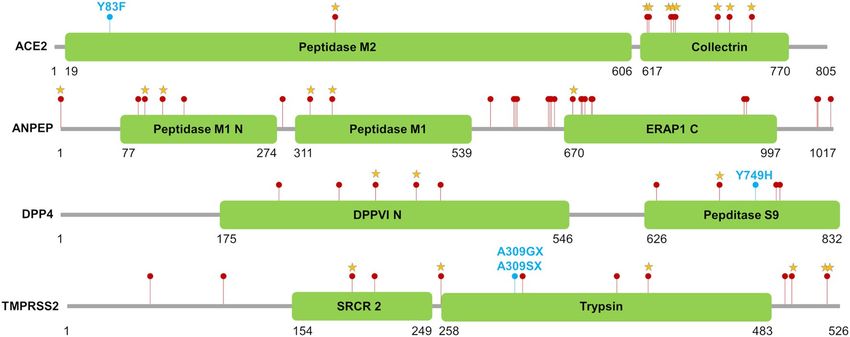
www.nature.com/scientificreports/

                                  tification of protein residues functionally relevant for coronavirus disease infections in humans (SARS, MERS
                                  and the novel COVID-19) was carried out through a survey of the literature that focused on human ACE2,
                                  ANPEP, DPP4 and TMPRSS2 proteins. Our attention was focused on all protein residues either interacting
                                  with coronavirus proteins or functional for the biological activity of the selected proteins, including active sites,
                                  substrate sites, ions binding sites, residues in interaction patches and glycosylation sites. These protein residues
                                  were selected according to 3D structural analyses and related literatures that identified key roles of these sites in
                                  the interaction with the virus spike proteins and the functions of the host protein in virus infections (see Sup-
                                  plementary material for details and the extensive references). We analyzed whether the identified residues were
                                  conserved in the porcine proteins via protein sequence alignments as reported above.

                                  Results
                                  Candidate gene polymorphisms detected in European pig breeds and wild boars. We identi-
                                  fied a total of 2229 variants (single nucleotide polymorphisms: SNPs; and insertion/deletions: indels) in the
                                  four candidate genes and their flanking regions (ACE2 = 837; ANPEP = 173, DPP4 = 460 and TMPRSS2 = 759)
                                  by mining whole genome resequencing data produced from 22 European pig breeds and European wild boars
                                  (Supplementary Table S3). On average, 90% of the detected variants were SNPs and the remaining 10% were
                                  indels (Fig. 1a). About 26% of these variants were novel and detected for the first time in this study whereas 74%
                                  of the identified polymorphisms were already deposited in dbSNP. ANPEP, DPP4 and TMPRSS2 genes included
                                  a comparable fraction of novel variants (from 9 to 14%) whereas about 50% of the ACE2 gene variants was novel
                                  (Fig. 1b; Supplementary Table S3). We further evaluated the distribution of variants considering different gene
                                  features. Overall, the largest proportion of polymorphisms (~ 78%) was within introns whereas variants in the
                                  coding regions represented only 3% of the total number of polymorphic sites. Untranslated (UTRs) and flank-
                                  ing regions had a similar number of DNA polymorphisms (~ 9%; Fig. 1c; Supplementary Table S3). Variant
                                  density (number of variants/100 bp of gene length) was analysed for all genes and all gene regions. TMPRSS2
                                  had the highest variant density, considering the total length of the gene, whereas DPP4 had the lowest density
                                  of polymorphic sites (Fig. 1d). ACE2 had the highest density of variants in the coding regions (about 1 variant
                                  every 100 bp).
                                      Allele frequency distribution of the identified variants over the four genes in the 22 pig breeds and wild boars,
                                  estimated on the number of reads carrying alternative forms as obtained from the sequenced DNA pools, are
                                  reported in Fig. 2.

                                  Analysis of porcine ACE2, ANPEP, DPP4 and TMPRSS2 deduced protein sequence vari‑
                                  ants. Protein variants might play important roles in receptor-driven host-virus interactions and in the func-
                                  tion of the host proteinases involved in the progression of coronavirus infections. The human ACE2, ANPEP,
                                  DPP4 and TMPRSS2 proteins have been extensively studied and several key residues have been identified in
                                  the corresponding proteins (see references cited in the Supplementary Material for a complete analysis of the
                                  available studies). To infer potential effects of the deduced variants identified using DNA sequencing data in the
                                  porcine ACE2, ANPEP, DPP4 and TMPRSS2 translated proteins (constituted by 805, 1017, 833 and 526 residues,
                                  respectively), we first compared the pig protein sequences with those of the human homologous proteins. Then,
                                  we evaluated the impact of protein coding variants identified in pigs and derived by combining the different
                                  datasets explored in this study (DNA pools from European breeds and wild boars; Asian pig genomes; Ensembl
                                  database). Figure 3 reports the position of the identified and analysed protein coding variants located in the four
                                  encoded proteins.

                                  Pig vs human protein sequence comparisons. Overall sequence homology between the pig and human ACE2,
                                  ANPEP, DPP4 and TMPRSS2 proteins showed that the two species share 81.7, 74.7, 81.0, 68.5% identical resi-
                                  dues, respectively. In these proteins, a total of 82 (ACE2), 19 (ANPEP), 24 (DPP4) and 30 (TMPRSS2) key resi-
                                  dues are considered essential either for the virus-host interaction or for the functional activity (Supplementary
                                  Table S4). At these key positions, the pig and human proteins showed a total of 62/82 (76%), 14/24 (58%), 21/30
                                  (70%) and 8/8 (100%) identical residues, respectively.
                                      In more details and considering the different functions of the protein positions, the analysis of the ACE2
                                  residues essential for the virus-host interaction showed 25/35 identical residues between the two species (Supple-
                                  mentary Table S4). ANPEP and DPP4 have 3/10 and 8/15 identical residues needed for the virus-host interaction
                                  (Supplementary Table S5–S6). The active and binding sites of the four proteins were all conserved across species
                                  (13/13 for ACE2, 5/5 for ANPEP, 6/6 for DPP4 and 8/8 for TMPRSS2; Supplementary Table S4–S7). Other sites,
                                  such as cleavage, glycosylation and host protein–protein interaction sites showed different degrees of conserva-
                                  tion between the human and pig sequences (Supplementary Table S4–S7).

                                  Protein coding variants deduced from whole genome resequencing datasets. A total of 25 variants affecting
                                  the protein sequence of the four candidate genes were identified by mining whole genome resequencing data
                                  obtained from the 22 European pig breeds and from the European wild boars (Table 2). Variants were located in
                                  all four investigated candidate genes: 11 were in the ACE2 gene (10 were then considered; see below), four in the
                                  ANPEP gene, two in the DPP4 gene and eight in the TMPRSS2 gene. Allele frequencies of these protein coding
                                  variants in the analysed pig breeds and wild boars are reported in Fig. 4 and Supplementary Table S8. All these
                                  variants were reported in the European pig breeds and nine segregated in the European wild boars.
                                       Based on this information, European breeds and wild boars were represented in multidimensional scaling
                                  plots that showed some contrasting differences among breeds for the information derived by the four genes

Scientific Reports |   (2021) 11:3359 |                https://doi.org/10.1038/s41598-021-82956-0                                                    5

                                                                                                                                                Vol.:(0123456789)
www.nature.com/scientificreports/
Figure 1. Variants in candidate genes discovered in the analysis of European pig breeds and wild boars. (a)
Number of called single nucleotide polymorphisms (SNP) and insertions/deletions (indel); (b) Classification
of variants as novel or already known (deposited in dbSNP); (c) Variant location at the gene level (untranslated
region: UTR); (d) Expected distance of discovered variants stratified by gene feature. Gene length includes UTRs
and flanking regions of 5 kbp upstream [flanking (5′-UTR)] and downstream [flanking (3′-UTR)]. Variant
counts can differ since variants can co-locate or have multiple consequences as predicted with VEP tool. Details
are given in Supplementary Table S3.
separately (Fig. 5a). Pig populations were more dissimilar when considering the TMPRSS2 gene, as points in the
plot (i.e. populations) did not form a very compact cloud.
Cluster analysis (Fig. 5b) highlighted similarities among breeds that resembled in part their geographical
distribution, including (i) two Lithuanian breeds (Lithuanian indigenous wattle and Lithuanian White old type)
and (ii) two Portuguese breeds (Alentejana and Bísara). Wild boars clustered together with Apulo-Calabrese
breed. It is worth to note that two breeds from the Balkan Peninsula (Swallow-Bellied Mangalitsa and Turopolje)
formed a small cluster completely separated from the rest of the European breeds/populations.
In the porcine ACE2 gene, as two SNPs (rs703692808 and rs713746699) affect the same residue S657 and
that manual inspection of sequenced reads highlighted complete linkage disequilibrium between these two
polymorphic sites, they were considered as one variant which caused a novel SAP (p.S675K). Another novel
Scientific Reports | (2021) 11:3359 | https://doi.org/10.1038/s41598-021-82956-0 6
Vol:.(1234567890)www.nature.com/scientificreports/
Figure 2. Representations of variant allele frequency values in the analyzed candidate genes plotted for each
breed and position, considering the alternative (ALT) allele (defined considering the corresponding nucleotides
on Sscrofa11.1 genome version). (a) ACE2, (b) ANPEP, (c) DPP4 and (d) TMPRSS2. Acronyms of the breed
name are the following: Alentejana, AL; Apulo-Calabrese, AC; Basque, BA; Bísara, BI; Black Slavonian, BS;
Casertana, CA; Cinta Senese, CS; Gascon, GA; Krškopolje, KR; Lithuanian Indigenous Wattle, LIW; Lithuanian
White Old Type, LWOT; Majorcan Black, MB; Mora Romagnola, MR; Moravka, MO; Nero Siciliano, NS; Sarda,
SA; Schwäbisch-Hällisches Schwein, SHS; Swallow-Bellied Mangalitsa, SBMA; Turopolje, TU; Italian Duroc,
IDU; Italian Large White, ILW; Italian Landrace, ILA; Wild Boar, WB.
protein coding variant in this gene (X:g.12136848 T > A; p.Y83F) was detected only in Gascon (alternative allele
frequency, AF = 0.056), Basque (AF = 0.172) and Bísara (AF = 0.095) breeds.
A novel variant was also identified in the DPP4 protein (15:g.68673354A > G; p.Y749H). The alternative allele
was detected only in Basque (AF = 0.04) and Bísara (AF = 0.09) breeds.
A few frameshift mutations were identified in the TMPRSS2 gene. Variant rs789572246 (13:g. 204877719del)
introduces a stop gain codon (p.P519X) near the C-terminal end of the protein, outside the peptidase domain
Scientific Reports | (2021) 11:3359 | https://doi.org/10.1038/s41598-021-82956-0 7
Vol.:(0123456789)www.nature.com/scientificreports/
Figure 3. Protein coding variants affecting the ACE2, ANPEP, DPP4 and TMPRSS2 proteins. Red dots indicate
the variants retrieved from Ensembl database. Light blue dots and stars indicate novel and known variants
identified from the resequencing datasets, respectively. Protein domains and their coordinates are based on the
Pfam database (https://pfam.xfam.org/) considering the protein identifiers provided in Table 1.
(Fig. 3). The P519 allele was also affected by a second missense variation (rs789944785). A manual inspec-
tion of sequenced reads highlighted that these two variants (rs789572246 and rs789944785) were not in
complete linkage disequilibrium. Two other novel frameshift mutations (13:g.204881920_20488192insT and
13:g.204881920_20488192insG) would completely change the peptidase coding region of the canonical gene
transcript. However, considering an alternative transcript for this gene (transcript ENSSSCT00000026685.3; Uni-
ProtKB I3LBF8), these two variants would be annotated as splice donors (as they might change the 2 nd base pair
region at the 5′-end of an intron). It is worth to mention that at this position, the reference allele was not found in
any resequencing dataset in which, instead, all three genotypes insG/insG, insG/insT and insT/insT were called.
Mining whole genome resequencing data retrieved from the Chinese Meishan breed and from Asian wild
boars identified other four variants affecting protein sequences (DPP4: p.I383V and p.S704L; ANPEP: p.V32A
and p.E359D). Considering also the other variants described above for the European breeds and wild boars, a
total of 15 and 14 variants affecting proteins were identified in the Chinese Meishan breed and in the Asian wild
boars, respectively (Table 2 and Fig. 4).
Putative functional effects of the porcine protein variants. For a comprehensive analysis of the effects of protein
coding variants in the four analysed genes, the 29 variants affecting proteins and identified in the European pig
breeds and wild boars and in the Asian pig populations (described above) were combined with information on
polymorphic sites available in the Ensembl database for the same genes. The Ensembl database reported a total
of 60 functional coding variants (10 in the ACE2 gene, 28 in the ANPEP gene, 9 in the DPP4 gene and 13 in the
TMPRSS2 gene) that combined with the mentioned variants accounted for a total of 64 variants affecting the
protein encoded by the four genes (11 in the ACE2 gene, 28 in the ANPEP gene, 10 in the DPP4 gene and 15 in
the TMPRSS2 gene; Supplementary Table S9). Figure 3 shows the position of all these variants.
Of the 11 ACE2 protein coding variants, p.P738L was the only one predicted to be deleterious (low confi-
dence). Variants affecting the residues p.N653, p.S657 and p.A658 were located in a protein region interacting
with the ADAM17 sheddase whereas variants of the residues p.K702 and p.R716 belong to a domain interacting
with the serine proteases TMPRSS1 and TMPRSS2 (Supplementary Table S4). The novel variant p.Y83F detected
only in a few European pig breeds (i.e. Gascon, Basque and Bísara) is located within a protein region (human
M82-Y83-P84) suggested to participate in SARS-CoV-2 S-protein a ssociation34.
Of the 27 ANPEP protein coding variants, 22 were classified as tolerated, four were classified as deleterious
and one was a frameshift variant (rs431825257) at the C-terminal end of the protein. Based on annotations com-
ing from the human ANPEP protein, none of these SAPs affected sites were relevant for the virus-host interac-
tion or for the functional activity of the protein (Supplementary Table S5). Porcine variants p.M663V, p.F645S,
p.A647V and p.R651Q were located in a protein region not homologous to the human protein (i.e. they were
included in an alignment gap).
Two out of ten DPP4 protein coding variants were predicted to be deleterious whereas the other seven
missense mutations were classified as tolerated. A stop gained variant that eliminates 60 amino acids of the
C-terminal end was also identified among the annotated variants in Ensembl. Key sites identified in the compara-
tive analysis did not overlap with any of these variants (Supplementary Table S6). However, the variants p.I383V
(p.L316Human) and p.A409V (p.A342 Human) were close to the p.R317 Human, p.R336 Human, p.I346 Human and p.Q344
Human
residues that constitute the MERS-CoV receptor-binding domain (Supplementary Table S7;84).
TMPRRS2 protein was affected by a total of 12 missense substitutions (5 tolerated, 6 deleterious and one not
classified) and three frameshift mutations. Based on annotations coming from the human TMPRRS2 protein, the
variant p.I258V (p.I256Human , Supplementary Table S6) may affect the proteolytic cleavage site (human R255-I256
bond), where auto-cleavage of TMPRSS2 occurs at p.R255 resulting in the release of the active p rotease85.
Scientific Reports | (2021) 11:3359 | https://doi.org/10.1038/s41598-021-82956-0 8
Vol:.(1234567890)www.nature.com/scientificreports/
Gene SSC1 Position2 Ref/Alt3 AFPigs(Europe)4 AFWB(Europe)5 Meishan6 WB(Asia)7 RefSNP8 SAP9 SIFT10 SIFT-score11
ACE2 X 12103359 G/A 0.011 0.000 0/5 0/2 rs713862336 P738L Deleterious-LC 0.04
ACE2 X 12103425 C/T 0.043 0.000 0/5 0/2 rs323807708 R716H Tolerated-LC 0.08
ACE2 X 12105547 T/C 0.276 0.000 2/5 0/2 rs322684836 K702E Tolerated-LC 1.00
ACE2 X 12107234 G/A 0.230 0.167 0/5 0/2 rs696938608 A658V Tolerated-LC 1.00
ACE2 X 12107236 A/T 0.225 0.167 2/5 0/2 rs703692808* S657K† Tolerated-LC 0.10
ACE2 X 12107237 C/T 0.225 0.167 2/5 0/2 rs713746699* S657K† Tolerated-LC 0.09
ACE2 X 12107248 A/C 0.254 0.167 4/5 0/2 rs345377857 N653K Tolerated-LC 1.00
ACE2 X 12109953 T/A 0.309 0.333 4/5 2/2 rs321042645 E631D Tolerated 0.52
ACE2 X 12109958 T/C 0.319 0.286 4/5 2/2 rs328679136 K630E Tolerated 0.40
ACE2 X 12120704 T/C 0.061 0.000 0/5 0/2 rs334297294 I305V Tolerated 0.27
ACE2 X 12136848 T/A 0.015 0.000 0/5 0/2 - Y83F Tolerated 1.00
ANPEP 7 55360022 T/C 0.610 0.133 5/5 2/2 rs322932309 I675V Tolerated 0.5
ANPEP 7 55363723 G/C 0.000 0.000 0/5 1/2 rs695736506 E359D Deleterious 0.00
ANPEP 7 55363906 G/A 0.048 0.000 5/5 1/2 rs331380848 P330S Tolerated 1.00
ANPEP 7 55365462 G/A 0.014 0.000 5/5 1/2 rs323965258 S164L Tolerated 0.27
ANPEP 7 55365619 G/A 0.033 0.000 1/5 1/2 rs334494411 P112S Tolerated 0.66
ANPEP 7 55365858 T/C 0.000 0.000 0/5 1/2 rs342665405 V32A Tolerated 0.09
DPP4 15 68673354 A/G 0.005 0.000 0/5 0/2 – Y749H Tolerated 0.70
DPP4 15 68676800 C/T 0.000 0.000 0/5 1/2 rs697343146 S704L Deleterious 0.00
DPP4 15 68696930 A/G 0.000 0.000 1/5 0/2 rs697267964 I383V Deleterious 0.04
DPP4 15 68704861 G/A 0.016 0.000 0/5 0/2 rs325595747 T340I Tolerated 0.16
TMPRSS2 13 204877719 A/– 0.297 0.000 3/5 1/2 rs789572246 P519X – –
TMPRSS2 13 204877721 G/T 0.005 0.000 0/5 1/2 rs789944785 P519T – –
TMPRSS2 13 204877772 A/T 0.015 0.000 2/5 1/2 rs341813954 C502S Deleterious-LC 0.04
TMPRSS2 13 204878494 A/G 0.013 0.000 0/5 0/2 rs697132526 M400T Tolerated 0.58
TMPRSS2 13 204881920 G/GC 0.598 0.700 2/5 2/2 – A309GX§ – –
TMPRSS2 13 204881920 G/GT 0.402 0.300 5/5 1/2 – A309SX§ – –
TMPRSS2 13 204883347 T/C 0.030 0.000 0/5 0/2 rs699066732 I258V Deleterious 0.02
TMPRSS2 13 204887942 A/T 0.011 0.000 0/5 0/2 rs703753915 F195I Deleterious 0.02
Table 2. Protein coding variants identified in the European and Asian pig breeds and wild boars. 1 Sus scrofa
chromosome; 2 Genomic coordinate on the Sscrofa11.1 reference genome; 3 Reference/Alternative alleles;
4
Frequency of the alternative allele in European pigs (estimated from sequencing data); 5 Frequency of
the alternative allele in European wild boars (estimated from sequencing data); 6 Number of Meishan pigs
carrying the variants; 7 Number of Asian wild boars carrying the variant; 8 dbSNP identification number; 9
Single Amino-acid Polymorphism. Protein coordinates refer to UniProtKB accession number listed in Table 1;
10
SIFT prediction. LC means low confidence prediction; 11 SIFT prediction score. *Variants rs703692808
and rs713746699, both affecting residue S657, are in complete linkage disequilibrium resulting in the SAP
p.S675K†. § The reference allele was not present in our sequencing. data.
Discussion
Genetic resistance to diseases is a complex trait that is re-emerging as a fundamental objective for sustainable
programs in animal breeding and selection plans in all livestock species. As a medium to long term selection
goal, this objective should be considered as part of a “One Health” strategy that requires more resistant or less
susceptible animals to diseases that could be passed to the humans or that could be derived from humans. A few
cases, caused by viruses, that also involved the pig in this two-directions transmission route, have been already
described (e.g.86). Conservation strategies of animal genetic resources should also consider the level of variability
within breeds and populations conferring resistance or determining susceptibility to diseases in the context of
a global “One Health” perspective.
In animals, genetic resistance to diseases cannot be easily measured and monitored and for these reasons it
is difficult to identify any appropriate phenotypic traits as descriptors or proxies of an animal state (related to
the diseased or susceptible condition) useful for their inclusion in breeding programs87. Alternative strategies
or shortcuts that use DNA markers in linkage disequilibrium to causative variants or directly implicated in
conferring different levels of susceptibility/resistance or that could be involved (as part of the host response or
driven mechanisms) in the infection processes, have been proposed88. One of the problems encountered in this
strategy is that genetic resistance to diseases is usually a complex quantitative trait that should be considered
according to the type of infection agent. Other questions related to this strategy are how it is possible to fill the
gaps among the level of the natural genetic variability segregating in the animal populations, the relevance and
the effects of these variants in conferring a desired effect against the pathogenic agents and the potential genetic
progress against a particular disease that could be achieved (based on the segregating variability). Results that
Scientific Reports | (2021) 11:3359 | https://doi.org/10.1038/s41598-021-82956-0 9
Vol.:(0123456789)www.nature.com/scientificreports/
Figure 4. Frequency and genotype information related to the alternative allele of the variants affecting the
protein of the four candidate genes (ACE2, ANPEP, DPP4 and TMPRSS2) in the 23 European and two Asian
populations (autochthonous pig breeds, commercial pig breeds and wild boars). Detailed information is
provided in Supplementary Table S8. Information for the European breeds and wild boars is obtained from the
sequenced DNA pools. Information for the Asian populations is obtained from whole genome sequencing data
of individual animals and the right part of the figure reports the carrier status of the alternative allele.
could be obtained in this context can also define risk levels in different populations, as already demonstrated for
some diseases in other livestock species (e.g.89).
Genomic technologies are opening new opportunities to analyse the host genome at a large scale and then to
identify potential candidate mutations conferring resistance to diseases by applying comparative genome analyses
across species. This approach takes the advantage from what is known in one species and transfers information
in another one. Even if caution should be applied for the interpretation of results, our study provided some
information in this direction by describing variability in a few candidate genes of the host (the pig) genome.
Whole genome resequencing data that we have generated for many pig genetic resources and the comparative
approach that we applied in this study can be further expanded by analysing several other genes for other similar
contexts by targeting other diseases and related potential genetic resistance.
In this study, the selected host genes (ACE2, ANPEP, DPP4 and TMPRSS2) are well known to be involved in
the infection mechanisms of coronaviruses: three of them encode for receptors of a few viruses of this group and
another one encodes for a key proteinase involved in the initiation of the infection after the invasion of the host
susceptible cells32–43. The comparative analysis was based on what is known for the human corresponding gene
products. The extensive genomic data that we mined in pigs gave the possibility to identify the most frequent
variants that can impact on the structure of the encoded proteins.
In many cases of coronavirus infection mechanisms, the entry into the target cell is mediated by the interac-
tion between some cellular receptors and the surface spike (S) g lycoprotein20. Few of these variants might change
the 3D structure or the function of the protein domain in which they are inserted and may potentially modify,
at least in part, their role in the infection routes of the targeted coronaviruses in pigs. It is worth to mention that
Scientific Reports | (2021) 11:3359 | https://doi.org/10.1038/s41598-021-82956-0 10
Vol:.(1234567890)www.nature.com/scientificreports/
Figure 5. (a) Over-imposed multidimensional scaling (MDS) plots and (b) cluster analysis of European pig
breeds and wild boars determined with information on the polymorphic sites in the ACE2, ANPEP, DPP4 and
TMPRSS2 genes. Acronyms of the breed name are given in Fig. 2 and Supplementary Table S1.
most of the DNA polymorphisms identified in the three genes are located in non-coding regions or do not affect
the encoded proteins. It could be possible that some of these variants play regulatory roles but here we did not
analyze the sequencing data for this purpose. Gene expression analyses in porcine target tissues would be needed
to evaluate the role of these variants in altering the expression of these genes and, in turn, to potentially affect
the level of susceptibility to the infection from coronaviruses of pigs with different genotypes.
We studied a large number of autochthonous pig breeds that constitute important genetic resources in Europe.
Mutations that we identified in the investigated genes enriched substantially the list of polymorphic sites already
described in the Sus scrofa for these loci. A large contribution for novel variants derived from the ACE2 gene.
All polymorphisms in the four genes together and their frequencies estimated in 23 European pig populations
(22 breeds and one wild boar population) were able to identify substantial differences that made it possible to
obtain meaningful clusters of these populations.
Among the 11 variants identified in the ACE2 protein, seven (p.Y83F, p.N653, p.S657, p.A658, p.K702, p.R716
and p.P738L) could potentially modify the protein function. Their effects could be inferred from the informa-
tion retrieved from the in silico analyses (from SIFT and from their position in specific domains). Particularly,
a novel variant (p.Y83F), identified only in a few autochthonous European breeds (Gascon, Basque and Bísara)
raised in France and in Portugal, might change the potential association between SARS-CoV-2 S-protein and
the host receptor. All studies that thus far have investigated the susceptibility of the pig to SARS-CoV-2 did not
consider the possibility of intraspecies variability in the ACE2 receptor p rotein12,60,61 that, actually, exists and
could be the source of potential variability in the response to artificial infection experiments. Therefore, in such
studies it will be important to report results with a sequence characterization of the host receptor and other key
proteins involved in the progression of the infections.
Other potential functional variants were identified in the remaining three proteins. Five of the 27 ANPEP
protein variants, two out of 10 DPP4 single amino acid substitutions and nine out of 15 TMPRRS2 protein mis-
sense substitutions or frameshift mutations could be deleterious or might change the protein structure and func-
tions. It will be important to evaluate, with in vitro experiments, the role of these variants in the corresponding
protein function, including for the receptors, their affinity with the coronavirus S-proteins. These analyses will
give the opportunity to also describe the interaction between host variants and with virus variants that could
further complicate the infection mechanisms and related pathogenic effects.
The comparative analysis with the human corresponding proteins will be also useful to further acquire ele-
ments to describe the pig as a valuable animal model to define genetic mechanisms associated to disease resist-
ance and susceptibility.
Genomic analyses of other breeds and populations could identify additional variants in these four genes
that might have a functional relevance, providing a general picture of the variability at these loci. The different
levels of variability for these genes can contribute, at least in part, to the potential genetic progress that could
be reached against coronavirus infections in pigs once it is established a direct relationship between variants
and virus determined diseases. Additional host genes might be also involved in the infection mechanisms of
coronaviruses in pigs as gene expression analyses have d emonstrated90. Moreover, the genetic characterization
at the selected loci and additional genes in large number of genetic resources might provide information useful
to define how the different breeds could contribute to these aims. Marker assisted selection programs designed
to increase genetic resistance to coronaviruses could be based on some of the described polymorphic sites if it
will be demonstrated their role in affecting susceptibility of the Sus scrofa species. The obtained results will con-
stitute a first step towards the inclusion of conservation and selection programs based on genomic information
Scientific Reports | (2021) 11:3359 | https://doi.org/10.1038/s41598-021-82956-0 11
Vol.:(0123456789)www.nature.com/scientificreports/
in this livestock species as part of a comprehensive “One Health” approach against coronaviruses. Risk analysis
for coronavirus infections might also consider the variability of the host genome whose level is different across
breeds and populations, as it might be derived from their genetic histories.
Data availability
Sequence data generated and analysed in the current study from DNA pools are available in the EMBL-EBI Euro-
pean Nucleotide Archive (ENA) repository (http://www.ebi.ac.uk/ena), under the study accession PRJEB36830.
From the same repository we retrieved sequence data of five Meishan pigs (samples: ERS804949, ERS804950,
ERS804951, ERS804953 and ERS804955) and two Asian wild boars (samples: ERS804971 and ERS805009)
deposited with the study accession PRJEB9922. The datasets generated and/or analysed during the current study
are available from the corresponding author on reasonable request.
Received: 14 August 2020; Accepted: 25 January 2021
References
1. Ma, C. Bovine coronavirus. Br. Vet. J. 149, 51–70 (1993).
2. Peiris, J. S. M. et al. Coronavirus as a possible cause of severe acute respiratory syndrome. Lancet 361, 1319–1325 (2003).
3. van der Hoek, L. et al. Identification of a new human coronavirus. Nat. Med. 10, 368–373 (2004).
4. Weiss, S. R. & Navas-Martin, S. Coronavirus pathogenesis and the emerging pathogen severe acute respiratory syndrome coro-
navirus. Microbiol. Mol. Biol. Rev. 69, 635–664 (2005).
5. Woo, P. C. Y. et al. Discovery of seven novel Mammalian and avian coronaviruses in the genus deltacoronavirus supports bat
coronaviruses as the gene source of alphacoronavirus and betacoronavirus and avian coronaviruses as the gene source of gam-
macoronavirus and deltacoronavirus. J. Virol. 86, 3995–4008 (2012).
6. Fehr, A. R., Channappanavar, R. & Perlman, S. Middle east respiratory syndrome: emergence of a pathogenic human coronavirus.
Annu. Rev. Med. 68, 387–399 (2017).
7. Wang, Q., Vlasova, A. N., Kenney, S. P. & Saif, L. J. Emerging and re-emerging coronaviruses in pigs. Curr. Opin. Virol. 34, 39–49
(2019).
8. Leopardi, S., Terregino, C. & Paola, D. B. Silent circulation of coronaviruses in pigs. Vet. Rec. 186, 323 (2020).
9. Munster, V. J., Koopmans, M., van Doremalen, N., van Riel, D. & de Wit, E. A novel coronavirus emerging in China—key questions
for impact assessment. N. Engl. J. Med. 382, 692–694 (2020).
10. Huang, C. et al. Clinical features of patients infected with 2019 novel coronavirus in Wuhan, China. Lancet 395, 497–506 (2020).
11. Wang, C., Horby, P. W., Hayden, F. G. & Gao, G. F. A novel coronavirus outbreak of global health concern. Lancet 395, 470–473
(2020).
12. Zhou, P. et al. A pneumonia outbreak associated with a new coronavirus of probable bat origin. Nature 579, 270–273 (2020).
13. Zhu, N. et al. A novel coronavirus from patients with pneumonia in China, 2019. N. Engl. J. Med. 382, 727–733 (2020).
14. Guan, Y. et al. Isolation and characterization of viruses related to the SARS coronavirus from animals in southern China. Science
302, 276–278 (2003).
15. Lau, S. K. P. et al. Severe acute respiratory syndrome coronavirus-like virus in Chinese horseshoe bats. Proc. Natl. Acad. Sci. U.S.A.
102, 14040–14045 (2005).
16. Li, W. et al. Bats are natural reservoirs of SARS-like coronaviruses. Science 310, 676–679 (2005).
17. Wang, L.-F. et al. Review of bats and SARS. Emerg. Infect. Dis. 12, 1834–1840 (2006).
18. Shi, Z. & Hu, Z. A review of studies on animal reservoirs of the SARS coronavirus. Virus Res. 133, 74–87 (2008).
19. Graham, R. L. & Baric, R. S. Recombination, reservoirs, and the modular spike: mechanisms of coronavirus cross-species transmis-
sion. J. Virol. 84, 3134–3146 (2010).
20. Li, F. Structure, function, and evolution of coronavirus spike proteins. Annu. Rev. Virol. 3, 237–261 (2016).
21. Li, F., Li, W., Farzan, M. & Harrison, S. C. Structure of SARS coronavirus spike receptor-binding domain complexed with receptor.
Science 309, 1864–1868 (2005).
22. Krempl, C., Schultze, B., Laude, H. & Herrler, G. Point mutations in the S protein connect the sialic acid binding activity with the
enteropathogenicity of transmissible gastroenteritis coronavirus. J. Virol. 71, 3285–3287 (1997).
23. Peng, G. et al. Crystal structure of bovine coronavirus spike protein lectin domain. J. Biol. Chem. 287, 41931–41938 (2012).
24. Promkuntod, N., van Eijndhoven, R. E. W., de Vrieze, G., Gröne, A. & Verheije, M. H. Mapping of the receptor-binding domain
and amino acids critical for attachment in the spike protein of avian coronavirus infectious bronchitis virus. Virology 448, 26–32
(2014).
25. Liu, C. et al. Receptor usage and cell entry of porcine epidemic diarrhea coronavirus. J. Virol. 89, 6121–6125 (2015).
26. Godet, M., Grosclaude, J., Delmas, B. & Laude, H. Major receptor-binding and neutralization determinants are located within the
same domain of the transmissible gastroenteritis virus (coronavirus) spike protein. J. Virol. 68, 8008–8016 (1994).
27. Wong, S. K., Li, W., Moore, M. J., Choe, H. & Farzan, M. A 193-amino acid fragment of the SARS coronavirus S protein efficiently
binds angiotensin-converting enzyme 2. J. Biol. Chem. 279, 3197–3201 (2004).
28. Hofmann, H. et al. Highly conserved regions within the spike proteins of human coronaviruses 229E and NL63 determine recogni-
tion of their respective cellular receptors. J. Virol. 80, 8639–8652 (2006).
29. Lin, H.-X. et al. Identification of residues in the receptor-binding domain (RBD) of the spike protein of human coronavirus NL63
that are critical for the RBD-ACE2 receptor interaction. J. Gen. Virol. 89, 1015–1024 (2008).
30. Du, L. et al. Identification of a receptor-binding domain in the S protein of the novel human coronavirus Middle East respiratory
syndrome coronavirus as an essential target for vaccine development. J. Virol. 87, 9939–9942 (2013).
31. Mou, H. et al. The receptor binding domain of the new Middle East respiratory syndrome coronavirus maps to a 231-residue region
in the spike protein that efficiently elicits neutralizing antibodies. J. Virol. 87, 9379–9383 (2013).
32. Li, W. et al. Angiotensin-converting enzyme 2 is a functional receptor for the SARS coronavirus. Nature 426, 450–454 (2003).
33. Hofmann, H. et al. Human coronavirus NL63 employs the severe acute respiratory syndrome coronavirus receptor for cellular
entry. Proc. Natl. Acad. Sci. U.S.A. 102, 7988–7993 (2005).
34. Li, W. et al. Receptor and viral determinants of SARS-coronavirus adaptation to human ACE2. EMBO J. 24, 1634–1643 (2005).
35. Kuba, K. et al. A crucial role of angiotensin converting enzyme 2 (ACE2) in SARS coronavirus-induced lung injury. Nat. Med. 11,
875–879 (2005).
36. Hoffmann, M. et al. SARS-CoV-2 cell entry depends on ACE2 and TMPRSS2 and Is blocked by a clinically proven protease inhibi-
tor. Cell 181, 271-280.e8 (2020).
37. Delmas, B., Gelfi, J., Sjöström, H., Noren, O. & Laude, H. Further characterization of aminopeptidase-N as a receptor for corona-
viruses. Adv. Exp. Med. Biol. 342, 293–298 (1993).
Scientific Reports | (2021) 11:3359 | https://doi.org/10.1038/s41598-021-82956-0 12
Vol:.(1234567890)You can also read