Influenza virus surveillance in Switzerland Season 2018-2019 - 1211 GENEVA 14 - SWITZERLAND National Reference Centre of Influenza Laboratory of ...
←
→
Page content transcription
If your browser does not render page correctly, please read the page content below

Influenza virus surveillance in Switzerland
            Season 2018–2019

     National Reference Centre of Influenza
            Laboratory of Virology
         Geneva University Hospitals,
         4 Rue Gabrielle-Perret-Gentil
      1211 GENEVA 14 – SWITZERLAND

                                        © NRCI
Contacts
Dr Ana Rita Gonçalves Tel: +41/22 372 40 81
Cabecinhas Fax: +41/22 372 49 90
: agnv@hcuge.ch
Ms Patricia Boquete-Suter Tel : +41/22 372 40 91
Fax: +41/22 372 49 90
: patricia.suter@hcuge.ch
Professor Laurent Kaiser Tel: +41/22 372 98 01
Fax: +41/22 372 40 97
: laurent.kaiser@hcuge.ch
Cover: Contribution of the National Reference Centre of Influenza to the influenza field.
2/69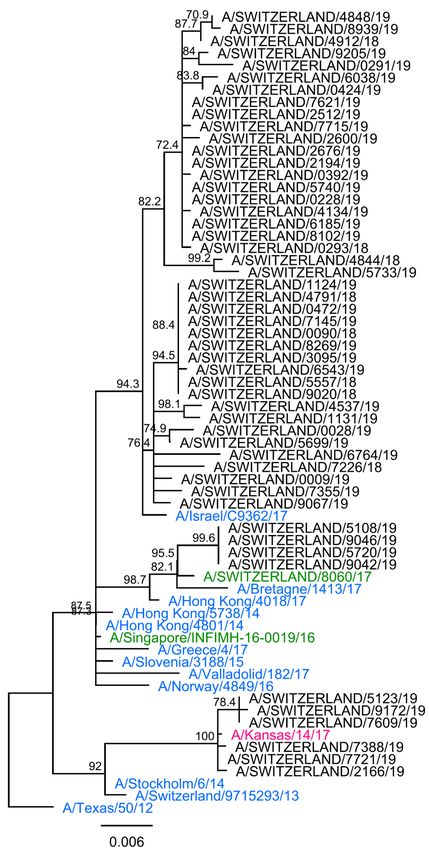
Contents
ABBREVIATIONS AND ACRONYMS ................................................................................................................... 5
ACKNOWLEDGEMENTS .................................................................................................................................... 6
RÉSUMÉ – ZUSAMMENFASSUNG – SUMMARY ................................................................................................ 7
   1       INTRODUCTION ........................................................................................................................................... 10
   2       THE INFLUENZA VIRUS ................................................................................................................................... 10
   3       METHODOLOGY .......................................................................................................................................... 11
        3.1    Clinical identification of influenza cases ............................................................................................ 11
        3.2    Sampled population data ................................................................................................................... 12
        3.3    Virological detection of influenza viruses ........................................................................................... 12
        3.4    Antigenic and genetic characterization of influenza viruses .............................................................. 13
           3.4.1        Cell culture ................................................................................................................................................ 14

           3.4.2        Hemagglutination inhibition assay ........................................................................................................... 16

           3.4.3        Antiviral resistance ................................................................................................................................... 16

   4       2018/19 INFLUENZA SEASON ........................................................................................................................ 19
        4.1    Sentinel population demographics..................................................................................................... 19
        4.2    Detection of influenza in nasopharyngeal samples............................................................................ 20
        4.3    Epidemiology of influenza viruses detected by the Sentinel network ................................................ 23
           4.3.1        Stratification by gender and age ............................................................................................................... 23

           4.3.2        Stratification by influenza vaccination status ........................................................................................... 25

        4.4    Antigenic and genetic characterization of influenza viruses .............................................................. 26
           4.4.1        Characterization of influenza A(H1N1)pdm09 .......................................................................................... 28

           4.4.2        Characterization of influenza A(H3N2) ..................................................................................................... 32

           4.4.3        Characterization of influenza B viruses ..................................................................................................... 37

        4.5    Antiviral resistance ............................................................................................................................. 38
           4.5.1        Sentinel isolates ........................................................................................................................................ 38

           4.5.2        Non-Sentinel isolates ................................................................................................................................ 38

   5       WHO RECOMMENDATION FOR THE COMPOSITION OF INFLUENZA VIRUS VACCINES FOR THE 2019/20 INFLUENZA SEASON39
   6       A(H1N2) VIRUSES ....................................................................................................................................... 39
   7       HUMAN INFECTION WITH ANIMAL INFLUENZA VIRUSES ........................................................................................ 39
        7.1    Swine-to-human influenza virus transmission ................................................................................... 40
        7.2    Avian influenza A subtypes in humans ............................................................................................... 41
                                                      14
   8       AVIAN INFLUENZA A IN ANIMALS .................................................................................................................. 42
   9       DISCUSSION ................................................................................................................................................ 44
   10      OTHER ACTIVITIES OF THE NRCI ..................................................................................................................... 48
        10.1        Validation and/or evaluation of assays ......................................................................................... 48
        10.2        Sharing of influenza cell-cultured isolates and/or reference strains ............................................. 49
        10.3        Collaborative projects/publications .............................................................................................. 50

3/69
10.4         Work in progress ........................................................................................................................... 51
    11       REFERENCES ............................................................................................................................................... 54
ANNEX 1: WEEKLY REPORT OF INFLUENZA VIRUS DETECTION AND VIRUS CHARACTERISTICS ........................ 56
ANNEX 2A: HEMAGGLUTINATION INHIBITION DATA OF INFLUENZA A(H1N1)PDM09 VIRUSES ...................... 57
ANNEX 2B: HEMAGGLUTINATION INHIBITION DATA OF INFLUENZA A(H1N1)PDM09 VIRUSES ...................... 58
ANNEX 2C: HEMAGGLUTINATION INHIBITION DATA OF INFLUENZA A(H1N1)PDM09 VIRUSES ...................... 59
ANNEX 3A: HEMAGGLUTINATION INHIBITION DATA OF INFLUENZA A(H3N2) VIRUSES ................................. 60
ANNEX 3B: HEMAGGLUTINATION INHIBITION DATA OF INFLUENZA A(H3N2) VIRUSES.................................. 61
ANNEX 3C: HEMAGGLUTINATION INHIBITION DATA OF INFLUENZA A(H3N2) VIRUSES .................................. 62
ANNEX 4: HEMAGGLUTINATION INHIBITION DATA OF INFLUENZA B YAMAGATA LINEAGE VIRUSES ............. 63
ANNEX 5: HEMAGGLUTINATION INHIBITION DATA OF INFLUENZA A(H1N1)PDM09 VIRUSES, WIC 22.01.2019
....................................................................................................................................................................... 64
ANNEX 6A: HEMAGGLUTINATION INHIBITION DATA OF INFLUENZA A(H3N2) VIRUSES, WIC 08.02.2019 ....... 65
ANNEX 6B: PLAQUE REDUCTION NEUTRALIZATION DATA OF INFLUENZA A(H3N2) VIRUSES, (MDCK-SIAT), WIC
24.01.2019 ..................................................................................................................................................... 66
ANNEX 6C: PLAQUE REDUCTION NEUTRALIZATION DATA OF INFLUENZA A(H3N2) VIRUSES, (MDCK-SIAT), WIC
28.01.2019 ..................................................................................................................................................... 67
ANNEX 7: LIST OF REFERENCE ANTISERA PROVIDED BY THE WIC FOR THE 2018/19 SEASON ......................... 68
ANNEX 8: SEQUENCING PRIMERS USED DURING THE 2018/19 SEASON ......................................................... 69

4/69
Abbreviations and Acronyms
CDC:          Centers for Disease Control and Prevention
CPE:          cytopathic effect
Ct:           cycle threshold
ECDC :        European Centre for Disease Prevention and Control
EEA:          European Economic Area
EEIQAP:       European External Influenza Virus Quality Assessment Programme
EQAP:         (WHO) External Quality Assessment Programme (for the Detection of
              Influenza Viruses)
EU:           European Union
FOPH:         Federal Office of Public Health
HA:           hemagglutinin
HEF:          hemagglutinin-esterase-fusion
HI:           hemagglutination inhibition
H/LPAI:       high/low pathogenic avian influenza
ILI:          influenza-like illness
M:            matrix
MDCK:         Madin-Darby canine kidney cells
MDCK-SIAT1:   sialic acid-enriched MDCK cells
MN:           microneutralization
MUNANA:       2’-(4-methylumbelliferyl)-a-D-N-acetylneuraminic acid
NA:           neuraminidase
NAI:          neuraminidase inhibitor
NEP:          nuclear export protein
NRCI:         National Reference Centre of Influenza
NS:           non-structural
PA:           acidic protein
PB:           basic protein
RBC:          red blood cells
RFU:          relative fluorescent units
RNA:          ribonucleic acids
RNP:          ribonucleoprotein
rRT-PCR:      real-time reverse-transcription polymerase chain reaction
USA:          United States of America
Vic, Yam:     Victoria, Yamagata
WHO:          World Health Organization
WIC:          Worldwide Influenza Centre

5/69
Acknowledgements
We would like to take this opportunity to extend our grateful thanks to:
       -   The Sentinel network and practitioners.
       -   Damir Perisa, Rita Born, Diana Guido, Clara Greiner, Raphael Rytz, Jean-
           Luc Richard, Andreas Birrer, Sabine Basler and Daniel Koch; Federal
           Office of Public Health.
       -   John McCauley, Rodney Daniels, Yi Pu Lin, Zheng Xiang, Lynne
           Whittaker, Karen Cross, Aine Rattingan, Burcu Ermetal, Mian Dai, Michael
           Bennett and Saira Hussain for their constant support and help during the
           epidemic; World Health Organization Collaborating Centre for Reference &
           Research on Influenza, the Crick Worldwide Influenza Centre, the Francis
           Crick Institute.
       -   Maja Lièvre, Christian Fuster, Sylvie Briand, Wenqing Zhang and all the
           members of the Global Influenza Surveillance and Response System,
           World Health Organization.
       -   The Global Initiative on Sharing All Influenza Data for all the sequences we
           use as references.
       -   Caroline S Brown, Sonja Olsen, Dmitriy Pereyaslov, Hannah Segaloff
           Tamara Meerhoff; World Health Organization Regional Office for Europe,
           and Angeliki Melidou for their support and promotion of European influenza
           surveillance in non-European Union member countries; European Centre
           for Disease Prevention and Control.
       -   Christiane Monnet-Biston and Danielle Massimino for their valuable
           ongoing administrative support; Laboratory of Virology, Geneva University
           Hospitals.
       -   Colette Nicollier for her contribution to the National Reference Centre of
           Influenza website; Geneva University Hospitals.
       -   Isabella Eckerle for her contribution to this report; Centre for Emerging Viral
           Diseases, Geneva University Hospitals.
       -   All members of the Swiss National Reference Centre for Emerging Viruses,
           Geneva University Hospitals, who regularly collaborate with the National
           Reference Centre of Influenza.
       -   All members of the Laboratory of Virology, Geneva University Hospitals,
           who collaborate with the National Reference Centre of Influenza.
6/69
Résumé – Zusammenfassung – Summary Résumé En Suisse, l'épidémie de grippe de 2018/19 a duré onze semaines, de la semaine 2/2019 à la semaine 12/2019, avec un pic pendant la semaine 6/2019. Mille et un prélèvements nasopharyngés ont été dépistés pour la grippe, parmi ces derniers 40,1% étaient positifs. Les virus de l’influenza A étaient largement dominants cette saison, seuls deux virus de l’influenza B ont été détectés. Les virus de influenza A(H1N1)pdm09 étaient prévalents de la semaine 45/2018 à la semaine 7/2019. La souche A(H3N2) devenant dominante dès la semaine 8/2019. La plupart des virus de l’influenza A(H1N1)pdm09 étaient antigéniquement proches de la souche vaccinale A/Michigan/45/2015 (clade 6B.1); avec une majorité des isolats (64,7%) se regroupant dans le sous-groupe génétique 6B.1A5. Les données antigéniques obtenues avec des panels de sera humains indiquent que les virus portant la substitution S183P HA1 sont moins bien inhibés par ces derniers. De ce fait, la souche d’influenza A(H1N1)pdm09 contenue dans les vaccins antigrippaux a été mise à jour pour la saison 2019/20. Les souches vaccinales recommandées étant des virus antigéniquement similaires à la souche A/Brisbane/02/2018. Tous les virus A(H3N2) isolés en Suisse étaient antigéniquement proches de la souche A/Singapore/INFIMH-16-0019/2016 (3C.2a1). Cependant, la reconnaissance était réduite pour certains isolats. Au niveau génétique, la plupart des isolats de A (H3N2) appartenaient au sous-groupe 3C.2a1b du clade 3C.2a1. Quelques virus 3C.3a « récents » et 3C.2a2 ont également été observés. Comme les virus 3C.3a «récents» se sont révélés antigéniquement distincts des virus 3C.2a1 et 3C.3a « antérieurs », le composant A(H3N2) du vaccin antigrippal pour 2019/20 a également été mis à jour pour. La souche A/Singapore/INFIMH-16-0019/2016 sera remplacée par un virus antigéniquement similaire à A/Kansas/14/2017. Le seul virus de l’influenza B appartenant à la lignée B/Yamagata/16/1988 isolé au cours de cette saison était antigéniquement similaire à la souche vaccinale 2018/19, B/Phuket/3073/2013 (clade 3). Aucun des isolats testés au Centre National de Référence de l’influenza dans le cadre de la surveillance saisonnière de l'influenza n'a présenté d'inhibition réduite par l'oseltamivir et le zanamivir. Cependant, un isolat provenant d'un patient hospitalisé 7/69
sous traitement à l'oseltamivir portait la substitution H275Y associée à une inhibition
fortement réduite par ce médicament antiviral.

Peu    de   cas   humains    d'infection   par   des   virus   de   l’influenza   aviaire
hautement/faiblement pathogène ont été identifiés depuis 2017/18.

Zusammenfassung
Dieses Jahr hat die Grippeepidemie in der Schweiz 11 Wochen gedauert. Das heisst
von Woche 2/2019 bis Woche 12/2019, mit einem Maximum im Verlauf der Woche
6/2019. Insgesamt wurden 1001 Proben auf Influenza-Viren untersucht, 40,1%
davon waren positiv. Influenza-A-Viren waren weitgehend dominant, nur zwei
Influenza-B-Viren wurden nachgewiesen. Influenza A (H1N1)pdm09 Viren waren von
Woche 45/2018 bis Woche 7/2019 verbreitet, dann wurden A(H3N2) der
dominierende      Stämme.    Die     meisten     A(H1N1)pdm09-Viren       waren     dem
Impfstoffstamm A/Michigan/45/2015 (Klade 6B.1) antigenisch ähnlich; mit einer
Mehrheit von Isolaten (64,7%), die sich in der genetischen Untergruppe 6B.1A5
gruppieren. Antigendaten, die mit Humanseren erhalten wurden, zeigten, dass Viren
mit der S183P HA1-Substitution, weniger gut gehemmt wurden. Die A(H1N1)pdm09
Impfstoffkomponente für 2019/20 wurde daher auf A/ Brisbane/02/2018 aktualisiert.

Alle A(H3N2)-Viren, die in der Schweiz isoliert wurden, waren antigenisch verwandt
mit dem Stamm A/Singapore/INFIMH-16-0019/2016 (3C.2a1). Die Erkennung war
jedoch für einige Isolate vermindert. Auf genetischer Ebene gehörten die meisten
A(H3N2)-Isolate zur Subklasse 3C.2a1b der Klasse 3C.2a1. "Aktuelle" 3C.3a- und
3C.2a2-Viren wurden auch bei niedrigeren Fallzahlen beobachtet. Da gezeigt wurde,
dass sich die „aktuellen“ 3C.3a-Viren antigenisch von 3C.2a1- und „früheren“ 3C.3a-
Viren unterscheiden, wurde auch die Impfstoffkomponente A(H3N2) für 2019/20 auf
A/Kansas/14/2017     aktualisiert.   Das    einzige    Influenza-B/Yamagata/16/1988-
Abstammungsvirus, das während dieser Saison isoliert wurde, war dem 2018/19
Impfstoffstamm, B/Phuket/3073/2013 (Klade 3) antigenisch ähnlich.

Keines der am National Referenz Zentrum von Influenza im Rahmen der Influenza-
Saisonüberwachung getesteten Isolate zeigte eine verminderte Hemmung durch
Oseltamivir und Zanamivir. Ein Isolat, das von einem Krankenhauspatienten unter
Oseltamivir-Behandlung stammte, trug jedoch die H275Y-Substitution, welche mit
einer stark reduzierten Empfindlichkeit gegenüber Oseltamivir verbunden ist.

8/69
Summary
The Swiss 2018/19 influenza epidemic lasted 11 weeks, from weeks 2/2019 to
12/2019, with a peak at week 6/2019. Of 1001 samples screened for influenza,
40.1% were positive. Influenza A viruses were largely dominant and only two
influenza B viruses were detected. The influenza A(H1N1)pdm09 virus was prevalent
from weeks 45/2018 to 7/2019, and then A(H3N2) became the dominant strain. Most
A(H1N1)pdm09 viruses were antigenically similar to the A/Michigan/45/2015 vaccine
strain (clade 6B.1), with a majority of isolates (64.7%) clustering in the 6B.1A5
genetic subgroup. Antigenic data obtained with human sera indicated that viruses
carrying the S183P HA1 substitution were less well inhibited. The A(H1N1)pdm09
strain for 2019/20 vaccine was therefore updated to A/Brisbane/02/2018.

All of the A(H3N2) viruses isolated in Switzerland were antigenically related to the
A/Singapore/INFIMH-16-0019/2016 strain (3C.2a1). However, recognition was
reduced for some isolates. At the genetic level, most A(H3N2) isolates belonged to
the subclade 3C.2a1b of clade 3C.2a1. “Recent” 3C.3a and 3C.2a2 viruses were
also observed at lower numbers. As “recent” 3C.3a viruses were shown to be
antigenically distinct from 3C.2a1 and former 3C.3a viruses, the A(H3N2) strain for
2019/20 vaccine was also updated to A/Kansas/14/2017. The only influenza
B/Yamagata/16/1988-lineage virus isolated during this season was antigenically
similar to the 2018/19 vaccine strain, B/Phuket/3073/2013 (clade 3).

None of the isolates tested at the National Reference Centre of Influenza in the
context of influenza seasonal surveillance exhibited a reduced inhibition by
oseltamivir and zanamivir. However, one isolate originating from a hospitalized
patient under oseltamivir treatment carried the H275Y substitution associated with
highly-reduced inhibition by oseltamivir. Only a few human cases of low and highly
pathogenic avian influenza infections have been identified since 2017/18.

9/69
1 Introduction
Influenza virus infections are a major clinical and economic burden worldwide.1 In
Switzerland, the Sentinel surveillance system is a community-based network of
primary care medical practitioners who report suspected cases of influenza or
influenza-like illness (ILI) to the Federal Office of Public Health (FOPH). A subgroup
of Sentinel practitioners collects respiratory samples from patients diagnosed with ILI
that are sent to the National Reference Centre of Influenza (NRCI) in Geneva for
further characterization. This report summarizes the demographic, epidemiologic and
virus characterization data gathered from samples processed and analyzed by the
NRCI during the 2018/19 influenza season.

   2 The influenza virus
Influenza viruses are orthomyxoviruses, a family of enveloped, negative, single-
stranded ribonucleic acid (RNA) viruses (Figure 1), known to be causative agents of
respiratory tract infections and referred to as influenza disease or “flu”. Influenza
viruses are divided into four genera, A, B, C and D.1

      NA          HA                                           HEF

                                    NEP
                    PB2                                           PB2
                    PB1                                          PB1                       PB2,PB1,PA/3
                    PA               M1                          P3
                    HA                                         HEF
                   NP                                          NP
                  NA
                                                            M1/2
                                                                                           RNA + NP
                M1/2
                NS1/NEP                                    NS1/NEP
                                    M2

        Influenza A/B                               Influenza C/D
Figure 1. The structure of influenza viral particles. Basic protein 2 (PB2), 1 (PB1) and acidic
protein or 3 (PA or P3) form a complex that corresponds to the RNA-dependent polymerase. The
hemagglutinin (HA) and the hemagglutinin-esterase-fusion (HEF) play a role in virus attachment to
sialic acids present at the surface of host cells and in fusion. The neuraminidase (NA) is crucial for
virion detachment from the cellular surface by cleaving the HA on the virus surface. In influenza B, the
NA gene also encodes the NB ion channel (not shown). The matrix protein 1 (M1) protein forms the
viral capsid. The ion channel M2 allows virion acidification required for fusion. The nuclear export
protein (NEP), also named “non-structural protein 2”, is implicated in the export of the virus
polymerase – RNA + nucleoprotein (NP) complex – to the cell nucleus. The RNA + NP is also called
ribonucleoprotein RNP. The RNA segments PB1, PB2, PA/3, HA or HEF, NP, NA (not present in
influenza C and D), M and NS are present inside the viral capsid, protected by NPs. Only non-
structural protein 1 is not present in the viral particle, but it is expressed upon infection of the host cell.
Influenza D is structurally closer to influenza C than to A and B.

10/69
Influenza A viruses have a wide host tropism, while influenza B viruses are mainly
found in humans2 and in harbour seals (human origin).3 These two influenza types
are responsible for the annual influenza epidemics. Influenza C viruses can be
isolated from swine and humans, in whom they can cause mostly limited symptoms,
and the epidemiological pattern is not well studied. Influenza D viruses are mainly
found in swine and cattle.4 Even if the pathogenic potential of influenza D virus in
humans remains unknown, a recent study estimated that specific influenza D
antibodies could be found in approximately 1.3% of the general human population.5
3 Methodology
3.1 Clinical identification of influenza cases
During the Swiss influenza surveillance period, starting at week 40 and lasting until
week 16 of the following year, 150 to 200 primary care practitioners participate in the
epidemiological national influenza surveillance network. They are requested to notify
ILI cases on a weekly basis. Within the Swiss sentinel system, ILI cases are defined
as sudden fever (>38°C) onset and cough or sore throat. The presence of other
symptoms such as malaise, myalgia, joint pain and headache, as well as
gastrointestinal symptoms is optional. Patients presenting with a secondary disease
(pneumonia, bronchitis, otitis, etc.) consecutive to an influenza not yet notified are
also expected to be reported.
A subgroup of Sentinel practitioners collects nasopharyngeal swabs from patients
with ILI for subsequent viral detection and characterization. The sampling procedure
of specimens is performed according to the following protocol:
1) during the pre- and post-epidemic phases: when the number of ILI reported
by Sentinel practitioners remains below the annual pre-defined epidemic
threshold, screening for influenza viruses is performed in all cases that fulfill
the case definition;
2) during the epidemic phase, defined as when the number of ILI is above the
epidemic threshold: screening is only performed in a subgroup of cases; in
general, every fifth ILI case per practitioner is sent to the NRCI and
screened for the presence of influenza.
11/69The threshold value is defined by the FOPH based on data collected over the past 10
years (excluding the pandemic season 2009/10). For the 2018/19 influenza season, it
corresponded to 68 suspected influenza cases per 100,000 inhabitants.
3.2 Sampled population data
The Sentinel practitioners who send samples to the NRCI are asked to complete a
case report form to collect the following data: sample type; age; gender; time of
symptoms onset; pneumonia; hospitalization; travel within the previous 14 days; and
influenza vaccination status.
3.3 Virological detection of influenza viruses1
Nasopharyngeal swabs received at the NRCI are submitted to virus screening and
subtyping tests. For screening, a one-step real-time reverse transcription polymerase
chain reaction (rRT-PCR) adapted from the 2009 USA Centers for Disease
Prevention and Control (CDC) protocol is used to detect the presence of influenza A
and B viral genomes in the clinical samples. The duplex rRT-PCR targets are the M
protein and the non-structural (NS) protein genes for influenza A and B viruses,
respectively.
Influenza A positive samples are subtyped since the 2017/18 season using an in-
house-developed quadruplex rRT-PCR targeting the HA (H1 and H3) and the NA (N1
and N2) genes in order to discriminate between influenza A(H1N1)pdm09 and
A(H3N2) strains. This new assay is a mix of already validated (in-house H1 and H3
CDC) and newly-designed (N22) rRT-PCR combinations, adapted from the one used
in the study by Henritzi et al6 (N1). The quadruplex detection limit is similar to that of
the diagnostic rRT-PCR. The N1 combination was able to detect the H1N1v3,
swH1N14 and H5N15 isolates tested during the assay validation process. The H3 and
N2 rRT-PCR combinations are also able to detect the A/Wisconsin/12/2010 H3N2
1 The evaluation of the proficiency of the Laboratory of Virology at Geneva University Hospitals in
performing molecular detection of influenza viruses is accessed through the World Health
Organization (WHO) External Quality Assessment Programme for the Detection of Influenza Viruses
by RT-PCR initiated in 2007 by the WHO.
https://www.who.int/influenza/gisrs_laboratory/external_quality_assessment_project/en/
2 Human N2 sequences from 2009-2017 were used for the N2 rRT-PCR design.
3 H1N1v: A/Switzerland/***2244/2011 and A/Berne/****6552/2017, variants isolated from Swiss pig
breeders.
4 swH1N1 35 (2008): virus isolated from a Swiss pig.
5 H5N1: A/Hong Kong/6841/2010 (EQAP panel 16) and A/goose/Qinghai/1A/05*A/PR8/34(INT).
12/69triple reassortant (H3N2tr),7 although the latter virus is not known to circulate in
Switzerland. Nevertheless, if needed, additional tests are available at the NRCI to
discriminate seasonal H3N2 from H3N2tr viruses. Influenza B Yamagata (Yam) and
B Victoria (Vic) lineages are determined using a duplex rRT-PCR.
The quality of the NRCI influenza A/B detection and subtyping was successfully
evaluated during 2018 by the European External Influenza Virus Quality Assessment
Programme 2018 (EEIQAP 2018) and the WHO External Quality Assessment
Programme for the Detection of Influenza Viruses by RT-PCR (EQAP panel 17).
During the pre- and post-epidemic phases, the majority of rRT-PCR-negative
specimens are inoculated on cells for viral culture. This strategy allows to detect
potential influenza strains that may have “escaped” rRT-PCR detection. For example,
this could be the case in the presence of drifted viruses carrying mutations in the
genomic regions targeted by rRT-PCR screening.
3.4 Antigenic and genetic characterization of influenza viruses
During the season, selection of influenza viruses are submitted to phenotypic and
genotypic characterization (Figure 2). In brief, during the pre- and post-epidemic
phases, all positive samples with sufficient HA titers are phenotypically characterized
using the HA inhibition (HI) assay, which evaluates the antigenic similarity between
the reference and circulating influenza strains. HIs are performed with
glutaraldehyde-fixed guinea pig (Charles River, Lyon, France) red blood cells (RBC).
Of note, a microneutralization (MN) test can be used for samples that do not (or only
poorly) hemagglutinate RBC. Reference antisera (Annex 8) and corresponding viral
reference strains used for the HI and MN are kindly provided by the WHO
Collaborating Centre Reference Laboratory at the Francis Crick Worldwide Influenza
Centre (WIC), London, United Kingdom. Reference virus stocks for the current
influenza season are produced on cells (Madin-Darby canine kidney [MDCK] and
MDCK-sialic acid-enriched [MDCK-SIAT]). During the epidemic phase, up to 5
positive samples per week with a cycle threshold (Ct) value ≤30 and sufficient HA
titers are analyzed. When judged relevant, samples with Ct values ≥30 can also be
selected for characterization.
To assess the phylogeny of the circulating strains and to determine how genetically
close they are to vaccine strains, the HA1 part of the HA gene is sequenced.
13/69Samples previously chosen for phenotypic characterization or judged to be of interest
are submitted to Sanger sequencing (up to five per week, generally with a Ct ≤30).
The corresponding NA genes are also sequenced. Whenever needed, additional
genes as influenza A M and influenza B NS genes can also sequenced. The NA
gene sequence allows to detect key mutations previously described as conferring
resistance to NA inhibitors (NAI). M and NS gene sequencing allow to control the
adequacy of rRT-PCR influenza A and B screening, respectively. In addition, we are
currently working on a sequencing protocol for the PA gene, in order to identify amino
acids substitutions associated with a reduced susceptibility to PA inhibitors as
baloxavir marboxyl.
3.4.1 Cell culture
As mentioned in chapter 3.3, most of the received samples, both positive and
negative, are cultured on MDCK and MDCK-SIAT1 cells in parallel during the pre-
and post-epidemic phases. This allows, to some extent, to control that a low positivity
rate observed outside the epidemic phase is not due to a rRT-PCR detection default.
As HI analysis requires a sufficient concentration of influenza virus, a viral
amplification step is performed by inoculating the clinical samples on MDCK and
MDCK-SIAT1 cells in parallel. A sampling of five rRT-PCR positive specimens per
week with Ct valuesPre-analytical
Nasopharyngeal swabs from individuals
with ILI were collected by 79 Sentinel
practitioners
rRT-PCR screening for influenza A
rRT-PCR screening for influenza B
Screening and subtyping
n=1001 (998 individuals)
rRT-PCR
subtyping/lineage Positive n=401 Negative n=600
n=393 successfully
subtyped
Sampling of specimens, ± 5 per week
for HAI and sequencing
Random
sampling
n=261
Genotypic 23 M genes NAI antiviral
Phenotypic and genotypic characterization
characterization sequenced resistance assessment Cell culture
HA gene sequencing NA gene sequencing n= 104
n=102 n=103
Phenotypic characterization by HI all negative for
n= 83 influenza by cell
successfully characterized
culture
Figure 2. Flow chart of Sentinel sample collection and processing. Numbers (n) represent the
number of samples submitted to the described step during the 2018/19 season.
15/693.4.2 Hemagglutination inhibition assay
A two-fold serial dilution is performed using 50 µl of viral suspension in SALK buffer
and 25 µl of glutaraldehyde-fixed guinea pig RBC (1.5%) are added for 1 h incubation
at 4°C. HA titer is defined as the last dilution in which the complete HA is still
observed. After titer determination, HI is performed according to the following
procedure: 25 µl of reference antisera are added in the first two wells of a 96-well
plate. Two-fold dilutions are prepared by adding 25 µl of SALK buffer in the second
well. Twenty-five µl are then collected from the same well and the procedure is
repeated to the end of each line. Twenty-five µl of viral suspension containing four
HA units are added to the ferret antisera dilution and incubated for 1 h at room
temperature. Then, 25 µl of guinea pig RBC are added to each well and the plates
are incubated for 1 h at 4°C. The HI titer corresponds to the last antiserum dilution for
which HA is still inhibited. This titer is compared to the homologous titer obtained with
reference strains submitted to their corresponding ferret antisera (antigenic table).
The antigenic tables are influenza strain-specific (Figure 3) and are therefore
adjusted each year. As the ferret serum is initially diluted 1/8, the titers provided in
Figure 3 and Annexes 2 to 4 should be multiplied by eight to obtain the final titers.
Since we started using fixed RBCs (more than 10 years’ expertise) instead of fresh
ones, the requirement for the addition of 20 nM oseltamivir during the HI test in order
to prevent NA-mediated hemagglutination of A(H3N2) viruses, is evaluated at the
beginning of each season. It is also checked regularly during the season or in the
case of unexpected test results.
3.4.3 Antiviral resistance
The evolution of influenza viruses is known to be very rapid, thus allowing them to
escape from immune responses and/or infection inhibition by therapeutic molecules.
Known mutations conferring antiviral resistance to a given influenza
type/subtype/lineage can be monitored by sequencing the NA genes for NAI
resistance and M genes for the M2 inhibitors. Viral sequences are manually and
semi-automatically (FluSurver: http://flusurver.bii.a-star.edu.sg/) screened for the
presence of mutations known to be associated with antiviral resistance as reported in
the WHO “Summary table of neuraminidase amino acid substitutions associated with
reduced inhibition by neuraminidase inhibitors” (https://www.who.int/influenza
16/69/gisrs_laboratory/antiviral_susceptibility/NAI_Reduced_Susceptibility_Marker_Table_ WHO.pdf?ua=1). New antiviral resistance to NAIs can be identified by combining NA genotyping/sequencing and phenotypic NA enzyme-inhibitor (NAI) assays. At the NRCI, phenotypic antiviral resistance of influenza stains are performed if needed and/or upon request using the NA-Fluor™ Influenza Neuraminidase Assay Kit (Thermo Fisher Scientific, Ecublens, Switzerland). In brief, a titration of the viral NA activity is performed for each test by serial two-fold dilutions. The optimum virus dilution to be used in subsequent inhibition assays is determined by plotting the virus dilutions against the relative fluorescent units (RFU) minus background values. In black 96-well plates, 25 μl of each NAI dilution to be tested are mixed with 25 μl of diluted virus; the plates are then covered and incubated for 30 min at 37°C. After incubation, 50 μl of 200 μM NA-Fluor™ substrate working solution are added to each well and the plates incubated again for 1 h at 37°C. The substrate-enzyme reaction is terminated by adding 100 μl of NA-Fluor™ Stop Solution to each well. The plates are read using a Fluoroskan Ascent™ FL Microplate Fluorometer (Thermo Fisher Scientific, Ecublens, Switzerland). The excitation/emission wavelengths were 355 nm and 460 nm, respectively. Data are plotted as the log inhibitor concentration against fluorescence inhibition and the IC50s are read from the graph. The quality of the NRCI sequencing and antiviral resistance (also phenotypic) assessment was successfully evaluated during 2018 by the European External Influenza Virus Quality Assessment Programme 2018 (EEIQAP 2018) and the WHO External Quality Assessment Programme for the Detection of influenza Viruses by RT-PCR (EQAP panel 17). 17/69
a. H1N1pdm09 / antisera                  A/California/7/09                          A/Michigan/45/15                            A/St Petersburg/27/11
      A/California/7/09                           128                                         128                                            128
      A/Michigan/45/15                             64                                          64                                             64
    A/St Petersburg/27/11                         128                                         128                                            128

        b. H3N2 / antisera            A/Switzerland/9715293/13                       A/Hong Kong/4801/14                      A/Singapore/INFIM-16-0019/16
  A/Switzerland/ 9715293/13                       128                                         128                                            128
    A/Hong-Kong/4801/14                            32                                          64                                             64
    A/Singapore/INFIM-16-
                                                  128                                         128                                            128
           0019/16

                                                                                                                                                      B/Colorado/0
                                B/Wisconsin      B/Novosibirsk/      B/Phuket/307       B/Brisbane        B/Hong Kong/         B/Johannesburg/
          c. B / antisera                                                                                                                                6/2017
                                   /1/10             1/12                3/13             /60/08           514/11 (1B)             3964/12
                                                                                                                                                         (1AΔ2)
      B/Wisconsin/1/10               128                64                 64
      B/Novosibirsk/1/12             128                256               128
4 2018/19 influenza season
The 2018/19 influenza surveillance started on 30 September 2018 (week 40/2018)
and ended on 20 April 2019 (week 16/2019). The first influenza positive case of the
season was detected during week 44 (Annex 1). The epidemic threshold of 68 ILI
cases reported per 100,000 inhabitants was exceeded from weeks 2/2019 to
12/2019. The epidemics peak was reached during week 6/2019. The epidemic phase
of the 2018/19 influenza season lasted 11 weeks and was four weeks shorter than in
2017/18.
4.1 Sentinel population demographics
A total of 998 individuals presenting with ILI in the community were sampled during
the 2018/19 influenza season. Among them, 505 (50.6%) were female and 493 were
male. Four hundred and one participants (40.2%) were positive for influenza A or B
(Table1). Three individuals were sampled twice during the surveillance period.
Data on age were available for 994 of 998 individuals (median, 37 years [range, 5
months to 92 years]; 95% CI, 35-39). Median age of females was 39 years (range, 8
months to 90 years; 95% CI, 36-42) and 35 years (range, 5 months to 92 years; 95%
CI, 32-39) for males. Individuals were stratified into different age groups defined by
the FOPH, i.e. 0-4 years (50; 5%), 5-14 years (127;12.8%), 15-29 years (203;
20.4%), 30-64 years (514; 51.7%), and ≥65 years (100; 10.1%) (Table 1).
Most individuals were sampled three days (median) after first symptoms onset (Table
1). Twenty-four (14 influenza positive and 10 negative) were reported as having a
swab collection time of more than 10 days after symptoms onset. Nineteen
individuals were reported as having pneumonia, among those seven were influenza
positive (four A(H1N1)pdm09 and three A(H3N2)).
Twenty-nine of 994 subjects experienced an ILI episode within 14 days of their return
from a stay outside Switzerland. Twenty were returning from European countries,
three from the Caribbean, three from Thailand, two from Africa, and one from Brazil.
Among these individuals, seven were A(H1N1)pdm09 (clade 6B.1) positive and three
A(H3N2) clade (3C.2a1b) positive.
One hundred and twenty-six (12.6%) subjects were vaccinated against influenza
during the 2018/19 season. Fifty-four (42.9%) out of 126 revealed to be influenza-
positive (Table 1), and 22 (40.7%) out of 54 individuals were ≥65 years old (Figure 9).
19/69During the 2018/19 season, only 41.8% (n=98) of individuals ≥65 years old in the
Sentinel population were vaccinated (Figure 9). No information on vaccination status
was provided for 39 participants.
Table 1. Description of the subgroup of the Sentinel population whose samples were
submitted to laboratory confirmation for influenza
Influenza A- Influenza B- Negative for
Total
positive positive influenza
Gender
Female 209 296 505
Male 190 2 301 493
Total 399 2 597 998
Age group distribution (y), n=994
0-4 18 32 50
5-14 58 1 68 127
15-29 72 131 203
30-64 205 1 308 514
≥65 44 56 100
Age was missing for 4 individuals.
Time to disease onset 3 (1-30) 5 (3 and 7) 3 (1-30) 3 (1-30)
median, (range, days) n=385 n=2 n=580 n=967
Information was lacking for 31 individuals.
Vaccination status, n=959
Vaccinated 54 72 126
Non-vaccinated 332 2 499 833
No information on the vaccination status was provided for 39 individuals.
4.2 Detection of influenza in nasopharyngeal samples
A total of 1001 samples were screened for influenza at the NRCI during the 2018/19
season. Overall, 401 (40.1%) swabs were positive for influenza by rRT-PCR (Figure
4a; Annex 1). Almost all samples (99.5%) were influenza A-positive; 217 (54.4%)
were A(H3N2) and 175 (43.9%) were A(H1N1)pdm09 strains (Figure 4b). Seven
(1.8%) influenza A positive samples could not be subtyped due to a low viral load
(Figure 4b). Only two (0.2% of the total and 0.5% of positive samples) swabs were
influenza B-positive, one B/Yamagata/16/88 and the other remained unsubtyped due
to low viral load.
20/69a.
rRT-PCR A +
rRT-PCR A/B - 39.9%
59.9%
rRT-PCR B +
0.2%
A undet.
1.8%
b.
H3N2 H1N1pdm09
54.4% 43.9%
Figure 4. Distribution of influenza viruses detected in nasopharyngeal specimens collected
during the 2018/19 season. a) Percentage of rRT-PCR A and B-positive (+) versus rRT-PCR-
negative (-) specimens (n=1001). b) Distribution (%) of the different influenza A subtypes (n=399). All
positive samples were submitted to subtyping. A undet.: subtype not determined (negative subtyping).
Frequent detection of positive samples (positivity rate >10%) started at week 48/2018
and lasted until week 15/2019. A maximum number of 93 (positivity rate, 64.5%)
samples were received during week 6/2019, which also corresponded to the peak of
ILI reported per 100,000 inhabitants. A positivity rate ≥50% was observed from
weeks 4/2019 to 11/2019, with a peak at 66.7% during weeks 7 and 8/2019. From
weeks 45/2018 to 8/2019, the number of influenza A(H1N1)pdm09-positive viruses
outnumbered the A(H3N2). The latter then became the dominant strain. Two
influenza B viruses were detected during weeks 50/2018 and 5/2019 respectively.
(Figure 5).
21/69Figure 5. Schematic illustration of the 2018/19 influenza season. A undet.: influenza A, but the type could not be determined. B undet.: influenza B, but the type could not be determined; B-Yam: influenza B of Yamagata lineage; ILI 18/19: ILI cases reported during the 2018/19 season (‰); red stars (sampling period): indicate the weeks when Sentinel practitioners were requested to send only 1 out of 5 ILI case samples for influenza screening (weeks 4 to 12/2019). 22/69
4.3    Epidemiology of influenza viruses detected by the Sentinel network
          4.3.1 Stratification by gender and age
The samples received were analyzed by gender and age. No significant differences
in the numbers of positive or negative samples were observed among males and
females (data not shown). The positivity rate, as well as the distribution of negative
and positive samples, was similar among age groups (Figures 6 & 7). Information
about age was lacking for four swabs (two positive and two negative).

          0-4 years old                                                     5-14 years old

                        POS
                                                                        NEG            POS
                        36%
         NEG                                                            54%            46%
         64%
                                         15-29 years old

                                                        POS
               n=50                                     35%
                                                                               n=127
                                          NEG
                                          65%
        30-64 years old
                                                                          ≥65 years old

                       POS                      n=203
        NEG            40%                                                             POS
                                                                         NEG           44%
        60%
                                                                         56%

               n=514
                                                                               n=100

              Figure 6. Influenza prevalence per age group. POS: positive; NEG: negative

23/69
0-4
5.0%
≥65
10.0% 5-14
12.8%
15-29
30-64 20.4%
51.8%
Positive n=997 Negative
0-4 0-4
4.5% 5.4%
≥65 ≥65 5-14
11.0% 5-14 9.4%
14.8% 11.5%
15-29 15-29
18.0% 21.9%
30-64 30-64
51.6% 51.8%
n=399 n=598
Figure 7. Distribution of the total influenza-positive and -negative samples in the different age
groups.
Influenza A(H3N2) was dominant in the age groups ≥65 years (79.5%; n=44), 15-29
years (59.7%; n=72) and 5-14 years (57.6%; n=58). Equivalent proportions of
A(H1N1)pdm09 and A(H3N2) were observed for the 30-64 years’ group (49.3% and
47.8%, respectively; n=205). Influenza A(H1N1)pdm09 was the most prevalent strain
in the 0-4 years’ (72.2%; n= 18) group (Figure 8). Influenza B viruses were observed
in the groups 5-14 years (n=1) and 30-64 years (n=1) (Figure 8).
24/6965
3 0 -6 4
Y e a r s o ld
1 5 -2 9
A u n d e t.
A (H 3 N 2 )
5 -1 4 A (H 1 N 1 )p d m 0 9
B (Y a m a g a ta )
0 -4 B u n d e t.
0
0
0
0
0
0
0
0
0
0
0
0
2
4
6
8
0
2
4
6
8
0
2
1
1
1
1
1
2
2
N u m b e r o f p o s it iv e s a m p le s
A u n d e t.
65
B (Y a m a g a ta )
3 0 -6 4 B u n d e t.
Y e a r s o ld
A (H 3 N 2 )
1 5 -2 9 A (H 1 N 1 )p d m 0 9
5 -1 4
0 -4
0
0
0
0
0
0
0
0
0
0
0
1
2
3
4
5
6
7
8
9
0
1
In f lu e n z a s u b t y p e s a n d lin e a g e s ( % )
Figure 8. Distribution of influenza virus subtypes/lineages per age group. Upper panel: number
of positive samples per subtype per age group. Lower panel: subtypes/lineages proportions per age
group (%); A/B undet.: not able to be subtyped.
4.3.2 Stratification by influenza vaccination status
Vaccination status was available for 959 individuals, among whom 126 were reported
as being vaccinated (Table 1). As mentioned previously, 54 (42.9%) of 126 were
influenza-positive; 38 (70.4%) were A(H3N2) and 16 (29.6%) A(H1N1)pdm09 (data
not shown). Of the 22 positive swabs originating from vaccinated participants of ≥65
years old (Figure 9), 90.9% (n=20) were A(H3N2) and 9.1% (n=2) A(H1N1)pdm09).
Among the 24 positive samples isolated from vaccinated individuals of the age group
30-64 years (Figure 9), eleven were identified as A(H1N1)pdm09 and thirteen as
A(H3N2) viruses.
25/69450 n=514
40%
400 60%
350
Number of samples
300
n=203
250 35%
65%
n=127
200
54% 46%
150 n=100
n=50
36% 56% 44%
100 64%
50
0
VAC UNVAC VAC UNVAC VAC UNVAC VAC UNVAC VAC UNVAC
0-4 5-14 15-29 30-64 ≥65
NEG 3 29 4 63 3 116 43 254 19 35
POS 2 16 3 52 3 65 24 177 22 22
n=50 n=122 n=187 n=498 n=98
Figure 9. Number of positive and negative samples, per age group and per individuals’
vaccination status. The pie charts correspond to Figure 6. VAC: vaccinated, UNVAC: unvaccinated.
POS: positive. NEG: negative. 0-4, 5-14, 15-29, 30-64 and ≥65 years old groups. n=x below the table
corresponds to the total number of samples, per age group, from individuals for whom a vaccination
status was reported. The vaccination status was not available for 39 participants.
Only few individuals from age groups 0-4, 5-14 and 15-29 years were reported as
being vaccinated (Figure 9).
4.4 Antigenic and genetic characterization of influenza viruses
One hundred and four influenza-positive samples were cultured on MDCK and
MDCK-SIAT cells. Among these, 83 (43 A(H1N1)pdma09, 39 A(H3N2) and 1
B/Yamagata/16/1988) grew on MDCK and/or MDCK-SIAT cells and were submitted
to antigenic characterization by HI. All isolates were successfully subtyped (Figure
10; Annexes 2 to 4).
One hundred and four samples were chosen for sequencing of the HA and NA genes
(52 A(H1N1)pdm09 and 52 A(H3N2)). One hundred and two HA (51 A(H1N1)pdm09
and 51 A(H3N2)) and 103 NA (51 A(H1N1)pdm09 and 52 A(H3N2)) sequences were
successfully recovered. One sample could not be sequenced at all due to a low viral
load and sample degradation.
26/69HI characterization Genotyping (clades)
(according to the used ferret antisera) n=*
A/Singapore/INFIM/0019/16-like 3C.2a1b, 80.4%
59%, 3C.2a1
A/Switzerland/9715293/13-like A(H3N2)
3C.3a «recent» , 11.8%
33.3%, 3C.3a «old» n= 39/*51
A/Hong Kong/4801/14-like 3C.2a2, 7.8%
7.7%, 3C.2a
A/St. Petersburg/27/11-like 6B.1A5, 64.7%
62.8%, 6
A/Michigan/45/15-like A(H1N1)pdm 09
6B.1A6, 13.7%
27.9%, 6B.1 n= 43/*51
A/California/07/09-like 6B.1A7, 13.7%
9.3%, 6B
6B.1A3, 5.9%
B/Yamagata B/Phuket/3073/13-like
n=1 100%, 3
6B.1A2, 2%
Figure 10. Antigenic and genetic characterization of selected influenza viruses isolated during
the 2018/19 season. a) Antigenic characterization by HI (n=83 culture positive samples). The
antigenic characterization was assessed on the basis of the antigenic tables for 2018/19 season
(Figure 3). Red : 2018/19 vaccine strains. b) Genetic characterization by HA1 sequence analysis
(n=102 positive influenza samples). Reference viruses for the genetic subgroups are found in the HA1
phylogenic trees (Figures 11 & 13). A(H3N2) 3C.2a1 (subclade of the 2018/19 northern hemisphere
vaccine strain), 3C.2a1b and 3C.2a2 (subclade of the 2019 southern hemisphere vaccine strain) are
genetic subgroups of subclade 3C.2a corresponding to the 2016/17 northern hemisphere vaccine
strain. : recent and old 3C.3a viruses are antigenically distinct. A(H1N1)pdm09 6B, 6B.1, 6B.1A1-7
are genetic subgroups of clade 6. B/Yamagata clade 3 corresponds to the 2018/19 vaccine strain
genetic group.
Twenty-three M genes (11 A(H1N1)pdm09 and 12 A(H3N2)) were also partially
sequenced. As observed during the 2017/18 season, no significant changes were
observed in the sequenced M region. Of note, one mismatch (C/T) in the fifth
nucleotide from the 3’ end of the forward primer (InfA-CDC) of the diagnostic rRT-
PCR (A/B-CDC) has been observed with increasing frequency since 2015 in
A(H3N2) strains only. This substitution does not have a visible impact on our
diagnostic capacity. However, this event will be closely monitored during the
forthcoming seasons.
A total of 40 Sentinel samples (20 in January and 20 in April 2019) were sent to the
Francis Crick Worldwide Influenza Centre (WIC) in London, UK, which is also a WHO
27/69Collaborating Centre for Reference and Research on Influenza. One A(H1N1)pdm09
positive sample (A/Switzerland/1860/2019) originating from a pig breeder swabbed in
April 2019 was also sent. Preliminary results for the first shipment are available in the
document: “Report prepared for the WHO annual consultation on the composition of
influenza vaccine for Northern hemisphere 2019-2020”.8 However, the results for the
second dispatch are still pending as of July 2019.
4.4.1 Characterization of influenza A(H1N1)pdm09
Forty-three (H1N1)pdm09 strains were successfully characterized by HI (Figure 10;
Annexes 2a-c). On the basis of our antigenic tables, 27 isolates were defined as
A/Saint-Petersburg/27/2011-like, 12 as A/Michigan/45/2015-like, and four as
A/California/07/2009-like. Thirty-three (76.8%) isolates were recognized by the
antiserum (F32/16) directed against the currently-used vaccine strain
A/Michigan/45/2015 as equal, two-fold or four-fold higher the homologous titer.
Interestingly, five (11.6%) isolates were recognized as eight-fold higher than the
homologous titer by the antiserum raised against A/Michigan/45/2015 and equal to
two-fold the homologous titer of the antisera (F07/16 and F23/11, respectively)
targeting A/California/07/2009 and A/Saint-Petersburg/27/2011 strains. Finally, five
(11.6%) isolates had titers two- to four-fold lower than the homologous titer of
A/Michigan/45/2015 antiserum. These five isolates were also poorly recognized by
antisera against A/California/07/2009 and A/Saint-Petersburg/27/2011 strains.
Fifty-one HA1 genes were successfully sequenced (Figures 10 & 11). All isolates
belonged to subclades (6B.1A2 [2%], 6B.1A3 [5.9%], 6B.1A5 [64.7%], 6B.1A6
[13.7%] or 6B.1A7 [13.7%]) of clade 6B.1, the current A/Michigan/45/2015 vaccine
strain genetic group, defined by the presence of S84N, S162N and I216T HA1 amino
acid substitutions (Figures 10 & 11). Clade 6B.1A isolates bear S74R, S164T
(glycosylation is altered from positions 162 to 164) and I295V additional mutations
compared to A/Michigan/45/2015. All, except the isolate A/Switzerland/5541/19,
carried the substitution S183P. Isolates of the 6B.1A5 subgroup carried substitutions
N260D in combination with N129D plus (all but one) T185I, or N260D plus E235D as
the reference strain A/Switzerland/3330/17. Viruses from 6B.1A3 and 6B.1A6
subgroups carried the T120A substitution, and those in the subclade 6B.1A7, the
mutation K302T.
28/69Fifty-one NA genes were successfully sequenced (Figure 12). All isolates clustered similarly to the respective HA1 genes. As for last year, when compared to A/Michigan/45/2015, all isolates carried the additional substitutions G77R, V81A and N449D; mutations I188T, D416N and I389K were also observed among some of the samples analyzed (Figure 12). Data from WIC: A(H1N1)pdm09 viruses8 Of 13 A(H1N1)pdm09 viruses from the first NRCI dispatch (January 2019), 12 were recovered and tested by HI assay. All viruses were recognized well by A/Michigan/45/15 Egg (6B.1) and A/Switzerland/3330/17 Egg (6B.1A5) antisera. At the genetic level, NRCI isolates sent to the WIC clustered in the subclades 6B.1A5 and 6B.1A6. All of the A(H1N1)pdm09 isolates (worldwide origin) tested by the WIC were antigenically similar to the egg-propagated A/Michigan/45/15 (clade 6B.1) (Annex 5) and all were divided into genetic subgroups of clade 6B.1A (data not shown). A(H1N1)pdm09 viruses All A(H1N1)pdm09 viruses isolated during the 2018/19 influenza season were antigenically and genetically similar to the vaccine strain A/Michigan/45/2015 (clade 6B.1); 64.7% of the Swiss isolates clustered in the 6B.1A5 genetic subgroup. 29/69
N260D
                                            N129D
                                             T185I

                                                                                       6B.1A5

                                                                                                   6B.1A
                                                V19I, P322Q

                                                          V272A                        6B.1A6
                                                T120A

                                    S74R                                                   6B.1A7
                                    S183P            K302T
                                    S164T                                               P183S
                                                        A48S
                                    I295V
                                                T120A
                                                                               6B.1A3

                                                                               6B.1A2
                                                                               6B.1A1
                                         N260D, E235D                             6B.1A5
                                                                        P183S
                                                                                  6B.1A4
                                                                            N260D, N129D
                                                                                                6B.1
                                                                                                6B.2
                                                                                                6A

Figure 11. Phylogenetic analysis of the HA1 genes of A(H1N1)pdm09 viruses. Black: influenza
virus detected in the Sentinel network during the 2018/19 season; strain names are
A/Switzerland/isolate number/year (e.g. A/Switzerland/5214/19). Green: 2018/19 vaccine strain. Pink:
2019/20 vaccine strain. Blue: reference strains. 6A, 6B, 6B.1 (A1-7) and 6B.2: A(H1N1)pdm09 genetic
clades and subclades. Purple: typical mutations described by the WIC and/or observed at the NRCI.
Italic: non-Sentinel sample isolated from a pig breeder. Sequences were aligned using Geneious 6.1.8
MAFFT alignment (v7.017) with default settings. A consensus tree was built from 1000 original trees in
ML (70% support threshold) constructed using Geneious 6.1.8 PHYML default settings.

30/69
P93H H93P
S286G
I264T
Q51K
F74S
D416N
A81T 6B.1A5
A81D
S74Y
6B.1A
S74Y
S74F
T188I
M314I
6B.1A7
A81V
T72I
6B.1A6
G77R I365T
V81A D416N
I188T P93H 6B.1A3
N449D I264T
* 6B.1A5
*N222D F74L
*D416N 6B.1A2
6B.1A1
F74S
T188I
6B.1A4
6B.1
6B.2
6A
Figure 12. Phylogenetic analysis of the NA genes of A(H1N1)pdm09 viruses. Black: influenza
virus detected in the Sentinel network during the 2018/19 season; strain names are
A/Switzerland/isolate number/year (e.g. A/Switzerland/5214/19). Green: 2018/19 vaccine strain. Pink:
2019/20 vaccine strain. Blue: reference strains. 6A, 6B, 6B.1 (A1-7) and 6B.2: A(H1N1)pdm09 genetic
clades and subclades. Purple: typical mutations described by the WIC and/or observed at the NRCI.
Sequences were aligned using Geneious 6.1.8 MAFFT alignment (v7.017) with default settings. A
consensus tree was built from 1000 original trees in ML (70% support threshold) constructed using
Geneious 6.1.8 PHYML default settings.
31/694.4.2 Characterization of influenza A(H3N2) Similar to the previous influenza seasons, WIC reported that antigenic characterization of A(H3N2) viruses by HI was difficult due to a variable agglutination of RBC from guinea pig, turkey and humans, and the NA-mediated agglutination of RBCs. As for the last season, all A(H3N2) viruses could successfully be tested by HI using our glutaraldehyde-fixed RBCs. Thirty-nine A(H3N2) viruses were analyzed using the HI assay. All were successfully characterized (Figure 10). Twenty-three (59%) of 39 were identified as A/Singapore/INFIM-16-0019/16-like viruses, the current vaccine strain. Thirteen were (33.3%) classified as A/Switzerland/9715293/13-like, the 2015/16 vaccine component. The three (7.7%) remaining isolates were A/Hong Kong/4801/14-like viruses, the 2016/18 vaccine strain. All viruses were recognized by the egg- propagated A/Singapore/INFIM-16-0019/16 antiserum (F46/17); 15 with equivalent, 19 within two-fold (15 reduced two-fold) and five within four-fold (four reduced four- fold) of the titer obtained with the A/Singapore/INFIM-16-0019/16 (E5/E2 10-4/MDCK- SIAT1) virus. Of note, test viruses classified as A/Switzerland/9715293/13-like were recognized by antiserum (F46/17) at titers systematically two- to four-fold lower than the homologous virus. Fifty-one HA1 genes were successfully sequenced (Figures 10 & 13). Most (88.2%) isolates belonged to different subclades of the 3C.2a genetic group, with 80.4% of 3C.2a1b a 3C.2a1 subclade (reference: A/Singapore/INFIM-16-0019/16) and 7.8% of 3C.2.a2 viruses (reference: A/Switzerland/8060/17, the 2019 Southern Hemisphere vaccine strain). Finally, 11.8% of the test viruses belonged to the “recent” 3C.3a clade (references: A/Switzerland/9715293/13 for the “old” 3C.3a, A/England/538/18 and A/Kansas/14/17 for the “recent 3C.3a strains). A HA1-based phylogenic tree, with representative mutations (in violet) of A(H3N2) clades and subclades is shown as Figure 13. Viruses of the genetic group 3C.2a usually carry substitutions L3I, N128T, N144S, N145S, F159Y, K160T, P198S, F219S, N225D and Q311H in their HA1; those of subclade 3C.2a1b have the additional K62R, R142G and H311Q substitutions in HA1. T128A and T135K mutations were also present in a subgroup of the 3C.2a1b genetic subgroup. Isolates from the “recent 3C.3a clade are mainly characterized by mutations L3I, S91N, N144K, F193S and K326R, in addition to the “old” 3C.3a HA1 substitutions T128A, A138S and R142G (Figure 13). 32/69
You can also read