Pre-analytical considerations in biomarker research: focus on cardiovascular disease - De Gruyter
←
→
Page content transcription
If your browser does not render page correctly, please read the page content below
Clin Chem Lab Med 2021; 59(11): 1747–1760

Review

Elena Revuelta-López, Jaume Barallat, Adriana Cserkóová, Carolina Gálvez-Montón,
Allan S. Jaffe, James L. Januzzi and Antoni Bayes-Genis*

Pre-analytical considerations in biomarker
research: focus on cardiovascular disease
https://doi.org/10.1515/cclm-2021-0377                                     a biological sample is subject before being analyzed,
Received March 29, 2021; accepted June 28, 2021;                           namely sample collection, handling, processing, and stor-
published online July 6, 2021
                                                                           age. Pre-analytical errors can induce systematic bias and
                                                                           imprecision, which may compromise research results, and
Abstract: Clinical biomarker research is growing at a fast
                                                                           are easy to avoid with an adequate study design. Academic
pace, particularly in the cardiovascular field, due to the
                                                                           clinicians and investigators must be aware of the basic
demanding requirement to provide personalized precision
                                                                           considerations for biospecimen management and essential
medicine. The lack of a distinct molecular signature for
                                                                           pre-analytical recommendations as lynchpin for biological
each cardiovascular derangement results in a one-size-
                                                                           material to provide efficient and valid data.
fits-all diagnostic and therapeutic approach, which may
partially explain suboptimal outcomes in heterogeneous                     Keywords: biomarker; blood handling; hemolysis; plasma;
cardiovascular diseases (e.g., heart failure with preserved                serum.
ejection fraction). A multidimensional approach using
different biomarkers is quickly evolving, but it is neces-
sary to consider pre-analytical variables, those to which                  Introduction
                                                                           Clinical use of biomarkers is increasing rapidly, and bio-
*Corresponding author: Antoni Bayes-Genis, MD, PhD, FESC, Heart
Institute, Hospital Universitari Germans Trias i Pujol, Carretera de       markers have become key for screening, diagnosis, prog-
Canyet s/n, 08916 Badalona, Barcelona, Spain; CIBERCV, Instituto de        nosis, and management in cardiovascular diseases. In the
Salud Carlos III, Madrid, Spain; Heart Failure and Cardiac                 case of heart failure, the cardiac response to acute and
Regeneration (ICREC) Research Program, Health Sciences Research            chronic injury is characterized by a complex series of tran-
Institute Germans Trias i Pujol (IGTP), Badalona, Barcelona, Spain;
                                                                           scriptional, signaling, structural, electrophysiological, and
and Department of Medicine, Universitat Autònoma de Barcelona,
Barcelona, Spain, Phone: +34 934 978915,
                                                                           functional alterations ultimately leading to myocardial
E-mail: abayesgenis@gmail.com                                              remodeling. This variety of pathophysiological processes
Elena Revuelta-López, Heart Failure Unit and Cardiology Department,        makes it difficult for a single biomarker to define the stage of a
Hospital Universitari Germans Trias i Pujol, Badalona, Spain;              disease, and multi-marker approaches using different bio-
CIBERCV, Instituto de Salud Carlos III, Madrid, Spain; and Heart Failure   markers are strongly needed. However, before thinking
and Cardiac Regeneration (ICREC) Research Program, Health Sciences
                                                                           which biomarkers may better define a cardiac disorder it is
Research Institute Germans Trias i Pujol (IGTP), Badalona, Barcelona,
Spain                                                                      necessary to focus on the origin of the biological material, the
Jaume Barallat, Biochemistry Service, University Hospital Germans Trias    state of which will be decisive for the results obtained. The
i Pujol, Badalona, Spain. https://orcid.org/0000-0003-3493-5958            contribution of biospecimens for research is a generous and
Adriana Cserkóová, Heart Failure and Cardiac Regeneration (ICREC)          voluntary action to advance scientific discovery and disease
Research Program, Health Sciences Research Institute Germans Trias i
                                                                           management; therefore, researchers must use this biological
Pujol (IGTP), Badalona, Barcelona, Spain
Carolina Gálvez-Montón, CIBERCV, Instituto de Salud Carlos III,
                                                                           material in the most correct and efficient way possible.
Madrid, Spain; and Heart Failure and Cardiac Regeneration (ICREC)               The establishment of a study protocol, which includes
Research Program, Health Sciences Research Institute Germans Trias i       traceability between laboratory and clinical personnel, is
Pujol (IGTP), Badalona, Barcelona, Spain                                   crucial for obtaining reliable results in cardiovascular
Allan S. Jaffe, Department of Cardiovascular Medicine, Mayo Clinic,        biomarker research. This protocol must summarize the study
Rochester, MN, USA
                                                                           objectives and the biospecimen life cycle (Figure 1). The
James L. Januzzi, Cardiology Division, Massachusetts General
Hospital Harvard Medical School, Harvard University, Boston, MA,           Biospecimen Reporting for Improved Study Quality (BRISQ)
USA                                                                        guidelines are an important resource for improving the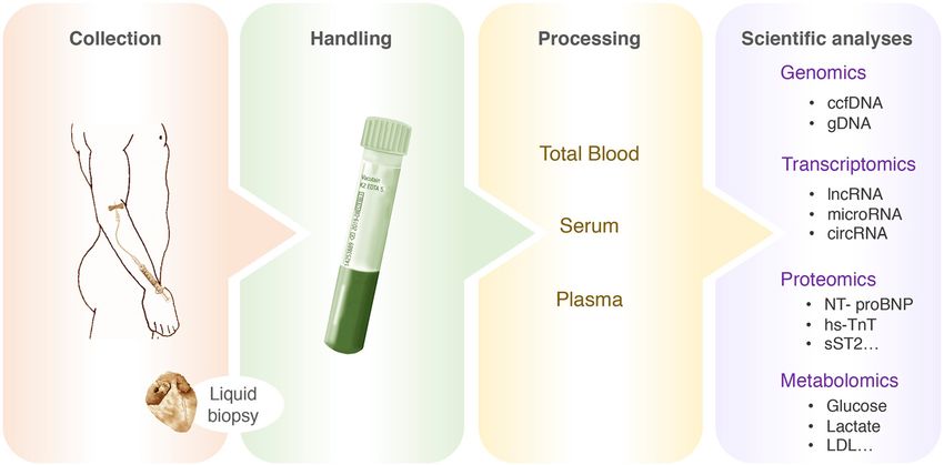
1748 Revuelta-López et al.: Pre-analytical considerations in biomarker research
quality of scientific biomarker research [1]. It is recommended defined an order for blood collection to avoid cross-
that at least collection, handling, processing, and storage of contamination of additives [7]. The current guidelines have
the biospecimens shall be defined [2]. Another interesting slightly modified these recommendations, but basically
tool is the Standard PREanalytical Code (SPREC), a code indicate that it is preferable to collect in tubes without
system that allows to identify and to record the impact that additive first [8].
pre-analytical variables have on biospecimens integrity
during collection, processing and storage [3].
Hemolysis
Studying circulating blood biomarkers by different ap-
proaches may allow a molecular understanding of the
Hemolysis is the most frequent pre-analytical problem that
myocardial status, as if a liquid biopsy were performed
may interfere with biomarker analysis in blood-derived
(Figure 2). Cardiologists and investigators need to be intro-
specimens [9, 10]. It subsumes 40–70% of all pre-analytical
duced to basic considerations and recommendations for the
confounds [11]. The hemolytic process involves rupturing
management of biospecimens, since imperfect biospecimen
the erythrocyte membrane and releasing some of its
handling or processing may introduce systematic bias and
contents, such as hemoglobin, potassium, or lactate
lead to reporting incorrect biomarker conclusions.
dehydrogenase. Less than 2% of biological samples with
We will review the importance of understanding the
hemolysis are due to in vivo hemolysis or endogenous
pre-analytical variables of blood biospecimens with a
causes [12, 13], whereas in vitro hemolysis may occur during
cardiovascular approach. First, we will point out the
the collection, handling, processing, transportation, or
important stages of the pre-analytical process. Then, we
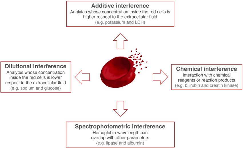
storage process [13, 14]. Hemolysis may interfere with
will review these variables, emphasizing those biomarkers
biochemistry results through additive, spectral, chemical,
approved by European and American clinical guidelines,
and dilutional mechanisms [13] (Figure 3). The presence of
used in clinical practices, and emerging biomarkers that
hemolysis in the sample can be detected visually when the
are being investigated in large cohorts stored at low
free hemoglobin concentration is 0.2–0.3 g/L; however,
temperatures.
the cut-off of at which free hemoglobin can cause inter-
ference clinically in sample analysis has been defined by
some authors as 0.5 g/L [15]. The interference caused by
Pre-analytical variables hemolysis in classical cardiac biomarkers has been
extensively studied [16–20].
Blood collection The IFCC Committee on Cardiac Biomarkers has
compiled information from existing tests for troponins and
Mistakes in sample collection procedures are the most natriuretic peptides (NPs) and designed a series of informa-
frequent failures in the pre-analytical phase [4–6]. Any tive tables to help solve problems of discordant analytical
defect during the collection process can alter the analysis results due to hemolysis [21]. In any case, it is important to
of blood-derived samples. In 1982, Calam and Cooper reference the assay inserts of each immunoassay technique
Figure 1: Illustration of the blood-derived
samples life cycle.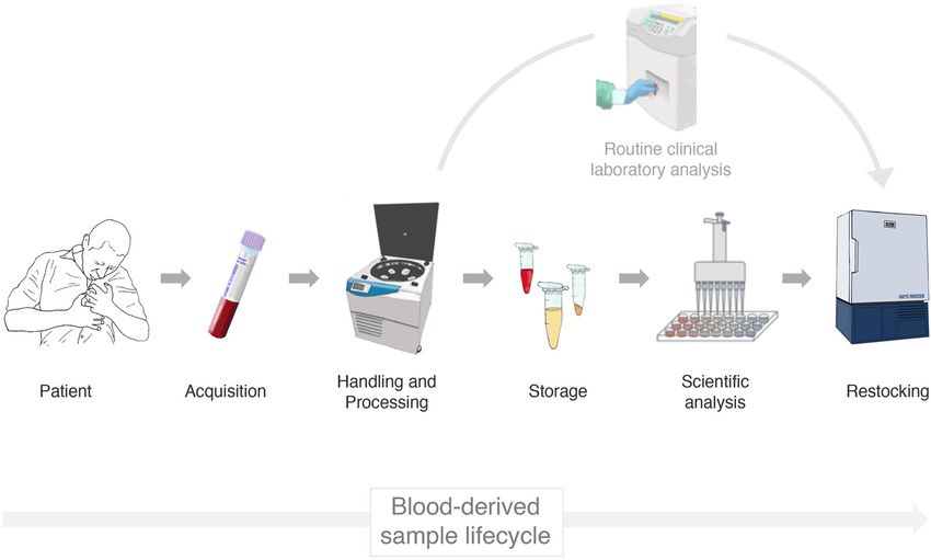
Revuelta-López et al.: Pre-analytical considerations in biomarker research         1749

                                                                                          Figure 2: Flowchart of myocardium liquid
                                                                                          biopsy.
                                                                                          ccfDNA, circulating cell free DNA; gDNA,
                                                                                          genomic DNA; lncRNA, long non-coding
                                                                                          RNA; circRNA, circular RNA; NT-proBNP,
                                                                                          N-terminal pro-brain natriuretic peptide;
                                                                                          hs-TnT, high-sensitivity cardiac troponin T;
                                                                                          LDL, low density lipoprotein.

                                                                                          Figure 3: Hemolysis interference.
                                                                                          LDH, lactate dehydrogenase.

and to carry out a literature search in order to review the       immunochemistry testing; however, Demonte et al.
latest specific interference studies. In the event that there is   described that citrate-plasma could be used on exceptional
no literature associated with the biomarker to be studied, it     occasions if there is no other option by applying a correction
would be best not to work with hemolyzed samples.                 factor [26]. EDTA and citrate inhibit coagulation via chelate
                                                                  formation with ion-dependent enzymes. In contrast, heparin
Anticoagulants                                                    accelerates the inhibition of Xa factor by antithrombin III,
                                                                  preventing fibrinogen formation from fibrin [27, 28]. Lithium,
The appropriate anticoagulant must be selected when               sodium, and ammonium salts are used in conjunction with
plasma samples are used in analytical processes. Different        heparin as an anticoagulant. Heparin may interact with
types of anticoagulants prevent clot formation through            several proteins [29], interfere in the antigen-antibody union
several mechanisms, and their choice depends on the type          [30], and interfere with liquid chromatography/mass spec-
of study to be carried out. The anticoagulant may affect the      trometry (LC-MS/MS) techniques. Therefore, heparin-plasma
measurement of small molecules [22], metabolic [23] and           is not recommended for peptide or proteomic analysis. Also,
lipidomic profiles [24], and other clinical parameters [25].       Glinge et al. do not recommend the use of heparin-plasma
Generally, the most widely used anticoagulants are                when measuring miRNAs [31]. In contrast, the use of heparin-
ethylenediaminetetraacetic acid (EDTA), heparin, and              plasma is highly recommended for metabolomics studies
citrate (Table 1). Typically, EDTA- or heparin-plasma are         using nuclear magnetic resonance (NMR) spectroscopy or
the sample matrix used for clinical chemistry and                 different mass spectrometry assays [32–34].
1750 Revuelta-López et al.: Pre-analytical considerations in biomarker research
Table : Principal additives, sample matrix, assay test, and matrix after collection, enabling rapid analysis and reducing
usage reference examples. turnaround time. For example, the 2020 ESC Guidelines
for the management of acute coronary syndromes in pa-
Additive Sample Test References
tients presenting without persistent ST-segment elevation
matrix
recommend a turnaround time of 1 h or less for cTn [43].
None/clot Serum Clinical chemistry, [, , , ]
This cannot be achieved with serum samples, where it is
activator immunochemistry
necessary to wait 30–60 min for the correct clot formation
Heparin Plasma Clinical chemistry, [, , ]
immunochemistry [44] or 20 min if BD RST tubes are used [45]. Moreover, the
EDTA Plasma/ Hematology, [, , , ] time lapse to obtain serum is not always easy to stan-
whole blood immunochemistry dardize in real world conditions. The use of plasma is also
Sodium Plasma/ Hemostasis, [, –] advantageous because is more stable than serum in several
citrate whole blood platelets
processes [46]. However, there are disadvantages associ-
EDTA, ethylenediaminetetraacetic acid. ated with plasma use: (a) the collection tube must be well
mixed for the anticoagulant to be effective, (b) plasma does
Protease inhibitors not always withstand freeze-thaw cycles well [47, 48], and
(c) most routine laboratories use serum. In addition, spe-
Some cardiovascular analytes are avidly bound and cifically heparin- and EDTA-plasma interfere with many
degraded by circulating proteases that are not well- LC-MS/MS and potentiometric methods, respectively.
inactivated in vitro by EDTA. Therefore, understanding
sensitivity to degradation is important when evaluating
new biomarkers. A prime example of an important cardiac
Blood handling
marker subject to rapid in vitro decay is atrial natriuretic
peptide (ANP) whose concentrations rapidly fall following
The pre-analytical phase known as handling refers to the
phlebotomy in part due to degradation by neprilysin; brain
time from extraction to processing. It is important to ensure
natriuretic peptide (BNP) is also subject to in vitro degra-
the optimal time and temperature handling is to avoid
dation but this is slower. For biomarkers vulnerable in
impacts on precision and quality.
this manner, collection in tubes with specialized protease
inhibitor cocktails, cold handling and processing, and
storage at −80° may provide optimal results [35]. Ease of obtaining samples
Not only can traumatic blood draws cause hemolysis but
Serum vs. plasma
traumatic draws will stimulate tissue thromboplastin and
by doing so, activate clotting. Thus, samples obtained for
Plasma and serum are obtained by centrifugation of whole
the measurement of coagulation related tests may be
blood; however, to obtain serum, blood clotting is neces-
influenced despite an appropriate anticoagulant in the
sary before centrifuging. Importantly, this makes the pro-
tube [49].
tein profile different between plasma and serum [36].
Respect to serum, during clot formation, several pro-
teins are non-specifically adsorbed or randomly captured Blood handling time
in the clot, and cellular elements can secrete components
[36, 37], which could lead to obtaining false positives for Classic standard guidelines for blood sample handling
differentially expressed proteins in serum [38]. In the other recommend that plasma and serum should be separated
hand, human plasma contains the most complex and from cells within the first 2 h after drawing the blood [50],
complete representation of the human proteome [39, 40], though some current guidelines specify that the time from
3,500 detectable proteins according to the last HUPO PPP collection to centrifugation should not exceed 1 h, or as
[41]. Accordingly, the use of serum samples is not recom- quickly as possible for plasma biomarker studies [28]. If the
mended for peptidomic biomarker discovery, although it centrifugation process is delayed, keeping the sample at
can be useful for validation purposes in order to improve 4 °C is better to maintain the stability of certain analytes,
specificity [27, 39, 42]. while in other cases it produces a negative effect [51]. For
Traditionally, plasma has been recommended in this reason, it is important to consider which biomarkers
emergency clinical laboratories over serum because anti- should be evaluated to assess if the sample can be kept
coagulated blood samples can be centrifuged immediately refrigerated in case of delay or not.Revuelta-López et al.: Pre-analytical considerations in biomarker research 1751
For example, in the specific case of cytokines, which number of freeze-thaw cycles, duration of thaw events,
play an important role in the progression of cardiovascular time from last thaw to processing, and temperature
disease, it has been proven that keeping blood samples for between last thaw and processing. This is usually accom-
a long time at room temperature or lower can significantly plished by processing the sample and aliquoting it into
alter plasma cytokine levels, so is crucial to minimize the multiple different tubes with somewhere between 250 and
blood handling time in these cases [52, 53]. 500 μL so that individual samples can be thawed for
measurement without impairing the ability to measure
Blood handling temperature other analytes that might be impacted by the thawing
process per se. It is important to differentiate samples that
Storage temperature before centrifugation is also an will be processed following centrifugation in the routine
important factor to consider. Several studies have clinical laboratory and those that will be stored in biobanks
demonstrated that the concentration of some biomarkers for future studies.
can vary depending on the storage temperature prior to the
centrifugation process [28, 54, 55]. Current standard Routine clinical laboratory
guidelines for blood sample handling recommend that, if
the sample can be centrifuged within a short time, the If the samples are not going to be stored, samples must be
samples should be kept at room temperature to minimize transferred directly to the analyzer in the routine clinical
the platelet activation that occurs at low temperatures. In laboratory to avoid alterations in the stability of the
some situations, in response to temperature, the analyte of analytes, considering that sample stability depends on
interest may be cleaved from its normal molecular position temperature and it is different depending on the analyte.
affecting measured values [56]. For example, N-terminal proBNP (NT-proBNP), cardiac
troponins (cTn), cancer antigen 125 (CA125), soluble ST2
(sST2), and galectin-3 are stable analytes under a wide-
Blood processing ranging of storage temperatures [51, 65–68], whereas BNP
is only stable in whole blood for at about 4 h at room
Various guidelines based on expert opinion or manufac- temperature [69] and ANP is even more unstable [70–73].
turer recommendations are available for sample process- Sometimes results from clinical laboratories are
ing [50, 57, 58]. To avoid disturbances in the blood sample collected in databases directly from routine analysis. For
during processing, the speed, temperature, and centrifu- instance, parameters as cTn, NT-proBNP, or C-reactive
gation time must be considered. To obtain serum, once the protein are included in many clinical profiles and analyzed
clot formation time has passed, the classic recommenda- in fresh samples for patient management. This has the
tion is to centrifuge the sample at 1,500–2,000 g at room advantage of not been subjected to freeze-thaw cycles, but
temperature for 10 min [46]. To obtain plasma, blood several considerations have to been made: (a) results may
samples are typically centrifuged at 1,000–1,200 g for be obtained from different laboratories or analyzers; (b) in
10 min; however, often a second centrifugation at a higher long lasting studies reactive and calibrators lots may
speed of 2,000–3,000 g is required to obtain platelet-poor change, and (c) operators and quality specifications may
plasma. Residual platelet contamination is a significant vary.
confounding source of circulating miRNAs [59–61]; there- Taking this into consideration, for stable analytes it
fore, in this case obtaining platelet-poor plasma is crucial should be recommended to process all samples in a same
for obtaining reliable results. Similarly, sample processing batch and analyzer, regardless previous results. This is
plays a crucial role in circulating cell free DNA (ccfDNA) particularly useful in novel immunoassays, that may not
studies to avoid genomic DNA contamination, and samples have an established standard for calibration.
must be completely cell free [62–64].
Biospecimen biobanking
Storage The banking of samples should not be neglected. The use of
retrospective serum and plasma samples may introduce
Proper specimen storage is critical to maintaining spec- significant variability in the molecular composition of the
imen integrity, as inappropriate specimen storage can samples solely because of heterogeneity in storage pro-
change the sample properties and affect the final results. cedures. Storage temperature, storage time, and freeze-
Appropriate storage conditions include controlling the thaw cycles may interfere with the sample composition.1752 Revuelta-López et al.: Pre-analytical considerations in biomarker research
– Storage temperature and time. To lessen the effects of Myocyte stress
storage temperature, it must remain stable over
time. Liquid nitrogen maintains stable ultra-low Natriuretic peptides
temperatures; however, most samples are stored in non-
cycling freezers with temperatures ranging between −70 Nowadays, NPs are the gold standard clinical indicators in
and −80 °C. Temperature variations in −20 °C or −40 °C the diagnosis and prognosis of heart failure.
freezers are more likely to affect sample stability to a As we have discussed previously, ANP concentrations
greater degree, particularly as many of these freezers are decrease rapidly following phlebotomy, it is very unstable
cycling, meaning their temperature rises and falls to [70–73], like BNP, although to a lesser degree. In these
reduce frost formation [74]. In any case, it is important to cases it is essential to carry out the sample collection in
have a centralized record of freezer temperatures, with tubes with specialized protease inhibitor cocktails and cold
measures taken from independent probes. Furthermore, handling to provide optimal results [35]. In addition, BNP
the short-term thermal exposure that may occur while must be measured in EDTA-plasma or whole blood drawn
removing individual samples from a biobank needs to into plastic tubes due to proteolytic degradation in vivo [79]
be considered. In this case, cooling systems, such as dry and it is only stable in whole blood for at about 4 h at room
ice, or working in low temperature rooms to avoid temperature [69].
heating the samples while searching for specific sam- NT-proBNP is less vulnerable to degradation. NT-pro
ples is recommended. Zander et al. demonstrated that BNP can be measured in serum or heparin-plasma with
short-term thermal exposures of 5 min while working in similar results [80] and it can be also evaluated in
low temperature rooms do not alter the levels some EDTA-plasma, but in this case, it may yield lower values
biomarkers while others are affected [75]. compared to serum and heparin-plasma, depending on the
– Freeze-thaw cycles. Typically, it is recommended to assay method [80, 81]. In addition, NT-proBNP is stable
aliquot the samples and store at −80 °C. It is important under a wide-range of storage temperatures [51, 65–68],
to find a balance between the number of aliquots and analyte levels are not influenced by moderate hemolysis
the volume of each and the available storage space. (0.6 g/L) [16] and it is not significantly affected by freeze-
The availability of several aliquots allows the effect of thaw cycles [66].
the number of freeze-thaw cycles to be mitigated, NPs are a clear example of the importance of under-
allowing analysis of different biomarkers and analytes standing sensitivity to degradation and the correct selec-
in different time frames. Most of the commercial tion of the blood collection tube to evaluate new
manuals specify avoiding several defrost cycles biomarkers.
without specifying an exact number, and studies are
not always available in the literature. Repeated freeze-
thaw cycles affectation will also depend on the anal-
Inflammation
ysis technique used [76–78].
Cytokines
Cytokines have been reported to participate in different
pathophysiological mechanisms of cardiovascular disease.
Pre-analytics impact in Cytokines play diverse roles with both, detrimental and
cardiovascular research positive effects on the cardiovascular system. Pro-
inflammatory cytokines, such interleukin-1 (IL-1) and
As described in the previous section, there are multiple pre- IL-18, are involved in the development of cardiac pathol-
analytical variables that can interfere with or alter the ogies and are suggested to be potential therapeutic targets.
results of clinical investigations carried out by our staff in There are also cytokines with pleiotropic functions,
hospitals, primary care and research centers. Table 2, including IL-6, that play duals role in CVD. All of them
summarizes the pre-analytical recommendations for the could be predictors of adverse outputs. Determine the
most relevant cardiac biomarkers to date; however, we will implication of each cytokine in the progression of cardio-
try to go deeper into it depending on its clinical value. vascular disease is key to developing new therapeuticRevuelta-López et al.: Pre-analytical considerations in biomarker research 1753
Table : Recommended pre-analytical variables of relevant cardiac biomarkers.
Biomarker Clinical value Sample matrix Storage temperature Freeze-thaw cycles References
stability stability
BNP Myocyte stress EDTA-plasma h at RT Affected very significantly [, ]
NT-proBNP Myocyte stress EDTA-plasma days at RT Not significantly affected [, , ]
Heparin-plasma serum days at – °C
months at − °C
ANP Myocyte stress EDTA-plasma Few days at − °C Affected very significantly [–]
month at − °C
sST Stress, inflammation, fibrosis EDTA-plasma h at RT Up to three cycles [, –]
Heparin-plasma serum days at – °C
months at − °C
hsTn Injury EDTA-plasma h at – °C Up to four cycles [–]
Heparin-plasma seruma months at − °C
CA Congestion EDTA-plasma days at – °C Up to three cycles [, ]
Heparin-plasma
Serum
Galectin- Fibrosis EDTA-plasma serum days at RT Up to six cycles [, , , ]
days at – °C
months at − °C
a
EDTA- and heparin-plasma samples should not be exchanged with serum samples. EDTA, ethylenediaminetetraacetic acid; BNP, brain
natriuretic peptide; NT-proBNP, N-terminal pro-brain natriuretic peptide; ANP, atrial natriuretic peptide; sST, soluble ST; CA, cancer
antigen ; hsTn, high-sensitivity cardiac troponin T.
agents that aim to treat the inflammatory processes asso- sST2
ciated with this pathology. Therefore, it is important to
know how pre-analytical variables can affect cytokine sST2, also known as interleukin 1 receptor-like 1 (IL1RL-1), is
concentrations in clinical research. produced in response to stress and overload by cardiac
Cytokines are highly labile to temperature, so it is fibroblasts and cardiomyocytes [86]. sST2 plays an important
advisable to measure them as soon as possible after role in many inflammatory diseases and prevents fibrosis
collection [82]. Binnington et al. described absence of and inflammatory response when interacting with IL-33.
alterations in cytokine concentrations in whole blood or Especially, sST2 is gaining attention in the manage-
plasma stored refrigerated up to 10 days [83]; however, also ment of chronic heart failure. Lupón et al. developed The
it has been proven that keeping the samples for a long time Bio-Heart Failure Risk Calculator (BCN BIO-HF Calculator)
at room temperature or lower can significantly alter plasma that incorporated available biomarkers, including ST2,
cytokine levels [52, 53]. Then, minimizing the blood reflecting different pathophysiological pathways in heart
handling time seems crucial in cytokine research. failure [87]. Really, sST2 is a novel biomarker that could be
Regarding freeze-thaw cycles, it has been shown that used for diagnosis and management of patients with
IL-6, IL-10, Interferon-γ, and IL-2 levels are stable in several cardiovascular diseases [88, 89].
plasma over three freeze-thaw cycles [84]; but, there are Regarding the pre-analytical variables that we have
other cytokines whose concentrations increase or decrease been commenting on, sST2 is a very stable biomarker in
due to thawing cycles. IL-1β, IL-4, and IL-10 circulating different aspects. The effects produced by hemolysis on
levels increase at least 3–5-fold following one freeze/thaw sST2 levels are minimal and not significant [90]. In addi-
cycle, stabilizing at these levels during a subsequent nine tion, serum, lithium heparin-, and EDTA-plasma are vali-
freeze/thaw cycles [85]. dated samples for the Presage ST2 assay, the only approved
Therefore, minimizing the pre-analytical variables as test by the FDA for clinical use [65, 91, 92]; however, the
much as possible in the study of interleukins is very new SEQUENT-IA ST2 assay has yet been evaluated only in
important to obtain valid and reliable results. serum and EDTA-plasma samples.1754 Revuelta-López et al.: Pre-analytical considerations in biomarker research
Respect to storage, sST2 is stable under a wide-ranging degrees of hemolysis decrease its concentrations in a he-
of storage temperatures, 48 h at RT, 7 days at 2–8 °C and moglobin concentration-dependent manner [18], causing
18 months at −20 °C [65, 67, 68] and it is stable up to three false negatives that make it difficult to interpret results
freeze-thaw cycles [65]. [98, 100], surely because hemolysis could release proteases
The pre-analytical stability of ST2 makes it easier to that cleave the cTnT antigenic regions recognized by the
investigate this promising biomarker in cardiovascular immunoassays [17]. In contrast, Harley et al. reported that
physiopathology. hemolysis interference can be positive or negative
depending on the high-sensitivity assay and the concen-
GDF-15 tration range for cTnI [101]. This positive interference has
been corrected in most immunoassays; however, it has
Growth differentiation factor 15 (GDF-15) is a member of the recently been published that hs-cTnI concentrations in
transforming growth factor-cytokine superfamily that has heparin-plasma are increased by hemolysis (>4 g/L) with
emerged as a stress potential biomarker in cardiovascular the Ortho hs-cTnI assay only at the low concentration range
disease with potential implications for risk and patient [101, 102]. The release of multiple proteins from erythro-
management. It has been recognized as a biomarker of cytes increases matrix complexity and this may influence
mortality in patients with acute coronary syndrome immunoassays in different ways, depending on the
[93–95]. monoclonal antibodies, solid phase characteristics, and
GDF-15 has favorable pre-analytic characteristics. The incubation times [100].
anticoagulant matrix does not affect the analyte measurement cTn are also a good example to demonstrate how the
since no differences are observed in the concentration of use of serum or plasma can influence concentration levels.
GDF-15 between serum and EDTA-, heparin-, or citrate- Initial reports indicated that for some assays cTn
plasma [96, 97]. In addition, GDF-15 immunoreactivity is levels were significantly lower in heparin-plasma samples
stable in serum and whole blood at room temperature for at than in serum, probably because heparin may bind to
least 48 h, which facilitates its use in routine laboratories. troponin, decreasing immunoreactivity [103, 104]. Roche
Regarding the analysis of GDF-15 in frozen samples, it must be Diagnostics, manufacturer of cTnT assay, recommended
considered that it is stable until the fourth freeze-thaw cycle customers to avoid using heparin-plasma samples in their
[96, 97]. assay until new test formulations corrected this variability
Both the pre-analytical characteristics of GDF-15 and [103, 105]. At present, Elecsys Troponin T hs package insert
its analytical stability make research with it advantageous. specify that plasma (EDTA and heparin) and serum sam-
ples cannot be interchanged because there are differences
in the values that are obtained [106]. With the increasing
Injury number of cTn assays reaching the market, this issue needs
to be revisited whenever a new assay is put in use.
Cardiac troponins
Cardiac circulating cell free DNA
Cardiac troponins I and T (cTnI, cTnT) are the primary
cardiac biomarkers used for the diagnosis of myocardial Dying cells release nucleosome-size fragments of genomic
injury, as they are sensitive and specific markers of car- DNA to the blood system. Identification of specific circu-
diomyocyte injury and more specific than creatine kinase. lating cell free DNA (ccfDNA) from cardiomyocytes would
Serial measurements of cTn are useful to understand allow a liquid biopsy to be performed, reflecting the
kinetics, peak values and percentage changes. Clinical physical state of myocardial tissue.
implications of high-sensitivity cTn (hs-cTn) assays are Recently, Zemmour et al. identified specific car-
widely known and detailed in the 2020 ESC Guidelines for diomyocyte methylation markers that allow to identify
the management of acute coronary syndromes in patients ccfDNA release by death cardiomyocytes after ischemia
presenting without persistent ST-segment elevation. ensues [64].
Daves et al. reported in cTnI and cTnT were not influ- Cardiac ccfDNA could be a promising non-invasive
enced by moderate hemolysis (0.6 g/L) [16]; however, the clinical marker and diagnostic tool in acute myocardial
development of very high sensitive methods could lead to infarction but more clinical studies are required and it is
notable differences [98]. In the recent years, it has been important to consider different pre-analytical variables.
described that hemolysis can produce both negative and Free hemoglobin has been reported to correlate with
positive interferences in cTn [99]. Respect cTnT, higher ccfDNA concentrations, then ccfDNA levels are increasedRevuelta-López et al.: Pre-analytical considerations in biomarker research 1755
in hemolyzed samples [107]. However, Streleckiene et al. Actually, research with this type of biomarker requires
demonstrated that the effect of hemolysis on ccfDNA levels a thorough bibliographic review. Faraldi et al. demon-
depended on the isolation kit used [108]. In this regard, it strated that different miRNAs are differently affected even
would be interesting to see if hemolysis has any effect on if the same collection method or storage condition are
the concentrations of ccfDNA released specifically from performed [119]. Establishing standardization protocols for
cardiomyocytes. the study of each miRNA independently is crucial for the
Several studies have shown higher ccfDNA concen- clinical implementation of miRNAs as biomarkers.
trations in serum than in plasma samples due to the clot-
ting process [109–112]. For that, to study ccfDNA it is more CA125
advisable to use plasma samples, and more specifically,
EDTA-plasma [110]. CA125 is emerging as a novel congestion biomarker in heart
Regarding sample storage, DNA is stable at 4 °C for failure. It has traditionally been a biomarker of ovarian and
several weeks, at −20 °C for months, and at −80 °C for years endometrial cancer, however, recent publications demon-
[113, 114]. Specifically for ccfDNA, storage time has been strate the usefulness of CA125 to monitor or guide the
reported to have no influence on the detection of specific treatment of heart failure [124, 125].
sequences or mutations after several years [115]. As its use is established for the diagnosis and man-
agement of different cancers, the implication of pre-
analytical variables in its stability is well studied.
Other Sandhu et al. found that CA125 levels were lower if samples
handled at 4 °C, so it is advisable to keep the samples at
microRNAs room temperature before centrifugation [51]. They also
showed that plasma or serum samples can be used inter-
microRNAs (miRNAs) are endogenous and conserved non- changeably and that CA125 remains stable under a wide-
coding RNAs that are involved in several pathways in the range of storage temperatures after centrifugation [51].
cardiovascular system. miRNAs are characterized by great Respect to freeze-thaw cycles, it has been described
stability, resisting degradation in blood-derived specimens that CA125 is stable up to three cycles of freezing and
kept at room temperature for up to 24 h after collection thawing [51].
[116, 117]. The stability of miRNAs in serum and plasma and
their resistance to degradation make them promising bio- Galectin-3
markers for diagnosis and prognosis of cardiovascular
diseases [118]; however, several pre-analytical variables Galectin-3 is a β-galactoside-binding member of the lectin
could influence in circulating miRNA identification and family implicated in cardiac fibrosis. It is considered a
quantification [119]. prognostic biomarker that identifies increased risk of death
Regarding sample matrix, it has been demonstrated and heart failure [126].
that EDTA is the best anticoagulant for studying circulating To evaluate circulating levels of galectin-3 it is crucial
miRNA profiling [120], as heparin and citrate may interfere to avoid hemolyzed samples. Hemolysis produces a posi-
with the enzyme activity in PCR-based assays. However, in tive interference due to intracellular release of galectin-3
the event that EDTA-sample is not available, Basso et al. from white blood cells [19]. Assessment of galectin-3 levels
demonstrated that miRNA quality is comparable in serum in hemolyzed specimens may confuse the identification of
and EDTA- and citrate-plasma [121]. the risk of death and heart failure.
Processing in this case is also a crucial step due to Both serum and EDTA-plasma have been validated for
differential processing could alter miRNAs quantitation galectin-3 measurement [65]. La’ulu et al. demonstrated
[122]. In addition, platelets represent a source of contami- similar galectin-3 levels between serum and EDTA-plasma
nation for circulating miRNAs, then obtaining platelet- [20], while Gaze et al. showed higher galectin-3 values in
poor plasma is crucial to avoid miRNA contamination. plasma than serum samples [127] using the same type of
Platelets also can interfere with stability during storage, assay and analyzer, so we would not recommend
despite several miRNAs resist degradation in plasma that is exchanging both types of sample until further evidence is
frozen and thawed multiple times [116, 123], Muth et al. published.
reported that freeze-thaw cycles can affect miRNA stability Respect analyte stability, it has been reported that
if platelet depletion is inadequate [61]. This is another galectin-3 is stable under a wide-range of storage temper-
reason why proper platelet removal is necessary. atures, and it is stable up to six freeze-thaw cycles [65].1756 Revuelta-López et al.: Pre-analytical considerations in biomarker research
Discussion References
There is a great need to discover and validate new bio- 1. Moore HM, Kelly AB, Jewell SD, McShane LM, Clark DP,
markers that help clinicians to manage the patient with Greenspan R, et al. Biospecimen reporting for improved study
quality (BRISQ). J Proteome Res 2011;119:92–101.
cardiovascular disease; however, it is necessary to
2. Hall JA, Salgado R, Lively T, Sweep F, Schuh A. A risk-management
consider that there are multiple pre-analytical variables approach for effective integration of biomarkers in clinical trials:
that can interfere in the clinical investigations carried out perspectives of an NCI, NCRI, and EORTC working group. Lancet
by our staff in hospitals, primary care, and research centers Oncol 2014;14:e184–93.
(Table 2). The ability to manage and track pre-analytical 3. Lehmann S, Guadagni F, Moore H, Ashton G, Barnes M, Benson E,
et al. Standard preanalytical coding for biospecimens: review
variations impacting biospecimen integrity is crucial to
and implementation of the sample PREanalytical Code (SPREC).
high quality and reliable results. Biopreserv Biobanking 2012;10:366–74.
As we have seen in this review, there are multiple 4. Plebani M, Carraro P. Mistakes in a stat laboratory: types and
differences between the different types of plasma and the frequency. Clin Chem 1997;43:1348–51.
concentration of certain analytes can vary between serum 5. Lima-Oliveira G, Volanski W, Lippi G, Picheth G, Guidi GC. Pre-
and plasma. The different levels of circulating cTn between analytical phase management: a review of the procedures from
patient preparation to laboratory analysis. Scand J Clin Lab Invest
plasma and serum found with the first automated immu-
2017;77:153–63.
noassays are a clear example of the importance of correctly 6. Lippi G, von Meyer A, Cadamuro J, Simundic AM. Blood sample
selecting the sample matrix, in the same way that the BNP quality. Diagnosis 2019;6:25–31.
instability exemplifies the importance of standardizing the 7. Calam RR, Cooper MH. Recommended “order of draw” for
handling time. collecting blood specimens into additive-containing tubes. Clin
Chem 1982;28:1399.
The stability and matrix effect of promising new bio-
8. NCCLS. Procedures for the collection of diagnostic blood
markers needs to be studied to facilitate clinical trials. The specimens by venipuncture; approved standard, 5th ed. NCCLS
analysis methodology and the objective must also be document H3-A5. Wayne, PA, USA: NCCLS; 2003.
considered, because if the study biomarker will be used in 9. Azman WNW, Omar J, Koon TS, Ismail TST. Hemolyzed specimens:
emergency laboratories, it should be validated with plasma major challenge for identifying and rejecting specimens in
for the reasons we have previously explained. clinical laboratories. Oman Med J 2019;34:94–8.
10. Goyal T, Schmotzer CL. Validation of hemolysis index thresholds
Nowadays, large multicenter global studies where
optimizes detection of clinically significant hemolysis. Am J Clin
samples are collected in different recruitment centers and Pathol 2015;143:579–83.
sent to the promoting center for analysis, are increasingly 11. Lippi G, Blanckaert N, Bonini P, Green S, Kitchen S, Palicka V,
abundant. For a correct reproducibility between derivation et al. Haemolysis: an overview of the leading cause of unsuitable
and validation cohorts it is necessary that the process of specimens in clinical laboratories. Clin Chem Lab Med 2008;46:
764–72.
obtaining and handling the sample is completely stan-
12. Carraro P, Servidio G, Plebani M. Hemolyzed specimens: a reason
dardized between centers. The formation of working for rejection or a clinical challenge? Clin Chem 2000;46:306–7.
groups that standardize and control the quality of pre- 13. Marques-Garcia F. Methods for hemolysis interference study
analytical handling of blood samples would be advisable. in laboratory medicine - a critical review. EJIFCC 2020;31:
It is necessary to introduce academic clinicians and 85–97.
14. Bush V, Mangan L. The hemolyzed Specimen : causes, effects,
investigators to basic considerations for biospecimen
and reduction. BD Vacutainer Syst Preanalytical Solut 2003;1–8.
management and essential pre-analytical recommenda-
15. Tóth J, Oláh AV, Petercsák T, Kovács T, Kappelmayer J. Detection
tions to use this biological material in the most appropriate of haemolysis, a frequent preanalytical problem in the serum of
and efficient way to obtain valid conclusions from new newborns and adults. EJIFCC 2020;31:6–14.
promising cardiovascular biomarker research. 16. Daves M, Salvagno GL, Cemin R, Gelati M, Cervellin G, Guidi GC,
et al. Influence of hemolysis on routine laboratory cardiac marker
testing. Clin Lab 2012;58:333–6.
Research funding: The authors received no specific 17. Sodi R, Darn S, Stott A. Time for troponin T? Implications from
funding for this work. newly elucidated structure. Clin Chem 2004;50:786–7.
Author contributions: All authors have accepted 18. Li A, Brattsand G. Stability of serum samples and hemolysis
responsibility for the entire content of this manuscript interference on the high sensitivity troponin T assay. Clin Chem
Lab Med 2011;49:335–6.
and approved its submission.
19. Christenson RH, Duh SH, Wu AHB, Smith A, Abel G, DeFilippi CR,
Competing interests: Authors state no conflict of interest. et al. Multi-center determination of galectin-3 assay performance
Informed consent: Not applicable. characteristics: anatomy of a novel assay for use in heart failure.
Ethical approval: Not applicable. Clin Biochem 2010;43:683–90.Revuelta-López et al.: Pre-analytical considerations in biomarker research 1757
20. La’ulu SL, Apple FS, Murakami MAM, Ler R, Roberts WL, valsartan in heart failure with reduced ejection fraction. JACC
Straseski JA. Performance characteristics of the ARCHITECT Heart Fail 2021;9:127–36.
Galectin-3 assay. Clin Biochem 2013;46:119–22. 36. Sapan CV, Lundblad RL. Considerations regarding the use of
21. Saenger AK, Jaffe AS, Body R, Collinson PO, Kavsak PA, Lam CSP, blood samples in the proteomic identification of biomarkers for
et al. Cardiac troponin and natriuretic peptide analytical cancer diagnosis. Cancer Genomics Proteomics 2006;3:227–30.
interferences from hemolysis and biotin: educational aids from 37. Wong HL, Pfeiffer RM, Fears TR, Vermeulen R, Ji S, Rabkin CS.
the IFCC Committee on Cardiac Biomarkers (IFCC C-CB). Clin Chem Reproducibility and correlations of multiplex cytokine levels in
Lab Med 2019;57:633–40. asymptomatic persons. Cancer Epidemiol Biomark Prev 2008;17:
22. Mei H, Hsieh Y, Nardo C, Xu X, Wang S, Ng K, et al. Investigation of 3450–6.
matrix effects in bioanalytical high-performance liquid 38. Ignjatovic V, Geyer PE, Palaniappan KK, Chaaban JE, Omenn GS,
chromatography/tandem mass spectrometric assays: Baker MS, et al. Mass spectrometry-based plasma proteomics:
application to drug discovery. Rapid Commun Mass Spectrom considerations from sample collection to achieving translational
2003;17:97–103. data. J Proteome Res 2019;18:4085–97.
23. Barton RH, Waterman D, Bonner FW, Holmes E, Clarke R, 39. Tammen H, Schulte I, Hess R, Menzel C, Kellmann M, Mohring T,
Nicholson JK, et al. The influence of EDTA and citrate et al. Peptidomic analysis of human blood specimens:
anticoagulant addition to human plasma on information recovery comparison between plasma specimens and serum by
from NMR-based metabolic profiling studies. Mol Biosyst 2009; differential peptide display. Proteomics 2005;5:3414–22.
6:215–24. 40. Anderson NL, Anderson NG. The human plasma proteome:
24. Gonzalez-Covarrubias V, Dane A, Hankemeier T, Vreeken RJ. history, character, and diagnostic prospects. Mol Cell Proteomics
The influence of citrate, EDTA, and heparin anticoagulants to 2002;1:845–67.
human plasma LC-MS lipidomic profiling. Metabolomics 2013; 41. Schwenk JM, Omenn GS, Sun Z, Campbell DS, Baker MS, Overall
9:337–48. CM, et al. The human plasma proteome draft of 2017: building on
25. Yi J, Craft D, Gelfand CA. Minimizing preanalytical variation of the human plasma PeptideAtlas from mass spectrometry and
plasma samples by proper blood collection and handling. complementary assays. J Proteome Res 2017;16:4299–310.
Methods Mol Biol 2011;728:137–49. 42. Misek DE, Kuick R, Wang H, Galchev V, Deng B, Zhao R, et al. A
26. Demonte D, Pucci M, Salvagno GL, Lippi G. Can citrate plasma be wide range of protein isoforms in serum and plasma uncovered
used in exceptional circumstances for some clinical chemistry by a quantitative intact protein analysis system. Proteomics
and immunochemistry tests? Diagnosis 2019;6:369–75. 2005;5:3343–52.
27. Tammen H, Schulte I, Hess R, Menzel C, Kellmann M, Schulz- 43. Collet J-P, Thiele H, Barbato E, Barthélémy O, Bauersachs J, Bhatt
Knappe P. Prerequisites for peptidomic analysis of blood DL, et al. 2020 ESC Guidelines for the management of acute
samples: I. Evaluation of blood specimen qualities and coronary syndromes in patients presenting without persistent
determination of technical performance characteristics. Comb ST-segment elevation. Eur Heart J 2021;42:1289–367.
Chem High Throughput Screen 2005;8:725–33. 44. Tuck MK, Chan DW, Chia D, Godwin AK, Grizzle WE, Krueger KE,
28. Guder WG, Narayanan S, Wisser H, Zawta B. Samples: from the et al. Standard operating procedures for serum and plasma
patient to the laboratory: the impact of preanalytical variables on collection: early detection research network consensus
the quality of laboratory results, 3rd Revised ed. Weinheim, statement standard operating procedure integration working
Germany: Wiley-VCH Verlag GmbH; 2003. group. J Proteome Res 2009;8:113–7.
29. Capila I, Linhardt RJ. Heparin - protein interactions. Angew Chem 45. Kocijancic M, Cargonja J, Delic-Knezevic A. Evaluation of the BD
Int Ed 2002;41:391–412. Vacutainer® RST blood collection tube for routine chemistry
30. Eskinazi DP, Perna JJ, Ershow AG, Sharrow SO. Effects of heparin analytes: clinical significance of differences and stability study.
on in vitro immune parameters. J Biol Response Modif 1988;7: Biochem Med 2014;24:368–75.
173–84. 46. Guder WG, Banfi G, Bauer K, Buchberger W, Deom A,
31. Glinge C, Clauss S, Boddum K, Jabbari R, Jabbari J, Risgaard B, World Health Organization, et al. Use of anticoagulants in
et al. Stability of circulating blood-based microRNAs-Pre-Analytic diagnostic laboratory: stability of blood, plasma and serum
methodological considerations. PloS One 2017;12:e0167969. samples. Geneva: WHO; 2002:1–62 pp.
32. Catalán Ú, Rodríguez MÁ, Ras MR, MacIá A, Mallol R, Vinaixa M, 47. Comstock GW, Burke AE, Norkus EP, Gordon GB, Hoffman SC,
et al. Biomarkers of food intake and metabolite differences Helzlsouer KJ. Effects of repeated freeze-thaw cycles on
between plasma and red blood cell matrices; A human concentrations of cholesterol, micronutrients, and hormones in
metabolomic profile approach. Mol Biosyst 2013;9:1411–22. human plasma and serum. Am J Epidemiol 2008;168:827–30.
33. Barri T, Dragsted LO. UPLC-ESI-QTOF/MS and multivariate data 48. Lee JE, Kim SY, Shin SY. Effect of repeated freezing and thawing
analysis for blood plasma and serum metabolomics: effect of on biomarker stability in plasma and serum samples. Osong
experimental artefacts and anticoagulant. Anal Chim Acta 2013; Public Health Res Perspect 2015;6:357–62.
768:118–28. 49. Eisenberg PR, Sherman LA, Schectman K, Perez J, Sobel BE, Jaffe
34. Zhou QY, Wang YL, Li X, Shen XY, Li KJ, Zheng J, et al. AS, et al. A marker of acute coronary thrombosis. Circulation
Metabolomics investigation of cutaneous T cell lymphoma 1985;71:912–8.
based on UHPLC-QTOF/MS. Asian Pac J Cancer Prev 2014;15: 50. Calam RR, Bessman JD, Ernst DJ, Smith S, Szamosi DI, Warunek DJ,
5417–21. et al. Procedures for the handling and processing of blood
35. Murphy SP, Prescott M, Camacho A, Iver S, Maisel A, Felker G, specimens , Approved Guideline — Third Edition. CLSI Doc H18-A3;
et al. Atrial natriuretic peptide and treatment with sacubitril/ 2004;24.1758 Revuelta-López et al.: Pre-analytical considerations in biomarker research
51. Sandhu N, Karlsen MA, Hogdall C, Laursen IA, Christensen IJ, high-sensitivity assay for measurement of soluble ST2 in human
Hogdall EVS. Stability of HE4 and CA125 in blood samples from plasma - the PresageTM ST2 assay. Clin Chim Acta 2009;409:
patients diagnosed with ovarian cancer. Scand J Clin Lab Invest 33–40.
2014;74:477–84. 69. Gobinet-Georges A, Valli N, Filliatre H, Dubernet MF, Dedeystere
52. Aguilar-Mahecha A, Kuzyk MA, Domanski D, Borchers CH, Basik O, Bordenave L. Stability of brain natriuretic peptide (BNP) in
M. The effect of pre-analytical variability on the measurement of human whole blood and plasma. Clin Chem Lab Med 2000;38:
MRM-MS-based mid- to high-abundance plasma protein 519–23.
biomarkers and a panel of cytokines. PloS One 2012;7:e38290. 70. Bhaggoe UM, Boomsma F, Admiraal PJJ, in t Veld AJM,
53. Cao Z, Kamlage B, Wagner-Golbs A, Maisha M, Sun J, Schalekamp MADH. Stability of human plasma atrial natriuretic
Schnackenberg LK, et al. An integrated analysis of metabolites, peptide during storage at -80°C. Clin Chim Acta 1993;223:
peptides, and inflammation biomarkers for assessment of 179–84.
preanalytical variability of human plasma. J Proteome Res 2019; 71. Lijnen P, Huysecom J, Fagard R, Staessen J, Amery A. Effects of
18:2411–21. haemolysis and prolonged cold storage of human plasma on the
54. Clark S, Youngman LD, Palmer A, Parish S, Peto R, Collins R. α-atrial natriuretic peptide concentration. Clin Chim Acta 1988;
Stability of plasma analytes after delayed separation of whole 171:333–4.
blood: implications for epidemiological studies. Int J Epidemiol 72. Tsuji T, Masuda H, Imagawa K, Haraikawa M, Shibata K, Kono M,
2003;32:125–30. et al. Stability of human atrial natriuretic peptide in blood
55. Heins M, Heil W, Withold W. Storage of serum or whole blood samples. Clin Chim Acta 1994;225:171–7.
samples? Effects of time and temperature on 22 serum analytes. 73. Nelesen RA, Dimsdale JE, Ziegler MG. Plasma atrial natriuretic
Clin Chem Lab Med 1995;33:231–8. peptide is unstable under most storage conditions. Circulation
56. Oliver LK, Voskoboev N, Heser D, McConnell JP, Hodel-Hanson S, 1992;86:463–6.
Callanan H, et al. Assessment of clinical performance without 74. Elliott P, Peakman TC. The UK Biobank sample handling and
adequate analytical validation: a prescription for confusion. Clin storage protocol for the collection, processing and archiving of
Biochem 2011;44:1247–52. human blood and urine. Int J Epidemiol 2008;37:234–44.
57. Lippi G, Salvagno GL, Montagnana M, Guidi GC. Preparation of a 75. Zander J, Bruegel M, Kleinhempel A, Becker S, Petros S, Kortz L,
quality sample: effect of centrifugation time on stat clinical et al. Effect of biobanking conditions on short-term stability of
chemistry testing. Lab Med 2007;38:172–6. biomarkers in human serum and plasma. Clin Chem Lab Med
58. Kiechle FL, Betson F, Blackeney J, Calam RR, Catalasan IM, Raj P, 2014;52:629–39.
et al. Procedures for the handling and processing of blood 76. Shen Q, Björkesten J, Galli J, Ekman D, Broberg J, Nordberg N,
specimens for common laboratory tests. Approved Guideline - et al. Strong impact on plasma protein profiles by
Fourth Edition. CLSI Doc H18-A4; 2010;30. precentrifugation delay but not by repeated freeze-thaw cycles,
59. Cheng HH, Yi HS, Kim Y, Kroh EM, Chien JW, Eaton KD, et al. as analyzed using multiplex proximity extension assays. Clin
Plasma processing conditions substantially influence circulating Chem Lab Med 2018;56:582–94.
microRNA biomarker levels. PloS One 2013;8:e64795. 77. Mitchell BL, Yasui Y, Li CI, Fitzpatrick AL, Lampe PD. Impact of
60. Mitchell AJ, Gray WD, Hayek SS, Ko YA, Thomas S, Rooney K, et al. freeze-thaw cycles and storage time on plasma samples used in
Platelets confound the measurement of extracellular miRNA in mass spectrometry based biomarker discovery projects. Canc Inf
archived plasma. Sci Rep 2016;6:32651. 2005;1:98–104.
61. Muth DC, Powell BH, Zhao Z, Witwer KW. MiRNAs in platelet-poor 78. Wang F, Debik J, Andreassen T, Euceda LR, Haukaas TH, Cannet C,
blood plasma and purified RNA are highly stable: a confirmatory et al. Effect of repeated freeze-thaw cycles on NMR-measured
study. BMC Res Notes 2018;11:273. lipoproteins and metabolites in biofluids. J Proteome Res 2019;
62. Chiu RWK, Poon LLM, Lau TK, Leung TN, Wong EMC, Lo YMD. 18:3681–8.
Effects of blood-processing protocols on fetal and total DNA 79. Lippi G, Fortunato A, Salvagno GL, Montagnana M, Soffiati G,
quantification in maternal plasma. Clin Chem 2001;47:1607–13. Guidi GC. Influence of sample matrix and storage on BNP
63. Swinkels DW, Wiegerinck E, Steegers EAP, De Kok JB. Effects of measurement on the Bayer Advia Centaur. J Clin Lab Anal 2007;
blood-processing protocols on cell-free DNA quantification in 21:293–7.
plasma. Clin Chem 2003;49:525–6. 80. Lippi G, Salvagno GL, Montagnana M, Guidi GC. Measurement of
64. Zemmour H, Planer D, Magenheim J, Moss J, Neiman D, Gilon D, Elecsys NT-proBNP in serum, K2 EDTA and heparin plasma. Clin
et al. Non-invasive detection of human cardiomyocyte death Biochem 2007;40:747–8.
using methylation patterns of circulating DNA. Nat Commun 81. Januzzi JL, Lewandrowski KB, Bashirians G, Jackson S, Freyler
2018;9:1443. D, Smith K, et al. Analytical and clinical performance of the
65. Mueller T, Dieplinger B. Soluble ST2 and galectin-3: what we Ortho-Clinical Diagnostics VITROS® amino-terminal pro-B
know and don’t know analytically. EJIFCC 2016;27:224–37. type natriuretic peptide assay. Clin Chim Acta 2008;387:
66. Sokoll LJ, Baum H, Collinson PO, Gurr E, Haass M, Luthe H, et al. 48–54.
Multicenter analytical performance evaluation of the Elecsys® 82. Thavasu PW, Longhurst S, Joel SP, Slevin ML, Balkwill FR.
proBNP assay. Clin Chem Lab Med 2004;42:965–72. Measuring cytokine levels in blood. Importance of
67. Dieplinger B, Egger M, Poelz W, Haltmayer M, Mueller T. Long- anticoagulants, processing, and storage conditions. J Immunol
term stability of soluble ST2 in frozen plasma samples. Clin Methods 1992;153:115–24.
Biochem 2010;43:1169–70. 83. Binnington B, Sakac D, Yi Q, Tong TN, Parmar N, Duong TT, et al.
68. Dieplinger B, Januzzi JL, Steinmair M, Gabriel C, Poelz W, Stability of 40 cytokines/chemokines in chronically ill patients
Haltmayer M, et al. Analytical and clinical evaluation of a novel under different storage conditions. Cytokine 2020;130:155057.You can also read