Molecular and Cellular Mechanisms of Vascular Development in Zebrafish
←
→
Page content transcription
If your browser does not render page correctly, please read the page content below
life

Review
Molecular and Cellular Mechanisms of Vascular Development
in Zebrafish
Jean Eberlein † , Lukas Herdt † , Julian Malchow † , Annegret Rittershaus † , Stefan Baumeister
and Christian SM Helker *

                                          Cell Signaling and Dynamics, Faculty of Biology, Philipps-University Marburg, 35043 Marburg, Germany;
                                          jean.eberlein@biologie.uni-marburg.de (J.E.); lukas.herdt@biologie.uni-marburg.de (L.H.);
                                          julian.malchow@biologie.uni-marburg.de (J.M.); annegret.rittershaus@biologie.uni-marburg.de (A.R.);
                                          baumeist@staff.uni-marburg.de (S.B.)
                                          * Correspondence: christian.helker@biologie.uni-marburg.de
                                          † Equal contribution, alphabetical order.

                                          Abstract: The establishment of a functional cardiovascular system is crucial for the development
                                          of all vertebrates. Defects in the development of the cardiovascular system lead to cardiovascular
                                          diseases, which are among the top 10 causes of death worldwide. However, we are just beginning
                                          to understand which signaling pathways guide blood vessel growth in different tissues and organs.
                                          The advantages of the model organism zebrafish (Danio rerio) helped to identify novel cellular and
                                          molecular mechanisms of vascular growth. In this review we will discuss the current knowledge of
                                          vasculogenesis and angiogenesis in the zebrafish embryo. In particular, we describe the molecular
                                          mechanisms that contribute to the formation of blood vessels in different vascular beds within the
                                          embryo.


                                          Keywords: zebrafish; vasculogenesis; angiogenesis; blood vessels; endothelial cells; signaling path-
Citation: Eberlein, J.; Herdt, L.;
                                          ways; Vegf; Apelin
Malchow, J.; Rittershaus, A.;
Baumeister, S.; Helker, C.S. Molecular
and Cellular Mechanisms of Vascular
Development in Zebrafish. Life 2021,
11, 1088. https://doi.org/10.3390/        1. Introduction
life11101088                                    The formation of a functional cardiovascular system is essential for the development
                                          of all vertebrates. During development, a network of blood vessels is formed to supply
Academic Editor: Einar Ringø              organs and tissues with oxygen, nutrients, signaling molecules and metabolites while
                                          simultaneously removing waste products contained in the blood. In addition to blood
Received: 10 September 2021
                                          carrying vessels, the cardiovascular system is also comprised of a network of lymphatic
Accepted: 13 October 2021
                                          vessels. These lymphatic vessels drain lymph from the capillary bed back to circulation
Published: 15 October 2021
                                          and play a major role in fluid homeostasis and immunity [1].
                                                The first artery and vein are formed de novo during embryonic development through
Publisher’s Note: MDPI stays neutral
                                          differentiation of endothelial cell progenitors from the mesoderm in a process called vascu-
with regard to jurisdictional claims in
                                          logenesis [2]. In contrast, during angiogenesis, new branches of blood vessels arise from
published maps and institutional affil-
                                          pre-existing blood vessels [2]. The newly formed blood vessels then become lumenized
iations.
                                          and mature. The inner cell layer, facing the lumen, is formed by endothelial cells (ECs)
                                          that are enclosed by a basal lamina and mural cells (pericytes and smooth muscle cells)
                                          which regulate the vascular tone [3,4]. Unlike vasculogenesis, angiogenesis not only takes
                                          place during embryonic development, but is also reinitiated during wound repair/tissue
Copyright: © 2021 by the authors.
                                          regeneration [5] and several diseases, most notably cancer [4,6].
Licensee MDPI, Basel, Switzerland.
                                                Since the introduction of the teleost fish Danio rerio (zebrafish) as a model system
This article is an open access article
                                          by Georg Streisinger in the 1970s [7], the zebrafish is a widely used model for analyzing
distributed under the terms and
                                          developmental as well as vascular biology. As a vertebrate, 71% of human proteins have
conditions of the Creative Commons
                                          an orthologue in the zebrafish genome [8] and, most importantly, the signaling pathways
Attribution (CC BY) license (https://
                                          that drive organ formation are conserved to mammals [9–11]. The rapid external devel-
creativecommons.org/licenses/by/
4.0/).
                                          opment as well as large, synchronized clutches allow for the continuous monitoring of

Life 2021, 11, 1088. https://doi.org/10.3390/life11101088                                                      https://www.mdpi.com/journal/life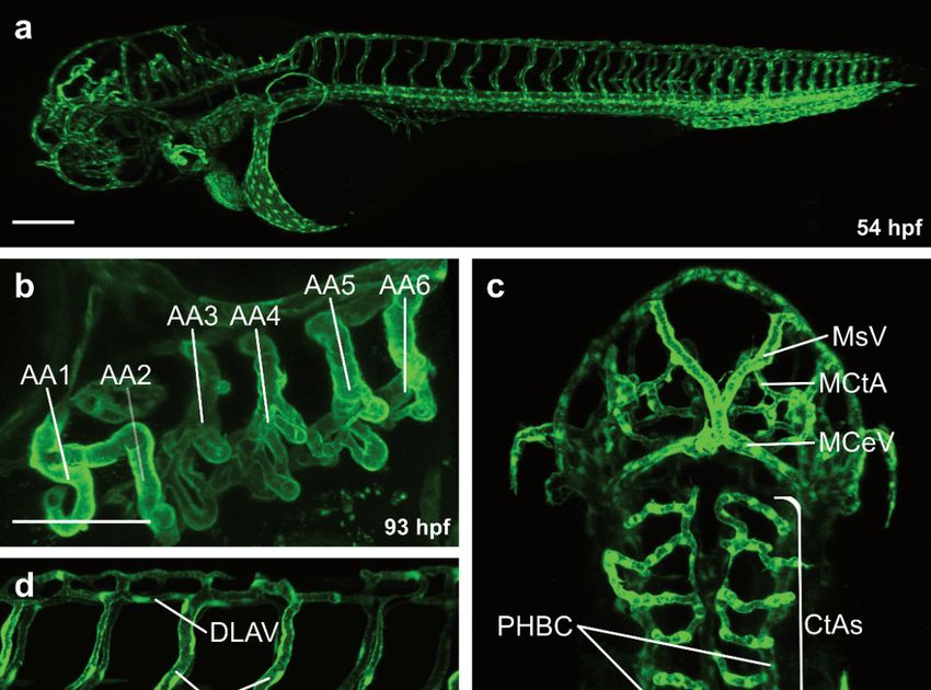
Life 2021, 11, 1088 2 of 17
embryonic development at a large scale. Already at 24 h post fertilization (hpf) the major
blood vessels are formed and heart contractility is initiated [12,13]. Whereas a defective
cardiovascular system would be lethal in mammals, zebrafish larvae can survive up to
five days without blood flow [14,15], making them an ideal model to study cardiovascular
development. Already at 48 hpf the embryos develop the characteristic vertebrate body
plan [12] and by pharmacological inhibition of pigmentation the embryos stay transpar-
ent [16], which enables the visualization of organ development even deep inside the tissue.
Those properties, in addition to the diploid genome, make the zebrafish a valuable model
for genetic [15,17,18] and pharmacological screens [11,19,20]. The first screens performed
in zebrafish uncovered several genes that are important for the development of the cardio-
vascular system and other organs [15,17,18]. Moreover, the establishment of CRISPR/Cas9
as a standard method for gene editing in zebrafish [21] further simplified the analysis of
gene function. In addition, the genetic accessibility facilitated the generation of transgenic
lines that are shared within the community [22–24]. These transgenic lines enable precise
observation of endothelial cell behavior, organelles, or even activities of enzymes [25] and
second messengers like Ca2+ [26] in vivo.
2. Vasculogenesis in Zebrafish
During vasculogenesis, new blood vessels are formed de novo from mesoderm-
derived endothelial progenitor cells, called angioblasts [2]. These angioblasts are initially
specified within the lateral plate mesoderm (LPM) and start to migrate between the somites
towards the embryonic midline [27]. Once the angioblasts reach the midline, they form
the first circulatory loop consisting of the dorsal aorta (DA) and the cardinal vein [27–30].
So far, only a few pathways have been demonstrated to be required for vasculogenesis.
In 1995, the cloche gene was identified as the first gene to be required for the specification
of ECs [31]. Mutants for the cloche gene lack almost all endothelial as well as hematopoietic
cells [31–35]. Hence, cloche is an upstream master-regulator, initiating signaling cascades
driving the specification of endothelial and hematopoietic cell lineages [31]. Due to the
location of the cloche gene on the telomere of chromosome 13, it took over two decades
from the first identification of cloche to clone the gene cloche. Finally, the group of Didier
Stainier, who first described the cloche mutant in 1995, identified cloche as the basic helix-
loop-helix/Per-ARNT-SIM (bHLH-PAS) domain containing transcription factor neuronal
PAS domain protein 4 like (npas4l) [36]. The expression of npas4l already starts during late
gastrulation, where it acts upstream of the ETS1-related protein (Etsrp) and T-cell acute
lymphocytic leukemia protein 1 (Tal1). [36].
Besides Npas4l, the ETS domain transcription factor Etsrp has been shown to be
indispensable for vasculogenesis [37,38]. Expression analysis revealed that etsrp mRNA
is already expressed in the LPM during the early somitogenesis in the zebrafish em-
bryo [38,39]. Embryos deficient for etsrp exhibit a downregulation of endothelial-specific
markers [38,40].
While EC specification is mediated by Npas4l and Etsrp function [31,35–37], the guid-
ance of angioblasts towards the embryonic midline is mediated by Apelin receptor early
endogenous ligand Apela (previously known as Elabela or Toddler), which signals through
the G protein-coupled receptor Apelin receptor (Aplnr) [27]. Apela is secreted by the
notochord and functions as a guidance cue for the angioblasts [27]. Depletion of both aplnr
or both ligands apela and apelin (apln) resulted in an impaired angioblast migration [27].
3. Sprouting Angiogenesis in Zebrafish
3.1. Formation of the Intersegmental Vessels
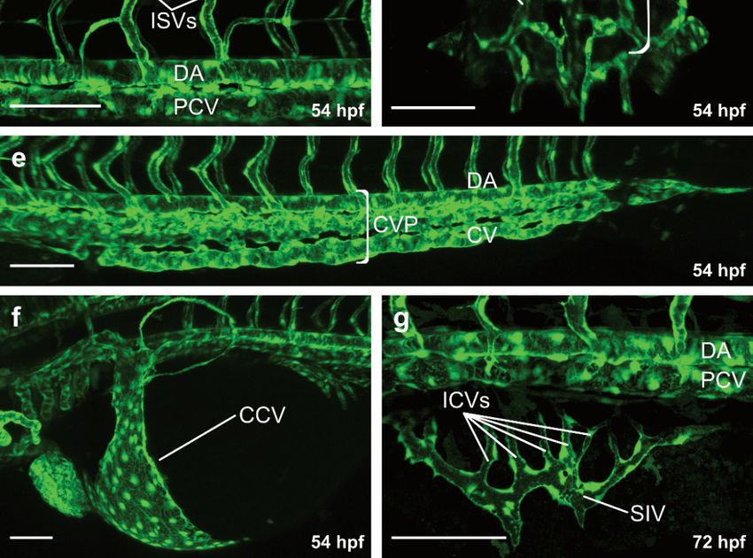
At 48 hpf, blood is circulating through a series of aortic arches (Figure 1a,b), entering
an anterior and posterior circulatory loop [13] (Figure 1a). The anterior vascular loop is
connected to the brain vasculature (Figure 1a,c), whereas the posterior loop is connected
to the DA [13] (Figure 1a,d). In the trunk, a series of intersegmental vessels (ISVs) is
formed in-between the somites (Figure 1a,d). Arterial ISVs connect the DA to the dorsal
longitudinal anastomotic vessel (DLAV) [13] (Figure 1a,d). Blood flows through the arterial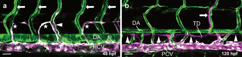
Life 2021, 11, 1088 3 of 17
ISVs into the DLAV and back through adjacent venous ISVs into the posterior cardinal
vein (PCV) [13] (Figure 1a,d). Both the anterior and the posterior circulatory loop merge
into the common cardinal vein (CCV), the largest vein in the embryo at this developmental
Life 2021, 11, x FOR PEER REVIEW timepoint [13,41] (Figure 1a,f). Blood then flows through the CCV over 4the yolk back to the
of 17
heart, closing the circulatory loop [13,41].
Figure 1. Overview of the vasculature of transgenic zebrafish larvae. Confocal projection images of
Figure 1. Overview
the vasculature in lateralof(a,b,d–g)
the vasculature of transgenic
and dorsal view zebrafish
(c). (a) Overview of thelarvae. Confocal
vasculature projection
at 54 hpf; (b) images of
the vasculature
Magnification inaortic
of the lateral (a,b,d–g)
arches and
at 93 hpf; dorsal viewof(c).
(c) Magnification the(a) Overview
blood vessels in of
thethe vasculature
brain at 54 at 54 hpf;
hpf;Magnification
(b) (d) Magnification ofofthetheaortic
trunk arches
vasculature
at 93 at hpf;
54 hpf;(c)(e) Magnification of
Magnification ofthe
theposterior trunk in the brain at
blood vessels
vasculature including dorsal aorta, caudal vein plexus and caudal vein at 54 hpf; (f) Magnification
54 hpf;common
of the (d) Magnification
cardinal vein at of54
the trunk
hpf; vasculatureofat
(g) Magnification the54subintestinal
hpf; (e) Magnification
vein plexus at 72 ofhpf.
the posterior trunk
vasculature including
AA, aortic arches; dorsal
CtA, central aorta,
artery; MsV,caudal vein plexus
mesencephalic and caudal
vein; MCeV, vein at
mid-cerebral 54 MCtA,
vein; hpf; (f) Magnification
mesencephalic
of the common central artery;vein
cardinal DLAV, dorsal
at 54 hpf;longitudinal anastomotic
(g) Magnification of vessel; ISV, intersegmental
the subintestinal vein plexus at 72 hpf.
vein; DA, dorsal aorta; PCV, posterior cardinal vein; CVP, caudal vein plexus; CCV, common car-
AA,
dinalaortic arches;
vein; SIV, CtA, central
subintestinal vein; ICV,artery; MsV, mesencephalic
interconnecting vein;
vessel; Scale bars: MCeV,
overview mid-cerebral
image 200 µm; vein; MCtA,
mesencephalic
all magnificationscentral
100 µm.artery; DLAV, dorsal longitudinal anastomotic vessel; ISV, intersegmental vein;
DA, dorsal aorta; PCV, posterior cardinal vein; CVP, caudal vein plexus; CCV, common cardinal
vein; SIV, subintestinal vein; ICV, interconnecting vessel; Scale bars: overview image 200 µm; all
magnifications 100 µm.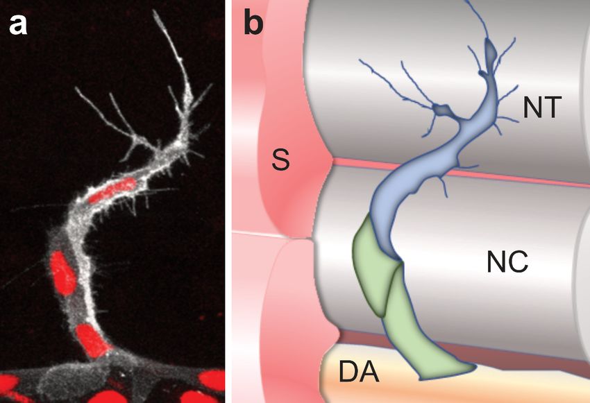
Life 2021, 11, 1088                                                                                             4 of 17

                             During primary angiogenesis in the trunk of the zebrafish embryo, single endothelial
                      cells within the DA sprout and migrate dorsally to form the ISVs (Figure 1a,d). This process
                      requires a variety of signal cues. At around 19 hpf Vascular endothelial growth factor a
                      (Vegfa) signaling through its receptor Kinase insert domain receptor like (Kdrl, previously
                      known as Vegfr2 or Flk1) stimulates the sprouting of the ISVs in the zebrafish trunk.
                      Binding of the ligand Vegfaa, which is expressed by the somites [42,43], modulates the
                      phosphorylation of Kdrl. This in turn activates the Mitogen-activated protein kinase (Mapk)
                      cascade and thus stimulates the phosphorylation of the Extracellular signal-regulated
                      kinase (Erk) in a subset of ECs within the DA [44]. Zebrafish embryos which are deficient
                      for either Vegfa or Kdrl exhibit an impaired ISV development [45–47].
                             Besides Kdrl, another Vegfr, termed the Fms Related Receptor Tyrosine Kinase 4 (Flt4,
                      previously known as Vegfr3), is critical to promote angiogenic sprouting in zebrafish as
                      well as in mammals [48,49]. However, in contrast to kdrl mutants, zebrafish mutants for
                      flt4 display defects in lymphatic and venous angiogenesis, while arterial development is
                      mainly unaffected [50,51].
                             It has been recently reported that Tm4sf18, a member of the Transmembrane 4 L6
                      protein family, is activated by Vegfa signaling and is specifically expressed in sprouting
                      ISVs [52]. A positive feedback loop between Tm4sf18 and Kdrl enhances Kdrl activity,
                      which leads to robust sprouting of ISVs [52]. Consequently, the lack of Tm4sf18 function
                      in zebrafish results in reduced Vegf signaling activity, vessel hypoplasia and truncated
                      ISVs [52].
                             During sprouting angiogenesis, endothelial cells arrange as tip cells, leading the devel-
                      oping sprout and following stalk cells (Figure 2). Tip cells can be distinguished from stalk
                      cells based on their morphology [53], expression of marker genes [54] and their metabolic
                      state [55–57] (Figure 2). The determination of tip and stalk cells in ISVs is achieved by the
                      Kdrl mediated downstream activation of Notch-Dll4 (Delta-like 4) signaling [49,58–60].
                      Vegf signaling in tip cells induces the expression of dll4 [61]. Subsequently, Dll4 activates
                      Notch signaling in adjacent stalk cells and represses the expression of both dll4 and kdrl
                      through lateral inhibition [49,58–61]. Thus, cells with reduced protein levels of Dll4 and
                      Kdrl become stalk cells, whereas cells with active Vegf signaling are determined as tip
                      cells [49,58–60]. In contrast, ECs deficient for Notch signaling exhibit a hyper = sprouting
                      phenotype with more than one tip cell guiding the sprout [49,58–60]. However, recent
                      studies showed that Notch signaling controls arterial angiogenesis by regulating the expres-
                      sion of C-X-C chemokine receptor type 4 (Cxcr4) and Vegfa [62,63]. In this model, Notch
                      signaling is rather required in the tip cells and directs them into developing arteries [62].
                             During angiogenesis, both Kdrl and Flt4 exert their pro-angiogenic signaling by
                      activating the MAPK cascade [44,64]. Inhibition of Erk activity prevents primary sprouting
                      of ISVs and expression of flt4, as well as sprouting of the trunk lymphatics [44,64]. A recent
                      study described a novel mechanism by which Vegfc/Flt4 signals via Erk induced G1 cell
                      cycle arrest of lymphatic endothelial cells (LECs), thereby enhancing lymphatic sprouting
                      efficiency [65]. In the ISVs Erk activity is higher in tip than in stalk cells, which is likely
                      caused by higher Kdrl signaling in tip cells [25,64]. Interestingly, after division of the tip
                      cell, this imbalance of Erk activity is maintained [25,66] due to the asymmetric cell division
                      and thus partitioning of the kdrl mRNA [66].
                             While Kdrl and Flt4 are positive regulators of angiogenesis, Flt1 has been shown
                      to restrict blood vessel growth [67,68]. In the zebrafish trunk, flt1 is expressed in the
                      developing vasculature, as well as in neurons [67,69,70]. Mutants for the soluble flt1 (sFlt1)
                      isoform exhibit a hyper-sprouting phenotype of venous ECs at the level of the neural
                      tube [69,70]. Both, neuronal sFlt1 [69] or radial glia promoting sFlt1 expression in the ECs
                      themselves [70] balance Vegfa signaling to modulate patterning of the trunk vasculature.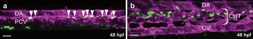
Life 2021, 11, 1088                                                                                                                                   5 of 17
 Life 2021, 11, x FOR PEER REVIEW                                                                                                                     5 of 17

                                      Figure 2. Schematic overview of a sprouting intersegmental vessel. (a) Magnification of a single ISV
                                    Figure 2. Schematic overview of a sprouting intersegmental vessel. (a) Magnification of a single ISV in
                                      in a double transgenic zebrafish embryo. The cell membrane is visualized in white (Tg(kdrl:GFP-
                                    a double transgenic zebrafish embryo. The cell membrane is visualized in white (Tg(kdrl:GFP-CAAX))
                                      CAAX)) and the nuclei are labeled in red (Tg(kdrl:NLS-mCherry)). Note the long cell protrusions/fi-
                                    and   the nuclei
                                      lopodia   of thearetiplabeled   in red
                                                              cell, while      (Tg(kdrl:NLS-mCherry)).
                                                                             the  following stalk cells only Noteexhibit
                                                                                                                    the long   cellfilopodia;
                                                                                                                            short   protrusions/filopodia
                                                                                                                                               (b) Schematic
                                    ofoverview
                                        the tip cell,
                                                 of a sprouting ISV and the surrounding tissues. The tip cell is labeledSchematic
                                                       while   the  following     stalk cells  only  exhibit short   filopodia;   (b)   in blue andoverview
                                                                                                                                                      the stalk
                                    ofcells
                                        a sprouting
                                            in green.ISVECsand    theISV
                                                              of the   surrounding
                                                                            sprout from tissues.  The tip
                                                                                            the dorsal     cell(DA)
                                                                                                         aorta  is labeled   in blueatand
                                                                                                                       to migrate      the the stalkboundary
                                                                                                                                           somite     cells in
                                      around
                                    green.     theofnotochord
                                             ECs                   (NC),from
                                                      the ISV sprout       eventually    leading
                                                                                 the dorsal   aortato(DA)
                                                                                                      the formation
                                                                                                           to migrateofatthethedorsal
                                                                                                                                 somitelongitudinal    anasto-
                                                                                                                                         boundary around
                                      motic  vessel   (DLAV)     at  the level   of the  neural  tube  (NT);  (c) The   tip  cell and  stalk
                                    the notochord (NC), eventually leading to the formation of the dorsal longitudinal anastomotic vessel    cells can  be dis-
                                      tinguished   morphologically        and   by  the expression    of marker   genes.  While    the
                                    (DLAV) at the level of the neural tube (NT); (c) The tip cell and stalk cells can be distinguished whole  ISV  is express-
                                      ing Kdrl, Aplnrb,and
                                    morphologically         PlexinD1
                                                                by the and     exhibit Ca
                                                                         expression
                                                                                           2+ oscillations, only the tip cell is expressing high levels of
                                                                                        of marker    genes. While the whole ISV is expressing Kdrl,
                                      the Notch ligand dll4 and the ligand apln. In contrast, the Notch receptor is highly expressed by the
                                    Aplnrb, PlexinD1 and exhibit Ca2+ oscillations, only the tip cell is expressing high levels of the Notch
                                      stalk cells. In addition, the surrounding tissue is also providing attractive signals such as vegfa and
                                    ligand dll4 and the ligand apln. In contrast, the Notch receptor is highly expressed by the stalk cells.
                                      repulsive signals such as semaphorin ligands. S, somite; NT, neural tube; NC, notochord; DA, dorsal
                                        addition, the surrounding tissue is also providing attractive signals such as vegfa and repulsive
                                    Inaorta.
                                    signals such as semaphorin ligands. S, somite; NT, neural tube; NC, notochord; DA, dorsal aorta.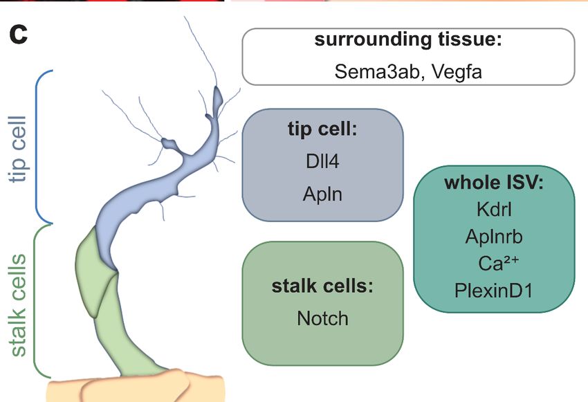
Life 2021, 11, 1088                                                                                           6 of 17

                            Further advancements in live imaging and transgenic tools uncovered Ca2+ oscil-
                      lations during development of the ISVs [26,71] and capillaries of the brain [72]. During
                      sprouting of the ISVs, Ca2+ oscillations are elevated in actively sprouting tip and stalk
                      cells [26] and while Vegf signaling positively regulates calcium oscillations, Dll4/Notch
                      signaling is required to suppress calcium oscillations in ECs adjacent to the stalk cell [26].
                      The transmembrane protein 33 (Tmem33), a three-pass transmembrane domain protein
                      located on the endoplasmic reticulum, has been demonstrated to be required for Ca2+
                      oscillations in response to Vegf [71]. Zebrafish tmem33 mutants exhibit a reduced number
                      of filopodia and impaired sprouting of ISVs [71]. Calcium signaling activity was also
                      found to be important for vascular pathfinding in the developing zebrafish brain [72].
                      Here, high and low frequency calcium oscillations are modulated by the mechanosensitive
                      channel Piezo1 and regulate extension or retraction of vascular branches [72].
                            Another pathway that has emerged as an important regulator of angiogenesis is the
                      Apelin signaling pathway. In contrast to vegfaa, which is expressed in the somites [42,43]
                      and neuronal cells [69], apln expression can be observed in the developing ISVs and is
                      enriched in tip cells [54,56]. In zebrafish, the Apelin pathway consists of two peptide
                      ligands Apela and Apelin that signal through the G protein-coupled receptors Aplnra
                      and Aplnrb. As mentioned in Section 2, Apela mainly regulates angioblast migration
                      during vasculogenesis [27]. In contrast, Apelin mediated activation of Apelin receptors
                      is required for ISV sprouting [56]. Interestingly, the expression of apln is downregulated
                      in mature blood vessels, but is reactivated in sprouting ECs during regeneration [73] and
                      tumor angiogenesis [74–76]. Furthermore, apln expression is regulated by Notch signaling
                      and Apelin deficiency prevents, similar to the knockdown of flt4, the hyper-sprouting
                      phenotype induced by knockdown of dll4 [50,56]. To facilitate sprouting, Apelin signaling
                      positively regulates EC metabolism by modulating the expression of the pro-metabolic
                      proteins PFKFB3 and C-MYC in human umbilical vein endothelial cells (HUVECs) [56].
                            Whereas Vegf and Apelin signaling are important pathways involved in ISV sprouting,
                      repulsive signals by Semaphorin-Plexin signaling restrict the migration path of growing
                      ISVs to the somite boundary [77]. While the plexinD1 receptor is specifically expressed
                      by endothelial cells, its ligand semaphorin3ab (sema3ab) is expressed by the surrounding
                      somites and thereby prevents ECs invasion into the somite [77]. Consequently, plexinD1
                      mutant embryos exhibit ectopic ISV sprouts which exceed the somite boundary [77,78].
                            In the zebrafish trunk, neighboring ISVs fuse in a process called anastomosis to form
                      the DLAV at around 30 hpf. Initially, all ISV sprouts are arterially derived. However, venous
                      sprouts emerging from the PCV migrate dorsally to contact the arterial ISVs. This process
                      is driven by vegfc expression by ECs in the DA and flt4 expression by ECs in the PCV [79].
                      Venous sprouts then fuse to the arterial ISVs and migrate against the direction of blood flow,
                      displacing arterial ECs and transforming the arterial ISV into a venous ISV [80] (Figure 3a).
                      Activation of Notch signaling in some ISVs protects those ISVs from being transformed
                      into venous ISVs [80,81]. Some 65% of the venous sprouts express the transcription factor
                      Prox1 [82], and 50% of these will become LECs forming the parachordal lymphangioblasts
                      (PLs) or parachordal cells (PACs) [83] (Figure 3a). These LECs will continue to migrate
                      along the arteries to form the main lymphatic vessels in the trunk of the zebrafish [83]
                      (Figure 3b).
                            Endothelial cell-to-extracellular matrix (ECM) interactions play an important role
                      during sprouting as well as maintenance of blood vessels. During sprouting, ECs adhere
                      to the ECM to facilitate their movement [84]. In addition, perivascular fibroblasts express
                      ECM components, such as the collagens col1a2 and col5a1 to stabilize blood vessels [85].
lymphangioblasts (PLs) or parachordal cells (PACs) [83] (Figure 3a). These LECs will con-
tinue to migrate along the arteries to form the main lymphatic vessels in the trunk of the
zebrafish [83] (Figure 3b).
Endothelial cell-to-extracellular matrix (ECM) interactions play an important role
Life 2021, 11, 1088 during sprouting as well as maintenance of blood vessels. During sprouting, ECs adhere 7 of 17
to the ECM to facilitate their movement [84]. In addition, perivascular fibroblasts express
ECM components, such as the collagens col1a2 and col5a1 to stabilize blood vessels [85].
Figure 3. Overview of arterial, venous and lymphatic vessels in the trunk of transgenic zebrafish larvae. Confocal projec-
Figure 3. Overview of arterial, venous and lymphatic vessels in the trunk of transgenic zebrafish larvae. Confocal projection
tion images of the trunk vasculature of a double transgenic zebrafish line (Tg(kdrl:EGFP); Tg(-5.2lyve1b:DsRed)) at 48 hpf
images of the trunk vasculature of a double transgenic zebrafish line (Tg(kdrl:EGFP); Tg(-5.2lyve1b:DsRed)) at 48 hpf (a)
(a) and 120 hpf (b) in lateral view. Arterial endothelial cells (ECs) are labeled in green (GFP), venous ECs are labeled in
and 120 hpf magenta
green and (b) in lateral
(GFPview. Arterial
and DsRed endothelial
double cells
positive) and(ECs) are labeled
lymphatic ECs arein labeled
green (GFP), venous
in magenta ECs are(a)labeled
(DsRed). in green
Magnifica-
and magenta
tion (GFP and
of a zebrafish trunkDsRed double
showing positive)
the DA and lymphatic
and arterial ECsvenous
(arrows) and are labeled in magenta as
ISVs (arrowhead) (DsRed).
well as (a) Magnification
lymphatic sprouts of a
(stars); (b)
zebrafish Magnification
trunk showing the of aDA
zebrafish trunk(arrows)
and arterial showing and
the main lymphatic
venous vessels: theas
ISVs (arrowhead) thoracic
well asduct (TD) (arrowheads)
lymphatic sprouts (stars);
and one intersegmental lymphatic vessel (ISLV) (arrow); DA, dorsal aorta; PCV, posterior cardinal
(b) Magnification of a zebrafish trunk showing the main lymphatic vessels: the thoracic duct (TD) (arrowheads)vein; ISV, intersegmen-
and one
tal vessel; TD, thoracic duct; Scale bars: 30 µm.
intersegmental lymphatic vessel (ISLV) (arrow); DA, dorsal aorta; PCV, posterior cardinal vein; ISV, intersegmental vessel;
TD, thoracic duct; Scale bars: 30 µm.
3.2. Formation of the Caudal Vein
3.2. Formation
The caudal of vein
the Caudal
(CV) isVeinlocated in the caudal part of the PCV, starting with the end
of the yolk extension [13]. At
The caudal vein (CV) is located aroundin27the
hpf, ECs of
caudal theofCV
part thesprout ventral and
PCV, starting withstart to of
the end
form the caudal vein plexus (CVP) [86]. This ventral sprouting of venous
the yolk extension [13]. At around 27 hpf, ECs of the CV sprout ventral and start to form theECs is highly
dependent
caudal veinon bone(CVP)
plexus morphogenetic
[86]. Thisprotein
ventral(BMP) signaling
sprouting [86,87],
of venous ECswhile Vegfadependent
is highly signaling on
has no effect on ventral sprouting of venous ECs [87]. Vice versa, arterial
bone morphogenetic protein (BMP) signaling [86,87], while Vegfa signaling has no effect on angiogenic
sproutssprouting
ventral do not respond
of venousto BMP
ECs signaling, but arearterial
[87]. Vice versa, highlyangiogenic
dependent sprouts
on Vegfa dosignaling
not respond
[87]. Downstream of BMP signaling, Cdc42 activates Formin-like 3 (Fmnl3), an actin-reg-
to BMP signaling, but are highly dependent on Vegfa signaling [87]. Downstream of BMP
ulating protein, leading to filopodia extension to facilitate angiogenic sprouting of the
signaling, Cdc42 activates Formin-like 3 (Fmnl3), an actin-regulating protein, leading to
CVP [86]. Inhibition of filopodia formation by either Latrunculin A treatment or injection
filopodia extension to facilitate angiogenic sprouting of the CVP [86]. Inhibition of filopodia
of a fmnl3 morpholino (MO) leads to defects in the development of the CVP [86,88]. Dur-
formation by either Latrunculin A treatment or injection of a fmnl3 morpholino (MO) leads
ing ventral sprouting venous ECs exhibit high β-Catenin-dependent transcriptional activ-
to defects in the development of the CVP [86,88]. During ventral sprouting venous ECs
ity [89]. Disturbance of β-Catenin-dependent transcription, either by overexpression of
exhibit high β-Catenin-dependent transcriptional activity [89]. Disturbance of β-Catenin-
axin or a dominant-negative T-cell factor (Tcf), results in impaired CV formation [89]. By
dependent transcription, either by overexpression of axin or a dominant-negative T-cell
36 hpf ventral sprouting of ECs led to the formation of a vascular plexus, named CVP.
factor (Tcf), resultspart
ECs in the ventral in impaired
of the CVP CVmaintain
formation high[89]. By 36 hpf
β-Catenin ventral
activity andsprouting
form the of lu-ECs
led to the formation of a vascular
menized definitive CV by 48 hpf [89]. plexus, named CVP. ECs in the ventral part of the CVP
maintain high β-Catenin activity and form the lumenized definitive
The formation of the CV-primordium, as well as sprouting from the CV, are both CV by 48 hpf [89].
The formation
dependent of the CV-primordium,
on hemodynamic as well
forces by blood flow as sprouting
[90–92]. Blocking from
cardiacthecontraction
CV, are both
dependent on hemodynamic forces by blood flow [90–92]. Blocking
and thereby blood flow in zebrafish embryos, either by treatment with ion channel cardiac contraction
block-
and
ers (nifedipine, BDM and tricaine) or by injection of troponin T type 2a (tnnt2a) MO,blockers
thereby blood flow in zebrafish embryos, either by treatment with ion channel re-
(nifedipine, BDM and
sults in an impaired CVPtricaine) or by[90–92].
formation injection of troponin T type 2a (tnnt2a) MO, results in
an impaired CVP formation [90–92].
3.3. Formation of the Brain Vasculature
Among all organs, the brain has the highest oxygen and nutrient consumption. Hence,
a complex network of blood vessels in the brain is necessary to ensure an appropriate
supply of oxygen and nutrients [93,94]. The vascularization of the brain is initiated at 32
hpf by angiogenic sprouting from the primordial hindbrain channels (PHBC) forming the
central arteries (CtA) (Figure 1c) [95–97]. Subsequently, CtA sprouts anastomose with the
basilar artery (BA) and lumenize between 36 and 48 hpf (Figure 1c) [95–97]. In contrast,
the mesencephalic CtAs (MCtA) in the midbrain sprout from the perineural vascular plexus
(PVP) into the brain parenchyma at 36 hpf (Figure 1c) [13,98]. Once sprouting and perfusion
of the brain is completed, ECs start to establish the blood-brain-barrier (BBB) [99–101].
The BBB controls the selective transport of molecules into the brain parenchyma, while it
also protects the brain by limiting the entry of pathogens and harmful molecules (reviewed
in [102,103]).Life 2021, 11, 1088 8 of 17
Similar to its role during the formation of the trunk vasculature, Vegf signaling plays a
crucial role for the development of the cerebral vasculature. Zebrafish embryos treated with
SU5416, an inhibitor of the tyrosine kinase activity of all Vegf receptors, exhibit defects in
the formation of the BA, CtAs, PHBC and mid-cerebral vein (MCeV) [47,97]. In particular,
Vegfa signaling is essential for sprouting of the CtAs in the hindbrain as demonstrated
by the absence of CtAs in mutants for kdrl and vegfaa [96]. Furthermore, the combination
of vegfab, vegfc and vegfd is required for the development of fenestrated vessels in the
myelencephalic choroid plexus (mCP) [104]. Triple mutants for vegfab, vegfc and vegfd
exhibit impaired mCP vascularization, while the formation of non-fenestrated brain vessels
was unaffected [104].
CXC-receptors are G protein-coupled receptors that are activated by small CXC-
motif containing chemokines (as reviewed in [105,106]). CXC chemokines function as
guidance cues for migrating cells [107–109]. During brain angiogenesis of the zebrafish
embryo, Cxcr4-Cxcl12 signaling plays a crucial role for the vascularization of the hindbrain.
In embryos deficient for the receptor cxcr4a or ligand cxcl12b, sprouting of the CtAs is
unaffected [96]. However, both mutants exhibit defects in pathfinding of the CtAs towards
the BA, resulting in an increased number of unperfused CtA [96,97]. In contrast, sprouting
and pathfinding of intersegmental vessels in the zebrafish trunk is unaffected in both cxcl12b
and cxcr4a mutants, highlighting the distinct role of Cxcr4 signaling during hindbrain
vascularization [96]. Subsequently, perfusion of the CtAs leads to a downregulation of
cxcr4a expression, while unperfused CtAs maintain high cxcr4a expression levels [96],
indicating that cxcr4a mRNA expression is negatively regulated by blood flow.
Canoncial Wnt/β-Catenin signaling plays a pivotal role both during brain angiogen-
esis and BBB formation [110–112]. In sprouting ECs of the brain, active Wnt/β-Catenin
signaling counteracts Sphingosine-1-phosphate receptor 1-(S1pr1) induced BBB formation
but decreases after lumen formation [112]. This decrease in Wnt/β-Catenin signaling then
leads to the activation of S1pr1-signaling regulating the localization of the cell-cell junction
molecules Ve-cadherin and Esama and therefore BBB formation [112].
The Adhesion G protein-coupled receptor A2 (Adgra2, previously known as Gpr124),
is a membrane-bound G protein-coupled receptor that plays a key role in the development
of the brain vasculature in zebrafish [113]. These adgra2 mutants exhibit vascular defects in
the brain, especially in the formation of the CtAs [113]. Together with the GPI-anchored
protein Reversion-inducing cysteine-rich protein with Kazal motifs (Reck), Adgra2 forms
a receptor complex that promotes Wnt7a/Wnt7b-dependent canonical Wnt/β-Catenin
signaling during sprouting of the CtAs in the brain [113–115]. Here, Adgra2 binds to the
extracellular domain of Reck to enable the formation of a complex consisting of Adgra2,
Reck, Frizzled (Fz) and Lrp5/6 [114]. This complex is required to deliver Reck-bound Wnt7
to the Frizzled receptors [114]. In addition, Adgra2 binds to the intracellular domain of the
scaffolding protein Dishevelled (Dvl) [114]. Dvl functions as a bridge for Fz and Adgra2 to
trigger Wnt/β-Catenin signaling through the Fz receptor and Lrp5/6 co-receptors [114].
Moreover, transplantation experiments of wildtype endothelial cells into adgra2 deficient
embryos revealed that Adgra2/Reck is specifically required in CtA tip cells but not ISV tip
cells [113].
4. Lumen Formation
4.1. Cord Hollowing and Cell Hollowing
A crucial step in blood vessel morphogenesis is the transition from an early sprout
to a tubular structure that enables fluid transport. The lumen of these tubes is formed ei-
ther extracellularly (cord hollowing or budding), opening up a lumen in-between the
ECs [29,116,117], or intracellularly by “cell hollowing”, forming a lumen within the
cell [118]. During cord hollowing, ECs align to each other as a cord, which can be ob-
served by the presence of cell-cell contacts [29,116,117]. Subsequently, ECs open the lumen
in between them [29,116,117]. The concept of cell hollowing [118] has been proposed in
particular for lumen formation during anastomosis. Here, the pressure from blood flowLife 2021, 11, 1088 9 of 17
leads to tunnel- like membrane invaginations at the distal growing end of the sprouting
vessels leading to the formation of a lumen within a single cell [118]. Recently, it was shown
that this process is accompanied by transient recruitment and contraction of actomyosin
resulting in inverse membrane blebbing, which appears to be dependent on differences
between intra and extracellular pressure [119]. Once blood flow is established within the
new vessel, ECs align in the direction of blood flow and reorient their Golgi apparatus
against the direction of blood flow [120]. This process has been shown to be dependent on
Aplnr signaling via ß-Arrestin [120].
4.2. Alternative Ways of Lumen Formation
A novel mechanism of extracellular lumen formation, named lumen ensheathment,
was shown for the formation of the CCV [41]. Here individual ECs align in a sheath-like
manner around an initially virtual tube lumen [41]. Another way of lumen formation
has recently been described for the CV-primordium [90]. Together with the CCV, the CV-
primordium is one of the largest vessels observed during embryonic development, being 5
times larger than the DA [47,90]. At 18 hpf endothelial struts coalesce in the future lumen
of the CV [90]. Consequently, single ECs start to form the vessel wall around the network
of endothelial struts [90]. At around 26–28 hpf, endothelial struts prune and integrate
into the vessel wall [90]. Laser ablation of endothelial cell struts results in a collapse of
the CV upon circulation, indicating that these elements provide structural support to the
CV [90]. Inhibition of BMP signaling causes a failure of ECs to coalesce into struts, leading
to defective CV development [90]. Conversely, global overexpression of bmp2b leads to
pruning defects of endothelial struts, which are still remaining at 48 hpf, resulting in an
only partially formed CV wall [90]. Along with the mechanism of lumen ensheathment [41],
endothelial struts [90] provide another way for ECs to form large-caliber blood vessels.
5. Vascular Remodeling
During organ growth, the vascular system is permanently adapting to the chang-
ing requirements of the growing organism by remodeling of the vascular network [121].
Through remodeling of the vascular network, shortcuts and loops are removed to ensure
an optimal and unidirectional blood flow that efficiently supplies all organs with [98,122].
On the cellular level, pruning is reminiscent of anastomosis in reverse, i.e., the pruning
segment reduces its lumen diameter until the lumen collapses and cell-cell contacts are
removed until ECs separate and migrate back into the adjacent vessel branches [98,123].
The selection of vessels to be pruned is triggered by low or fluctuating blood flow and
in this way supports stabilized segments with constant blood flow [98,123–126]. On the
molecular level it has been shown that the pruning process is accompanied by an activation
of Rac1 triggered by low blood flow, which contributes to the increased migratory capacity
of ECs [98]. Recently, klf6a and tagln2 were identified to regulate cell-cell contacts and
cytoskeleton rearrangement to support pruning of the caudal vein [127].
6. Development of Hematopoietic Stem Cells
6.1. Emergence of Hematopoietic Stem Cells
Hematopoietic stem cells (HSCs) generate all blood lineages during adult life [128,129].
The hemogenic endothelium (HE), a specialized subpopulation of endothelial cells (EC),
located in the ventral floor of the DA (Figure 4a), generates HSCs during development
in a process known as endothelial-to-hematopoietic transition (EHT) [130–133]. EHT is
controlled by a variety of signaling pathways such as Notch-[134,135], TGFβ [136,137],
BMP [138], Wnt9a/β-Catenin [139], Hif [140,141], YAP [142] and NOS-signaling [143].
Following their specification within the HE, nascent HSCs enter circulation and colonize
the CVP [144,145] (Figure 4b).sate to the abluminal side of the vascular wall and trigger the surrounding ECs to form a
pocket like structure in a process called “endothelial cuddling” [146]. Within the pocket
like structure HSCs dynamically interact with mesenchymal stromal cells (MSC) [146].
The complex environment within the niche, created by ECs, MSCs and also immune cells,
like macrophages and neutrophils, tightly regulates HSC proliferation, differentiation and
Life 2021, 11, 1088 egression of the HSCs from the CHT from 72 hpf onward [145,148,150]. After leaving the 10 of 17
vascular niche HSCs eventually populate the adult lymphopoietic- and hematopoietic or-
gans, the thymus and the kidney in zebrafish [144,145].
Figure4.4.Hematopoietic
Figure Hematopoietic stem
stemcell
celldevelopment
developmentinin
transgenic
transgeniczebrafish larvae.
zebrafish Confocal
larvae. projection
Confocal imagesimages
projection of the trunk
of the(a)
trunk
and the tail (b) of a 48 hpf zebrafish embryo in lateral view. HSCs are visualized in green (Tg(itga2b:GFP)) and the vascu-
(a) and the tail (b) of a 48 hpf zebrafish embryo in lateral view. HSCs are visualized in green (Tg(itga2b:GFP)) and the
lature in magenta (Tg(kdrl:HsHRAS-mCherry)). (a) HSCs (green) emerging from the DA (arrowheads); (b) HSCs (green)
vasculature
residing in in
themagenta
CHT. HSCs,(Tg(kdrl:HsHRAS-mCherry)). (a) dorsal
Hematopoietic stem cells; DA, HSCs aorta;
(green) emerging
PCV, from
posterior the DA
cardinal vein;(arrowheads); (b) HSCs
CHT, caudal hema-
(green) residing in the CHT. HSCs,
topoietic tissue; scale bars: 30 µm. Hematopoietic stem cells; DA, dorsal aorta; PCV, posterior cardinal vein; CHT, caudal
hematopoietic tissue; scale bars: 30 µm.
7. Perspective
6.2. Homing of Hematopoietic Stem Cells
Over the past four decades, experiments conducted in zebrafish have contributed to
The vascular plexus of the CVP is also known as caudal hematopoietic tissue (CHT),
a deeper understanding of the morphogenetic processes that shape the vertebrate body.
which is the zebrafish equivalent to the mammalian fetal liver [145] and serves as a vascular
The initially simple blueprint of the zebrafish vasculature allows for fast gain and loss of
niche for HSCs [144–148]. Upon emerging from the ventral floor of the DA (Figure 4a),
function analyses. The optical clarity of the zebrafish embryo in combination with trans-
HSCs enter the circulation [130,131,144]. From 48 hpf onwards circulating HSCs colonize
genic techniques enables the analysis of the behavior of cells and signaling processes live
the CHT [146], where they interact with Vcam1-expressing macrophages [149]. This inter-
at a single cell resolution using confocal microscopy. The development of new imaging
action between HSCs and Vcam1-expressing macrophages requires Integrin alpha 4 and
technologies, such as light sheet microscopy, new fluorophores and biosensors, will allow
is
even morefor
crucial homing
detailed of HSCs
images to betoacquired
small venous capillaries
over longer periods[149]. Next,
of time HSCs
without extravasate
bleaching
to the abluminal side of the vascular wall and trigger the surrounding ECs
or photo toxicity. Technologies such as small molecule screens [151–153] and protein-pro- to form a
pocket like structure
tein interaction assaysin(e.g.,
a process
BioID)called “endothelial
[154,155] cuddling”
are extending [146]. Within
the toolbox the the
for using pocket
like
zebrafish model. The emergence of single cell sequencing technologies within the last[146].
structure HSCs dynamically interact with mesenchymal stromal cells (MSC)
The complex environment within the niche, created by ECs, MSCs and also immune cells,
like macrophages and neutrophils, tightly regulates HSC proliferation, differentiation and
egression of the HSCs from the CHT from 72 hpf onward [145,148,150]. After leaving the
vascular niche HSCs eventually populate the adult lymphopoietic- and hematopoietic
organs, the thymus and the kidney in zebrafish [144,145].
7. Perspective
Over the past four decades, experiments conducted in zebrafish have contributed to
a deeper understanding of the morphogenetic processes that shape the vertebrate body.
The initially simple blueprint of the zebrafish vasculature allows for fast gain and loss
of function analyses. The optical clarity of the zebrafish embryo in combination with
transgenic techniques enables the analysis of the behavior of cells and signaling processes
live at a single cell resolution using confocal microscopy. The development of new imaging
technologies, such as light sheet microscopy, new fluorophores and biosensors, will allow
even more detailed images to be acquired over longer periods of time without bleaching or
photo toxicity. Technologies such as small molecule screens [151–153] and protein-protein
interaction assays (e.g., BioID) [154,155] are extending the toolbox for using the zebrafish
model. The emergence of single cell sequencing technologies within the last years enabled
the dissection of the developmental trajectories of cells and their heterogeneity within one
cell lineage [156].
Author Contributions: Conceptualization, J.E., L.H., J.M., A.R., S.B. and C.S.H.; investigation, J.E.,
L.H., J.M., A.R., S.B. and C.S.H.; writing—original draft preparation, J.E., L.H., J.M., A.R., S.B.
and C.S.H.; writing—review and editing, J.E., L.H., J.M., A.R., S.B. and C.S.H.; visualization, J.E., L.H.,
J.M., A.R., S.B. and C.S.H.; supervision, C.S.H.; project administration, C.S.H.; funding acquisition,
C.S.H.; All authors have read and agreed to the published version of the manuscript.
Funding: This research was funded by the DFG (SFB 834/4), the DFG (GRK 2213) and the Forschungscam-
pus Mittelhessen.
Institutional Review Board Statement: All zebrafish housing and husbandry were performed under
standard conditions in accordance with institutional (University of Marburg) and national ethicalLife 2021, 11, 1088 11 of 17
and animal welfare guidelines approved by the ethics committee for animal experiments at the
Regierungspräsidium Gießen, Germany, as well as the FELASA guidelines (Aleström et al., 2020).
Acknowledgments: We apologize for not being able to cite all of the original research articles and
related references due to space limitations.
Conflicts of Interest: The authors declare no conflict of interest.
References
1. Karpanen, T.; Alitalo, K. Molecular Biology and Pathology of Lymphangiogenesis. Annu. Rev. Pathol. Mech. Dis. 2008, 3, 367–397.
[CrossRef] [PubMed]
2. Risau, W.; Flamme, I. Vasculogenesis. Annu. Rev. Cell Dev. Biol. 1995, 11, 73–91. [CrossRef] [PubMed]
3. Armulik, A.; Abramsson, A.; Betsholtz, C. Endothelial/Pericyte Interactions. Circ. Res. 2005, 97, 512–523. [CrossRef] [PubMed]
4. Carmeliet, P. Angiogenesis in health and disease. Nat. Med. 2003, 9, 653–660. [CrossRef] [PubMed]
5. Gurtner, G.C.; Werner, S.; Barrandon, Y.; Longaker, M.T. Wound repair and regeneration. Nature 2008, 453, 314–321. [CrossRef]
[PubMed]
6. Hanahan, D.; Weinberg, R.A. The Hallmarks of Cancer. Cell 2000, 100, 57–70. [CrossRef]
7. Streisinger, G.; Walker, C.; Dower, N.; Knauber, D.; Singer, F. Production of clones of homozygous diploid zebra fish (Brachydanio
rerio). Nature 1981, 291, 293–296. [CrossRef] [PubMed]
8. Howe, K.; Clark, M.D.; Torroja, C.F.; Torrance, J.; Berthelot, C.; Muffato, M.; Collins, J.E.; Humphray, S.; McLaren, K.; Matthews, L.;
et al. The zebrafish reference genome sequence and its relationship to the human genome. Nature 2013, 496, 498–503. [CrossRef]
9. Phillips, J.B.; Westerfield, M. Zebrafish models in translational research: Tipping the scales toward advancements in human
health. Dis. Model. Mech. 2014, 7, 739–743. [CrossRef]
10. Bradford, Y.M.; Toro, S.; Ramachandran, S.; Ruzicka, L.; Howe, D.G.; Eagle, A.; Kalita, P.; Martin, R.; Moxon, S.A.T.; Schaper, K.;
et al. Zebrafish Models of Human Disease: Gaining Insight into Human Disease at ZFIN. ILAR J. 2017, 58, 4–16. [CrossRef]
11. MacRae, C.A.; Peterson, R.T. Zebrafish as tools for drug discovery. Nat. Rev. Drug Discov. 2015, 14, 721–731. [CrossRef] [PubMed]
12. Kimmel, C.B.; Ballard, W.W.; Kimmel, S.R.; Ullmann, B.; Schilling, T.F. Stages of embryonic development of the zebrafish. Dev.
Dyn. 1995, 203, 253–310. [CrossRef] [PubMed]
13. Isogai, S.; Horiguchi, M.; Weinstein, B.M. The Vascular Anatomy of the Developing Zebrafish: An Atlas of Embryonic and Early
Larval Development. Dev. Biol. 2001, 230, 278–301. [CrossRef] [PubMed]
14. Sehnert, A.J.; Huq, A.; Weinstein, B.M.; Walker, C.; Fishman, M.; Stainier, D.Y.R. Cardiac troponin T is essential in sarcomere
assembly and cardiac contractility. Nat. Genet. 2002, 31, 106–110. [CrossRef]
15. Stainier, D.Y.; Fouquet, B.; Chen, J.N.; Warren, K.S.; Weinstein, B.M.; Meiler, S.E.; Mohideen, M.A.; Neuhauss, S.C.; Solnica-Krezel,
L.; Schier, A.F.; et al. Mutations affecting the formation and function of the cardiovascular system in the zebrafish embryo.
Development 1996, 123, 285–292. [CrossRef]
16. Whittaker, J.R. An analysis of melanogenesis in differentiating pigment cells of ascidian embryos. Dev. Biol. 1966, 14, 1–39.
[CrossRef]
17. Haffter, P.; Granato, M.; Brand, M.; Mullins, M.C.; Hammerschmidt, M.; Kane, D.A.; Odenthal, J.; van Eeden, F.J.; Jiang, Y.J.;
Heisenberg, C.P.; et al. The identification of genes with unique and essential functions in the development of the zebrafish, Danio
rerio. Development 1996, 123, 1–36. [CrossRef]
18. Driever, W.; Solnica-Krezel, L.; Schier, A.F.; Neuhauss, S.C.; Malicki, J.; Stemple, D.L.; Stainier, D.Y.; Zwartkruis, F.; Abdelilah, S.;
Rangini, Z.; et al. A genetic screen for mutations affecting embryogenesis in zebrafish. Development 1996, 123, 37–46. [CrossRef]
19. Peterson, R.T.; Link, B.A.; Dowling, J.E.; Schreiber, S.L. Small molecule developmental screens reveal the logic and timing of
vertebrate development. Proc. Natl. Acad. Sci. USA 2000, 97, 12965–12969. [CrossRef] [PubMed]
20. Zon, L.I.; Peterson, R.T. In vivo drug discovery in the zebrafish. Nat. Rev. Drug Discov. 2005, 4, 35–44. [CrossRef]
21. Jao, L.-E.; Wente, S.R.; Chen, W. Efficient multiplex biallelic zebrafish genome editing using a CRISPR nuclease system. Proc. Natl.
Acad. Sci. USA 2013, 110, 13904–13909. [CrossRef]
22. Schuermann, A.; Helker, C.S.M.; Herzog, W. Angiogenesis in zebrafish. Semin. Cell Dev. Biol. 2014, 31, 106–114. [CrossRef]
23. Okuda, K.S.; Hogan, B.M. Endothelial Cell Dynamics in Vascular Development: Insights From Live-Imaging in Zebrafish. Front.
Physiol. 2020, 11, 842. [CrossRef]
24. Lawson, N.; Weinstein, B.M. In Vivo Imaging of Embryonic Vascular Development Using Transgenic Zebrafish. Dev. Biol. 2002,
248, 307–318. [CrossRef]
25. Okuda, K.S.; Keyser, M.S.; Gurevich, D.B.; Sturtzel, C.; Mason, E.A.; Paterson, S.; Chen, H.; Scott, M.; Condon, N.D.; Martin, P.;
et al. Live-imaging of endothelial Erk activity reveals dynamic and sequential signalling events during regenerative angiogenesis.
eLife 2021, 10, e62196. [CrossRef]
26. Yokota, Y.; Nakajima, H.; Wakayama, Y.; Muto, A.; Kawakami, K.; Fukuhara, S.; Mochizuki, N. Endothelial Ca2+ oscillations
reflect VEGFR signaling-regulated angiogenic capacity in vivo. eLife 2015, 4, e08817. [CrossRef]
27. Helker, C.S.M.; Schuermann, A.; Pollmann, C.; Chng, S.C.; Kiefer, F.; Reversade, B.; Herzog, W. The hormonal peptide Elabela
guides angioblasts to the midline during vasculogenesis. eLife 2015, 4, e06726. [CrossRef] [PubMed]Life 2021, 11, 1088 12 of 17
28. Fouquet, B.; Weinstein, B.M.; Serluca, F.C.; Fishman, M.C. Vessel Patterning in the Embryo of the Zebrafish: Guidance by
Notochord. Dev. Biol. 1997, 183, 37–48. [CrossRef] [PubMed]
29. Jin, S.-W.; Beis, D.; Mitchell, T.; Chen, J.-N.; Stainier, D. Cellular and molecular analyses of vascular tube and lumen formation in
zebrafish. Development 2005, 132, 5199–5209. [CrossRef] [PubMed]
30. Kohli, V.; Schumacher, J.A.; Desai, S.P.; Rehn, K.; Sumanas, S. Arterial and Venous Progenitors of the Major Axial Vessels Originate
at Distinct Locations. Dev. Cell 2013, 25, 196–206. [CrossRef]
31. Stainier, D.Y.R.; Weinstein, B.M.; Iii, H.W.D.; Zon, L.I.; Fishman, M.C. Cloche an early acting ZF gene is reqd by both endothelial
and hematopoietic lineages. Development 1995, 3150, 3141–3150. [CrossRef]
32. Liao, E.C.; Paw, B.H.; Oates, A.C.; Pratt, S.J.; Postlethwait, J.H.; Zon, L.I. SCL/Tal-1 transcription factor acts downstream of cloche
to specify hematopoietic and vascular progenitors in zebrafish. Genes Dev. 1998, 12, 621–626. [CrossRef]
33. Thompson, M.A.; Ransom, D.G.; Pratt, S.J.; Mac Lennana, H.; Kieran, M.; Detrich, H.; Vaila, B.; Huber, T.L.; Paw, B.; Brownlie, A.J.;
et al. TheclocheandspadetailGenes Differentially Affect Hematopoiesis and Vasculogenesis. Dev. Biol. 1998, 197, 248–269.
[CrossRef]
34. Parker, L.; Stainier, D.Y. Cell-autonomous and non-autonomous requirements for the zebrafish gene cloche in hematopoiesis.
Development 1999, 126, 2643–2651. [CrossRef]
35. Liao, W.; Bisgrove, B.W.; Sawyer, H.; Hug, B.; Bell, B.; Peters, K.; Grunwald, D.J.; Stainier, D.Y. The zebrafish gene cloche acts
upstream of a flk-1 homologue to regulate endothelial cell differentiation. Development 1997, 124, 381–389. [CrossRef]
36. Reischauer, S.; Stone, O.; Villasenor, A.; Chi, N.; Jin, S.-W.; Martin, M.; Lee, M.T.; Fukuda, N.; Marass, M.; Witty, A.; et al. Cloche is
a bHLH-PAS transcription factor that drives haemato-vascular specification. Nature 2016, 535, 294–298. [CrossRef]
37. Sumanas, S.; Jorniak, T.; Lin, S. Identification of novel vascular endothelial–specific genes by the microarray analysis of the
zebrafish cloche mutants. Blood 2005, 106, 534–541. [CrossRef]
38. Sumanas, S.; Lin, S. Ets1-Related Protein Is a Key Regulator of Vasculogenesis in Zebrafish. PLoS Biol. 2005, 4, e10. [CrossRef]
39. Pham, V.N.; Lawson, N.; Mugford, J.W.; Dye, L.; Castranova, D.; Lo, B.; Weinstein, B.M. Combinatorial function of ETS
transcription factors in the developing vasculature. Dev. Biol. 2007, 303, 772–783. [CrossRef]
40. Chestnut, B.; Chetty, S.C.; Koenig, A.L.; Sumanas, S. Single-cell transcriptomic analysis identifies the conversion of zebrafish
Etv2-deficient vascular progenitors into skeletal muscle. Nat. Commun. 2020, 11, 1–16. [CrossRef] [PubMed]
41. Helker, C.; Schuermann, A.; Karpanen, T.; Zeuschner, D.; Belting, H.-G.; Affolter, M.; Schulte-Merker, S.; Herzog, W. The zebrafish
common cardinal veins develop by a novel mechanism: Lumen ensheathment. Development 2013, 140, 2776–2786. [CrossRef]
42. Liang, D.; Xu, X.; Chin, A.; Balasubramaniyan, N.V.; Teo, M.A.L.; Lam, T.J.; Weinberg, E.S.; Ge, R. Cloning and characterization of
vascular endothelial growth factor (VEGF) from zebrafish, Danio rerio. Biochim. Biophys. Acta Gene Struct. Expr. 1998, 1397, 14–20.
[CrossRef]
43. Liang, D.; Chang, J.R.; Chin, A.; Smith, A.; Kelly, C.; Weinberg, E.S.; Ge, R. The role of vascular endothelial growth factor (VEGF)
in vasculogenesis, angiogenesis, and hematopoiesis in zebrafish development. Mech. Dev. 2001, 108, 29–43. [CrossRef]
44. Shin, M.; Male, I.; Beane, T.J.; Villefranc, J.A.; Kok, F.O.; Zhu, L.J.; Lawson, N.D. Vegfc acts through ERK to induce sprouting and
differentiation of trunk lymphatic progenitors. Development 2016, 143, 3785–3795. [CrossRef]
45. Nasevicius, A.; Larson, J.; Ekker, S.C. Distinct Requirements for Zebrafish Angiogenesis Revealed by a VEGF-A Morphant. Yeast
2000, 1, 294–301. [CrossRef]
46. Covassin, L.D.; Siekmann, A.F.; Kacergis, M.C.; Laver, E.; Moore, J.C.; Villefranc, J.A.; Weinstein, B.M.; Lawson, N.D. A genetic
screen for vascular mutants in zebrafish reveals dynamic roles for Vegf/Plcg1 signaling during artery development. Dev. Biol.
2009, 329, 212–226. [CrossRef]
47. Covassin, L.D.; Villefranc, J.; Kacergis, M.C.; Weinstein, B.M.; Lawson, N.D. Distinct genetic interactions between multiple
Vegf receptors are required for development of different blood vessel types in zebrafish. Proc. Natl. Acad. Sci. USA 2006, 103,
6554–6559. [CrossRef] [PubMed]
48. Tammela, T.; Zarkada, G.; Wallgard, E.; Murtomäki, A.; Suchting, S.; Wirzenius, M.; Waltari, M.; Hellström, M.; Schomber,
T.; Peltonen, R.; et al. Blocking VEGFR-3 suppresses angiogenic sprouting and vascular network formation. Nature 2008, 454,
656–660. [CrossRef]
49. Siekmann, A.F.; Lawson, N. Notch signalling limits angiogenic cell behaviour in developing zebrafish arteries. Nature 2007, 445,
781–784. [CrossRef]
50. Hogan, B.; Herpers, R.; Witte, M.; Heloterä, H.; Alitalo, K.; Duckers, H.J.; Schulte-Merker, S. Vegfc/Flt4 signalling is suppressed
by Dll4 in developing zebrafish intersegmental arteries. Development 2009, 136, 4001–4009. [CrossRef] [PubMed]
51. Kok, F.O.; Shin, M.; Ni, C.-W.; Gupta, A.; Grosse, A.S.; van Impel, A.; Kirchmaier, B.C.; Peterson-Maduro, J.; Kourkoulis, G.; Male,
I.; et al. Reverse Genetic Screening Reveals Poor Correlation between Morpholino-Induced and Mutant Phenotypes in Zebrafish.
Dev. Cell 2014, 32, 97–108. [CrossRef]
52. Page, D.J.; Thuret, R.; Venkatraman, L.; Takahashi, T.; Bentley, K.; Herbert, S.P. Positive Feedback Defines the Timing, Magnitude,
and Robustness of Angiogenesis. Cell Rep. 2019, 27, 3139–3151.e5. [CrossRef]
53. Gerhardt, H.; Golding, M.; Fruttiger, M.; Ruhrberg, C.; Lundkvist, A.; Abramsson, A.; Jeltsch, M.; Mitchell, C.; Alitalo, K.; Shima,
D.; et al. VEGF guides angiogenic sprouting utilizing endothelial tip cell filopodia. J. Cell Biol. 2003, 161, 1163–1177. [CrossRef]
[PubMed]Life 2021, 11, 1088 13 of 17
54. Del Toro, R.; Prahst, C.; Mathivet, T.; Siegfried, G.; Kaminker, J.S.; Larrivee, B.; Breant, C.; Duarte, A.; Takakura, N.; Fukamizu, A.;
et al. Identification and functional analysis of endothelial tip cell-enriched genes. Blood 2010, 116, 4025–4033. [CrossRef]
55. De Bock, K.; Georgiadou, M.; Schoors, S.; Kuchnio, A.; Wong, B.; Cantelmo, A.R.; Quaegebeur, A.; Ghesquière, B.; Cauwenberghs,
S.; Eelen, G.; et al. Role of PFKFB3-Driven Glycolysis in Vessel Sprouting. Cell 2013, 154, 651–663. [CrossRef] [PubMed]
56. Helker, C.S.M.; Eberlein, J.; Wilhelm, K.; Sugino, T.; Malchow, J.; Schuermann, A.; Baumeister, S.; Kwon, H.-B.; Maischein,
H.-M.; Potente, M.; et al. Apelin signaling drives vascular endothelial cells toward a pro-angiogenic state. eLife 2020, 9, e55589.
[CrossRef]
57. Wilhelm, K.; Happel, K.; Eelen, G.; Schoors, S.; Oellerich, M.F.; Lim, R.; Zimmermann, B.; Aspalter, I.M.; Franco, C.; Boettger, T.;
et al. FOXO1 couples metabolic activity and growth state in the vascular endothelium. Nature 2016, 529, 216–220. [CrossRef]
[PubMed]
58. Hellström, M.; Phng, L.-K.; Hofmann, J.J.; Wallgard, E.; Coultas, L.; Lindblom, P.; Alva, J.; Nilsson, A.-K.; Karlsson, L.; Gaiano, N.;
et al. Dll4 signalling through Notch1 regulates formation of tip cells during angiogenesis. Nature 2007, 445, 776–780. [CrossRef]
59. Leslie, J.D.; Ariza-McNaughton, L.; Bermange, A.L.; McAdow, R.; Johnson, S.L.; Lewis, J. Endothelial signalling by the Notch
ligand Delta-like 4 restricts angiogenesis. Development 2007, 134, 839–844. [CrossRef]
60. Suchting, S.; Freitas, C.; le Noble, F.; Benedito, R.; Breant, C.; Duarte, A.; Eichmann, A. The Notch ligand Delta-like 4 negatively
regulates endothelial tip cell formation and vessel branching. Proc. Natl. Acad. Sci. USA 2007, 104, 3225–3230. [CrossRef]
61. Lobov, I.B.; Renard, R.A.; Papadopoulos, N.; Gale, N.W.; Thurston, G.; Yancopoulos, G.D.; Wiegand, S.J. Delta-like ligand 4 (Dll4)
is induced by VEGF as a negative regulator of angiogenic sprouting. Proc. Natl. Acad. Sci. USA 2007, 104, 3219–3224. [CrossRef]
62. Hasan, S.S.; Tsaryk, R.; Lange, M.; Wisniewski, L.; Moore, J.C.; Lawson, N.; Wojciechowska, K.; Schnittler, H.; Siekmann, A.F.
Endothelial Notch signalling limits angiogenesis via control of artery formation. Nature 2017, 19, 928–940. [CrossRef] [PubMed]
63. Pitulescu, M.E.; Schmidt, I.; Giaimo, B.D.; Antoine, T.; Berkenfeld, F.; Ferrante, F.; Park, H.; Ehling, M.; Biljes, D.; Rocha, S.F.; et al.
Dll4 and Notch signalling couples sprouting angiogenesis and artery formation. Nature 2017, 19, 915–927. [CrossRef] [PubMed]
64. Shin, M.; Beane, T.J.; Quillien, A.; Male, I.; Zhu, L.; Lawson, N.D. Vegfa signals through ERK to promote angiogenesis, but not
artery differentiation. Development 2016, 143, 3796–3805. [CrossRef] [PubMed]
65. Jerafi-Vider, A.; Bassi, I.; Moshe, N.; Tevet, Y.; Hen, G.; Splittstoesser, D.; Shin, M.; Lawson, N.D.; Yaniv, K. VEGFC/FLT4-induced
cell-cycle arrest mediates sprouting and differentiation of venous and lymphatic endothelial cells. Cell Rep. 2021, 35, 109255.
[CrossRef]
66. Costa, G.; Harrington, K.I.; Lovegrove, H.E.; Page, D.J.; Chakravartula, S.; Bentley, K.; Herbert, S.P. Asymmetric division
coordinates collective cell migration in angiogenesis. Nature 2016, 18, 1292–1301. [CrossRef]
67. Krueger, J.; Liu, D.; Scholz, K.; Zimmer, A.; Shi, Y.; Klein, C.; Siekmann, A.; Schulte-Merker, S.; Cudmore, M.; Ahmed, A.; et al.
Flt1 acts as a negative regulator of tip cell formation and branching morphogenesis in the zebrafish embryo. Development 2011,
138, 2111–2120. [CrossRef]
68. Shibuya, M. Vascular endothelial growth factor receptor-1 (VEGFR-1/Flt-1): A dual regulator for angiogenesis. Angiogenesis 2006,
9, 225–230. [CrossRef]
69. Wild, R.; Klems, A.; Takamiya, M.; Hayashi, Y.; Strähle, U.; Ando, K.; Mochizuki, N.; van Impel, A.; Schulte-Merker, S.; Krueger,
J.; et al. Neuronal sFlt1 and Vegfaa determine venous sprouting and spinal cord vascularization. Nat. Commun. 2017, 8, 13991.
[CrossRef]
70. Matsuoka, R.L.; Marass, M.; Avdesh, A.; Helker, C.; Maischein, H.-M.; Grosse, A.S.; Kaur, H.; Lawson, N.; Herzog, W.; Stainier,
D.Y. Radial glia regulate vascular patterning around the developing spinal cord. eLife 2016, 5, e20253. [CrossRef]
71. Savage, A.; Kurusamy, S.; Chen, Y.; Jiang, Z.; Chhabria, K.; Macdonald, R.B.; Kim, H.R.; Wilson, H.; Van Eeden, F.J.M.; Armesilla,
A.L.; et al. tmem33 is essential for VEGF-mediated endothelial calcium oscillations and angiogenesis. Nat. Commun. 2019, 10,
1–15. [CrossRef]
72. Liu, T.-T.; Du, X.-F.; Zhang, B.-B.; Zi, H.-X.; Yan, Y.; Yin, J.-A.; Hou, H.; Gu, S.-Y.; Chen, Q.; Du, J.-L. Piezo1-Mediated Ca2+
Activities Regulate Brain Vascular Pathfinding during Development. Neuron 2020, 108, 180–192.e5. [CrossRef]
73. Marín-Juez, R.; El-Sammak, H.; Helker, C.S.M.; Kamezaki, A.; Mullapuli, S.T.; Bibli, S.-I.; Foglia, M.J.; Fleming, I.; Poss, K.D.;
Stainier, D.Y.R. Coronary Revascularization During Heart Regeneration Is Regulated by Epicardial and Endocardial Cues and
Forms a Scaffold for Cardiomyocyte Repopulation. Dev. Cell 2019, 51, 503–515.e4. [CrossRef] [PubMed]
74. Zhao, H.; Tian, X.; He, L.; Li, Y.; Pu, W.; Liu, Q.; Tang, J.; Wu, J.; Cheng, X.; Liu, Y.; et al. Apj+ Vessels Drive Tumor Growth and
Represent a Tractable Therapeutic Target. Cell Rep. 2018, 25, 1241–1254.e5. [CrossRef] [PubMed]
75. Kidoya, H.; Kunii, N.; Naito, H.; Muramatsu, F.; Okamoto, Y.; Nakayama, T.; Takakura, N. The apelin/APJ system induces
maturation of the tumor vasculature and improves the efficiency of immune therapy. Oncogene 2011, 31, 3254–3264. [CrossRef]
[PubMed]
76. Uribesalgo, I.; Hoffmann, D.; Zhang, Y.; Kavirayani, A.; Lazovic, J.; Berta, J.; Novatchkova, M.; Pai, T.; Wimmer, R.A.; László, V.;
et al. Apelin inhibition prevents resistance and metastasis associated with anti-angiogenic therapy. EMBO Mol. Med. 2019, 11,
e9266. [CrossRef] [PubMed]
77. Torres-Vázquez, J.; Gitler, A.D.; Fraser, S.D.; Berk, J.D.; Pham, V.N.; Fishman, M.C.; Childs, S.; Epstein, J.A.; Weinstein, B.M.
Semaphorin-Plexin Signaling Guides Patterning of the Developing Vasculature. Dev. Cell 2004, 7, 117–123. [CrossRef]You can also read