Cancer Development and Damped Electromagnetic Activity
←
→
Page content transcription
If your browser does not render page correctly, please read the page content below
applied
           sciences
Article
Cancer Development and Damped
Electromagnetic Activity
Jiří Pokorný 1 , Jan Pokorný 1, *, Jitka Kobilková 2 , Anna Jandová 3 and Robert Holaj 4
 1    Institute of Physics of the Czech Academy of Sciences, Na Slovance 2, 182 21 Prague 8, Czech Republic;
      pokornyj@fzu.cz
 2    Department of Gynaecology and Obstetrics of the 1st Faculty of Medicine, Charles University and General
      Teaching Hospital, Apolinářská 18, 128 51 Prague 2, Czech Republic; jitka.kobilkova@centrum.cz
 3    Senior private researcher, 250 92 Šestajovice, Czech Republic; jandova.gubo@seznam.cz
 4    3rd Department of Medicine, Department of Endocrinology and Metabolism of the 1st Faculty of Medicine,
      Charles University and General University Hospital in Prague, U nemocnice 1, 128 08 Prague 2,
      Czech Republic; robert.holaj@vfn.cz
 *    Correspondence: pokorny@fzu.cz

 Received: 29 December 2019; Accepted: 27 February 2020; Published: 6 March 2020

 Featured Application: This work brings a framework of fundamental interdisciplinary
 mechanisms connected with cancer which may promote new methods of diagnostics, prevention,
 and treatment.

 Abstract: Cancer can be initiated in a cell or a fibroblast by short-circuiting of the cellular
 electromagnetic field by various fibers, parasitic energy consumption, virus infections, and
 mitochondrial defects, leading to a damped cellular electromagnetic field. Except short-circuiting (e.g.,
 by asbestos fibers), the central process is mitochondrial dysfunction in cancer cells (the Warburg effect)
 or in fibroblasts associated with a cancer cell (the reverse Warburg effect), critically lowered respiration,
 reversed polarity of the ordered water layers around mitochondria, and damped electromagnetic
 activity of the affected cells. Frequency and power changes of the generated electromagnetic field
 result in broken communication between cells and possibly in reduced control over chemical reactions,
 with an increased probability of random genome mutations. An interdisciplinary framework of
 phenomena related to cancer development is presented, with special attention to the causes and
 consequences of disturbed cellular electromagnetic activity. Our framework extends the current
 knowledge of carcinogenesis, to clarify yet unexplained phenomena leading to genome mutation and
 cancer initiation.

 Keywords: mitochondrial dysfunction; damped cellular electromagnetism; energy parasitism;
 Warburg effect; reverse Warburg effect; disturbed coherence

1. Introduction
      Processes related to energy and physical forces play a principal role in the world around us.
Energy plays a key role in chemical processes; periodic crystal structures occur due to the presence
of attractive and repulsive forces among their building units, etc. Theoretical models are routinely
used to characterize known structures and to predict new ones; for example, before an attempt to
synthesize a new compound, an energetic model is used to calculate whether the new compound could
exist. Unlike nonliving objects in a state of thermodynamic equilibrium, living biological systems
are in an excited state that is far from thermodynamic equilibrium [1], maintained by continuous
energy supply. To provide an energy supply, living systems perform metabolic activity. In eukaryotic
cells, energy in a form of adenosine and guanosine triphosphate (ATP and GTP, respectively) is

Appl. Sci. 2020, 10, 1826; doi:10.3390/app10051826                               www.mdpi.com/journal/applsci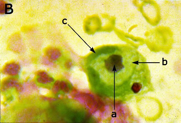
Appl. Sci. 2020, 10, 1826                                                                            2 of 17

provided mainly by double-membrane-bound organelles, mitochondria. Chemical energy of ATP
and GTP can be transformed to mechanical, electric, magnetic, or electromagnetic form for biological
utilization. Created forces can provide mass transfer, mechanical motion, and ordering of the system
itself. Elementary thermodynamic considerations imply that, without the presence of long-range
ordering forces, a complex biological system such as a human body would disintegrate within a
fraction of a second. It is then obvious that energy-related phenomena in living systems are of even
higher importance than in the nonliving world and that a disruption of energetic processes can result
in a number of pathologies, including the state of thermodynamic equilibrium called death.
      Cancer is an anti-evolutionary process, separating cells from an organized multicellular system,
forming their independence, and producing new generations of cells. Connection of the cancer process
with disturbed cellular energetics was first described by O. Warburg [2,3]. Cancer process is a pathology
connected with mitochondrial dysfunction—reduced oxidative metabolism in mitochondria to about
50% due to suspension of the oxidative processing of pyruvate (Warburg effect). The missing energy is
replaced by fermentative decomposition of glucose in the cellular volume, which is a similar process by
which, e.g., muscle cells use an additional energy source in case of increased physical exertion. Warburg
assessed differences of the ordered and disordered cells of the embryos and of the tumors, respectively,
with a conclusion that “The adenosine triphosphate synthesized by respiration therefore involves more
structure than adenosine triphosphate synthesized by fermentation”. However, disturbance of the
oxidative energy production has been accepted by the general biological scientific community as a
negligible side effect of cancer development, even after its explanation by mitochondrial dysfunction.
The origin, nature, and essence of the organization forces and energy transformation mechanisms for
their excitation were generally neglected for a long time, regardless of widely used cancer diagnostic
methods of cytology and histology, showing a strong disorder in cancer cells. It is well-known that
disorder can be a consequence of weakened ordering forces or of an effect of strong random activity
which overcomes the ordering measures (or a combination of both cases).
      Chemical bonds (covalent, ionic, hydrogen, and Van der Waals) have been supposed to be a
primary phenomenon to drive ordering and activities in biological systems. However, chemical
bonds are connected with short-range forces acting at nanometric distances, while biological systems
exhibit ordering at all length scales [4,5]. Centrally organized systems need at least ordering forces,
communication, and information processing; biological systems are not excluded. For example,
thousands of chemical reactions can occur simultaneously in a cell. At a given moment, only
some of them are prevailing, followed by others, etc. A sophisticated control over a large scale is
obviously in action which cannot be provided by neighbor-to-neighbor effects, such as diffusion, or
by purely chemical agents. Of the known mechanisms, such a control can be provided by electric or
electromagnetic field. Electric field can provide dielectrophoresis (polarization and motion of charged
particles), attraction or repulsion of charged particles, shift of resonant frequencies, and increase or
decrease of energy. Oscillating electric field may excite oscillations in microparticles and structures,
and in the case of resonance (equal frequencies of the field and of the structure), the acting forces
are enhanced. Electric or electromagnetic field being the only known phenomenon providing fast
interaction and strong enough forces at all length scales, an idea was introduced of ordering and
control by electromagnetic field generated by the living structures themselves.
      A theoretical background based on a hypothesis of coherent electrical polar vibrational states was
given by H. Fröhlich [6,7]. Coherent electromagnetic activity in a wide spectral region, from acoustic
frequencies to the UV region, can be considered a fundamental expression of life [5]. In multicellular
systems, an electromagnetic field is supposed to be generated by microtubules with a periodic structure
of tubulin heterodimers and oscillating electric dipoles [8]. At nanometric scales, the near component
of the electric field is supposed to reach extremely high values [9] so that it can excite valence electrons
to energies comparable with energy of chemical bonds. In such a configuration, biochemical reactions
might be controlled by the oscillating field providing selectivity of bonds [9].
Appl. Sci. 2020, 10, 1826                                                                            3 of 17

      The presence of an electromagnetic field can be visualized by its force effects on polarized and
charged particles and structures—their motion, changed distribution, oscillation, etc. The biological
electromagnetic field has been experimentally proved by Pohl et al. [10], Hölzel [11], Pokorný et al. [12],
and Sahu et al. [13]; its amplitude increases in important processes requiring energy supply, such as
cellular division (M phase). An experimental study of enzymatic reactions and their acceleration by
a factor up to 105 in a strong electric field [14] led to a conclusion that the electric-field-dependent
acceleration of elementary reactions might be a general concept in biological and chemical catalysis [15].
Both theoretical predictions and experimental data thus indicate that decreased power of the generated
field due to the dysfunction of energy-providing mitochondria could lead to a replacement of controlled
biochemical reactions by random chemical processes [9]. On the other hand, routinely observed
disturbed ordering in cancer cells is a clear evidence of changed organization forces, regardless of the
mechanism of their generation.
      The lifetime risk of cancers of many different types is strongly correlated with the total number
of divisions of the normal self-renewing cells maintaining the tissue homeostasis [16]. Therefore,
the total probability of cancer initiation depends on the sum of individual probabilities of division,
particularly in the periods of stem cells. Cancer transformation is assumed to be caused by an oncogene
mutation [17–22]. Nevertheless, understanding the origin of the genome somatic mutation is rather
limited. Thousands of somatic mutations (passengers) are accompanying the oncogene mutation
(driver) in a single cancer sample [17]. The experimental results very likely demonstrate a disturbed
protection of selectivity of chemical reactions and their randomization in cancer cells which may
involve genome mutations.
      The pathway of chemical bonding to the genome and transferring consequences of the oncogene
mutation involves a sequence of processes, each of which faces major obstacles. Access to DNA is heavily
restricted, e.g., by supercoiling, limiting full access to the cell-division period. Random mutations can
be corrected by DNA repair processes. If a specific oncogene mutation occurs, the oncogene expression
toward mitochondrial dysfunction meets another obstacle: the cell mobilizes tumor-suppressor
networks to avert the hazard of malignant transformation and to be withdrawn from the proliferative
pool. One of the mechanisms, oncogene-induced apoptosis, triggers a preprogrammed death of
a cell suffering from large enough DNA damage. Oncogene-induced senescence (OIS) represents
a complementary mechanism depending on the response to induction of the DNA damage [23].
OIS is induced by suppression of the PDH kinase and induction of PDH phosphatase, resulting in
increased respiration [24]. Mitochondrial dysfunction is an effector pathway of OIS [25]. Even if
tumor-suppressor networks become inactive, the tumor-suppressor genes can be reactivated [26]. All
the processes leading to cancer transformation of the cell must occur during a limited timescale of
the cellular life cycle. The resulting cancer cell must then evade the supervision of the cell-mediated
immunity (CMI) response involving T-lymphocytes (part CD4 Ly). A specific oncogene mutation
and/or overexpression, together with the loss of tumor suppressors, is a complex process which should
occur simultaneously in a large enough number of cells to initiate cancer. It is therefore reasonable to
assume that a production of mitochondrial dysfunction by an isolated oncogene mutation without
other serious pathologies has a low probability.
      Oncogenic viruses play a special role as experimental models for investigating cellular
networks [27]. A number of viruses associated with human cancer have been identified, e.g.,
Epstein–Barr virus, hepatitis B and C viruses, human herpes virus 8, human T-cell leukemia virus, and
HIV-1. More than 170 individual types of human papillomaviruses (HPV) have been classified [28–31].
Despite their different nature related to different virus families, oncogenic viruses share key features,
in particular, prolonged latency and their ability to establish long-term persistent infections instead of
killing their host cells [27]. To achieve that, they must employ strategies for evading immune response
of the host. A long-term interaction between a virus and its host seems to be an important factor for
cancer development. Cancer is assumed to be initiated when a viral coding sequence is integrated
into oncogene of the cellular genome as a result of infection. Carcinogenicity of oncogenic viruses
Appl. Sci. 2020, 10, 1826                                                                            4 of 17

may be connected with their capability to disturb or overcome protection of biochemical genome
bonding. Besides that, a virus as a living system requires energy to be supplied from the host cell.
A reduced energy supply utilized by the cell inevitably causes lowered amplitude of the generated
electromagnetic field, lowered organization forces, and possibly—at some critical stage—disturbed
write protection of the DNA [9]. About 15%–20% of all human cancers are estimated to be caused by
viral infections [27]; however, the role of viruses is supposed to be underappreciated [32]. In contrast
to many other carcinogenic agents, they provide a direct mechanism contributing to cancer initiation
(writing into DNA of the host cell), together with indirect ones (inflammation or other chronic effects).
Nevertheless, the probability of such a process depends on the virus activity and cellular protection
levels. It has to be admitted that the actual occurrence of cancers caused by viruses might be higher
than the so far estimated figure, e.g., if further oncogenic viruses are revealed.
     We suggest that disturbances of the energy levels possibly resulting in serious pathologies might be
caused by energy-consuming parasites, such as the lactate dehydrogenase (LDH) virus (or Riley virus,
now classified as NAD 1.1.1.27 Oxidoreductase). Our extensive immunological study [33–37] revealed a
response to the LDH virus antigen in (statistically) all cancer patients. However, establishing etiological
role of a pathogen in chronic diseases with decades-lasting incubation period is difficult. A possible
approach by Yerushalmy and Palmer [38] involves proving (A) the simultaneous presence of the
organism and disease and their appearance in the correct sequence, and (B) the specificity of effect of the
organism on the development of the disease. We discuss the postulate (A) in terms of our experimental
data and (B) by explanation of carcinogenesis by suggesting a biophysical model, based on published
experimental data to a significant extent. Our model extends currently acknowledged theories on
cancer initiation primarily by providing explanation of the processes preceding the oncogene mutation.

2. Materials and Methods
     Cell-mediated immunity (CMI) response is considered to correlate with adherence of T
lymphocytes to some solid-state surfaces [39] and interaction forces between human CD4 Ly cells with
the antigen [40]. Leukocyte adherence inhibition (LAI) assay is based on microscopic evaluation of
in vitro adherence of T lymphocytes to surfaces of specific glass or plastic materials in the presence and
in the absence of antigens. Two types of antigens were used. The specific antigen is an immunological
functional fraction prepared from a malignant tumor of the same type as the malignancy of the patient
from which the blood was taken. The nonspecific antigen is an immunological functional fraction
prepared from the serum of inbred laboratory mouse C3H H2k strain infected with the LDH virus. Both
antigens prepared by high-pressure gel chromatography are ribonucleic acids (RNA). A comprehensive
description of the experimental method including preparation of T lymphocytes and the antigens is
available in [9,33,34,37]. Sorption effects for separation of a complex naturally occurring mixture of
high-molecular-weight compounds is described in Vytášek et al. [41] A simple and sensitive test for
detection of tumor-specific antigen mediated by macrophage adherence inhibition is in Holáň et al. [42].
The gel chromatogram of antigen fractions prepared from human carcinoma and inbred mice (strain
C3H) infected with LDH virus is in Jandová et al. [37]. Besides chemical reactions, the antigens were
separated by a differential refractometer and a spectrophotometric detector, and the data were recorded,
using a two-channel Philips PM 8010 recorder [43]. The antigen is a 340 nm fraction. A schematic
picture with the receptor, ligand, and signaling pathway is in Jandová et al. [36].
     The CMI response by the described method was investigated on patients with cancer of the
cervix, endometrium, ovary, breast, and lung, as well as melanoblastoma and otorhinolaryngological
cancers. Cancer diagnosis was verified by histopathology, using cell samples taken during surgery.
Conditions for malignant transition in cancer development were studied on precancerous cervical
lesions. These lesions were diagnosed by cytological (PAP) smears, from examination by colposcopy
and from colposcopically directed ‘punch’ biopsy material by histology. The grades of development
of precancerous lesions LSIL and HSIL (low- and high-grade squamous intraepithelial lesions,
Appl. Sci. 2020, 10, 1826 5 of 17
respectively) were determined by cytology, and CIN 1, 2, and 3 (cervical intraepithelial neoplasia)
grades by histology—Jandová et al. [37].
Cells from healthy humans adhere both without and with the antigens, adherence of cancer cells
with either antigen is decreased, and the mutual difference is small. The results of LAI assay were
evaluated in three regions—healthy, noise, and pathological (e.g., cancer). A relative number is used:
M = 100 · m/m0 (in %) where m0 and m are numbers of cells in the suspension before and after the
sedimentation–adhesion process.
The LAI assay is a complex and tedious experimental method, and unequivocal results are
conditioned by a high purity of antigens. Chemical and physical agents can affect the cell-mediated
immunity response [34–36]. For example, magnetic field 0.5–10 mT decreases (increases) adherence of
CD4 Ly taken from healthy humans (cancer patients). Magnetic field can affect adherence surface of
glass, interaction forces between CD4 Ly, and properties of antigen, but the mechanism of such behavior
is unknown.
A positive response to the nonspecific antigen was also examined in patients with acute myocardial
infarction, schizophrenia, and recurrent spontaneous abortions in early pregnancy from unknown
reasons [44].
3. Results
3.1. Warburg Effect (Differentiated) Cancers
Energy transport, processing, and parceling out into small bits stored in ATP and GTP form a joint
fermentative and oxidative pathway crucial for life. Warburg disclosed that healthy tissues produce
the majority of energy by respiration, while cancer tissues produce approximately the same amount of
energy by fermentation as by respiration, i.e., about 50% by metabolizing glucose to lactic acid [2,3].
Transport of pyruvate, a product of glycolysis, to mitochondrial matrix is decreased by dysfunctional
pyruvate dehydrogenase (PDH) enzymes, and this deviation is called a mitochondrial dysfunction or
the Warburg effect.
Mitochondrial function is connected with a transfer of hydrogen (H+ ) ions across its inner
membrane and a formation of a charged layer of H+ ions in cytosol around mitochondria. Due to the
H+ transfer, pH values in matrix and cytosol change. H+ ions transferred to the cytosol form a layer
creating an electric potential across the inner membrane. Further in the text, we denote this electric
potential as actual potential, and it should be approximately proportional to the number of H+ ions
transferred to the cytosol, which is in turn related to the amount of the energy produced by oxidative
processes. The actual electric potential of the fully functional mitochondria evaluated from the transfer
of H+ ions is about −140 mV, and that of the dysfunctional mitochondria about −70 mV [45]. These
values contrast to the apparent potential values of −100 and −160 mV, respectively, measured by uptake
and retention of positively charged fluorescent dyes, such as Rhodamine 123 [46]. The differences can
be explained by the presence of an additional potential layer of ordered water formed in the region of
the high-intensity electric field of the H+ ion layer [47].
Water ordering in regions of a high electric field is a general phenomenon [48]. Ordered water
layers up to 0.5 mm thick are formed in areas of a large gradient of the electric potential and at charged
surfaces, regardless of their chemical composition. Ordered water layers can be formed at a plasma
membrane, at the layer of H+ ions around mitochondria, and at charged surfaces of macromolecules.
A theoretical model describes water ordering in terms of electronic excitation, i.e., coherent oscillations
and vortexing of electrons in molecules [49,50]. Electrons in water molecules are supposed to coherently
oscillate between a fundamental state with strongly bound electrons (where the energy to expel an
electron amounts to 12.60 eV) and the excited state with weekly bound electrons (where the expel
energy is about 0.54 eV). Therefore, an ordered water layer can be a donor of electrons. The thickness of
the ordered water layer depends on the pH value: it increases with increasing offset from an intermediate
point, pH0 , where the ordered water layer is not formed [48]. For pH < pH0 , the intensity of the electricAppl. Sci. 2020, 10, 1826 6 of 17
field in the ordered water layer is oriented in the opposite direction from that in the H+ layer forming
the actual potential of mitochondria (Figure 1), and positively charged particles are excluded from
the layer. For pH > pH0 , both intensities of the electric field are oriented in the same direction, and
negatively
Appl. Sci. 2020,charged
10, x FORparticles are excluded.
PEER REVIEW 6 of 17
Healthy Cancer
Electron
emission
Ordered
water
H+ H+
Matrix Matrix
Figure 1. Potential layers
1. Potential layers at at the
the inner
inner mitochondrial
mitochondrial membrane
membrane of functional (Healthy) and
dysfunctional (Cancer) mitochondria. The electric potential consists of two components—the components—the actual
electric potential of the
the electrochemical
electrochemical H H++ gradient and the potential of the ordered water layer. layer. The
vertical dimensions of the layers correspond to the electric potentials and the inner arrows arrows denote
denote
orientations of the
the intensity
intensity of ofthe
theelectric
electricfield.
field.For
ForpH
pH < pH
< pH 0 , the
0, the intensity
intensity of the
of the electric
electric fieldfield in
in the
the ordered
ordered water
water layer
layer hashas anan opposite
opposite directiontotothe
direction theelectric
electricpotential
potential of
of the electrochemical
electrochemical H H++
gradient. For
ForpHpH>>pH pH
0, 0 , both
both directions
directions areare
thethe same.
same. TheThe intermediate
intermediate valuevalue pH0 determines
pH0 determines the
the point
point where the ordered water layer is not formed. The arrows at the upper surface
where the ordered water layer is not formed. The arrows at the upper surface of ordered water layer of ordered water
layer
in thein the cancer
cancer case denote
case denote emission
emission of electrons
of electrons whichwhich
dampdamp the electromagnetic
the electromagnetic field.field.
The
The thickness
thickness and and the
the properties
properties of of the
the ordered
ordered water
water layer
layer at at the
the mitochondrial
mitochondrial membrane membrane
depend +
depend on on the
the difference
difference of of the
the electric
electric field
fieldgenerated
generatedby byH layer and
H+ layer and byby the
the surrounding
surrounding cytosolcytosol
(dependent on pH), and on the difference of proton concentration
(dependent on pH), and on the difference of proton concentration (forming the electrochemical(forming the electrochemical proton
gradient which exerts a proton motive force). Cytosol may have
proton gradient which exerts a proton motive force). Cytosol may have pH about 7.2 (neutral pH about 7.2 (neutral medium).
However,
medium). cells are able
However, to control
cells are ablethe pH of their
to control intracellular
the pH compartments
of their intracellular (e.g., even 5(e.g.,
compartments pH). even
Inside5
mitochondria the pH is higher than 7.2. The mitochondrial proton layer
pH). Inside mitochondria pH is higher than 7.2. The mitochondrial proton layer may correspond to may correspond to a difference
of about −1 pH.
a difference The measured
of about −1 pH. The values of the values
measured potentials andpotentials
of the dependence andofdependence
the thickness ofofthe
thethickness
ordered
layer
of theon pH make
ordered layerpossible
on pHtomake prepare an approximate
possible to preparemodel of healthy and
an approximate cancer
model cellsmutual
of the which explains
position
forming of ordered water layers with different polarizations. In the model,
of the healthy and cancer cells on pH and forming of ordered water layers with different polarization. a healthy cell has the
intermediate
In the model,point of about
a healthy cell6 has
pH and the cancer cellpoint
the intermediate higher—Pokorný
about 6 pH and et al.the
[47]. It should
cancer cell be noted
higher—
that this is only a crude approximation, as the pH processes inside cells
Pokorny et al. [47]. It should be noted that this is only a crude approximation, as the pH processes depend on many physical and
chemical factors in cytosol and inside mitochondria, as well as on biological
inside cells depend on many physical and chemical factors in cytosol and inside mitochondria, as activity.
well The
as onpotential
biological across the ordered water layer at the mitochondrial membrane can be obtained
activity.
as a The
difference of the apparent
potential across the ordered minus actual
water electric
layer at the potentials
mitochondrial for functional
membraneand can bedysfunctional
obtained as
mitochondria. Dependence of the intensity of the electric field in
a difference of the apparent minus actual electric potentials for functional and dysfunctionalthe ordered water layer on pH can
affect electromagnetic
mitochondria. Dependence activity in cells
of the [51]. For
intensity of the > pH0 ,field
pHelectric the ordered water has
in the ordered watera high
layertendency
on pH can to
release electrons [49]. The electrons are transported into cytosol and form
affect electromagnetic activity in cells [51]. For pH > pH0, the ordered water has a high tendency to a conductive environment
which
releasecan strongly
electrons damp
[49]. Thethe electromagnetic
electrons activity
are transported of cytosol
into the cell and
[47,52–55].
form a conductive environment
Cellular oscillating systems are strongly nonlinear
which can strongly damp the electromagnetic activity of the cell [47,52–55]. [56]. Resonant frequency of such a system
is notCellular
a constant; it depends on the energy stored in the oscillating
oscillating systems are strongly nonlinear [56]. Resonant frequency system. Changed amplitudes
of such a system of is
cellular oscillations due to their damping result in frequency shifts to
not a constant; it depends on the energy stored in the oscillating system. Changed amplitudes of higher values compared to those
in the healthy
cellular state. due
oscillations The interference
to their damping pattern of the
result inelectromagnetic
frequency shiftsfield is thenvalues
to higher rebuiltcompared
and shifted,to
resulting
those in the in disturbed
healthy state. organization forces. pattern
The interference Due to different oscillation frequencies
of the electromagnetic field is thenin healthy
rebuilt and
and
cancer
shifted,cells, their in
resulting mutual electromagnetic
disturbed organizationcommunication must be disrupted
forces. Due to different oscillation at least to some
frequencies extent.
in healthy
Disturbed
and cancer cells, their mutual electromagnetic communication must be disrupted at least tocancer
electromagnetic activity, a fundamental feature of all cancers [57], thus can enable a some
cell to liberate
extent. Disturbed itself from the tissue.activity, a fundamental feature of all cancers [57], thus can enable
electromagnetic
a cancer cell to liberate itself from the tissue.
A strong-enough electric field can heavily influence chemical reactions, e.g., enzymatic reaction
rate can be enhanced up to 105-fold by the presence of a static electric field [14]. Resonant effects of
oscillating fields can be even many orders of magnitude stronger. Photon absorption can modify the
electronic configuration of an atom or molecule and thus enable chemical reactions which wouldAppl. Sci. 2020, 10, 1826 7 of 17
A strong-enough electric field can heavily influence chemical reactions, e.g., enzymatic reaction
rate can be enhanced up to 105 -fold by the presence of a static electric field [14]. Resonant effects
of oscillating fields can be even many orders of magnitude stronger. Photon absorption can modify
the electronic configuration of an atom or molecule and thus enable chemical reactions which would
otherwise be impossible. By varying the oscillation field, chemical reactions might be efficiently
switched on and off by a suggested mechanism of the reverse vibrational Stark effect [9], particularly
when the photon energy absorbed by a valence electron is higher than the energy of chemical bonds
(van der Waals attraction to covalent bonding correspond to a photon frequency range of 1012 –1017 Hz).
It is reasonable to assume the mechanism to be involved in controlling information storage into DNA.
We suggest that if the oscillating field is damped, selective reactions might be replaced by standard
chemical reactions which may then lead to a number of random writings into DNA, causing massive
genomic mutations.
Similar effects can be caused by short-circuiting of the cellular generating structures by nanofibers or
other structures which have penetrated into the cell. Asbestos filaments are the most discussed structure
of the type. The filaments represent optical fibers or conducting wires that provide a short-circuit for
the cellular electric and/or electromagnetic signals [58,59]. Both pathological mechanisms, i.e., damping
and short-circuiting, are fundamental processes resulting in cancer. Decreased power, disturbed
coherence, and changed frequencies are conditions for disturbed order inside the cell and the tissue,
local invasion, and metastases. Gradual evolution of the pathological processes corresponds to gradual
morphological changes routinely observed.
3.2. Reverse Warburg Effect (Undifferentiated) Cancers
The reverse Warburg effect [60] is a tissue process of a pathological cooperation between a cancer
cell and associated fibroblasts. Pathological processes in inflicted fibroblasts associated with the cancer
cell are similar to those in the Warburg effect cancer cells. Pathological transformation of the fibroblast
is controlled by signaling from the tissue and from the cancer cell. Loss of stromal Caveoline-1 (Cav-1)
expression results in increased production of nitric oxide, enlarged reactive oxygen species (ROS)
production, increased oxidative stress, and mitochondrial dysfunction in fibroblasts. By altered model
for tumor–stroma co-evolution, cancer cells induce oxidative stress in adjacent fibroblasts and possibly
in other stroma cells. Oxidative stress in the tumor stroma mimics the effect of hypoxia under aerobic
condition and produces other processes for cancer development [61]. Excess stromal production of
reactive oxygen species is assumed to be a basis for the following chemical reactions, but energy
processes caused by increased damping are not assumed.
The cancer cell is fueled by energy-rich metabolites, such as lactate, pyruvate, glutamine, and
ketone BHB (beta-hydroxybutyrate), produced by the stroma cells. Their high energy supply leads
to a high excitation of the generated electromagnetic field. The reverse Warburg effect cancer cells
are shifted to an unstable highly nonlinear region, their working point may float with energy supply,
and coherence is disturbed. Their electromagnetic forces are strong but with varying amplitudes.
Oscillation frequencies are lower than those in the healthy state and fluctuating. Due to strong but
unstable acting forces, the morphological changes are pronounced so that the cancer cells do not
resemble their original counterparts. Advanced prostate cancers, for example, can transform to this
phenotype [60].
3.3. LDH Virus—An Energy Parasite
In 1960, V. Riley discovered LDH virus (lactate dehydrogenase elevating virus or Riley virus), an
agent that systematically affects experimental results in cancer research on mice [62]. It can be transferred
primarily by blood, including direct transfer from a mother to offspring. After approximately a week of
lasting acute infection, the virus reduces to its ribonucleic acid (RNA) chain, covered with a structure
similar to the membrane of the host cell, which helps in evading detection by the immune system. A
lifelong chronic infection follows. The virus is considered a perfect parasite [63–65]—the graduallyAppl. Sci. 2020, 10, 1826 8 of 17
growing population of RNA chains causes no apparent harm to the host cell, and no morphological
changes or obvious disease occur. The RNA chains “only” parasite on cellular energy resources,
invading energy processes prior to the oxidative stage. In infected cells, the level of LDH enzyme is
elevated five- to ten-fold, compared to noninfected cells. The LDH enzyme catalyzes a conversion of
reduced NADH (nicotinamide adenine dinucleotide + hydride ion H− ) to oxidized NAD+ and vice
versa [63]. Pyruvate, the final product of glycolysis, is converted to lactate before it can be transferred
Appl. Sci. 2020, 10, x FOR PEER REVIEW 8 of 17
to mitochondria, which forms a parallel pathway to inhibition of pyruvate transfer to mitochondria
by
PDH dysfunctional
(Figure 2) inPDH (Figure
Warburg 2) incancer
effect Warburg effect
cells. Thecancer
infectedcells.
cellThe
withinfected cell withofa viral
a population population
RNAs
of viral RNAs exceeding some critical level then behaves in a similar way as a Warburg
exceeding some critical level then behaves in a similar way as a Warburg effect cancer cell, or dies as effect cancer
cell,
in theorcase
dies of
as heart
in theinfarction.
case of heart
A infarction. A possibleofconnection
possible connection the LDH virus of thewith
LDHcancer
virus with
was cancer
studiedwasby
studied by Rowson and Mahy, and Riley and Wroblewski [63,66]; changes
Rowson and Mahy, and Riley and Wroblewski [63,66]; changes of the immune activity in the acuteof the immune activity in
the acute phase of the LDH virus infection are given in [67]. Cell-mediated immunity
phase of the LDH virus infection are given in [67]. Cell-mediated immunity (CMI) response to the (CMI) response
to
LDHthevirus
LDH antigen,
virus antigen, extensively
extensively studied studied by Jandová
by Jandová et al. [33–37],
et al. [33–37], indicates
indicates activityactivity
of theof the virus
LDH LDH
virus or a similar
or a similar pathogenpathogen in humans.
in humans.
Glucose
Glycolysis
(fermentation) { ATP
ATP
LDH
2x Pyruvate
Oncogene Lactate
mutation ATP
Fatty
Mitochondrion {
Oxidative
acids
phosphorylation
{ GTP
Figure 2. Energy transformation by glycolysis and oxidative phosphorylation in living cells. Cancer
development can result from inhibition of pyruvate transfer to mitochondrial matrix. Parasitic energy
consumption and transfer of pyruvate to lactate form a parallel
consumption parallel pathway
pathway to mitochondrial
mitochondrial dysfunction
caused by deactivated PDH enzymes.
3.4. Immune Response
3.4. Immune Response Study
Study
The immune system
The immune system is is one
one ofof the
the most
most complex
complexstructures
structuresininaahuman
humanbody. body.It Itresponds
respondstotoa
a presence of antigens, i.e., molecules or molecular fragments characterizing
presence of antigens, i.e., molecules or molecular fragments characterizing individual pathogens. individual pathogens.
The
The presence of antigens is detected either directly or indirectly through antibodies.
presence of antigens is detected either directly or indirectly through antibodies. There are two main There are two
main mechanisms
mechanisms of immunity—humoral
of immunity—humoral and cellular.
and cellular. The latter The latter is mediated
is mediated by T lymphocytes.
by T lymphocytes. Since the
Since the 1960s, a group of Czech scientists studied cell-mediated
1960s, a group of Czech scientists studied cell-mediated immunity (CMI) of healthy immunity (CMI) of healthy blood
blood donors
donors and patients
and patients with various
with various tumors.tumors. The study
The study was focused
was focused on theonimmune
the immune response
response of cytotoxic
of cytotoxic T-
T-lymphocytes, leukocytes capable of killing whole cells, such as bacteria,
lymphocytes, leukocytes capable of killing whole cells, such as bacteria, cells infected by cells infected by viruses,
viruses,
cancer
cancer cells,
cells, etc. Pathogenic cells
etc. Pathogenic cells are
are directly
directly detected
detected by by receptors
receptors on on the
the cellular
cellular membrane
membrane of of aa T
T
lymphocyte. Quantitative assessment can be based on the fact that the
lymphocyte. Quantitative assessment can be based on the fact that the cell-mediated immune cell-mediated immune response
by T lymphocytes
response correlates with
by T lymphocytes their adherence
correlates with their toadherence
an appropriate glass or plastic
to appropriate glasssurface. The surface.
or plastic method
is
Thereferred
method toisasreferred
leukocyte adherence
to as leukocyte inhibition
adherence (LAI) test. (LAI) test.
inhibition
The
The LAI method was used to study CMI response on
LAI method was used to study CMI response on over
over 12,000
12,000 patients
patients with
with cancer
cancer of
of the
the
cervix, endometrium, ovary, breast, and lung, as well as melanoblastoma and
cervix, endometrium, ovary, breast, and lung, as well as melanoblastoma and otorhinolaryngological otorhinolaryngological
cancers
cancers [9,33–37].
[9,33–37].T T cells were
cells separated
were separatedfrom venous blood, and
from venous threeand
blood, sample
threesolutions
sampleforsolutions
microscopicfor
evaluation
microscopic evaluation of non-adhering cells were prepared: (1) a solution containing T solution
of non-adhering cells were prepared: (1) a solution containing T cells only; (2) a of
cells only;
T
(2)cells with a of
a solution specific
T cellsantigen, i.e., immunological
with a specific functional fraction
antigen, i.e., immunological prepared
functional fromprepared
fraction tumors offrom
the
same type as the tumor found in the patient; (3) a solution of T cells with a nonspecific
tumors of the same type as the tumor found in the patient; (3) a solution of T cells with a nonspecific antigen, i.e.,
antigen, i.e., immunological functional fraction obtained from the serum of inbred laboratory mouse
strain C3H infected with the LDH virus. T cells of healthy blood donors did not show a significant
response to the presence of the antigens. On the contrary, the presence of both antigens elicited a
strong response for the vast majority of cancer patients. It should be noted that the specific antigen
elicited a response only for patients with tumors corresponding to the specific antigen obtained fromAppl. Sci. 2020, 10, 1826 9 of 17
immunological functional fraction obtained from the serum of inbred laboratory mouse strain C3H
infected with the LDH virus. T cells of healthy blood donors did not show a significant response to the
presence of the antigens. On the contrary, the presence of either antigen elicited a strong response for
the vast majority of cancer patients. It should be noted that the specific antigen elicited a response only
for patients with tumors corresponding to the specific antigen obtained from a tumor of the same type,
while the nonspecific antigen of the LDH virus elicited a response for all cancer patients. T cells of a
healthy blood donor (cancer patient) with antigen exhibited the relative number M = 96.68% (0.6%),
2.65% (2.05%), and 0.67% (97.35%) in healthy, noise, and pathological regions, respectively.
The LAI method was also applied to patients with various grades of development of precancerous
cervical lesions. The experiment revealed 27.7% and 34.8% of the cases corresponding to healthy
women for specific and nonspecific antigen, respectively, and 67.0% and 59.8% corresponding to
patients with a tumor. No statistically significant difference was revealed for different grades of lesion
development, which corresponds to the fact that behavior of precancerous events is unpredictable
and invasive carcinoma may develop from any level of precancerous abnormalities [68]. The results
indicate that the precancerous stage is a critical phase of cancer transformation of a cell.
3.5. Morphology of Healthy and Cancer Cells
Evaluation of morphological pictures of tissues, cells, nuclei, nucleoli, membranes, and chromatin
belongs to standard diagnostic methods for cancer. Histological and cytological pictures are routinely
used. Morphological changes and their evolution serve as significant criteria of cancer development.
The Warburg effect (differentiated) cancer cells resemble their original cells, and their morphological
changes develop gradually. The final stage of their development is indicated by local invasion
and metastases. Their behavior is predictable. A comprehensive assessment of their slow gradual
development seems to be a sign of increasingly disturbed and weakened organization forces. The
reverse Warburg effect (undifferentiated) cancer cells do not resemble their original counterparts. They
are small, with large nuclei, scant cytoplasm, and foci of necrosis. The most startling properties are their
violent growth, aggressiveness, and early invasion in surroundings and metastases, which indicates
strong forces in a disordered system.
Development of the Warburg effect (differentiated) cancers is shown in Figure 3:
• Figure 3A displays normal squamous cells from uterine cervical surface area of a healthy woman.
Cytoplasm is abundant and has distinct borders. The nuclear–cytoplasmic volume ratio is in
favor of cytoplasm and can be about 5/60. Possible physical view: This picture shows a healthy
state. The pathological development is hidden as long as the decrease of the oxidative energy
production does not result in damping of power of cellular electromagnetic field, organization
forces in the cell remain unchanged, and oncogenes not transformed. It should be emphasized
that a considerable part of the development of the Warburg effect is not visible by itself.
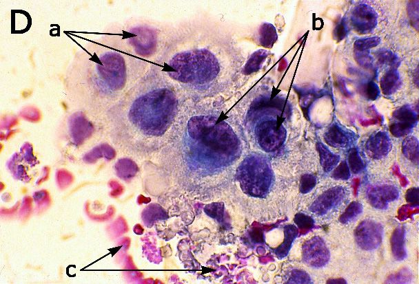
• Figure 3B shows cells of low-grade squamous intraepithelial cervical cell lesions which have
enlarged nuclei of about three times with respect to a healthy cell and well-defined cytoplasm.
Possible physical view: The power of the electromagnetic field in the cell is lowered, and the most
sensitive organization activities are disturbed, but the oscillation frequencies seem to coincide
with those of healthy cells. The power in the cell could be kept at a considerably high level by an
energy supply from neighboring cells connected through tubulin nanotubes [69].
• Figure 3C displays cells of high-grade squamous intraepithelial cervical cell lesions with variable
size of cells, variable size and shape of nuclei, quite irregular nuclear membrane, and reduced
cytoplasm. The degree of nuclei enlargement is more variable than that in the low-grade case.
Possible physical view: Connections by tubulin nanotubes between cells are disturbed and
electromagnetic power is low. The power and frequencies of oscillations differ between healthy
and cancer cells, and between cancer cells. Cancer cells are not selectively interacting with healthy
cells in the tissue and become independent entities.Appl. Sci. 2020, 10, 1826 10 of 17
• Figure 3D: Marked pleomorphism of cellular size and shape as a characteristic property of invasive
squamous cell – cancer can be recognized. Cell borders are poorly defined, cytoplasm is missing,
size and shape of nuclei manifest large differences, and chromatin in nuclei is coarsely clumped.
Debris of cells and vessels can be seen in the background.Possible physical view: The power of
the electromagnetic field in cancer cells is very low, and its level in similar cancer cells is different.
Variation of frequencies resulted in repulsion forces between cancer and healthy cells. Power and
frequency of oscillations have strong random components.
Appl. Sci. 2020, 10, x FOR PEER REVIEW 10 of 17
Figure 3. Cellular morphological disorder indicates pathologically mutated forces. (A) Healthy
uterine cervix cells: (a) superficial squamous cells. (b) Intermedial cells. (B) LSIL—low grade
Figure 3. Cellular
squamous morphological
intraepithelial disorder
cervical cell indicates
lesion (mild pathologically
precancerosis): (a) mutated
nucleoli areforces. (A) Healthy
generally absent,
uterineare
nuclei cervix
threecells:
times(a) superficial
larger compared squamous
to those cells. (b) Intermedial
of normal intermedial cells.
cells. (B) LSIL—low
A variable grade
degree of
squamous intraepithelial cervical cell lesion (mild precancerosis): (a) nucleoli are
nuclear hyperchromasia. (b) Perinuclear cavitation (koilocytosis) consisting of sharply delineated generally absent,
nucleiperinuclear
clear are three times
zone. larger compared cytoplasm.
(c) Well-defined to those of (C) normal intermedial
HSIL—high gradecells. A variable
squamous degree of
intraepithelial
nuclear hyperchromasia. (b) Perinuclear cavitation (koilocytosis) consisting of sharply
cervical cell lesion (severe precancerosis): (a) the cells are smaller than the cells in LSIL. Cell size delineated
clear perinuclear
varies. zone. of
(b) The degree (c) nuclear
Well-defined cytoplasm.
enlargement (C) HSIL—high
is variable grade squamous
too. (c) Contours of nuclear intraepithelial
membrane
are quite irregular. (d) Wrinkling of nuclear membrane. (D) Invasive carcinoma cells: (a) Cell
cervical cell lesion (severe precancerosis): (a) the cells are smaller than the cells in LSIL. cellssize
are
varies. (b) The degree of nuclear enlargement is variable too. (c) Contours of
frequently smaller than healthy ones and display most of the features of HSIL cells. (b) Nucleinuclear membrane are
quite irregular. (d) Wrinkling of nuclear membrane. (D) Invasive carcinoma cells:
demonstrate coarsely clumped chromatin. (c) Tumor diathesis consists of necrotic debris of cells and old (a) cells are
frequently
blood. smaller than
Magnification: (A) healthy ones350×;
200×; (B–D) and display
Staining:most of the features of [70],
(A,C,D)—Papanicolaou HSIL(B)—Pekárek
cells. (b) Nuclei
[71],
demonstrate coarsely clumped chromatin. (c) Tumor diathesis consists of necrotic
Kobilková and Siracký [72]. Location: (A) ectocervix of uterus; (B) vagina; (C,D) endocervical area. debris of cells and
old blood. Magnification: (A) 200×; (B–D)
Nuclei—dark colors; cytoplasm—light colors. 350×; Staining: (A,C,D)—Papanicolaou [69], (B)—Pekárek
[70], Kobilková and Siracký [71]. Location: (A) ectocervix of uterus; (B) vagina; (C,D) endocervical
Morphological
area. Nuclei—dark changes
colors;of cells and tissues
cytoplasm—light along the cancer transformation pathway are assessed
colors.
by comparison with morphology of a cell in the healthy state. Sizes, shapes, and structures of
Morphological
cell membranes, changes
nuclei, of cells
nucleoli, andand tissues are
chromatin along the cancer
changed. Cancertransformation
cells can have pathway
more than are
assessed by comparison with morphology of a cell in the healthy state. Sizes, shapes,
one nuclei and the nuclear–cytoplasmic ratio is different. Differences between equal cells, nuclei,and structures
of cell membranes, nuclei,
nuclear–cytoplasmic nucleoli,
ratio, and otherand chromatin
structures are changed.
and parameters Cancer
are cells can
significant. have more than
Morphological one
changes
nuclei
are and the nuclear–cytoplasmic
connected ratio is different.
with disturbed organization mechanism Differences between
and increased equal
effect of cells,
random nuclei, nuclear–
events. The
cytoplasmic structure
well-defined ratio, andofother structures
healthy cells andand
theirparameters
long-rangeare significant.
arrangement inMorphological changes
tissues require the are
existence
connected with disturbed organization mechanism and increased effect of random events. The well-
defined structure of healthy cells and their long-range arrangement in tissues require the existence of
long-range forces. A disturbed electromagnetic field and related forces in cancer cells are impressed
into morphological changes—the more disturbed the field, the more chaos in morphology.Appl. Sci. 2020, 10, 1826 11 of 17
of long-range forces. A disturbed electromagnetic field and related forces in cancer cells are impressed
into morphological changes—the more disturbed the field, the more chaos in morphology.
4. Discussion
This paper brings a comprehensive framework of fundamental interdisciplinary mechanisms
connected with cancer initiation and development, described by Pokorný et al. [9,47]. Typical
Appl. Sci. 2020, 10, x FOR PEER REVIEW 11 of 17
biophysical processes leading to cancer are based either on parasitic energy consumption, resulting in
emission of of
absorption freeconducting
electrons from ordered water
contaminants, bothlayer at dysfunctional
damping the cellularmitochondria, or by absorption
electromagnetic field. The
of conducting
damped cellular contaminants,
electromagnetic both field
damping the cellular
is followed electromagnetic
by changes field. The damped
in the biochemical region. cellular
Strong
electromagnetic field is followed by changes in the biochemical region.
controlling signals are missing and a large number of randomized chemical reactions and genome Strong controlling signals are
missing and a large number of randomized chemical reactions and genome
mutations is produced as coherent controlling signals are overcome by random features. The defects mutations is produced
as biophysical
in coherent controlling
and biochemicalsignalsregion
are overcome by random
are transferred to thefeatures.
biologicalTheregiondefects in biophysical
and result in the knownand
biochemical region are transferred to the biological region and result in
biological stage of cancer. Essential biophysical processes of the cancer transformation are mutuallythe known biological stage of
cancer. Essential biophysical processes of the
connected with other links of pathological development of cancer. cancer transformation are mutually connected with other
linksTheof pathological
decisive part development
of the cancer of transformation
cancer. pathway is caused by biophysical mechanisms
The decisive part of the cancer transformation
connected with damping by electrons in a Warburg effect cancer pathway is caused by biophysical
cell, in fibroblasts mechanisms
associated with a
connected
reverse with damping
Warburg by electrons
effect cancer cell, and in awith
Warburg effect cancerbycell,
short-circuiting in fibroblasts
filaments associateda with
contaminating cell,
a reverse Warburg effect cancer cell, and with short-circuiting by filaments
dividing cancers into three basic phenotype groups. The highest probability of cancer initiation seems contaminating a cell,
dividing cancers into three basic phenotype groups. The highest probability
to be connected with parasitic energy consumption in cells. Different cancer incidence in different of cancer initiation
seems
cell to be
types connected
[45], e.g., low with parasitic
incidence in energy
muscleconsumption
and liver cells in cells.
(most Different
liver tumors cancerareincidence
formed by in
different cell types [45], e.g., low incidence in muscle and liver cells (most
metastases) can be also explained by this mechanism. Due to abundant blood vessels, the cells have liver tumors are formed by
metastases) can
high-enough be also
energy explained
supply, even by thiscase
in the mechanism. Due to abundant
of comparatively high parasiticbloodenergy
vessels, the cells have
consumption. If
high-enough
the parasitic energy supply, even
consumption in theacase
exceeds of comparatively
critical rate, cellularhigh parasitic
death mayenergyoccur consumption.
instead of cancer If the
parasitic consumption
transformation. exceeds a critical
Cell-mediated immune rate,response
cellular death
to the may occurvirus
LDH instead of cancer
antigen in transformation.
patients with
Cell-mediated
myocardial immune
infarction response
supports thetoidea
the LDH
[44]. Byvirus antigenfundamental
including in patients with myocardial
biophysical infarction
processes and
supports the idea [44]. By including fundamental biophysical processes
their disturbances into a general picture, the ideas presented in this paper represent a novel attitude and their disturbances into
a general
to the cancerpicture, the ideas
problem, presented
shedding light on in yet
thisunexplained
paper represent a novel attitude
phenomena. Development to the pathways
cancer problem,
of the
shedding effect
Warburg light on andyetthe
unexplained
reverse Warburgphenomena. Development
effect cancers pathways
are presented in of the Warburg
Figures 4 and 5. effect and
Current
the reverse Warburg effect cancers are presented in Figures
knowledge enables us to explain biophysical mechanisms of cancer origin and to suggest4 and 5. Current knowledge enables us
to explain biophysical mechanisms of cancer origin and to suggest
continuation and development in biochemical and biological regions. Biochemical reactions and continuation and development
in biochemical
mechanisms areand biological
presented onlyregions. Biochemical
to demonstrate theirreactions and and
connections mechanisms
dependences. are presented only to
demonstrate their connections and dependences.
Figure 4. A schematic
schematic picture of physical pathological
pathological links
links in cancer development
development of of the
the Warburg
effect (differentiated)
(differentiated) phenotype.
phenotype. Damping of the cellular electromagnetic field is the main functional
link along the pathway of cancer development. The The electromagnetic
electromagnetic field can be damped by electrons
released from
fromthethe
repolarized ordered
repolarized waterwater
ordered layers layers
aroundaround
dysfunctional mitochondria
dysfunctional or by conducting
mitochondria or by
fibers insidefibers
conducting the cell, such
inside theascell,
asbestos.
such as The organization
asbestos. forces of electromagnetic
The organization nature innature
forces of electromagnetic a cell
are disturbed.
in the cell are disturbed.Appl. Sci. 2020, 10, 1826 12 of 17
Appl. Sci. 2020, 10, x FOR PEER REVIEW 12 of 17
Figure 5.
Figure 5. A schematic picture of physical pathological links in cancer development of the reverse reverse
Warburg
Warburgeffect
effect(undifferentiated)
(undifferentiated) phenotype.
phenotype. Pathological
Pathologicalprocesses in ainfibroblast
processes associated
a fibroblast withwith
associated the
cancer cell are
the cancer cellsimilar to those
are similar in a cancer
to those cell ofcell
in a cancer theofWarburg effect.effect.
the Warburg Pathological transformation
Pathological of the
transformation
fibroblast is controlled
of the fibroblast by signaling
is controlled from the
by signaling fromtissue
the and from
tissue andthe cancer
from cell. Loss
the cancer cell.ofLoss
stromal Cav-1
of stromal
expression is a keyiscondition
Cav-1 expression for development
a key condition of the reverse
for development Warburg
of the reverse effect. The
Warburg cancer
effect. The cell is fueled
cancer cell is
by energy-rich
fueled metabolites
by energy-rich produced
metabolites by fibroblasts.
produced by fibroblasts.
Our
Our model
modelisisbased
based ononexperimental
experimental data to ato
data significant extent.
a significant The LAI
extent. TheassayLAI data
assay[33–37] clearly
data [33–37]
indicate
clearly indicate that cancer development is connected with the LDH virus infection, as suggested or
that cancer development is connected with the LDH virus infection, as suggested in [63,66], in
with a pathological
[63,66], process with
or with a pathological a similar
process with CMI response.
a similar CMIChronical
response.infection
Chronical byinfection
the LDHby virus
the leads
LDH
to decreased
virus leads tooxidative
decreased processes
oxidative in mitochondria which corresponds
processes in mitochondria which to an equivalent
corresponds to anbehavior
equivalentof a
Warburg +
behavioreffect cancer cell.
of a Warburg Decreased
effect cancer cell.oxidative processes
Decreased lead toprocesses
oxidative a reducedlead transfer of H ions
to a reduced through
transfer of
the inner mitochondrial membrane, resulting in an altered membrane
H ions through the inner mitochondrial membrane, resulting in an altered membrane potential
+ potential [45,46]. The discrepancy
in the potential
[45,46]. values published
The discrepancy in the in [45,46] can
potential be explained
values publishedbyina presence
[45] and and [46] repolarization
can be explained of a layer
by a
of orderedand
presence water [48] formed on
repolarization ofthe a membrane
layer of ordered potential layer.[48]
water Theformed
repolarized water
on the layer can emit
membrane. The
electrons which increase conductivity of the cytosol. As a result, higher
repolarized water layer can emit electrons which increase conductivity of the cytosol. As a result, damping of electromagnetic
field
higher occurs,
damping and of
ordering forces must
electromagnetic fieldbeoccurs,
disrupted. The presence
and ordering forces ofmust
electromagnetic
be disrupted. field
Thegenerated
presence
by
of living cells was experimentally
electromagnetic field generatedconfirmed by living[10–12],
cells was and experimentally
increased damping in cancer
confirmed tissue was
[10–12], and
detected [52–55]. The idea of electromagnetic control over biochemical
increased damping in cancer tissue was detected [52–55]. The idea of electromagnetic control over reactions is a hypothesis only,
although
biochemical partially supported
reactions by a massive
is a hypothesis only,influence
although ofpartially
electric field on enzymatic
supported reactions
by a massive discussed
influence of
in [14,15]. If valid, the hypothesis would shed light to the origin of genome
electric field on enzymatic reactions discussed in [14,15]. If valid, the hypothesis would shed light to mutations in a cancer cell.
It is well-known
the origin that microparticle
of genome mutations in a cancer parameters
cell. are affected by an electric field [73], in particular,
frequency and energy that
It is well-known of oscillations—vibrational
microparticle parameters Stark
areeffect [74,75].
affected by an Weelectric
suggestfield that[72,73],
chemical in
reactions might be triggered and controlled by a coherent electromagnetic
particular, frequency and energy of oscillations—vibrational Stark effect [74,75]. We suggest that field (reverse vibrational
Stark
chemical effect), whichmight
reactions generates resonantand
be triggered frequency
controlled components [9]. In
by a coherent such a case, a field
electromagnetic virus(reverse
is able
to disturb the
vibrational bond
Stark if it which
effect), produces a resonant
generates resonantelectromagnetic field of sufficient
frequency components [9]. In such power (sufficient
a case, a virus
number of photons in the time of bonding) in comparison with the
is able to disturb the bond if it produces a resonant electromagnetic field of sufficient power biological source. If the biological
field is damped,
(sufficient number theofprobability
photons in of thevirus
timebonding
of bonding) is increased. In thewith
in comparison casetheof high-enough
biological source. damping,
If the
randomized biochemical reactions may also occur [9], together with disturbed
biological field is damped, the probability of virus bonding is increased. In the case of high-enough long-range organization
forces
damping, of anrandomized
electric and electromagnetic
biochemical reactions nature may
[76]. also
Numerous carcinogenic
occur [9], together with agents, such as long-range
disturbed tobacco tar,
might then contribute
organization forces oftoan specificity
electric and of the mutation and thus
electromagnetic accelerate
nature the process
[76]. Numerous of cancer initiation.
carcinogenic agents,
Genome mutations are known to drive cancer progression. However,
such as tobacco tar, might then contribute to specificity of the mutation and thus accelerate the their role in cancer initiation
is questionable,
process of canceras initiation.
specific oncogene mutations are not being found in all tumors in a given cancer type,
whileGenome
being absent in theirare
mutations healthy
known counterparts
to drive [77].cancer In progression.
other words, cancer
However, transformation
their role in cancancer
occur
without a specific mutation, while the mutation itself need not necessarily
initiation is questionable, as specific oncogene mutations are not being found in all tumors in a given lead to cancer. Our model
thus
cancer explains
type, the mechanism
while being of cancerin
absent initiation and suggests
their healthy a hypothesis
counterparts on its
[72]. In consequence—genome
other words, cancer
mutation.
transformation can occur without a specific mutation, while the mutation itself needvirus
Even though the model is based on data of CMI response to the LDH antigen, it
not necessarily
lead to cancer. Our model thus explains the mechanism of cancer initiation and suggests a hypothesis
on its consequence—genome mutation.You can also read