Analysis of human satellite cell dynamics on cultured adult skeletal muscle myofibers
←
→
Page content transcription
If your browser does not render page correctly, please read the page content below
Feige et al. Skeletal Muscle    (2021) 11:1
https://doi.org/10.1186/s13395-020-00256-z

 METHODOLOGY                                                                                                                                       Open Access

Analysis of human satellite cell dynamics
on cultured adult skeletal muscle myofibers
Peter Feige1,2,3, Eve C. Tsai1,2,4,5 and Michael A. Rudnicki1,2,3*

  Abstract
  Background: Maintaining stem cells in physiologically relevant states is necessary to understand cell and context-
  specific signalling paradigms and to understand complex interfaces between cells in situ. Understanding human
  stem cell function is largely based on tissue biopsies, cell culture, and transplantation into model organisms.
  Methods: Here, we describe a method to isolate post-mortem intact human muscle myofibers and culture muscle
  stem cells within the niche microenvironment to assay cellular dynamics, stem cell identity, stem cell hierarchy, and
  differentiation potential.
  Results: We show human myofiber culture maintains complex cell-cell contacts and extracellular niche
  composition during culture. Human satellite cells can be cultured at least 8 days, which represents a timepoint of
  activation, differentiation, and de novo human myofiber formation. We demonstrate that adult human muscle stem
  cells undergo apicobasal and planar cell divisions and express polarized dystrophin and EGFR. Furthermore, we
  validate that stimulation of the EGFR pathway stimulates the generation of myogenic progenitors and myogenic
  differentiation.
  Conclusions: This method provides proof of principle evidence for the use of human muscle to evaluate satellite
  cell dynamics and has applications in pre-clinical evaluation of therapeutics targeting muscle repair.
  Keywords: Human skeletal muscle, Satellite cell, Muscle stem cell, Myofiber culture

Background                                                                            Changes to satellite cell-intrinsic signalling, satellite
Skeletal muscle is a complex tissue, responsible for                                  cell-niche interactions, or the regenerative context in
mobility, thermoregulation, and breathing. Skeletal                                   conditions such as ageing or muscular dystrophy alter
muscle maintenance, growth, and repair are facilitated                                the kinetics of muscle repair (reviewed in [2]).
by muscle resident stem cells (satellite cells) which                                 Improved modeling of human satellite cell dynamics
reside within skeletal muscle, resting within a special-                              in a physiologically relevant context will improve our
ized cleft underneath the basal lamina [1]. Muscle                                    understanding of signalling pathways pertinent to
homeostasis requires a balance between satellite cell                                 muscle repair in humans.
self-renewal and differentiation to facilitate efficient                                 With age, progressive loss of satellite cell number is
repair over time and in response to injury [2].                                       observed with functional decline in skeletal muscle [3, 4].
                                                                                      In aged mice, extrinsic changes to soluble ligands in the
                                                                                      niche [5–7], increased fibrosis [8], and intrinsic changes in
* Correspondence: mrudnicki@ohri.ca
1
 Sprott Center for Stem Cell Research, Regenerative Medicine Program,                 satellite cells such as constitutively active p38α/β signalling,
Ottawa Hospital Research Institute, 501 Smyth Road, Ottawa, ON K1H 8L6,               or elevated JAK-STAT signalling alter cell fate, reduces re-
Canada                                                                                generative capacity in muscle [9–12] and biases aged satel-
2
 Department of Cellular and Molecular Medicine, Faculty of Medicine,
University of Ottawa, Ottawa, ON, Canada                                              lite cells to asymmetric modes of division producing
Full list of author information is available at the end of the article                committed progenitors and exhausting the muscle stem cell
                                       © The Author(s). 2021, corrected publication 2021. Open Access This article is licensed under a Creative Commons Attribution
                                       4.0 International License, which permits use, sharing, adaptation, distribution and reproduction in any medium or format, as
                                       long as you give appropriate credit to the original author(s) and the source, provide a link to the Creative Commons licence,
                                       and indicate if changes were made. The images or other third party material in this article are included in the article's Creative
                                       Commons licence, unless indicated otherwise in a credit line to the material. If material is not included in the article's Creative
                                       Commons licence and your intended use is not permitted by statutory regulation or exceeds the permitted use, you will need
                                       to obtain permission directly from the copyright holder. To view a copy of this licence, visit http://creativecommons.org/
                                       licenses/by/4.0/. The Creative Commons Public Domain Dedication waiver (http://creativecommons.org/publicdomain/zero/1.
                                       0/) applies to the data made available in this article, unless otherwise stated in a credit line to the data.
Feige et al. Skeletal Muscle   (2021) 11:1                                                                         Page 2 of 14

pool [10]. Chemical and physical cues present in young and       progenitors. Consequently, regeneration is impaired con-
healthy muscle act to balance satellite cell quiescence, self-   tributing to disease progression [40]. Dystrophin expres-
renewal, and asymmetric division to maintain the satellite       sion has been noted in dispersed fetal human myogenic
cell pool in a state amenable to rapid activation in response    cells [41], but has yet to been observed in adult muscle
to injury [9, 10, 13]. Understanding if these processes are      stem cells in situ.
conserved or altered in human satellite cells is important to      Dystrophin-mediated signalling influencing satellite
develop methods improving endogenous satellite cell-             cell polarity is one mechanism promoting myogenic
mediated repair.                                                 progenitor formation. We have previously shown that
  Applying laboratory insights to human disease is               activation of the EGFR pathway can stimulate the forma-
failure-prone, where roughly 1 in 10 pharmaceuticals             tion of apicobasal-oriented mitotic centrosomes in a
entering clinical trials gain approval by the FDA [14]           dystrophin-independent manner in dystrophin-deficient
with efficacy being a major concern [15]. The value of           mice [23]. Mechanistically, polarized phosphorylated
animal models for predicting clinical responses to ther-         EGFR can recruit the mitotic centrosome regulator
apy remains controversial and partially explained by             Aurora Kinase A to facilitate apicobasal-oriented mitotic
publication biases, methodological flaws in animal exper-        divisions and the formation of myogenic progeny
iments, non-transparent data reporting, and fundamental          through asymmetric divisions. Both dystrophin and
differences in human and animal physiology limiting              EGFR-mediated satellite cell polarity are influenced by
generalizability of results [16, 17].                            the myofiber 3D microenvironment, where apicobasal-
  Improved methods investigating satellite cell biology          oriented satellite cell divisions can be observed myofiber
such as satellite cell isolation [18], satellite cell trans-     culture in situ and in vivo in response to injury [22, 42].
plantation [19, 20], and myofiber culture [21] have              Whether this pathway similarly functions in human
improved our understanding of satellite cell heterogen-          muscle stem cells has remained an unanswered
eity [22–24], hierarchy [24–26], regenerative capacity           question.
[10, 27, 28], and the therapeutic potential of augmenting          Myofiber culture [21] has led to multiple seminal dis-
muscle stem cell repair [23, 29, 30].                            coveries in mouse satellite cell biology [10, 22, 23, 28, 40].
  Development of novel 3D culture systems [31], bio-             We hypothesized human myofiber isolation could be feas-
engineering approaches modeling human muscle [32, 33],           ible and provide insight into fundamental differences in
isolation of primary muscle cells from human cadavers            human and mouse satellite cell biology. Common
[34], humanized mouse models of muscular dystrophy               methods of isolating human muscle such as punch biopsy
[35], and advancements in iPSC-derived myogenic cells            are not suitable to culture as sarcolemma membrane dam-
[36, 37] have uncovered unique aspects of human muscle           age results in calcium overload and myofiber hyper con-
disease; however, no system robustly recapitulates the           traction [43]. Maintaining healthy myofibers amenable for
complexity in the adult human myofiber chemical com-             culture requires tendon to tendon isolation followed by
position, physical composition, or cellular composition          enzymatic digestion of the extracellular matrix to release
[33]. In model organisms, culturing myofibers harboring          single muscle fibers [21], where healthy non-contracted
satellite cells maintains extracellular matrix composition       myofibers are cultured to maintain endogenous niche
[1], myofiber rigidity [38], and endogenous niche interac-       interactions.
tions [22] to provide a more relevant ex vivo culture para-        Here, we report that by utilizing primary human
digm [2].                                                        myofibers, we can model human muscle stem cell
  Understanding the satellite cell-intrinsic changes             dynamics in a chemically, physically, and cellularly
occurring with disease provides important insight into           relevant context. This method is amendable to model
treatments addressing the etiology of muscle disease.            human muscle resident stem cells including satellite
Duchenne’s muscular dystrophy (DMD) is a devasting               cells and in the future feasibly other resident cell
lethal disease where loss of dystrophin leads to severe          types in muscle such as fibroadipogenic precursors,
deficit in myofiber structural integrity [39]. Our lab           mesenchymal cells, fibroblast, pericyte, endothelial
made the seminal discovery that in mice, muscle stem             cells, and tenocytes [44, 45]. Myofibers can be pre-
cells also express dystrophin where it is localized to-          pared within 3 h of surgical excision and can be cul-
gether with the dystrophin-associated glycoprotein com-          tured ex vivo for at least 8 days which reflects
plex at the basal cortex of a portion of dividing cells          satellite cell expansion, differentiation, and de novo
against the basal lamina. Dystrophin recruits Par1b/             human myofiber formation. The ability to directly
Mark2 to establish polarity that is required for asymmet-        assay genetic pathways in human satellite cells in a
ric apicobasal cell divisions. In the absence of dys-            relevant context provides an exciting opportunity for
trophin, loss of polarity leads to a significant reduction       pre-clinical testing and to develop causative relation-
in asymmetric divisions and reduced generation of                ships in human satellite cell signalling.
Feige et al. Skeletal Muscle (2021) 11:1 Page 3 of 14
We show adult human satellite cells undergo apicoba- Using blunt tipped micro scissors and blunt tipped twee-
sal and planar cell divisions and express polarized dys- zers, fascicle boundaries are gently retracted and peri-
trophin and EGFR. Moreover, we report that stimulation mysium is cut to free fascicle bundles continuing along
of the EGFR pathway augments satellite cell generation the fascicle boundaries without freeing bundles from the
of myogenic progenitors. Therefore, this method holds tendon. Fascicles are further dissociated to bundles con-
the potential to accelerate therapeutic development by taining ~ 200 muscle fibers by measuring with a preci-
evaluating genetic pathways relevant to human muscle sion micro ruler (TDI) to ensure ~ 2 mm in diameter
disease. bundles are prepared. Using sharp micro scissors,
tendons are cut to release free myofiber bundles avoid-
Methods ing injury to myofibers. Myofiber bundles are then
Experimental subjects placed in excess filtered myofiber culture media
This study was approved by the Ottawa Hospital (DMEM, 110 mg Pyruvate, 20% FBS, 2.5ng/ml bFGF, 1%
Research Ethics Board and informed consent was Chicken Embryo extract–25 ml/bundle) and cultured at
obtained prior to proceeding. Samples were harvested 37 °C in normoxia. For EGF treatment, human recom-
from patients that had already been consented for organ binant EGF was supplemented to the media at isolation
donation through the Trillium Gift of Life Network fol- at 100 ng/mL where 1% BSA in PBS served as vehicle
lowing neurological determination of death in compli- control. Media is changed every day. Following culture,
ance with guidelines from The Ottawa Hospital myofibers are fixed by placement in excess warmed 4%
Research Ethics Board. Donors did not present with paraformaldehyde for 5 min where excess 4% paraformal-
cachexia or muscular dystrophy by independent chart dehyde is then injected directly within myofiber bundles
review. using an insulin syringe. Bundles are fixed for 30 min in
4% paraformaldehyde. Bundles are then moved to 0.4%
Histological analysis of muscle cross sections paraformaldehyde for 12 h followed by extensive wash-
Minimum fiber Feret and myofiber surface area mea- ing. Samples are maintained in PBS containing Pro-
surements were performed using the semi-automated Clin950 at 4 °C for long-term storage. For analysis of full-
SMASH software plugin for MATLAB 2015a described length bundles, individual myofibers may be isolated by
previously [46]. Total myofiber count per cross section retraction using blunt tipped tweezers along the length of
was verified by manual validation of SMASH myofiber a myofiber bundle. For cross-sectional analysis of cul-
masks and original images. Myofiber types were counted tured bundles, bundles are frozen whole prior to fixing or
manually across each cross section studied. fixed bundles are segmented, hydrated for 12 h in a
sucrose gradient and frozen by imbedding in OCT and
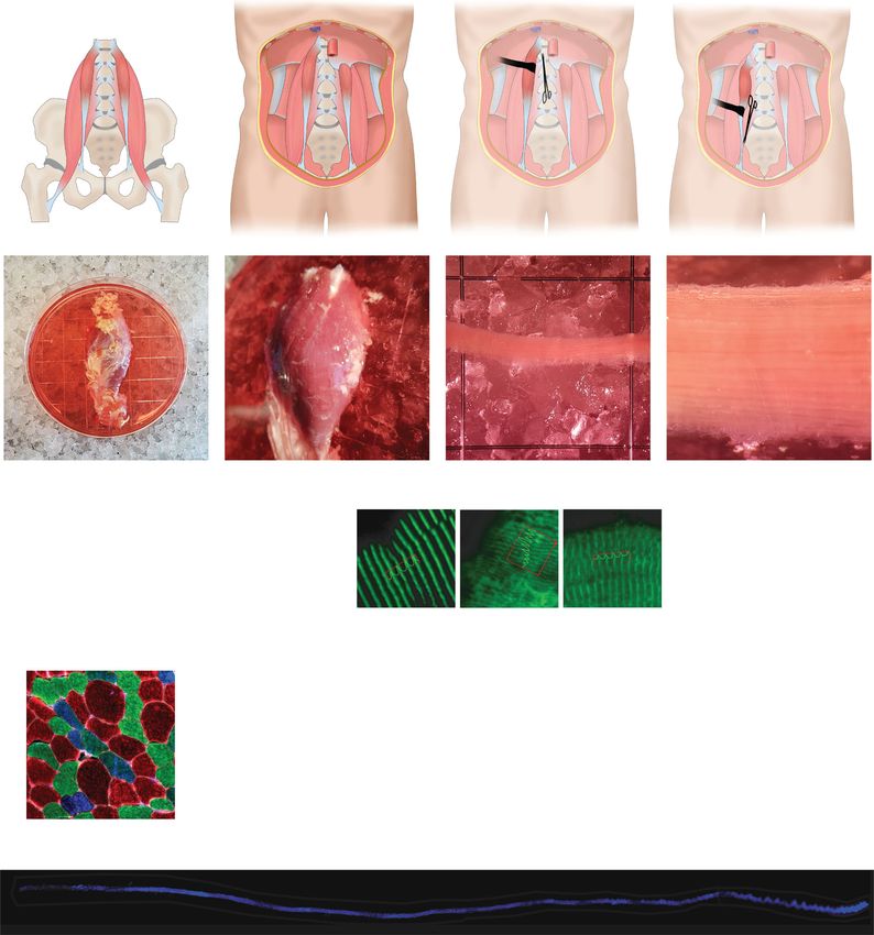
Human Psoas minor isolation freezing by nitrogen cooled isopentane. For bundle ana-
Immediately after the organ retrieval process was lysis, fixed cultured myofiber bundles are manually disso-
completed, the psoas minor muscle was exposed ciated to 5–15 myofiber bundles and segmented to
using Deaver retractors to visualize tendinous inser- length amendable to microscopic analysis and to improve
tions into the iliopectineal arch. A graphic overview antibody penetration.
of the procedure is provided in Fig. 1a. The distal as-
pect of the psoas tendon was cut, and the psoas dis- Immunostaining and antibodies
sected to its origin on the vertebral bodies and cut Myofibers and myofiber bundles are processed by identi-
immediately adjacent to the vertebra. It is critical to cal means. Fixed myofiber samples are washed in PBS
minimize tension on the Psoas minor during dissec- and permeabilized in 0.4% Triton-X100 for 1 h with
tion. The free Psoas minor was the immediately rocking. Samples are washed in PBS containing 125 mM
placed into sterile, cold transport media (DMEM, 110 glycine 3× in excess buffer until no appearance of deter-
mg/ml Pyruvate), and maintained on ice during gent remains. Samples are blocked using TrueBlack as
transport. per manufacturer’s instructions. Samples are then
blocked in blocking buffer containing 5% normal donkey
Human myofiber preparation serum for 3 h at room temperature to overnight at 4 °C
In a sterile environment, Psoas muscle is placed in an with rocking. Samples are washed in PBS and primary
appropriate vessel buffered with ice and containing antibodies are applied for 24 h at room temperature with
enough transport media to submerge. A photographic rocking. Samples are washed in excess PBS 5× 30 min
overview of the procedure is presented in Fig. 1a and each with rocking. Secondary antibodies are applied 24 h
S1A. Using a dissecting microscope and blunt micro at room temperature with rocking in the dark. Samples
scissors, adipose tissue, and free epimysium is dissected are washed in excess PBS for 30 min with rocking and
to visualize muscle fascicle tendon-tendon organization. DAPI is applied for 1 h with rocking. Samples are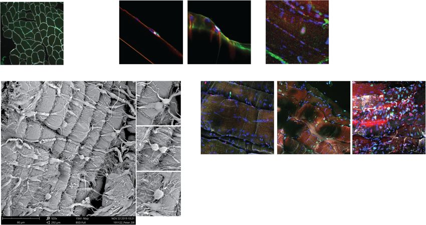
Feige et al. Skeletal Muscle                                                         (2021) 11:1                                                                                                                                                                                Page 4 of 14

  A
                                                 Psoas minor                                                                        Psoas minor
                                                 T12
                                                 L1

                                                 L2

                                                 L3

                                                 L4

                                                 L5

                                              Psoas major                                                                           Psoas major

  B

                                                                                                                                                            20mm                                     10mm                                                                        1mm

                                                                                                                                                                                                                  A-actinin spacing (nm)
                                                 Bundle diameter (mm)
         Fiber number / bundle

                                                                                                                                                                                                                                                                p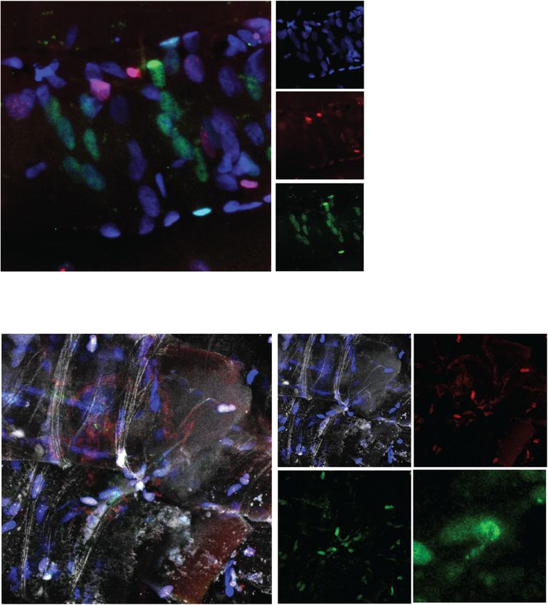
Feige et al. Skeletal Muscle   (2021) 11:1                                                                  Page 5 of 14

washed in excess PBS 5× 30 min each with rocking in        presented images represent maximum intensity projec-
the dark. Samples are then passed through a serial 20–     tions unless stated otherwise. Myofibers partially encom-
80% glycerol series and mounted onto slides in glycerol    passed in Z-stacks were excluded form analysis.
mounting media containing 0.1 M n-propyl gallate.
  Antibodies used in the study are as follows: Mouse       Quantification and statistical analysis
anti-Pax7 (1:2, DSHB, Cat. no. Ab528428; RRID: AB_         Compiled data are expressed as mean ± standard devi-
528428), rat anti-Laminin (1:1000, Sigma, Cat. no.         ation (SD) or mean ± standard error of the mean (SEM)
L0663; RRID: AB_477153), Mouse anti-Dystrophin (1:         as stated. Experiments were performed with a minimum
500, DSHB, Cat. no. MANEX1011B; RRID:AB_1157876),          of three biological replicates unless stated otherwise. For
Rabbit anti-p-EGFR (1:250, Cell Signaling technology,      statistical comparisons of two conditions, the Student’s t
Cat. no. 3777S; RRID: AB_2096270), Mouse anti-M-           test was used. Data is presented as paired (mean (control
Cadherin (1:500, BD Biosciences Cat. no. 611101, RRID:     vs treatment)) for direct comparison of biologically
AB_398414), Rabbit anti-MyoD (1:500, Abcam, Cat. no,       matched samples and unpaired (mean control vs mean
ab133627), Mouse anti-Syndecan-4 (1:500, Santa Cruz        treatment) where appropriate. Statistical testing was per-
Biotechnology Cat. no. sc-12766; RRID:AB_628314),          formed for each histogram where appropriate and was
Rabbit anti-Annexin-5 (1:500, Abcam, Cat. no. ab14196,     reported where significant. p values are provided for
RRID:AB_300979), Mouse anti-MyoG (1:500, Novus,            each statistical test performed. No data were removed as
Cat. no. MAB66861; RRID:AB_10973343), Mouse anti-          outliers. Experimental design incorporated user blinding
α-Actinin (1:1000, Sigma, Cat. no. A7732; RRID:AB_         when possible. Statistical analysis was performed in
2221571), Mouse anti-MHC (1:1000, DSHB, Cat. no. MF        GraphPad Prism or Microsoft Excel.
20; AB_2147781), Mouse anti-MyH2 (1:100, DSHB, Cat.
no. SC-71; AB_2147165), Mouse anti-MyH1 (1:100,            Key resources
DSHB, Cat. no. 6H1; AB_2314830), Mouse anti-MyH4           Key resources are listed in Table S2.
(1:100, DSHB, Cat. no. BF-F3; RRID: AB_2266724),
Mouse anti-MyH7 (1:100, DSHB, Cat. no. BA-F8; RRID:        Results
AB_10572253), Rat anti-Perlecan (1:500, NSU Biorea-        Human Psoas muscle is amenable for satellite cell culture
gents, Cat. no. V2600; RRID:AB_2119238), Rabbit anti-      in situ
Ki67 (1:1000, Abcam, Cat. no. ab15580; RRID:AB_            To evaluate our hypothesis that human myofibers could
443209), and Wheat Germ Agglutinin Alexa 488 conju-        be cultured in a laboratory setting, we decided to isolate
gate (1:1000, Fisher, Cat. no. W11261).                    primary human tissue from neurological determination
                                                           of death organ donors following ethics approval and in-
Scanning electron microscopy                               formed consent. We evaluated muscle groups for suit-
Samples were washed with water prior to dehydration in     ability in myofiber culture where surgical access to both
a 35–100% graded series of ethanol. Samples were crit-     tendinous insertions is feasible and where myofibers are
ical point dried in dry 100% ethanol using liquid CO2 as   short to facilitate manipulation in a laboratory setting
transition fluid. CO2 was exchanged at 5-min intervals     with common plasticware and reasonable reagent
for 8 rounds followed by a final 3 h release. Samples      volumes. We identified the small muscles of the hand,
were maintained under dust-free desiccation following      including the Abductor or Flexor Pollicis Brevis, the
critical point drying. Samples were mounted on             Flexor Digitorum Brevis of the foot or the Pronator
aluminum stages using double-sided carbon tape.            Quadratus of the forearm would be ideal candidates due
Mounted samples were sputter coated with gold for 1        to their size and accessibility; however, clinical availabil-
min (~ 10 nm) and stored under dust-free desiccation       ity of these muscles limited laboratory testing. We rea-
prior to imaging on a phenom Pro-X scanning electron       soned that an alternative strategy would involve isolating
microscope at an accelerating voltage of 15 kV.            muscle with large angles of pennation, where individual
                                                           myofibers are aligned at an angle oblique to the longitu-
Imaging and analysis                                       dinal angle of muscle contraction. These muscles pro-
Human Psoas myofiber bundles were analyzed on a            vide a mechanical advantage over shorter contraction
Leica TCS SP8 confocal microscope equipped with HC         distances and possess shorter myofiber fascicles attach-
PL APO 20× IMM CORR objective with HyD and PMT             ing to fibrous aponeuroses running along the periphery
detectors. Filters and detectors were set to maximal       of the muscle [47]. These muscle types would also allow
bandwidth and sensitivity to limit bleed through between   myofiber isolation from partial muscle dissections.
channels. Tile scans were stitched directly following         We identified the human Psoas muscle as a moder-
acquisition using the Leica LAS AF software. Manual        ately pennate muscle attached to the T12 vertebrae and
image analysis was performed using ImageJ and              the Lacunar ligament (Fig. 1a) with some variable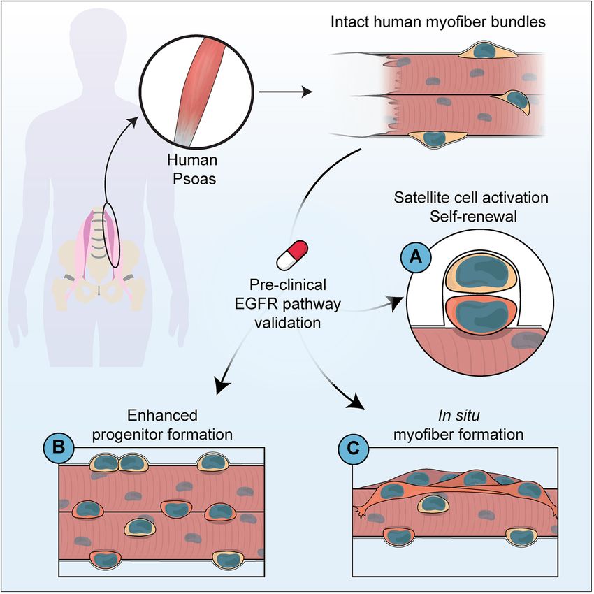
Feige et al. Skeletal Muscle   (2021) 11:1                                                                     Page 6 of 14

presence within populations [48, 49], as a good candi-        generally lack glycolytic myosin heavy chain type IIb
date to isolate myofibers. We obtained post-mortem            (MyHC IIb) muscle fibers in favor of type IIx [50].
muscle samples from neurologically determined                 Additionally, myofiber type has an impact on satellite
deceased organ donors (2♀, 1♂) with a mean age of 61          cell response to exercise, where satellite cells resident to
± 9.4 years (range 50–68) (Table S1). Donors did not          oxidative MyHC type I human muscles show augmented
have muscular dystrophy and did not exhibit muscle            expansion following aerobic training [51]. We hypothe-
cachexia. Muscle samples were 24.35 ± 13.56 g in mass         sized that myofiber type would influence satellite cell
and 11.42 ± 2.15 cm in length and isolated within 2–4 h       fate in human Psoas myofiber cultures and could con-
from cardiac death.                                           found translating signalling pathways identified in mice
   Mouse myofiber isolation requires enzymatic digestion      to human satellite cells.
of the extracellular components from isolated mouse              We evaluated myofiber type and histological profiles
Extensor digitorum longus muscle to release single myo-       of human Psoas myofiber bundles, mouse Extensor
fibers amenable for culture. To test if human Psoas myo-      digitorum longus (EDL) and mouse Psoas myofibers to
fibers could be prepared similarly, we subjected              better correlate difference in rodent and human satellite
myofiber isolated Psoas biopsies to collagenase digestion.    cell biology. Isolated human Psoas myofiber bundles
Isolated Psoas muscle was resistant to digestion using        were composed of mixed 36.5 ± 3.1% slow (type I) and
collagenase type 1, type 2, type 3, type 4, type 5, type 6,   63.5 ± 6% fast (type IIa, IIx) myofibers compared to 14.4
type 7, or elastase buffers due to the thick perimysium,      ± 2% type IIa and 62.5 ± 1.2% type IIb fast twitch mouse
endomysium, extracellular matrix (ECM), and vascular          Psoas or 11 ± 1.2% type IIa and 72.9 ± 0.7% type IIb
networks present (data not shown). Thus, the established      EDL muscles (Figure S1G). Human Psoas myofibers do
methods of myofiber isolation used in mouse studies are       not significantly differ in minimum fiber Feret (Fig. 1h)
not appropriate for human myofiber isolation.                 or myofiber surface area (Figure S1I) compared to
   Therefore, we manually dissected myofiber bundles          mouse Psoas or EDL fibers where a subset of human
from Psoas samples from tendon to tendon (Figure S1A).        Psoas myofibers are hypertrophic (Fig. 1I, S1J). Human
Manually dissected myofibril bundles contained 220 ± 63       myofibers however are roughly 10-fold longer than
myofibers (Fig. 1c) and were 1.9 ± 0.5 mm in diameter         mouse Psoas or EDL myofibers (Fig. 1j–k) (36.4 ± 0.4
(Fig. 1d). Myofiber bundles displayed heterogeneous via-      mm versus 3.34 ± 0.05 mm and 3.60 ± 0.12 mm). Taken
bility, where excess tension during surgical excision or      together, this data suggests that human Psoas myofibers
processing resulted in samples containing hyper               display distinct histological characteristics from mouse
contracted myofibers which were excluded from further         Psoas or EDL muscle likely due to requirements for hip
analysis (Figure S1A). Successful myofiber preparations       flexion and posture in an erect position.
contained intact myofibers where 83 ± 5% of myofibers
within a bundle did not exhibit any signs of hyper contrac-
tion or injury (Fig. 1e). Injured myofibers can be distin-    Human satellite cells expand in myofiber culture
guished into hypercontracted myofibers, moderately            Satellite cells remain mitotically quiescent but are
injured and minor injured subgroups. Hypercontracted          poised to activate and enter the cell cycle in response
myofibers (Figure S1B) show bisected myofiber segmenta-       to extrinsic cues such as exercise or trauma [2]. In ro-
tion within an extracellular matrix scaffold while myofi-     dents, this can be achieved by experimental models of
bers with moderate damage (Figure S1C) exhibit                injury, muscle digestion for stem cell isolation [52, 53]
widespread disorganization of sarcomeric banding and sig-     or in the case of myofiber preparation, digestion with
nificant autofluorescence. Myofibers with minor damage        collagenase and exposure to growth factors in cell cul-
(Figure S1D) exhibit focal autofluorescence and invagin-      ture [21]. To evaluate if human satellite cells from
ation of the extracellular matrix and maintain myofiber-      Psoas myofiber cultures spontaneously activate in vitro,
cell-ECM contact. Quantification of sarcomere spacing         we evaluated basal satellite cell numbers immediately
through α-Actinin staining (Fig. 1f, S1E-F) showed signifi-   following surgical excision and during culture where we
cantly shorter sarcomere banding in contracted myofibers,     analyzed an average 1.8 ± 1.1 mm length of myofibers
whereas healthy myofibers can be maintained in culture        per experiment (Figure S2A).
for at least 8 days without myofiber contraction or loss of     Human Psoas muscle has generally more satellite cells
sarcomere disorganization. Together, this data suggests       per mm2 (15.7 ± 3.0) compared to mouse Psoas (9.5 ±
that human Psoas myofiber bundles can be isolated and         0.5) or EDL myofiber cross-sections (10.0 ± 0.36) follow-
maintained in culture without consequence to myofiber         ing immunofluorescence staining for Pax7 (Fig. 2a). We
integrity.                                                    observed by immunofluorescence staining for Pax7 that
   Mouse and human myofibers exhibit different histo-         human Psoas myofibers with centrally located nuclei
logical characteristics, where human skeletal muscle          possess significantly increased (75.1%, 8.67 ± 1.6 versus
Feige et al. Skeletal Muscle (2021) 11:1 Page 7 of 14
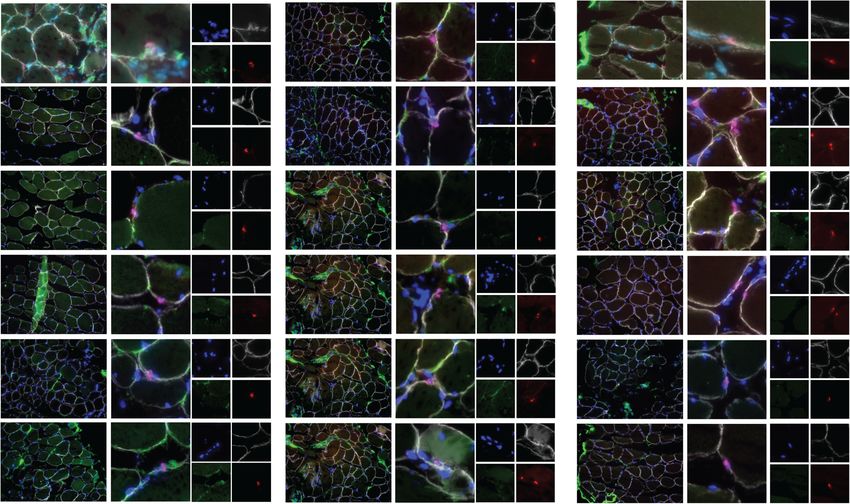
A B Mouse D0 Human D0 C Human D0 D
Human Psoas
% MCAD+ satellite cells
% SCD4+ satellite cells
DAPI LAM Dmd Pax7
30 100
DAPI PLC Pax7 LAM
100
DAPI SDC4 Pax7
Pax7 per mm2
80 80
60 60
15
40 40
20 20
0 0 0
DL S S
10um
d0 d0
10um 10um
10um
mESOASOA
E m hP
P
F Human D0 Human D4 Human D8
DAPI Ki67 Pax7 Dmd
10um 10um 10um
G H I J
Total Nuclei /mm fiber
150 40 50 p=0.02 100 pFeige et al. Skeletal Muscle (2021) 11:1 Page 8 of 14
EGF stimulation promotes myogenic progenitor asymmetric cell division to orient the satellite cell mi-
formation in human Psoas myofiber culture totic spindle in an apical-basal orientation [23]. To
To evaluate myogenic differentiation in Psoas myofiber evaluate if human Psoas satellite cells integrate EGFR
culture, we examined myofibers by immunofluorescence signalling, cultures were treated with EGF ligand
staining for Myogenin (MyoG), a transcription factor throughout culture to stimulate EGFR activation. Fol-
expressed at the onset of differentiation [54]. Culturing lowing EGFR stimulation, myofibers were stained for the
Psoas myofibers for 8 days results in significant presence presence of phosphorylated EGFR, where most satellite
of MyoG-expressing cells (20.8 ± 1.8 per millimeter cells express p-EGFR (Fig. 3d, Figure S3D), and its ex-
myofiber) concomitant with proliferation of satellite pression can become locally restricted as observed in ac-
cells, where we do not observe co-labelling of Pax7 and tivated mouse satellite cells [23]. This data suggests that
MyoG by immunofluorescence (Fig. 3a–c, S3A). the EGFR signalling pathway is activated following EGF
Additionally, by 8 days in culture, we can observe de treatment in human satellite cells in a manner analogous
novo myofiber formation occurring characterized by to mouse satellite cells.
multiple organized and aligned myocytes expressing To evaluate if EGFR augmented the production of
MyoG (Figure S3B-C) suggesting human Psoas myofiber myogenic progeny through the promotion of asymmetric
culture may represent a paradigm to model human satel- division as found previously in mouse muscle [23], we
lite cell activation, differentiation, and myofiber forma- examined fibers by immunofluorescence for Pax7,
tion. This data indicates that by 8 days in culture, MyoG, and Ki67. Quantifying total nuclei per millimeter
human Psoas satellite cells expand in number and of fiber show no significant change following 8 days of
express differentiation markers including MyoG. EGF treatment (Figure S3H); however, treatment with
We have previously established that mouse satellite EGF results in an appreciable increase in the number of
cells express EGFR that is polarized before an proliferating satellite cells per millimeter of myofiber
A Human myofiber Day 8 B C
40 40
p=0.008
per mm myofiber
per mm myofiber
DAPI MyoG Pax7
MyoG+ cells
30 30
Pax7+ cells
20 20
10 10
0 0
d0 d8 d0 d8
D E F G
50 8 50
Ki67- Pax7+ nuclei
Ki67+ Pax7+ Nuclei
DAPI pEGFR Pax7 Dmd
per mm myofiber
per mm fiber
40 40
per mm fiber
MyoG+ cells
6
30 30
4
20 20
10 2 10
0 0 0
d8 d8 d8 d8 d8 d8
EGF EGF EGF
Fig. 3 Human satellite cell expansion and differentiation can be tuned in situ. a Representative image (magnification from Figure S3A) of human
satellite cells and Myogenin (MyoG) expressing differentiating progenitors cultured on 8-day human Psoas minor myofiber cultures stained with
DAPI (blue), MyoG (green), and Pax7 (red). Quantification of b number of Pax7-expressing cells per millimeter of myofiber, c MyoG-expressing
cells per millimeter of myofiber. d Representative images of human satellite cells expressing phosphorylated active EGFR (p-EGFR) stained with
DAPI (blue), p-EGFR (green), Pax7 (red), and dystrophin (white). White arrow denotes localized p-EGFR expression. Quantification of e satellite cells
expressing Ki67 per millimeter of myofiber and f Ki67-negative satellite cells per millimeter of myofiber following EGF treatment or vehicle control
of human myofibers. g Quantification of number of MyoG-expressing cells per millimeter of myofiber following EGF treatment of human
myofibers. b, c, e–g Error bars represent means ± SD (EGF) and means ± SEM (control); b, c n = 3 biological replicates. d–g n = 3 biological
replicates control, n = 2 biological replicates EGFFeige et al. Skeletal Muscle (2021) 11:1 Page 9 of 14
(56.7% paired, 78% unpaired, n = 3 control, n = 2 EGF) examined myofibers by immunofluorescence staining
(Fig. 3e, S3F) and an increased proportion of satellite throughout culture. Interestingly, we observed rare apico-
cells per myofiber (29% increase unpaired, 15% increase basal and planar-oriented satellite cell doublets expressing
paired, n = 3 control, n = 2 EGF) (Figure S3G). Interest- Pax7 and residing in the niche from samples fixed at isola-
ingly, EGF treatment increased the number of Ki67- tion and stained with Pax7, Perlecan, and Dystrophin
negative satellite cells per millimeter of myofiber (161% (Fig. 4). As these cells are occupying the same niche space,
increase unpaired, 118% increase paired, n = 3 control, n we believe this is not due to random cell migration along
= 2 EGF) following 8 days in culture treated with EGF the myofiber, suggesting that homeostatic repair mecha-
or vehicle control (Fig. 3f, S3H). However, the propor- nisms may undergo either mode of division.
tion of Pax7/Ki67 expressing cells was similar following We additionally examined culture day 3 to day 4, a
EGF treatment (92% control vs 89% EGF) suggesting any time point reflecting the first division in human satellite
effect as a mitogen was negligible. Concomitant with an cells in culture. Strikingly, staining for the protein dys-
increase in proliferating satellite cells, EGF treatment re- trophin (DMD), which can be polarized in mouse satel-
sulted in an appreciable increase in MyoG-expressing lite cells to facilitate asymmetric division [40], shows
cells per millimeter of myofiber (69% increase unpaired, strong expression and polarity on the basal surface of a
78% increase paired n = 3 control, n = 2 EGF) (Fig. 3g, subset of cultured satellite cells (Fig. 5a), along with a
S3J). Therefore, we conclude that activation of EGFR subset of satellite cells expressing non-polar dystrophin
signalling in muscle stem cells promotes the generation (Fig. 5b). We observe asynchronous expression of DMD
of progenitors, likely by stimulating asymmetric divi- in satellite cells along myofibers, where cells in close
sions, similarly between human and mouse. proximity can express DMD perhaps due to region-
specific cues present within the myofiber microenviron-
Human satellite cells undergo apicobasal and planar cell ment. This suggests that human satellite cells can
divisions and express polarized dystrophin polarize dystrophin to their basal surface interfacing
To evaluate the possibility of human satellite cells under- with the extracellular matrix in an identical manner to
going apical-basal-oriented asymmetric division, we activated mouse satellite cells.
A Apicobasal B Planar
DAPI PLC Pax7 Dmd DAPI PLC Pax7 Dmd DAPI PLC Pax7 Dmd
Fig. 4 Human satellite cells can orient division angles. Representative images of a planar and b apicobasal-oriented human satellite cells in the
niche at isolation stained with DAPI (blue), Perlecan (green), Pax7 (red), and dystrophin (white). n = 3 biological replicatesFeige et al. Skeletal Muscle (2021) 11:1 Page 10 of 14 A Polar DAPI Dmd Pax7 DAPI Dmd Pax7 B Non-Polar DAPI Dmd Pax7 C Diffuse DAPI Dmd Pax7 Fig. 5 Human satellite cells express polarized dystrophin in culture. Representative image of human satellite cells cultured for four days and stained with DAPI (blue), Pax7 (red), and dystrophin (green) showing a polarized dystrophin localization, b non-polar dystrophin localization, and c diffuse dystrophin staining. n = 3 biological replicates Discussion essential to maintaining muscle repair over a lifetime. Evaluating human stem cell dynamics in a relevant con- Asymmetric stem cell division is one method to bal- text is critical to model biological phenomena and ance stem cell maintenance and the production of generalize results to benefit human health. New methods myogenic progeny, where following division one to improve the pre-clinical evaluation of therapeutic daughter cell maintains its stem cell state and one strategies to augment endogenous stem cell activity hold differentiates down the myogenic lineage. Asymmetric promise to improve regenerative medicine outcomes in cell division is established through cells integrating conditions such as Duchenne’s muscular dystrophy. extrinsic environmental cues to restrict cell fate deter- Here, we developed a novel system to evaluate human minants in a polarized manner such that when a cell satellite cell fate choices in a relevant context to interro- orients its mitotic centrosomes parallel with internally gate human satellite cell biology and evaluate pre- polarized cell fate determinants, daughter cells will re- clinical therapeutics in improving muscle regeneration ceive discrete cellular contents [2]. Typically, in (Fig. 6). mouse satellite cells, the daughter cell maintaining A balance in satellite cell proliferation, production niche interactions with the basal lamina maintains its of myogenic progeny, and return to quiescence is stem cell nature. Establishment of an apical-basal-
Feige et al. Skeletal Muscle    (2021) 11:1                                                                                         Page 11 of 14

 Fig. 6 Graphical outline of human Psoas culture. Schematic diagram summarizing the procedure for the characterization of human satellite cell
 dynamics on cultured intact myofibers from the human Psoas muscle

oriented mitotic spindle is in part facilitated by the                     studies exploring other resident cell types such as
PAR polarity complex, where we have previously                             fibroblast, pericyte, and fibroadipogenic precursor
shown EGFR is spatially restricted before mitotic divi-                    cells will better evaluate the specificity of EGF in cul-
sions to orient centrosomes through recruitment of                         ture. Additionally, studies exploring growth factor and
Aurora kinase A and spindle assembly [23]. We hy-                          oxygen penetration in myofiber bundles through
pothesized that as human Psoas myofibers maintain                          staining of growth factors and incubation in oxygen
myofiber and extracellular matrix composition that                         sensing compounds such as Hypoxy Probe labels
human satellite cells in culture could integrate three-                    would further evaluate the potential effect of nutrient
dimensional external cues to influence cell fate.                          availability on heterogeneity in satellite cell activity
  Our findings support that human Psoas myofiber                           within myofiber bundles. Studies exploring non-
cultures provide a new opportunity to culture human                        myogenic cell death occurring in culture day 4–8 may
satellite cells to explore fate choices during satellite                   provide insight into the cellular dynamics along a
cell activation and differentiation. In our system, myo-                   myofiber during early muscle repair.
fiber integrity is maintained (Fig. 1, S1) as well as cell                   The observation that EGF treatment increases the
polarity cues (Figs. 2b, e, 4, and 5) where treatment                      number of non-proliferative satellite cells following
with EGF results in augmented production of myo-                           treatment (Fig. 3f) suggests that the effect of EGF on
genic progeny (Fig. 3e–g). As EGF treatment also in-                       satellite cells may not be acting as a general mitogen.
creases the number of non-satellite cells expressing                       Taken with an increase in proliferating satellite cells
Ki67 (Figure S3I), it is possible that EGF influences                      (Fig. 3e) and increased formation of myogenic pro-
cell survival or is acting as a mitogen on the varied                      geny (Fig. 3g), supports the hypothesis that EGF
cell populations within myofiber cultures. Further                         treatment stimulates asymmetric division resulting in
Feige et al. Skeletal Muscle   (2021) 11:1                                                                                      Page 12 of 14

augmented production of non-cycling satellite stem                conserved mechanism first discovered in mouse. Future
cells (Pax7+Ki67−) and rapidly proliferating myogenic             studies with increased experimental size exploring the
satellite cells similar to that observed in mouse [23].           kinetics of cell cycle entry and quantification of division
Further studies are required to increase biological               angles following the first satellite cell division (likely days
sample sizes to fully delineate the role of EGF on hu-            2–3) will shed important insight into the extent of sym-
man satellite cells. Taken together, the human Psoas              metric and asymmetric satellite cell divisions occurring
muscle provides an exciting tool to explore in niche              in human muscle.
satellite cell biology and facilitate translation of pre-
clinical therapeutics from studies in model organisms             Conclusion
to humans.                                                        Human myofiber culture provides an exciting opportun-
   Previous studies have developed methods to assess              ity to evaluate pre-clinical drug efficacy in a relevant hu-
human satellite cell expansion in vitro from primary tis-         man context. This method provides an opportunity to
sue [55] through the culture of hypercontracted human             assay satellite cell heterogeneity, stem cell potential,
myofiber fragments from punch biopsies. Myofiber frag-            stem cell hierarchy, and activation kinetics with the po-
ments contained 1–8 bisected myofibers 2–3 mm in                  tential to therapeutically interrogate pathways of interest.
length and by 10 days in culture, 80% of nuclei were              Additionally, this method provides an additional tool to
Pax7+ with a significant amount of Desmin expressing              validate phenomena observed in other model organisms,
cells within the myofiber [55]. Transplantation of whole          validate lead compounds for drug discovery and testing,
myofiber fragments resulted in limited engraftment into           reduce animal model use, and accelerate the evaluation
mouse recipients. Differences in timepoints and muscle            of therapeutics improving human health. We envision
groups assessed between our study and others [55] limit           this technique will aid in generalizing pre-clinical strat-
comparisons; however, in our hands’ injury to the myofi-          egies into the clinical arena and may in the long term be
ber through hyper contraction or bisection results in dis-        appropriate as a personalized therapeutic tool.
organized myofiber sarcomeres (Fig. 1f) and altered
myofiber-ECM interactions (Figure S1B-C), where only
                                                                  Supplementary Information
minor focal damage is tolerated along myofibers to                The online version contains supplementary material available at https://doi.
maintain myofiber-cell-ECM interactions (Figure S1D).             org/10.1186/s13395-020-00256-z.
Typically, hypercontracted fibers are discarded in experi-
ments using mouse myofibers due to the abnormal                    Additional file 1: Supplemental figures related to figures 1-3, patient in-
                                                                   formation used in this study and key resource table. Figure S1: Myofi-
behavior of satellite cells [56]. Additionally, cell polarity      bers from human Psoas muscle can be maintained in situ, Related to Fig.
is maintained in Psoas myofiber culture (Figs. 3d and 5)           1. A) Photographic overview of human Psoas minor myofiber bundle iso-
and by 8 days ~ 30% of cells express Pax7 and ~ 20%                lation showing expanded images of intact myofiber bundles (panel 9)
                                                                   and hypercontracted myofiber bundles (panel 10). Representative images
represent committed progeny. Differences between the               of B) hypercontracted myofibers and C) myofibers with moderate dam-
models could be attributed to differential activation cues,        age stained for DAPI (Blue), α-Actinin (Green) and Myosin heavy chain
non-satellite cell survival, differential activation in differ-    (MF20, Red). D) Representative image of myofibers with minor damage
                                                                   stained for DAPI (Blue), Dystrophin (Green), Laminin (White) and IgG
ent muscle groups, or limitations in our study including           (Red). E) Representative images of single myofiber sarcomeres from intact,
exogenous culture of myofibers, modest sample size, or             contracted and cultured myofibers stained with α-actinin (Green) show-
differences in outbred human donors. This suggests                 ing representative histograms of staining intensity and sarcomere spa-
                                                                   cing. F) Representative image of disorganized sarcomeres from injured
human Psoas myofiber culture may reflect a model of                myofibers stained with α-Actinin (Green) and MF20 (Red). G) Representa-
homeostatic turnover or response to minor injuries such            tive images and quantification of myofiber type from mouse Extensor
as load-induced trauma or de-innervation, while human              digitorum longus and mouse Psoas muscle stained with Type 1 myofi-
                                                                   bers (Blue), Type 2a myofibers (Green), Type 2b myofibers (Red) and
myofiber fragments may represent a paradigm of rapid               Wheat germ agglutinin (White). H) Representative image of human Psoas
satellite cell activation in response to widespread myofi-         muscle cross sections stained with Laminin (Red) with I) quantification of
ber damage.                                                        average myofiber surface area and (J) myofiber surface area proportion
                                                                   from human Psoas myofibers compared to mouse Extensor digitorum
   Our findings provide proof-of-principle evidence to             longus and mouse psoas muscles using SMASH software. K) Representa-
support that in addition to the mature myofiber, human             tive image and quantification of mouse Extensor digitorum longus and
satellite cells express polarized dystrophin during satel-         mouse psoas myofiber lengths from isolated single myofibers. (K) Error
                                                                   bars represent mean ± SD, (G-J) Error bars represent mean ±SEM; (G, I-J)
lite cell activation and that human myofiber culture rep-          n = 3 biological replicates, (K) n = 40 myofibers per condition. Figure
resents a novel paradigm to explore human satellite cell           S2: Human satellite cells expand in situ, Related to Fig. 2. A) Quantifica-
self-renewal and myogenic differentiation. We further              tion of average length of myofiber analyzed per experiment, whiskers
                                                                   represent min and max. B) Representative image of human myofibers
validate that human satellite cells can undergo planar             showing centrally located nuclei stained with DAPI (Blue), Ki67 (Green),
and apicobasal-oriented divisions and demonstrate the              Pax7 (Red) and Dystrophin (White) and C) quantification of satellite cells
EGFR pathway to be of pre-clinical interest to augment             per mm myofiber present at isolation on centrally nucleated fibers (CNF).
                                                                   D) Representative image of myofibers stained with DAPI (Blue), SDC4
the production of myogenic progeny through a
Feige et al. Skeletal Muscle (2021) 11:1 Page 13 of 14
(Green) Pax7 (Red) and Annexin-5 (White) with E) bisected myofibers Ethics approval and consent to participate
serving as positive control stained for Annexin-5 (White) DAPI (Blue) and This study was approved by the Ottawa Hospital Research Ethics Board
Pax7 (Red). F) Quantification of satellite cells expressing SDC4 at day 8 in under the protocol number 20150544-01H.
culture. G) Representative image of satellite cells expressing M-Cadherin
after isolation stained for DAPI (Blue), MCAD (Green) and Pax7 (Red). H)
Consent for publication
Representative image of satellite cell expansion on myofibers following 8
Not applicable.
days in culture stained with DAPI (Blue), Ki67 (Green), Pax7 (Red) and Dys-
trophin (White) and quantification of I) Ki67 expression non-satellite cells
per mm of myofiber, J) number of KI67 negative satellite cells per mm of Competing interests
myofiber and K) Ki67 expressing satellite cells per mm of myofiber across M.A.R is CSO and Founder of Satellos Bioscience. P.F. and E.T. declare no
samples (s#). (A, C, K) Error bars represent mean ± SD, (F, I-K) Error bars competing interests.
represent mean ± SEM; (A) n = 351 myofibers. (C) n = averages from 20
(non-CNF) and 9 (CNF) myofibers. (F, I-K) n = 3 biological replicates. (K) n Author details
= averages from 4-22 myofibers, where individual data points represent 1
Sprott Center for Stem Cell Research, Regenerative Medicine Program,
individual myofibers. Figure S3: Myofiber culture unveils unique regen- Ottawa Hospital Research Institute, 501 Smyth Road, Ottawa, ON K1H 8L6,
erative phenomena, Related to Fig. 3. Representative images of A) Repre- Canada. 2Department of Cellular and Molecular Medicine, Faculty of
sentative image of cultured myofiber bundle stained for DAPI (Blue), Medicine, University of Ottawa, Ottawa, ON, Canada. 3Department of
MyoG (Green) and Pax7 (Red) (also presented in Figure 3A for reference). Medicine, Faculty of Medicine, University of Ottawa, Ottawa, ON, Canada.
B) Representative image of myogenic progenitors and C) in situ de novo 4
Department of Surgery, Division of Neurosurgery, Faculty of Medicine,
myofiber repair from fibers stained with DAPI (Blue), MyoG (Green) and University of Ottawa, Ottawa, ON, Canada. 5Ottawa Hospital Research
MyoD (Red) where white dotted arrows outline the myocyte alignment. Institute, Neuroscience Program, Ottawa, ON, Canada.
D) Representative images of cultured myofiber bundles stained for DAPI
(Blue), pEGFR (Green) and Pax7 (Red). Quantification of E) total nuclei per Received: 3 September 2020 Accepted: 6 December 2020
mm of myofiber and across samples. F) Quantification of human satellite
cells expressing Ki67 or Ki67 negative per mm of fiber across samples fol-
lowing culture in control or EGF containing media. G) Quantification pro-
portion of nuclei expressing pax7 per myofiber. Quantification of H) References
proportion of satellite cells (Pax7+) stained negative for Ki67 and I) pro- 1. Bentzinger CF, Wang YX, Dumont NA, Rudnicki MA. Cellular dynamics in the
portion of non-satellite cells (Pax7-) expressing Ki67 following culture in muscle satellite cell niche. EMBO Rep. 2013;14(12):1062–72.
control or EGF containing media. J) quantification of MyoG-expressing 2. Feige P, Brun CE, Ritso M, Rudnicki MA. Orienting muscle stem cells for
nuclei per mm of myofiber across samples (s#). (E,F, J) Error bars represent regeneration in homeostasis, aging, and disease. Cell Stem Cell. 2018;23(5):
mean ±SD, (E,G-I) Error bars represent means ± SD (EGF) and means ± 653–64.
SEM (Control); (E-I) n= 2 biological replicates EGF, 3 biological replicates 3. Carlson ME, Conboy IM. Loss of stem cell regenerative capacity within aged
control, (E, G, J) n = 4-32 myofibers, where individual data points repre- niches. Aging Cell. 2007;6(3):371–82.
sent individual myofibers. Table S1: Patient information used in this 4. Shefer G, Van de Mark DP, Richardson JB, Yablonka-Reuveni Z. Satellite-cell
study. Patient information including sex, age, clinical complication, Psoas pool size does matter: defining the myogenic potency of aging skeletal
muscle mass, length and prefusion solution used during isolation. Table muscle. Dev Biol. 2006;294(1):50–66.
S2: Key resource Table. 5. Conboy IM, Conboy MJ, Smythe GM, Rando TA. Notch-mediated restoration
of regenerative potential to aged muscle. Science. 2003;302(5650):1575–7.
6. Conboy IM, Rando TA. The regulation of Notch signaling controls satellite
Abbreviations cell activation and cell fate determination in postnatal myogenesis. Dev
DMD: Duchenne muscular dystrophy; ECM: Extracellular matrix; EDL: Extensor Cell. 2002;3(3):397–409.
digitorum longus; EGF: Epidermal growth factor; EGFR: Epidermal growth 7. Brack AS, Conboy MJ, Roy S, Lee M, Kuo CJ, Keller C, et al. Increased Wnt
factor receptor; FDA: Food and Drug Administration (USA); MyHC: Myosin Signaling During Aging Alters Muscle Stem Cell Fate and Increases Fibrosis.
heavy chain; MyoG: Myogenin Science. 2007;317(5839):807–10.
8. Brack AS, Rando TA. Intrinsic changes and extrinsic influences of myogenic
stem cell function during aging. Stem Cell Rev. 2007;3(3):226–37.
Acknowledgements 9. Bernet JD, Doles JD, Hall JK, Kelly-Tanaka K, Carter TA, Olwin BB. P38 MAPK
We would like to thank the patients and their families for the generous gift signaling underlies a cell autonomous loss of stem cell self-renewal in aged
of the tissues that were used in this study. skeletal muscle. Nat Med. 2014;20(3):265–71.
10. Price FD, von Maltzahn J, Bentzinger CF, Dumont NA, Yin H, Chang NC,
et al. Inhibition of JAK/STAT signaling stimulates adult satellite cell function.
Authors’ contributions
Nat Med. 2014;20(10):1174–81.
Conceptualization: P.F., E.T., and M.A.R.; methodology: P.F. and E.T.;
11. Chakkalakal JV, Jones KM, Basson MA, Brack AS. The aged niche disrupts
investigation: P.F., E.T., and M.A.R.; writing—original draft: P.F. and M.A.R.;
muscle stem cell quiescence. Nature. 2012;490(7420):355–60.
writing—review and editing: P.F., E.T., and M.A.R.; funding acquisition: E.T.
12. Rozo M, Li L, Fan C-M. Targeting β1-integrin signaling enhances
and M.A.R.; resources: E.T. and M.A.R.; supervision: E.T. and M.A.R. The
regeneration in aged and dystrophic muscle in mice. Nat Med. 2016;22(8):
author(s) read and approved the final manuscript.
889–96.
13. Cosgrove BD, Gilbert PM, Porpiglia E, Mourkioti F, Lee SP, Corbel SY, et al.
Funding Rejuvenation of the aged muscle stem cell population restores strength to
P.F. is supported by a fellowship from CIHR. M.A.R. holds the Canada injured aged muscles. Nat Med. 2014;20(3):255–64.
Research Chair in Molecular Genetics. These studies were carried out with 14. Hay M, Thomas DW, Craighead JL, Economides C, Rosenthal J. Clinical
grant support to M.A.R. from the US National Institutes for Health development success rates for investigational drugs. Nat Biotechnol. 2014;
(R01AR044031), the Canadian Institutes for Health Research (FDN-148387), E- 32(1):40–51.
Rare-2: Canadian Institutes of Health Research/Muscular Dystrophy Canada, 15. Van Norman GA. Phase II trials in drug development and adaptive trial
and the Stem Cell Network. design. JACC Basic Transl Sci. 2019;4(3):428–37.
16. van der Worp HB, Howells DW, Sena ES, Porritt MJ, Rewell S, O’Collins V,
et al. Can animal models of disease reliably inform human studies? PLoS
Availability of data and materials Med. 2010;7(3).
The datasets from the current study are available from the corresponding 17. Hackam DG, Redelmeier DA. Translation of research evidence from animals
author on request. to humans. JAMA. 2006;296(14):1727–32.Feige et al. Skeletal Muscle (2021) 11:1 Page 14 of 14
18. Pasut A, Oleynik P, Rudnicki MA. Isolation of muscle stem cells by 42. Gurevich DB, Nguyen PD, Siegel AL, Ehrlich OV, Sonntag C, Phan JMN, et al.
fluorescence activated cell sorting cytometry. In: DiMario JX, editor. Asymmetric division of clonal muscle stem cells coordinates muscle
Myogenesis: Methods and Protocols. Totowa: Humana Press; 2012. p. 53–64. regeneration in vivo. Science. 2016;353(6295).
19. Collins CA, Olsen I, Zammit PS, Heslop L, Petrie A, Partridge TA, et al. Stem 43. Gissel H. The role of Ca2+ in muscle cell damage. Ann N Y Acad Sci. 2006;
cell function, self-renewal, and behavioral heterogeneity of cells from the 1066(1):166–80.
adult muscle satellite cell niche. Cell. 2005;122(2):289–301. 44. Tedesco FS, Moyle LA, Perdiguero E. Muscle interstitial cells: a brief field
20. Feige P, Rudnicki MA. Isolation of satellite cells and transplantation into guide to non-satellite cell populations in skeletal muscle. In: Perdiguero E,
mice for lineage tracing in muscle. Nat Protoc. 2020;15(3):1082–97. Cornelison D, editors. Muscle Stem Cells: Methods and Protocols. New York:
21. Shefer G, Yablonka-Reuveni Z. Isolation and culture of skeletal muscle Springer; 2017. p. 129–47.
myofibers as a means to analyze satellite cells. In: Helgason CD, Miller 45. Giordani L, He GJ, Negroni E, Sakai H, Law JYC, Siu MM, et al. High-
CL, editors. Basic Cell Culture Protocols. Totowa: Humana Press; 2005. p. dimensional single-cell cartography reveals novel skeletal muscle-resident
281–304. cell populations. Mol Cell. 2019;74(3):609–621.e6.
22. Kuang S, Le Grand F, Rudnicki MA. Asymmetric self-renewal and 46. Smith LR, Barton ER. SMASH - semi-automatic muscle analysis using
commitment of satellite stem cells in muscle. Cell. 2007;129(5):999–1010. segmentation of histology: a MATLAB application. Skelet Muscle. 2014;4:21.
23. Wang YX, Feige P, Brun CE, Hekmatnejad B, Dumont NA, Renaud J-M, et al. 47. Aagaard P, Andersen JL, Dyhre-Poulsen P, Leffers A-M, Wagner A,
EGFR-Aurka signaling rescues polarity and regeneration defects in Magnusson SP, et al. A mechanism for increased contractile strength of
dystrophin-deficient muscle stem cells by increasing asymmetric divisions. human pennate muscle in response to strength training: changes in muscle
Cell Stem Cell. 2019;24(3):419–432.e6. architecture. J Physiol. 2001;534(Pt 2):613–23.
24. Yin H, Pasut A, Soleimani VD, Bentzinger CF, Antoun G, Thorn S, et al. 48. Dragieva P, Zaharieva M, Kozhuharov Y, Markov K, Stoyanov GS. Psoas minor
MicroRNA-133 controls brown adipose determination in skeletal muscle muscle: a cadaveric morphometric study. Cureus. 2018;10(4).
satellite cells by targeting Prdm16. Cell Metab. 2013;17(2):210–24. 49. Hanson P, Magnusson SP, Sorensen H, Simonsen EB. Anatomical differences
25. Seale P, Bjork B, Yang W, Kajimura S, Chin S, Kuang S, et al. PRDM16 controls in the psoas muscles in young black and white men. J Anat. 1999;194(Pt 2):
a brown fat/skeletal muscle switch. Nature. 2008;454(7207):961–7. 303–7.
26. Seale P, Sabourin LA, Girgis-Gabardo A, Mansouri A, Gruss P, Rudnicki MA. 50. Harrison BC, Allen DL, Leinwand LA. IIb or not IIb? Regulation of myosin
Pax7 Is Required for the Specification of Myogenic Satellite Cells. Cell. 2000; heavy chain gene expression in mice and men. Skelet Muscle. 2011;1:5.
102(6):777–86. 51. Fry CS, Noehren B, Mula J, Ubele MF, Westgate PM, Kern PA, et al. Fibre
27. Bentzinger CF, von Maltzahn J, Dumont NA, Stark DA, Wang YX, Nhan K, type-specific satellite cell response to aerobic training in sedentary adults. J
et al. Wnt7a stimulates myogenic stem cell motility and engraftment Physiol. 2014;592(Pt 12):2625–35.
resulting in improved muscle strength. J Cell Biol. 2014;205(1):97–111. 52. Machado L, Esteves de Lima J, Fabre O, Proux C, Legendre R, Szegedi A,
28. Le Grand F, Jones AE, Seale V, Scimè A, Rudnicki MA. Wnt7a activates the et al. In situ fixation redefines quiescence and early activation of skeletal
planar cell polarity pathway to drive the symmetric expansion of satellite muscle stem cells. Cell Rep. 2017;21(7):1982–93.
stem cells. Cell Stem Cell. 2009;4(6):535–47. 53. van Velthoven CTJ, de Morree A, Egner IM, Brett JO, Rando TA.
29. Judson RN, Quarta M, Oudhoff MJ, Soliman H, Yi L, Chang CK, et al. Transcriptional profiling of quiescent muscle stem cells in vivo. Cell Rep.
Inhibition of methyltransferase Setd7 allows the in vitro expansion of 2017;21(7):1994–2004.
myogenic stem cells with improved therapeutic potential. Cell Stem Cell. 54. Bentzinger CF, Wang YX, Rudnicki MA. Building muscle: molecular
2018;22(2):177–190.e7. regulation of myogenesis. Cold Spring Harb Perspect Biol. 2012;4(2):a008342.
30. von Maltzahn J, Renaud J-M, Parise G, Rudnicki MA. Wnt7a treatment 55. Marg A, Escobar H, Gloy S, Kufeld M, Zacher J, Spuler A, et al. Human
ameliorates muscular dystrophy. Proc Natl Acad Sci. 2012;109(50):20614–9. satellite cells have regenerative capacity and are genetically manipulable. J
31. Kasper AM, Turner DC, Martin NRW, Sharples AP. Mimicking exercise in Clin Invest. 2014;124(10):4257–65.
three-dimensional bioengineered skeletal muscle to investigate cellular and 56. Pasut A, Jones AE, Rudnicki MA. Isolation and culture of individual myofibers
molecular mechanisms of physiological adaptation. J Cell Physiol. 2018; and their satellite cells from adult skeletal muscle. J Vis Exp JoVE. 2013;73:
233(3):1985–98. e50074.
32. Afshar Bakooshli M, Lippmann ES, Mulcahy B, Iyer N, Nguyen CT, Tung K,
et al. A 3D culture model of innervated human skeletal muscle enables Publisher’s Note
studies of the adult neuromuscular junction. eLife. 8. Springer Nature remains neutral with regard to jurisdictional claims in
33. Li EW, McKee-Muir OC, Gilbert PM. Chapter five - cellular biomechanics in published maps and institutional affiliations.
skeletal muscle regeneration. In: Sassoon D, editor. Current Topics in
Developmental Biology: Academic Press; 2018. p. 125–76.
34. Latil M, Rocheteau P, Châtre L, Sanulli S, Mémet S, Ricchetti M, et al. Skeletal
muscle stem cells adopt a dormant cell state post mortem and retain
regenerative capacity. Nat Commun. 2012;3(1):903.
35. Yucel N, Chang AC, Day JW, Rosenthal N, Blau HM. Humanizing the mdx
mouse model of DMD: the long and the short of it. NPJ Regen Med. 2018;3.
36. Shelton M, Kocharyan A, Liu J, Skerjanc IS, Stanford WL. Robust generation
and expansion of skeletal muscle progenitors and myocytes from human
pluripotent stem cells. Methods. 2016;101:73–84.
37. Piga D, Salani S, Magri F, Brusa R, Mauri E, Comi GP, et al. Human induced
pluripotent stem cell models for the study and treatment of Duchenne and
Becker muscular dystrophies. Ther Adv Neurol Disord. 2019;12.
38. Gilbert PM, Havenstrite KL, Magnusson KEG, Sacco A, Leonardi NA, Kraft P,
et al. Substrate Elasticity Regulates Skeletal Muscle Stem Cell Self-Renewal in
Culture. Science. 2010;329(5995):1078–81.
39. Petrof BJ, Shrager JB, Stedman HH, Kelly AM, Sweeney HL. Dystrophin
protects the sarcolemma from stresses developed during muscle
contraction. Proc Natl Acad Sci. 1993;90(8):3710–4.
40. Dumont NA, Wang YX, von Maltzahn J, Pasut A, Bentzinger CF, Brun CE,
et al. Dystrophin expression in muscle stem cells regulates their polarity and
asymmetric division. Nat Med. 2015;21(12):1455–63.
41. Alexander MS, Rozkalne A, Colletta A, Spinazzola JM, Johnson S, Rahimov F,
et al. CD82 is a marker for prospective isolation of human muscle satellite
cells and is linked to muscular dystrophies. Cell Stem Cell. 2016;19(6):800–7.You can also read