Inner ear hair cells deteriorate in mice engineered to have no or diminished innervation
←
→
Page content transcription
If your browser does not render page correctly, please read the page content below
ORIGINAL RESEARCH
                                                                                                                                      published: 18 March 2015
                                                                                                                                 doi: 10.3389/fnagi.2015.00033

                                            Inner ear hair cells deteriorate in
                                            mice engineered to have no or
                                            diminished innervation
                                            Jennifer Kersigo and Bernd Fritzsch *

                                            Department of Biology, University of Iowa, Iowa City, IA, USA

                                            The innervation of the inner ear critically depends on the two neurotrophins Ntf3 and
                                            Bdnf. In contrast to this molecularly well-established dependency, evidence regarding
                                            the need of innervation for long-term maintenance of inner ear hair cells is inconclusive,
                                            due to experimental variability. Mutant mice that lack both neurotrophins could shed
                                            light on the long-term consequences of innervation loss on hair cells without introducing
                                            experimental variability, but do not survive after birth. Mutant mice with conditional
                                            deletion of both neurotrophins lose almost all innervation by postnatal day 10 and show
                                            an initially normal development of hair cells by this stage. No innervation remains after
                                            3 weeks and complete loss of all innervation results in near complete loss of outer and
                                            many inner hair cells of the organ of Corti within 4 months. Mutants that retain one allele
                                            of either neurotrophin have only partial loss of innervation of the organ of Corti and show
                                            a longer viability of cochlear hair cells with more profound loss of inner hair cells. By 10
                          Edited by:
                    Marta Magarinos,
                                            months, hair cells disappear with a base to apex progression, proportional to the residual
    Universidad Autonoma de Madrid,         density of innervation and similar to carboplatin ototoxicity. Similar to reports of hair cell
                               Spain        loss after aminoglycoside treatment, blobbing of stereocilia of apparently dying hair cells
                        Reviewed by:        protrude into the cochlear duct. Denervation of vestibular sensory epithelia for several
                   Fernando Giraldez,
     Universitat Pompeu Fabra, Spain        months also resulted in variable results, ranging from unusual hair cells resembling the
           Ricardo Romero-Guevara,          aberrations found in the organ of Corti, to near normal hair cells in the canal cristae. Fusion
           University of Florence, Italy
                                            and/or resorption of stereocilia and loss of hair cells follows a pattern reminiscent of Myo6
                   *Correspondence:
                          Bernd Fritzsch,
                                            and Cdc42 null mice. Our data support a role of innervation for long-term maintenance
     Department of Biology, College of      but with a remarkable local variation that needs to be taken into account when attempting
  Liberal Arts and Sciences, University     regeneration of the organ of Corti.
         of Iowa, 143 Biology Building,
     129 E Jefferson Street, Iowa City,     Keywords: inner ear, hair cells, degeneration, innervation, neurotrophins, conditional deletion
                  IA 52242-1324, USA
              bernd-fritzsch@uiowa.edu

                                            Introduction
          Received: 19 January 2015
         Accepted: 28 February 2015         It is estimated that over 900 million people worldwide will have at least a 25 dB reduction in hear-
          Published: 18 March 2015
                                            ing sensitivity by 2025 [http://hearinghealthmatters.org/hearinginternational/2011/incidence-of-
                              Citation:     hearing-loss-around-the-world/]. Even mild hearing loss (26–40 dB HL) may deprive people from
 Kersigo J and Fritzsch B (2015) Inner
                                            their accustomed way of communication (Yamasoba et al., 2013), promote cognitive decline (Lin
      ear hair cells deteriorate in mice
 engineered to have no or diminished
                                            et al., 2013), and possibly increase the risk for developing dementia, including Alzheimer’s dis-
                            innervation.    ease (Lin and Albert, 2014). Clinically, hearing loss is multifactorial in its etiology, having both
          Front. Aging Neurosci. 7:33.      genetic and environmental (noise exposure, ototoxic drugs, neurotoxic drugs, etc.) components
       doi: 10.3389/fnagi.2015.00033        (Kopecky and Fritzsch, 2011; Makary et al., 2011; Huisman and Rivolta, 2012; Rivolta, 2013).

Frontiers in Aging Neuroscience | www.frontiersin.org                              1                                          March 2015 | Volume 7 | Article 33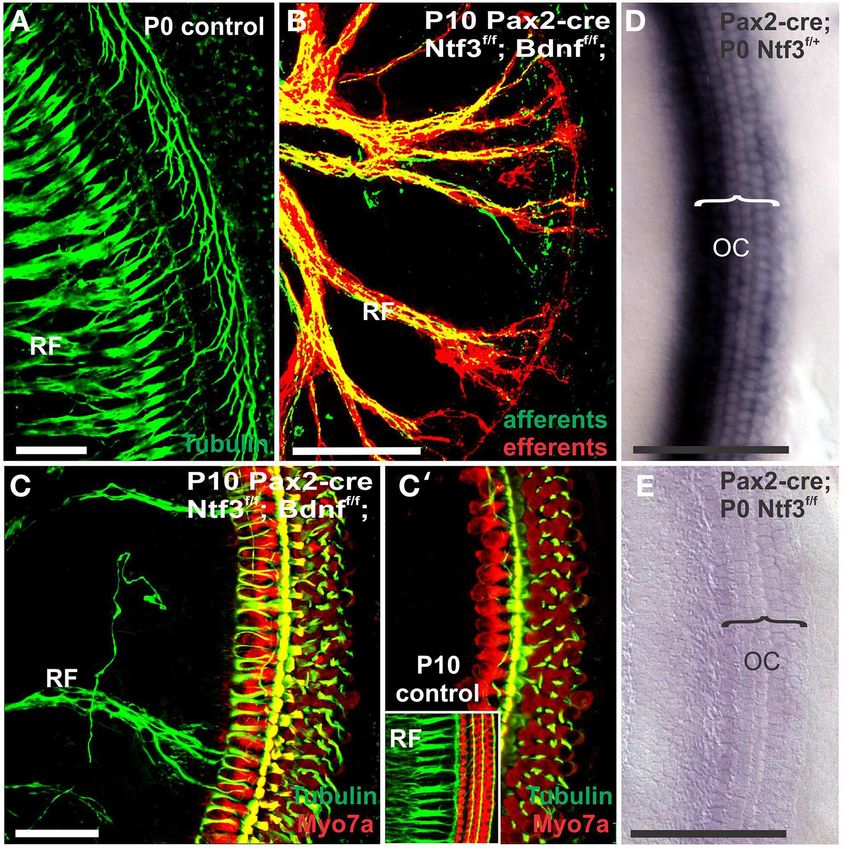
Kersigo and Fritzsch                                                                                                 Hair cells die without innervation

Sensorineural hearing loss is a common type of age-related hear-            is changed by mutating Sox2 (Okubo et al., 2006, 2009). These
ing loss (AHL) and mainly results from loss of cochlear hair cells          data suggest that initial formation of taste sensory cells occurs
(HCs) and/or spiral ganglion neurons (SGNs). Potentially more               autonomously, much like hair cells in the ear (Ma et al., 2000)
devastating is a decline in vestibular function which typically             but innervation is needed to maintain sensory cells. Similar to
starts about a decade after the onset of hearing loss (Rauch, 2001).        taste buds, electroreceptive sensory cells and organs depend on
Vestibular dysfunction can increase the risk of falling, resulting in       innervation for maintenance. Hair cells die within hours after
fractures and subsequent morbidity of the elderly. Recently the             severing the nerve and organs disappear rapidly through cell
hallmarks of aging were reviewed and cell communication was                 death after nerve fibers are cut and reappear rapidly upon rein-
found to be a major aspect that defines which cell will survive             nervation (Fritzsch et al., 1990). Transplantation studies have
and which will die (López-Otín et al., 2013), possibly underlying           shown that the initial development of electroreceptive organs
the diversity of cellular reactions that has been stressed in recent        may be autonomous (Northcutt et al., 1995), again suggesting
papers studying hair cell loss after ototoxic treatments (Taylor            that afferents maintain but do not induce electroreceptive sen-
et al., 2012). Here we evaluate the historically controversial influ-       sory cells. Neither taste buds nor electroreceptors have an efferent
ence of innervation on hair cell viability in the ear using newly           innervation, clearly indicating the role of afferents for mainte-
developed models of targeted deletion of neurotrophins.                     nance. Electroreceptive sensory cells are closely related to the
    Human temporal bone studies of the cochlea suggest that neu-            mechanosensory HCs of the lateral line, neuromasts (Duncan
ronal and HC loss are unrelated as over 90% of neuronal loss                and Fritzsch, 2012), which also seem to depend on innervation
can occur without loss of HCs (Otte et al., 1978; Makary et al.,            for long term viability. In bony fish and amphibians, the hair cells
2011). Animal studies also suggest that innervation and HC loss             of neuromasts are lost after months of denervation (Jones and
can happen independently of each other (Perez and Bao, 2011;                Singer, 1969). These data suggest that possibly all placode derived
Kidd and Bao, 2012) except for inconclusive data that report the            sensory cells can differentiate autonomously but require innerva-
loss of some HCs after afferent innervation to the inner ear was            tion for viability. Among placode derived sensory cells, inner ear
cut (Sugawara et al., 2005). In the vestibular system of humans,            hair cells appear to be unique: like other placode derived sensory
there appears to be a somewhat matching decline of both vestibu-            cells they have autonomous development in the absence of inner-
lar ganglion neurons (VGNs) and HCs over time (Rauch, 2001).                vation but may not depend on afferent innervation for long term
Earlier claims in studies involving adult guinea pigs show com-             viability.
plete loss of HCs in vestibular sensory epithelium 4 months after               A new approach using a transgenic mutation resulting in the
vestibular nerve transection (Favre and Sans, 1991) have not been           targeted deletion of neurotrophins to test the potential influ-
confirmed in other investigations in humans (Suzukawa et al.,               ence of afferents and efferents on HC viability, without com-
2005) leaving the loss of vestibular hair cells after loss of inner-        promising blood supply, could clarify this issue. Previous work
vation open to interpretation. To date, a large portion of the              has shown that mice lacking neurotrophins or their receptors
studies addressing the dependency of inner ear hair cell survival           are born with little to no innervation (Ernfors et al., 1995;
on innvervation utilize surgical techniques with potential flaws:           Silos-Santiago et al., 1997; Fritzsch et al., 2004) but these mice
either incomplete surgical denervation or inadvertent disruption            die soon after birth. Mice with ear-specific conditional dele-
of blood supplies may affect data (Sugawara et al., 2005). Despite          tions of the two relevant neurotrophins in the ear (Pax2-cre;
over 50 years of work on this subject, it is fair to say that no            Ntf3f /f , Bdnf f /f ) are viable for several months but are deaf and
unequivocal answer has been reached largely due to technical                show vestibular and cerebellar motor control defects. We raised
limitations in all but one study that shows complete loss of all            these mice for up to 10 months and investigated the pattern
vestibular hair cells after surgical denervation (Favre and Sans,           of remaining HCs and innervation using Myo7a, tubulin, and
1991).                                                                      neurofilament (NF) immunohistochemistry, myelinated nerve
    Of note, in contrast to these disputed effects related to sur-          fiber staining, and SEM. These mice show an age-dependent,
gical removal of innervation on adult hair cells, data on mutant            progressive loss of HCs that correlates with the reduction of
mice that lack all innervation to the ear by various mutations have         innervation in the cochlea and vestibular organs and sug-
established that absence of innervation has no short-term effect            gests a yet to be determined, variable threshold of innerva-
on HC development (Fritzsch et al., 1997a; Ma et al., 2000; Yang            tion for different organs and different hair cells within a given
et al., 2011). In fact, removing neurotrophin receptors eliminates          organ.
innervation without affecting hair cell development (Fritzsch
et al., 1997a). This absence of any apparent effect of denervation          Material and Methods
contrasts with all other sensory cells, which seemingly depend
on innervation either for complete differentiation or viability             Mouse Breeding and Collection
(Fritzsch et al., 1998). For example, severing gustatory nerves             Pax2-cre mice (Ohyama and Groves, 2004) were crossed with
results in rapid loss of taste bud sensory cells, which can reappear        floxed Ntf3 (Bates et al., 1999) (aka NT3) and floxed Bdnf mice
after nerve fibers grow back into the skin (Farbman, 2003; Fei              (Gorski et al., 2003) to generate conditional, ear-specific and
et al., 2014). However, some embryonic differentiation of taste             viable mutants that lack neurotrophin expression in the ear.
sensory cells can occur in the absence of innervation (Fritzsch             Breeding pairs consist of mice carrying the Pax2-cre together with
et al., 1997b; Ito et al., 2010) and gustatory nerve fibers cannot          heterozygosity of the floxed neurotrophins (Pax2-cre; Ntf3f /+ ;
induce taste buds if the molecular competence of the epidermis              Bdnf f /+ ). These mice were crossed with mice homozygotic for

Frontiers in Aging Neuroscience | www.frontiersin.org                   2                                          March 2015 | Volume 7 | Article 33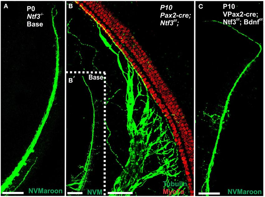
Kersigo and Fritzsch                                                                                                     Hair cells die without innervation

floxed alleles of both neurotrophins (Ntf3 f /f ; Bdnf f /f ). 1 in 8           stained with Hoechst stain according to existing protocols (Jahan
mice were doubly homozygotic for both floxed genes and also                     et al., 2013).
expressed cre. Combinations of cre with heterozygotic floxed
Bdnf and homozygosity of Ntf3 (Pax2-cre; Ntf3f /f ; Bdnf f /+ )                 Lipophilic Dye Tracing
or homozygosity for floxed Bdnf and heterozygosity for Ntf3                     Small pieces of dye-soaked filter paper (Fritzsch et al., 2005a;
(Pax2-cre; Ntf3f /+ ; Bdnf f /f ) are also included here. These mice            Tonniges et al., 2010) were implanted into the cochlear nuclei
lose much of their innervation of the organ of Corti and in
case of loss of all Bdnf, also all innervation to canal cristae                             ™
                                                                                (NeuroVue Maroon) or the efferent fiber tract (NeuroVue
                                                                                Red) in animals fixed in 4% PFA. Fixed tissue was kept at 60◦ C
                                                                                                                                                        ™
(Fritzsch et al., 2004). Because of the further delay in inner-                 for ∼48–96 h to allow for dye diffusion from the hindbrain to the
vation loss in the apical half of the cochlea, we concentrated                  ear. Ears were micro-dissected, split into a basal and apical turn
on the basal turn for this presentation except where stated                     and mounted on a slide in glycerol. To avoid diffusion of dye out
differently.                                                                    of the lipid bilayer into the glycerol used as mounting medium,
    Mice were genotyped within 3 days after birth. Non-desired                  images were taken immediately with a Leica TCS SP5 confocal
littermates were eliminated to increase the viability of vestibu-               microscope.
lar defected mutant mice (due to loss of Bdnf). Mice were raised
to the designated age of 1, 2, 4, 7–10 months and were sacri-
                                                                                Imaging
ficed. Six mutant animals were collected per the three genotypes
                                                                                Immunostained cochlea halves were mounted flat on a slide using
whenever possible together with age-matched control littermates
                                                                                glycerol, coverslipped and imaged using a Leica SP5 confocal
at the designated age to minimize genetic background effects.
                                                                                microscope. Data sets were generated by collecting stacks at 3–
Data were pooled across two or more litters to eliminate any
                                                                                6 µm steps (depending on the magnification) in 100–200 µm
genetic background bias. Given that we had to cross three dis-
                                                                                long segments at three different positions: the basal hook region,
tinct mutant lines carrying the Pax2-cre, the floxed Bdnf and the
                                                                                near the apical tip and at approximately the middle of the cochlea.
floxed Ntf3 into a mixed mouse line, we do not expect strain spe-
cific effects of time delay as previously reported (Taylor et al.,
2012). We cannot exclude that some strain specific background                   SEM Imaging
effects are present in our mixed lines. Nevertheless, we consider               Selected ears of animals at late stages of HC loss were imaged
the best comparison to be with littermates with a different geno-               using SEM to detail the loss and aberration of hair bundles
type but housed under identical circumstances in the same box                   and the reorganization of supporting cells after induced HC loss
to avoid undo bias introduced by unknown genetic background                     as recently described (Jahan et al., 2010). Ears designated for
effects.                                                                        SEM were postfixed in 2.5% glutaraldehyde followed by 1.0%
    For the present analysis we concentrated on three genotypes:                OsO4 fixation. The cochlea apex was cut away from the cochlear
Pax2-cre; Ntf3f /f ; Bdnf f /f ; Pax2-cre; Ntf3f /f ; Bdnf f /+ and Pax2-       base with fine scissors resulting in an apical turn and a basal
cre; Ntf3f /+ ; Bdnf f /f + . The latter two genotypes had each only            ¾ turn. OsO4 stains all myelinated nerve fibers black and images
one single allele of neurotrophin left whereas the first had no                 were taken after OsO4 staining to verify completion of nerve
neurotrophin expression left in the ear. In every case we used                  fiber loss or partial loss, depending on the genotype. Images
age matched controls to compare the effect of mutations. Ani-                   of osmicated ears were taken on a Leica dissection scope using
mals without a cre or without floxed neurotrophin alleles were                  identical settings for mutant and control animals. SEM prepara-
designated as control animals.                                                  tions were critical point dried, sputter coated and viewed with
    Control and mutant mice were raised together to a defined                   a Hitachi S-4800 scanning electron microscope. HC and sup-
age, euthanized by deep anesthesia with an intraperitoneal injec-               porting cell reorganization were interpreted according to known
tion of Avertin (1.25% tribromoethanol solution; 0.025 ml/g of                  effects of aminoglycoside toxicity (Taylor et al., 2012). Cochlea
body weight). Absence of blink and paw withdrawal reflex was                    and vestibular organs of age matched control and mutant litter-
used as evidence for proper depth of anesthesia. Once all reflexes              mates were processed together but differed in being either left or
had seized the chest was opened and the mouse was transcardially                right ear for easy identification.
perfused with 4% paraformaldehyde (PFA) in 0.1 M Phosphate
buffer using appropriate-sized needles assuring immediate death.                In situ Hybridization
After perfusion, ears were dissected and fixed overnight in 4%                  Both the floxed Bdnf and the floxed Ntf3 have been used in the ear
PFA and decalcified in 10% EDTA for 2–3 weeks, depending on                     for targeted deletion but only the Bdnf has been used before with
age. The method of anesthesia is consistent with AVMA Guide-                    Pax2-cre (Zilberstein et al., 2012; Zuccotti et al., 2012). We there-
lines on Euthanasia and is approved by the University of Iowa                   fore verified the absence of Ntf3 in Pax2-cre mice at birth to show
Institutional Animal Care and Use Committee (IACUC; protocol                    that indeed there was no detectable level of Ntf3 at this late stage,
#1403046).                                                                      consistent with the innervation phenotype. In situ hybridiza-
                                                                                tion was performed as described previously (Duncan et al., 2011)
Immunostaining                                                                  using a probe specific for Ntf3 (courtesy of L. Reichardt). Previ-
Cochleae were micro-dissected, split into near equal sized basal                ous work has already demonstrated the effectiveness of Pax2-cre
and apical turn sections and immunostained for Myo7a (HCs),                     to excise the floxed alleles of Bdnf (Zuccotti et al., 2012) and the
tubulin and neurofilament (neuronal processes). Cell nuclei were                effects agreed with previously described losses of Bdnf.

Frontiers in Aging Neuroscience | www.frontiersin.org                       3                                          March 2015 | Volume 7 | Article 33
Kersigo and Fritzsch Hair cells die without innervation
Results
Complete Absence of the Neurotrophins Ntf3 and
Bdnf (Pax2-cre; Ntf3f/f; Bdnf f/f )
Mice without any neurotrophins were difficult to maintain past
postnatal day 21 (P21). Morbidity past 2 months was very high
causing loss of all but one animal collected at 4 month of age.
Morbidity may relate to aberrations in the cerebellum previously
demonstrated in mutants lacking both neurotrophin receptors
(Silos-Santiago et al., 1997). Given the presence of the neu-
rotrophin Ntf3 in cochlear nuclei (Maricich et al., 2009) and the
delayed expression of Pax2-cre in the cochlear nuclei (Ohyama
and Groves, 2004), more afferent fibers should survive past birth
compared to the neurotrophin double null mutant mice which
lose all innervation at or around birth, depending on the back-
ground (Ernfors et al., 1995; Yang et al., 2011). Indeed, P10
mice had limited afferent and more profound efferent supply
to the cochlea (Figure 1) labeled through selective application
of lipophilic dyes to cochlear nuclei and efferent fibers bun-
dles, respectively (Simmons et al., 2011). No afferent or effer-
ent fibers were detected in these animals in the basal turn.
Immunostaining for tubulin also showed limited innervation at
FIGURE 1 | Conditional deletion of floxed Ntf3 and floxed Bdnf in the
P10. Development of HCs (shown by Myo7a immunostaining) ear using Pax2-cre leads to near complete loss of afferents by P10
appeared normal (Figure 1). Likewise, no alteration in support- (B,C,C′ ) compared to innervation in a control at birth (A) or 10 days
ing cells could be detected using the immunostaining for tubulin (insert in C′ ) indicating it is a true loss of afferents and not just delayed
(Figure 1). Complete absence of any innervation in these condi- innervation. Lipophilic dye labeling reveals that efferent fibers (red) reach to
tional null mutants for both neurotrophins (Pax2-cre; Bdnf f /f ; the IHCs (B) but also form tunnel-crossing fibers to OHCs. Afferents (green in
B) are nearly absent and reduced to few radial bundles in the middle turn.
Ntf3f /f ) seemed to occur around P21 as neither tracing nor Immunostaining for tubulin (green in B) shows very few fibers (both afferents
immunstaining for tubulin or neurofilament showed any fiber and efferents) left in the middle turn (C) but not in the base (C′ ). Note that all
to the ear in these (data not shown) and older mice (Figure 2). hair cells, immunostained for Myo7a, are normally developed and surrounded
However, even at this stage there was no change in hair cells, by supporting cells (stained in green with anti-tubulin). In situ hybridization for
Ntf3 (D,E) shows a strong signal in control animals (D) but no signal above
suggesting that at least until P21 partial or complete afferent and
background after conditional deletion of Ntf3 using Pax2-cre (E). Bar =
efferent loss has no effect on early differentiation of hair cells in 100 µm (A,B,D,E) and 50 µm in (C,C′ ).
the organ of Corti or the vestibular organs.
The most profound effect of the complete loss of innervation
was in a 4 month old double conditional null mouse, the oldest distribution of remaining HCs showed some aggregation near the
mouse of this genotype obtained thus far. Absence of innerva- very tip of the base but moving up only a few 100 µm toward
tion was verified with OsO4 as previously described (Retzius, the middle turn we found stretches of organ of Corti void of hair
1884; Postigo et al., 2002). OsO4 labels all myelinated nerve fibers cells (Figures 3, 4). In fact, throughout most of the basal turn the
in control littermates including a high density of radial fiber OHC/supporting cell area had nearly disappeared in this mutant
bundles connecting the spiral ganglion neurons with the organ (Figures 3A–C) as compared to conrol littermates (Figure 3A,
of Corti (Figures 1A,B). Neither the cochlea (Figures 2A′ ,B′ ) insert). Remaining OHCs showed reduced numbers of stereocilia
nor the vestibular region (Figure 2C) showed any myelinated with variable height, the more central stereocilia usually being
nerve fibers in the mutant mice, confirming the complete absence much shorter compared to those more lateral (Figure 5C). The
of innervation of vestibular and auditory organs by myelinated few remaining scattered inner hair cells showed a partial fusion
nerve fibers. Neither neurofilament and tubulin nor Myo7a of stereocilia, mostly organized as a single row with more or
showed positive immunostaining (data not shown). However, less extensive gaps between them (Figures 3, 4). Many remain-
such negative results could be due to numerous problems, includ- ing inner hair cells had only few stereocilia left on either side
ing loss of hair cells and fibers or their ability to express such whereas other stereocilia showed various stages of shortening and
epitopes in detectable amounts. We next investigated the degree fusion (Figures 4–6). Some IHCs showed a ballooning expan-
of differentiation of the organ of Corti HCs using SEM. sion protruding into the scala media, as previously described
The organ of Corti of 4 month old mice with no innerva- following otoxic treatment (Taylor et al., 2008), sometimes
tion showed a nearly complete loss of almost all outer hair cell accompanied by barely recognizable stereocilia in the same cell
(OHCs) and over 60% of inner hair cells (IHCs) throughout the (Figure 5A).
basal turn (Figures 3, 4). This contrasted with control littermates In some parts of the organ of Corti lacking any hair cells,
that showed a normal complement of three rows of OHCs and 1 the inner pillar cells also partially or completely dedifferentiated
row of IHCs with rarely any loss of HCs (Figure 3A, insert). The allowing continuity between the inner and outer spiral sulcus
Frontiers in Aging Neuroscience | www.frontiersin.org 4 March 2015 | Volume 7 | Article 33Kersigo and Fritzsch Hair cells die without innervation FIGURE 2 | Osmication labels all myelinated nerve fibers and is used horizontal (HC) or anterior canal cristae (AC) of the conditional mutants (C′ ) in here to assess completeness of nerve fiber loss. Inner ears of the 4 stark contrast to the control littermate (C). Pax2-cre; Ntf3f/f; Bdnf f/+ has month old control (A,B,C) and Pax2-cre; Ntf3f/f; Bdnf f/f mutant mice some innervation remaining after 11 weeks to the middle turn (D) and apex (A′ ,B′ ,C′ ,D,D′ ) of 7 months old Pax2-cre; Ntf3 f/f; Bdnf f/+ mutant mice, and (D′ ) while the basal hook region (top left in D) is devoid of any radial fibers. (E,F) are inner ears of 7 months old control and Pax2-cre; Ntf3f/+; Bdnf f/f Pax2-cre; Ntf3f/+; Bdnf f/f mutants have no nerve fibers to the canal cristae mutant mice. Note the myelin in the spiral ganglion neurons (SGN) in the (E,E′ ) and reduced innervation to the base and the apex (F,F′ ). These data control littermate (A,B) and complete absence of any myelin staining in both show that a single allele of Bdnf provides enough support to rescue many the basal (A′ ) and apical turn (B′ ) of mice with a conditional deletion of both neurons for at least several weeks (D,D′ ) and that a single allele of Ntf3 is less neurotrophins (A′ ,B′ ). Likewise, there are no myelinated nerve fibers to the effective for long term innervation (compare D,F). Bar indicates 500 µm. (ISS/OSS Figure 3B). We could not find a consistent relation- short microvilli resemble in detail those of IPhCs found between ship between loss of IHCs, OHCs and changes in inner pillar adjacent IHCs in areas that had IHCs. At places, the IPCs were cells (IPCs). In some areas where all HCs were lost IPCs were partially dedifferentiated (Figures 3–5) leaving their heads stand- near normal but were disrupted in others near remaining HCs ing freely over the remaining OPCs and the expanded OSS (Figures 3B, 4C). The IPCs had the bundle of tubulin filaments (Figures 3, 4). In other places we could not identify IPCs and BCs protruding as little bumps due to the steep inclination of the of the ISS seemed to approximate Claudius cells (CC) of the OSS IPC head toward the OSS. Where IHCs were lost, IPCs expanded (Figure 3B; asterisk). laterally to fill the reticular lamina gap left by lost IHCs. These Overall, the organ of Corti showed dramatic regional varia- lateral expansions of IPCs either abutted the border cells (BC) tion of cellular changes with profound variability along the length of the ISS (Figure 3) or appeared to have a remaining layer of of the basal turn, ranging from stretches of nearly flat epithe- inner phalangeal cells with numerous short microvilli between lium (Figure 3B) to near normal. Nevertheless, these changes the remaining IPCs and BCs (Figure 4). We presume these cells imply a putative temporal progression toward the more differ- are remaining inner phalangeal cells (IPhC) as their numerous entiated middle turn: OHCs were lost more rapidly and the outer Frontiers in Aging Neuroscience | www.frontiersin.org 5 March 2015 | Volume 7 | Article 33
Kersigo and Fritzsch Hair cells die without innervation
FIGURE 4 | Details of the organ of Corti reorganization after 4 months
of denervation indicate an uncoupling of changes of IPCs from either
IHC or OHC loss. IPC protrusions above the underlying outer pillar cells
FIGURE 3 | Four month old Pax2-cre; Ntf3f/f ; Bdnf f/f mutant shows (OPC) can be seen in the presence (A,B) or absence (C) of either IHCs, OHCs,
partial or complete loss of HCs along the organ of Corti (A–C) that or both IHCs and OHCs (C). Note that loss of the first row of OHCs leads to
contrasts sharply with control littermates that have no obvious defects an expansion of OPCs that becomes continuous (A,B). In certain areas, cells
in HCs (insert in A). Except for small regions (asterisk in B) where border are present between IPCs and BCs (C) with dense, short microvilli resembling
cells (BC) of the inner spiral sulcus (ISS) seem to approximate Claudius cells the inner phalangeal cells (IPhC) between IHCs (B). In other areas, medial
(CC) of the outer spiral sulcus (OSS), inner pillar cells (IPCs) are present even in expansions of the reticular head of IPCs seem to be in direct contact with
areas lacking all hair cells. Outer hair cells (OHC) are mostly lost at this stage in border cells (BC in A). Bar equals 10 µm.
the basal turn whereas inner hair cells (IHC) show partial loss. Numbers in (A)
indicate remaining HCs (5) and IPCs (23) that should normally form a ratio of
IPC:OHC:IHC of 5:4:3 as in control animals (insert in A). In mutants this ratio is a survival capacity of around 100 days in the complete absence
7: 3:1. (C) shows a tilted and enlarge version of (A) to reveal the single OHC of any innervation from around P12 forward. Some OHCs and
barely visible on the steep slope of the reticular lamina. Bar equals 10 µm.
more near normal IHCs remain scattered between profoundly
altered cellular organization of the OC indicates a large degree
of local variation to the effect of postnatal loss of innervation.
compartment dedifferentiates and was overgrown or replaced by To further investigate the effect of limited innervation on long
CCs of the OSS. Many IHCs survived much longer compared to term HC viability, we next investigated hair cell viability using
OHCs. Lost IHCs were replaced by expansions of the IPCs with littermates with varying genotypes and long term maintenance of
or without retention of the inner phalangeal cells. IPCs were the some innervation mainly to the middle turn of the cochlea.
longest remaining cells in the organ of Corti. When IPCs ded-
ifferentiated this allowed BCs of the ISS to approximate CCs of Complete Absence of Ntf3 and Incomplete
the OSS, constituting what has been termed a flat epithelium Absence of Bdnf (Pax2-cre; Ntf3f/f ; Bdnf f/+ )
(Izumikawa et al., 2008). Previous work has shown that loss of a given neurotrophin
In summary, mutants lacking two neurotrophins allow the has both a longitudinal and a radial effect. Loss of Ntf3 caused
study of HC maintenance in the complete absence of any innerva- absence of basal turn spiral ganglion neurons with residual inner-
tion (both afferent and efferent fibers) from approximately post- vation spiraling along the inner hair cells from the middle turn
natal day 12 (P12) onward. Within the limits of the delayed loss of spiral ganglion neurons (Figure 6). In contrast, loss of Bdnf
a few middle turn afferents and efferents, this model is consistent caused only a reduced density of innervation in the apex with a
with some previous claims of HC development being indepen- reduction of afferents to the OHCs (Fritzsch et al., 2004; Yang
dent of innervation. Consistent with the most controlled surgical et al., 2011). Either tubulin immunocytochemistry in neonates
approach to sever ear innervation (Favre and Sans, 1991), our (Figure 6) or osmication in adults (Figure 2D) showed condi-
data suggests that the HCs of the organ of Corti in mice have tional deletion of Ntf3 with conditional deletion of only one allele
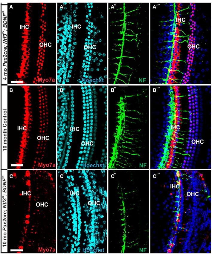
Frontiers in Aging Neuroscience | www.frontiersin.org 6 March 2015 | Volume 7 | Article 33Kersigo and Fritzsch Hair cells die without innervation FIGURE 5 | Both IHCs (A,B,D) and OHCs (C) show variability in the expanding into the scala media, occasionally from IHCs that bear some length of stereocilia with partial or complete fusion and what appears stereocilia (A,B). Note that IPCs typically have very short microvilli (A,D) but, to be resorption into the HCs. Some IHCs show globular protrusions at places, may entirely lack microvilli formation (B). Bar equals 5 µm. of Bdnf resulted in loss of all spiral ganglion neurons in the basal At 8 month, the SEM data revealed a less severe deficit com- turn. Middle turn spiral ganglion neurons had processes spiral- pared to 4 month old, denervated cochlea (Figures 3, 8). Thus, ing along the inner spiral bundle to the base. Even 10 month old a limited residual innervation maintains HCs under otherwise mutants (Figure 7) had some fibers innervating mostly IHCs and equal conditions for several more months compared to com- mostly in the upper middle turn. plete loss of innervation (Table 1). Most notable were differ- In contrast to control littermates, 10 month old Pax2-cre; ences in OHC vs. IHC loss and among the three rows of OHCs Ntf3f /f ; Bdnf f /+ mutant mice showed no Myo7a positive stain- (Figure 8). Whereas in many cases there was no loss of OHCs in ing throughout the basal turn (data not shown). We found the third and second row, the first row of OHCs and in partic- Myo7a positive staining HCs in the middle turn and in the apex ular IHCs showed a regionally specific, severe loss (Figure 8A). (Figure 7C). However, while control littermates (either no cre This was in stark contrast to littermates that showed the well- or no LoxP flanked neurotrophins) had near uniform Myo7a known patchy loss of single OHCs that were scattered across staining with occasional loss of one or two OHCs (Figure 7B), all rows with a tendency to be more profound in the third row Pax2-cre; Ntf3f /f ; Bdnf f /+ mutants showed a profound reduc- (Figure 8E). At places, most IHCs (Figures 8B,C) and nearly tion of Myo7a with very few, mostly IHCs normally labeled all OHCs of the first row (Figure 8D) were lost in mutants. In (Figure 7C). Hoechst nuclear staining confirmed the presence of fact, in many instances, multiple IHCs were lost instead of the OHCs and IHCs (and the occasional loss of OHC) in control typical single OHCs (Figure 8). Thus, overall hair cell loss in animals (Figure 7B′ ) while nuclear stain was difficult to use to the middle turn of mutants differed from age-matched litter- identify HCs in the mutant due to large gaps and distribution of mates in showing loss of multiple adjacent HCs. Notably, there nuclei at different levels (Figure 7C′ ). Numerous fibers could be was virtually no loss of IHCs in control animals even at this traced to IHCs and OHCs in control animals (Figures 7B′′ ,B′′′ ) age (Figure 8E). Closer examination showed fusion of multi- whereas very few tunnel-crossing fibers were found in mutants ple stereocilia in OHCs (Figure 9B) and IHCs (Figures 9C–F). (Figure 7C′′ ,C′′′ ). These data suggest a progressive loss of HCs This fusion in some IHCs was so advanced that only one or two in the mutant. However, it needs to be stressed that areas exist prominent protrusions reached from IHCs into the scala media in the middle turn of mutants with fairly normal HC distri- (Figures 9E,F). Loss of IHCs resulted in medial expansion of bution that seemingly correlated with apparent higher level of IPCs to either touch BCs (Figures 9C,D) or to leave inner pha- innervation density, though the details require more quantifica- langeal cells (IPhC) between them (Figure 9E). At places OPCs tion. Since 10 months seemed to be on the advanced end of HC expanded into the outer compartment between the first rows loss in these mutants, we concentrated the SEM study on the 8 of OHCs (Figure 9A). Loss of HCs in the first row of OHCs month old mutants to learn more about the cellular changes to was usually filled by expansion of the lateral process of the expand beyond the data obtained in double null neurotrophin OPCs (Figure 9A) but occasionally by a medial expansion of the mutant mice. first row of Deiter’s cells (Figure 9B) generating a continuity of Frontiers in Aging Neuroscience | www.frontiersin.org 7 March 2015 | Volume 7 | Article 33
Kersigo and Fritzsch                                                                                                            Hair cells die without innervation

 FIGURE 6 | Loss of innervation of the cochlear base is shown in                 immunocytochemistry with anti-tubulin, B′ ). Importantly, conditional
 mice with a complete deletion of Ntf3 (A). A conditional deletion of            deletion of Ntf3 using Pax2-cre requires the additional elimination of one
 Ntf3 using Pax2-cre (B) or a conditional deletion of both Bdnf and Ntf3         or two alleles of Bdnf before the phenotype approaches that of the
 in the ear. Note that there is, at the most, a few fibers spiraling along       unconditional null for Ntf3 (A,C). Note that the reduction of innervation
 the IHCs from the middle turn in either mutant and no matter the                has no apparent effect on early development of hair cells visualized with
 technique used (lipophilic dye tracing with NVMaroon, A,C;                      anti-Myo7a staining (B). Bar equals 50 µm.

different types of supporting cells without any HC between them.                 proposed simple quantitative compounding effects (Yang et al.,
This interpretation is consistent with a recent report showing that              2011). Each of these different combinations of partial Bdnf
Deiter’s cells function as scavengers that engulf dying hair cells               loss (Pax2-cre; Ntf3f /f ; Bdnf f /+ ) or partial Ntf3 loss (Pax2-cre;
(Anttonen et al., 2014).                                                         Ntf3f /+ ; Bdnf f /f ) have clear differences in the remaining inner-
                                                                                 vation (Figures 2D,F) pattern, despite the fact that in each case
Complete Absence of Bdnf and Incomplete                                          only one of the four neurotrophin alleles remains. We next con-
Absence of Ntf3 (Pax2-cre; Ntf3f/+ ; Bdnf f/f )                                  firmed the reduction of innervation in the Pax2-cre; Ntf3f /+ ;
Previous work had demonstrated a limited effect of loss of                       Bdnf f /f using immunocytochemistry (Figure 7A).
Bdnf on cochlear innervation but complete loss of canal cristae                     At 4 months the first effects of partial denervation in the
(Fritzsch et al., 2004; Yang et al., 2011). Consistent with these                basal turn appeared (Figure 7A). In fact, at this stage these mice
embryonic data, we find no innervation left to the canal cristae of              already showed a loss of OHCs that was more obvious compared
Pax2-cre; Ntf3f /+ ;Bdnf f /f (Figures 2E,E′ ). In contrast to the loss          to a 10 month old control animal (Figures 7A,B). Specifically,
of vestibular innervation and a severe reduction of the vestibu-                 multiple outer hair cells were missing, mostly in the base. As in
lar ganglion (data not shown) the innervation of the organ of                    other mutants, it appears that OHCs are preferentially missing
Corti was reduced (Figures 2F,F′ ). Interestingly enough, fewer                  in the first row compared to the second row (Figure 7). There
fibers were present to the basal turn but without the profound                   was also some limited effect in IHCs which were less regular
loss of all basal turn afferents characteristic for mice null for                in their distribution, making them more difficult to assess by
Ntf3 (Figure 2D). Bdnf expression changes from embryonic api-                    nuclear staining alone. Most of the remaining fibers that traced
cal to neonatal basal expression (Flores-Otero et al., 2007) and                 to OHCs showed features consistent with efferents (Figure 7A′′ ).
more profound effects in the basal turn have been noted before                   An occasional type II afferent fiber was identified (Simmons et al.,
in Pax2-cre; Bdnf f /f mice (Zuccotti et al., 2012). It appears that             2011).
heterozygosity of Ntf3 profoundly compounds the effect of sim-                      Our SEM data mostly confirmed previous changes in mutants
ple loss of Bdnf, resulting in reduced innervation of the cochlea                at a cellular level but also showed surprising longitudinal and
(Figures 2E,F). This confirms that long term loss of one neu-                    radial effects. Most interesting was that stretches of IHCs were
rotrophin combined with haploinsufficiency of the second neu-                    missing in the basal turn (Figure 10B) of 7 month old mice
rotrophin increases innervation defects suggestive of previously                 whereas all IHCs were usually present in the apex (Figure 10A).

Frontiers in Aging Neuroscience | www.frontiersin.org                        8                                                March 2015 | Volume 7 | Article 33
Kersigo and Fritzsch Hair cells die without innervation FIGURE 7 | Conditional deletion of Bdnf combined with missing at this age (B′ ) compared to loss of nuclei in 4 months old partially heterozygosity of Ntf3 using Pax2-cre leads to reduced innervation denervated cochlea (A′ ). The combined staining shows that nuclei (blue) and and sporadic OHC loss by 4 months (A′′ ,A′′′ ). In contrast, control HCs (red) are the clear target of the many fibers (B′′′ ). In mutants lacking littermates have a dense innervation and near continuous rows of IHC/OHCs, Ntf3 and retaining only one allele of Bdnf, very few nerve fibers remain (C′′ ), revealed with anti-neurofilament staining even at 10 months (B′′ ) of the basal many nuclei of OHC/IHCs are missing or are disorganized (C′ ). Only few part of the middle turn. All remaining HCs stain positive for anti-Myo7a (A–C). Myo7a positive IHCs or OHCs remain in mutants (C) and many have nearly Note the regular appearance of HC nuclei with only an occasional OHC undectable levels of Myo7a labeling (C,C′′′ ). Bar equals100 µm. There were many losses of HCs in the second and third row of Mice without Bdnf have severe loss of all vestibular inner- OHCs in the apex whereas the basal turn showed more losses vation at birth (Ernfors et al., 1995) but, in particular, the in the first row. In particular, IHCs showed similar phenotypes canal cristae loses all innervation (Fariñas et al., 2001; Fritzsch in terms of fusion of stereocilia as previously encountered in et al., 2004). Consistent with these known embryonic defects, the other mutations of this background (Figures 10C,C′ ,C′′ ). there was no innervation of canal cristae at any postnatal Such fusion and reduced length of stereocilia was also found stage (Figures 2C′ ,E′ ). Indeed, in 7 month old mutants we in OHCs were some cells showed short stereocilia on one side found only a limited innervation of the utricle but no inner- of the cell and only bumps of apparently fused stereocilia on vation of the canal cristae (Figures 11A′ ,B′ ). There was also the other side (Figure 10B). Overall, the changes in HCs and a noticeable reduction in size of the posterior canal cristae the pattern of loss with a large reduction in IHCs was simi- (Figures 11C,D) and the utricle (Figures 11A,B). Closer exam- lar to the other incompletely denervated mutant line analyzed ination showed numerous calyces around type I vestibular here. hair cells in the control littermates but only a very rare Frontiers in Aging Neuroscience | www.frontiersin.org 9 March 2015 | Volume 7 | Article 33
Kersigo and Fritzsch                                                                                                                  Hair cells die without innervation

 FIGURE 8 | The upper middle turn of an 8 month old Pax2-cre;                   are typically filled by medial expansions of IPCs that directly contact
 Ntf3f/f ; Bdnf f/+ mutant mouse (A–D) is compared with a control               border cells (BC) of the inner spiral sulcus (ISS). Lost OHCs of the first
 littermate (E; no Pax2-cre). Mutant mice lack many OHCs and IHCs,              row are mostly replaced by expansions of OPCs but sometimes by
 sometimes as single cells and sometimes as partial rows (asterisks in          medial expansions of the first row of Dieter’s cells. Due to the loss of
 A). In contrast, control animals show sporadic loss only of OHCs with a        OHCs and in particular IHCs, the ratio of IPC:OHC:IHC differs between
 tendency to be more profound in the third row (E). Spaces of lost IHCs         control (13:8:8) and mutants (14:7:6). Bar indicates 10 µm.

calyx in the mutants (inserts in Figures 11A′′ ,B′′ ) consistent
                                                                                TABLE 1 | Percent remaining hair cells quantified from three areas of
with a previous report that calyx formation requires normal                     200 µm length near the base.
Bdnf signaling through the TrkB receptor (Sciarretta et al.,
2010).                                                                          Genotype                          % remaining HC 4           % remaining HC 7–8
                                                                                                                   months (base)               months (base)
   SEM data also suggested a smaller utricular area compared
to the control littermates. Only minor changes were found                                                          IHC          OHC           IHC            OHC
in HCs such as incomplete stereociliary bundles. However,
                                                                                Pax2-cre; Ntf3f/f ; Bdnf f/f      44%           8%                      na
such changes were difficult to document due to the density of
                                                                                Pax2-cre; Ntf3f/f ; Bdnf f/+               na                 51%            62%
stereocilia in the utricle. However, the posterior canal cristae
                                                                                Pax2-cre; Ntf3f/+ ; Bdnf f/f               na                 11%            72%
appeared reduced in size compared to the anterior canal crista
(Figures 11C,D) and had stretches of hair cells without long                    control; Ntf3f/+ ; Bdnf f/+       100%          98%          100%            82%

stereocilia (Figure 12D), consistent with gaps in HCs shown by                  All data are means form 6 ears, except for the 4 months double null mutant that are from
immunostaining (Figure 11D). Some of the HCs in these areas                     a single mutant.
had partially fused stereocilia (Figures 12F,G) that were lying flat
on the surface of the epithelium (arrows in Figures 12F,G). Bun-                and shortening, both in IHCs and OHCs (Figure 13) and
dles were composed of stereocilia of uneven size and uneven                     the vestibular epithelia (Figure 12). The reorganizations of the
length. Further quantification is needed to verify how much of                  remaining supporting cells is more obvious in the organ of
the obvious shrinkage is due to loss of calyces and/or due to hair              Corti and shows medial expansion of IPCs into the territory of
cell loss.                                                                      lost IHCs and lateral expansion of OPCs into the territory of
   In summary, changes in HCs after partial denervation require                 the lost first row of OHCs. The simple fact that in our mixed
at least twice as long to develop compared to complete dener-                   background we find profound loss of IHCs even with partial
vation (Figure 13; Table 1). The overall changes at the hair cell               denervation, combined with the unusual phenotypes of reduced
level are somewhat similar and consist of fusion of stereocilia                 Myo7a immunopositivity, and fusion of stereocilia suggests that

Frontiers in Aging Neuroscience | www.frontiersin.org                      10                                                      March 2015 | Volume 7 | Article 33
Kersigo and Fritzsch Hair cells die without innervation
FIGURE 9 | Both OHCs (A,B,D) and IHCs (C–F) show various degrees of
resorption and fusion of stereocilia in this 8 month old Pax2-cre;
Ntf3f/f ; Bdnf f/+ as well as patchy loss and locally different degrees of
reorganization of supporting cells. IPCs always expand medially to close FIGURE 10 | Development of stereocilia and distribution of surviving
the reticular lamina over lost IHCs. However, IPCs may either directly contact hair cells is shown in a 7 month old Pax2-cre; Ntf3 f/+ ; Bdnf f/f null
(C,D) border cells (BC) or a layer of cells with numerous short microvilli, mutant mouse. Note that the apex shows a continuous row of IHCs (A)
presumably inner phalangeal cells (IPhC), may be wedged between IPCs and whereas the base has large stretches where there is only an occasional
BCs (A,E,F). OPCs usually expand to complete the reticular lamina if the first IHC left (B). OHCs show loss in either region but the appears is more
row of OHCs is lost (A) but sometimes may expand to the second row of obvious in the innermost row of OHCs in the base whereas it is more
OHCs (B). First row Deiter’s cells (D1 in B) may occasionally expand to close obvious in the two outermost rows in the apex. IHCs show various unusual
the reticular lamina in places of lost first row of OHCs (OHC1 in B). Bar fusions of stereocilia (C,C′ ,C′′ ) while others adjacent to these fused
indicates 5 µm. stereocilia appear normal. The few remaining OHCs in the upper middle
turn show very short stereocilia (D,D′ ) if stereocilia are present at all.
OHCs may show small bumps instead of stereocilia (D′ ). Bar indicates
these effects are mediated by yet to be determined compounds 20 µm in (A–C′ ) and 5 µm in (C′′ ,D′ ).
associated with innervation.
Discussion any innervation (Fritzsch et al., 1998, 2005b). Our data confirm a
normal complement of HCs at P12 even when little innervation
Denervation Defects HCs remains (Figures 1, 7). Contradictory data should be reconsid-
Overall, our data suggest a time line of innervation dependency ered in the light of partial and/or difficult to detect remaining
of cochlear HCs of ∼4–8 months with loss of all OHCs and innervation and the time lapse between denervation and analy-
many IHCs of the basal turn in the absence of any innervation sis as well as the time at which denervation is initiated (Sugawara
at 4 months (Figures 3, 4, 13). This is within the same range et al., 2005). Loss of hair cells in complete denervation cases
previously reported for the vestibular HCs after transection of should not be dismissed as likely due to blood supply problems
the vestibular nerve without compromising the blood supply (Sugawara et al., 2005; Suzukawa et al., 2005) as blood supply is
(Favre and Sans, 1991). In contrast, in mice, most vestibular HCs not an issue in our mutant mice.
require at least 7 months of complete denervation before notice- Recent data suggest that altered synaptic activity can induce
able changes can be identified (Figure 12). The time line of sev- inner ear HC loss over a long period of time (Kidd and Bao,
eral months of viability of denervated hair cells also agrees with 2012) but does not show a clear overall correlation between
published data on the lateral line mechanosensory cells in sala- loss of HCs and loss of neurons (Perez and Bao, 2011). Most
manders and frogs (Jones and Singer, 1969). Different to these recently physiological defects were found in OHCs after long
obvious effects on long term maintenance, both in vitro and term efferent disruption (Liberman et al., 2014). The molecular
in vivo data clearly demonstrate that maturation and short-term basis of neurotrophic support from sensory epithelia to sensory
survival of inner ear HCs is possible in the complete absence of neurons is well-known (Fritzsch et al., 2004; Bailey and Green,
Frontiers in Aging Neuroscience | www.frontiersin.org 11 March 2015 | Volume 7 | Article 33Kersigo and Fritzsch Hair cells die without innervation
FIGURE 12 | The 7 month old mice lacking Bdnfand one allele of Ntf3
(Pax2-cre; Bdnf f/f ; Ntf3f/+ ) by Pax2-cre shows reduced size of
sensory epithelia most obvious in a comparison of the utricle with that
of a control littermate (dotted line in A,B). Interestingly, there is a size
difference in the mutant between the anterior canal crista (C) and the posterior
canal crista (D). While the anterior canal crista is close to the remaining
FIGURE 11 | This comparison of 7 months old control mouse vestibular innervation of the utricle in this model and may transiently receive limited
organs (A,C) with a Pax2-cre; Ntf3f/+ ; Bdnf f/f littermate shows innervation, the posterior canal crista is removed from the limited innervation in
changes in size and innervation density. Hair cells revealed with Myo7a the basal turn of the cochlea. Vestibular hair cells in the control animal show
(A–D) and their innervation (A′ ,A′′ ,B′ ,B′′ ) shows smaller sensory epithelia in the normally bundled organization (E) whereas many aberrant bundles are
mice lacking Bdnf and one allele of Ntf3 (Pax2-cre; Bdnf f/f ; Ntf3f/+ ) found in the hair cells of the posterior canal crista around the “balding” region
compared to the control littermate. Note that only the utricle (U) receives shown in the right hemicrista (D,F,G). These hair cells show splayed bundles
limited innervation in the mutant (B′ ,B′′ ). In contrast to the frequent calyces of stereocilia with fused stereocilia, and stereocilia of variable length that
engulfing type I vestibular hair cells (insert in A′′ ), mutants have only rare and occasionally appear to be lying flat on the remaining epithelium (arrows in F,G).
partial calyces (insert in B′′ ). The reduction in size of sensory epithelium is Bar indicates 50 µm (A–D) and 20 µm (E–G).
most profound in the posterior canal crista (PC in C,D) that is completely
denervated and the only epithelium without any innervation throughout
development. Bar indicates 50 µm (A–D) and 10 µm (inserts).
if the neuronal loss spares over 10% of the SGNs (Makary et al.,
2011). Evaluating our model in other mammalian species could
2014). Neither the molecular basis of afferent support on devel- verify if the effects described here are unique to the genetic back-
oping auditory nucleus neurons (Levi-Montalcini, 1949; Rubel ground of our conditional deletion mice or can be expanded to
and Fritzsch, 2002) nor the molecular basis of innervation on the other mammals or even humans. Previous work on dependency
physiology of HCs (Liberman et al., 2014) or the long term via- of cochlear nucleus neurons on innervation shows a profound
bility of hair cells (Figure 13; Table 1) is known. The fact that critical phase and delayed loss of innervation has progressively
neurons die after embryonic (Pan et al., 2011) or adult HC loss less effects on cochlear nucleus neuron viability (Rubel and
in rodents (Alam et al., 2007) but not in humans (Linthicum and Fritzsch, 2002). Our denervation experiment is certainly earlier
Fayad, 2009) indicates some yet to be molecular defined species- and more complete compared to other attempts and our effects
specific differences. Adding to this emerging complexity of adult could indicate a critical phase of hair cell dependency on inner-
HC-SGN interactions are recent data on loss of afferent inner- vation. The longer viability of hair cells in partially denervated
vation and SGNs after frequent sound exposures that seemingly mice could indicate that targeted deletions of neurotrophins at
does not affect HCs (Kujawa and Liberman, 2009), at least not different time points are needed to exclude other interpretations.
Frontiers in Aging Neuroscience | www.frontiersin.org 12 March 2015 | Volume 7 | Article 33Kersigo and Fritzsch Hair cells die without innervation
in this model type II neurons and efferents remain and HC are
sensitive to ouabain (Fu et al., 2012).
Limited Innervation Can Provide Long Term HC
Support
Our data and those gathered in other systems (Fritzsch et al.,
1998) raise the possibility that compromised neuronal viability
provides some feedback for long-term integrity of mechanosen-
sory HCs in the inner ear but apparently with a large time delay,
that is even longer with a limited innervation of less than 10%.
We base this suggestion on quantification of spiral ganglion neu-
ron loss in Ntf3 null mice (∼85%) and Bdnf null mice (∼7%)
that combines to ∼92% loss of SGNs (Bianchi et al., 1996; Fariñas
et al., 2001). Assuming that there is a simple additive effect, this
suggests that most papers claiming no effect of severe reduction
of innervation on hair cell viability need to be revisited to deter-
mine exactly how much innervation was left when HCs appear
to be normal and at which age all innervation was indeed lost. In
addition, as innervation falls below 10% it appears that a very pro-
found time delay exists before HCs are compromised that would
be problematic for many studies dealing with mice that show
premature age related HC loss. How this support of HCs is dis-
tributed between efferents and afferents remains to be elucidated
but data on other sensory systems without efferents clearly point
out the importance of afferents (Fritzsch et al., 1990). In fact, the
unusual feature of our model is the effect on IHCs which receive
only transient innervation during development and in certain cir-
cumstances in the adult system (Simmons et al., 2011; Lauer et al.,
2012). Therefore, for IHCs, it appears likely that their high den-
sity of afferent innvervation plays a major role (Fritzsch et al.,
2015). Such an interpretation is consistent with the preferential
IHC loss in our models with diminished innervation. The appar-
ent preferential loss of the first row of outer hair cells could relate
to the difference in innervation density between IHCs and OHCs,
assuming that afferents release a diffusible factor that can reach
FIGURE 13 | This comparison shows the pertinent differences in
a short distance comparable to neurotrophins (Fritzsch et al.,
complete loss of the entire outer compartment and nearly all OHCs in
a 4 month old double neurotrophin conditional null mouse (A) as 2004).
compared to a 8 month old mutant that retains one allele of Bdnf (B)
and a 8 month old littermate control (C). Note that all mice have about 17 Reconciling Literature Discrepancies
IPCs but a variable number of HCs. Control littermates retain all IHCs for at Cutting the cochlear nerve has led to contradictory, variable and
least 8 months, forming an approximate 4:3 ratio of IPCs and IHCs (C). This
inconclusive data (Sugawara et al., 2005) possibly due to a difficult
changes to a 4:1 ratio in double null mutants (A) and a 3:1 ratio in partially
denervated mice (B). Note the variable loss of OHCs that is most profound in mix of surgery related blood supply disturbance and incomplete
the first row in a mutant with incomplete loss of innervation (B) whereas it is elimination of all innervation, differences between experimen-
more profound in the second and third row in control littermates. Bar equals tal animals and the possible effect of a critical phase of HCs on
20 µm. innervation. We reason that all these data could be reconciled
if it could be established that mechanosensory HCs of the ear
depend on a yet to be defined critical threshold of afferent and
Available evidence suggests presence of neurotrophin receptors efferent innervation during a critical phase, comparable to other
only on neurons (Ylikoski et al., 1993; Fariñas et al., 2001) but sensory cells (Fritzsch et al., 1998) and cochlear nucleus neurons
delayed expression of limited receptors needs to be verified using (Rubel and Fritzsch, 2002). However, neuronal dependency may
appropriate modern techniques to rule out any possible direct take a longer time to manifest itself in the case of mechanosen-
effect. Different cre lines such as a combination of Atoh1-cre sory HCs of the ear compared to other sensory systems (Favre and
(Matei et al., 2005) with induced delayed deletion in support- Sans, 1991) but is comparable in its timeline to the mechanosen-
ing cells using Fgfr3-creER (Anttonen et al., 2014) could result sory lateral line system (Jones and Singer, 1969). Consistent with
in more viable mice lacking all inner ear innervation. Another our anatomical data, long-term viability (Walsh et al., 1998) and
way to achieve denervation without affecting blood supply is by function of outer hair cells (OHCs) might depend on efferent
chemical treatment such as ouabain (Yuan et al., 2014). However, innervation (Liberman et al., 2014), whereas minor alterations
Frontiers in Aging Neuroscience | www.frontiersin.org 13 March 2015 | Volume 7 | Article 33Kersigo and Fritzsch Hair cells die without innervation
in synaptic transmission may affect viability of inner hair cells fusion or resorption of stereocilia (Figure 9) that resembles the
(IHCs) exposed to loud sound (Zuccotti et al., 2012). Ideally, degeneration of HCs in Myo7a mutant mice (Self et al., 1998).
one would like to eliminate a neurotrophic factor (Lindholm and This similarity is even more profound in Myo6 mutants that
Saarma, 2010; Bailey and Green, 2014) to show effects we describe show fusion of stereocilia of IHCs and OHCs (Self et al., 1999;
here in the presence of innervation, comparable to the loss of spi- Hertzano et al., 2008) remarkably similar to our data (Figures 5,
ral ganglion cells in neurotrophin mutants in the presence of hair 9, 10, 12). Conditional deletion of Cdc42 leads preferentially to
cells (Fritzsch et al., 2004) to verify the nature of this molecule IHC loss through stereocilia fusion (Ueyama et al., 2014) that
(or molecules). Whether such molecules could also be the basis is nearly identical to mice with incomplete denervation. How-
for the unknown support of developing cochlear nucleus neu- ever, while Cdc42 mice lose IHCs after 8 weeks, IHCs take over
rons (Levi-Montalcini, 1949; Rubel and Fritzsch, 2002) remains 4 months to degenerate even in completely denervated mice. It
to be seen. remains unclear if this correlation indicates some causality. If
so, it could indicate that altered expression of Myo6, Cdc42 and
Partial Denervation May Aid in the Study of other proteins associated with stereocilia homeostasis (Kitajiri
Age-related HC Loss et al., 2010; Sekerková et al., 2011) could underlie not only inner-
This mouse model, with no or with greatly diminished innerva- vation dependent hair cell maintenance but may play a role in
tion, allows us to test the hypothesis that cochlear HCs depend age dependent, variable loss of HCs through interference with the
on innervation but have a different time constant compared stereocilia homeostasis (Lenz et al., 2010). Like age dependent HC
to other sensory systems and that vestibular HCs are even loss (Otte et al., 1978; Keithley and Feldman, 1982; Wright et al.,
more resilient. Unfortunately, our partially denervated model 1986), loss of HCs in our model is highly variable with rather dif-
is more difficult to interpret. There is a well-known depen- ferent time lines for individual HCs that is even more different
dency of cochlear innervation on support provided by the nor- with residual presence of some innervation. Combined, our data
mally developed organ of Corti (Bailey and Green, 2014). This strongly support the conclusion of local variability of effects after
support will obviously decline as the organ of Corti dedif- aminoglycoside treatment (Taylor et al., 2012) and support the
ferentiates upon loss of HCs (Alam et al., 2007; Pan et al., argument raised by Taylor et al. (2012) that attempts to regenerate
2011, 2012). It is possible that additional loss of innervation an organ of Corti requires a close look at this variability of local
due to loss of HCs may accelerate regionally specific HC loss effects. Given the variable residual presence of IPCs, IPhCs or
(a possible negative feedback loop). However, this is of no con- nothing, strategies must be designed to take the local variations in
cern for the general problem investigated here, namely that supporting cell viability into account when attempting to restore
mechanosensory HCs depend on a limited level of innerva- a functional organ of Corti. Our data support the notion of
tion for long-term viability. Such feedback loops have not been molecular uniqueness and independence of IPCs (Fritzsch et al.,
apparent in previous work simply because the loss of neurons 2015) that have been shown to survive long term in other molec-
in most cases studied over long periods is far less (Makary ular models of HC loss (Pauley et al., 2008) as well as in our model
et al., 2011) compared to our presumed 93–100% loss of neu- (Figure 13).
rons. Only sparing HC loss has been reported after effer-
ent deletion (Walsh et al., 1998), in contrast to our massive Translational Aspects
loss of HCs within 4 months after all afferents and efferents Previous work has shown that subcritical sound levels can result
have been deleted (Figure 13). More recent work has suggested in long-term loss of some innervation (Kujawa and Liberman,
the existence of such a feedback loop regulating functional- 2009). It is possible that such retraction of nerve fibers is accel-
ity of OHCs after complete elimination of efferents (Liber- erated through compromised neurotrophin signaling (Wang and
man et al., 2014). Unfortunately efferents depend on afferents Green, 2011), accelerating additional loss of innervation caused
for homing (Simmons et al., 2011) and thus early afferent by sound. Such a possible loop can eventually result in loss
loss will result in efferent loss as well. Our model thus can- of HCs as consequences of mutation related to reduced signal-
not easily distinguish between afferent and efferent long term ing of neurotrophins or other molecules such as Igf-1 (Varela-
support. Nieto et al., 2013) combined with reduced density of inner-
vation. Given our data and those after aminoglycoside treat-
Comparison to Models of Induced HC Loss ment that indicate profound local variations (Taylor et al., 2012),
Overall, the loss of hair cells and reorganizations of the organ any treatment of the aging human cochlea to retain HCs or
of Corti to eventually turn into a flat epithelium presented here restore the organ of Corti needs to take local variation into
follows, to a large extent, changes observed after aminoglycoside account. This model could be used to develop expression pro-
treatment (Taylor et al., 2012, 2008). However, while hair cells are files of remaining HCs to eventually identify genes (Liu et al.,
lost rapidly after aminoglycoside treatment (Taylor et al., 2008) or 2014) responsible for their viability focusing on interactions
carboplatin treatment (Ding et al., 2012) it takes weeks to months between Cdc42 and Myo6, the two mutants (Self et al., 1999;
(depending on the mouse line) for the organ of Corti to reorga- Ueyama et al., 2014) that have the greatest similarities with our
nize (Taylor et al., 2012). In the case of complete denervation, denervation hair cell phenotype. Ultimately, we would need to
we see progression of HC loss and reorganization over several identify the molecular basis of the neuronal signal to gener-
months with profound local variation. Our data suggest that there ate small molecular analogs to rescue HCs in the absence of
is a correlation with changes in Myo7a expression (Figure 7) and innervation. Several candidate trophic factors exist (Bailey and
Frontiers in Aging Neuroscience | www.frontiersin.org 14 March 2015 | Volume 7 | Article 33You can also read