Bacterial and fungal endophyte communities in healthy and diseased oilseed rape and their potential for biocontrol of Sclerotinia and Phoma ...
←
→
Page content transcription
If your browser does not render page correctly, please read the page content below
www.nature.com/scientificreports
OPEN Bacterial and fungal endophyte
communities in healthy
and diseased oilseed rape
and their potential for biocontrol
of Sclerotinia and Phoma disease
C. S. Schmidt1, L. Mrnka1*, P. Lovecká2, T. Frantík1, M. Fenclová2, K. Demnerová2 &
M. Vosátka1
Phoma stem canker (caused by the ascomycetes Leptosphaeria maculans and Leptosphaeria
biglobosa) is an important disease of oilseed rape. Its effect on endophyte communities in roots
and shoots and the potential of endophytes to promote growth and control diseases of oilseed
rape (OSR) was investigated. Phoma stem canker had a large effect especially on fungal but also
on bacterial endophyte communities. Dominant bacterial genera were Pseudomonas, followed by
Enterobacter, Serratia, Stenotrophomonas, Bacillus and Staphylococcus. Achromobacter, Pectobacter
and Sphingobacterium were isolated only from diseased plants, though in very small numbers. The
fungal genera Cladosporium, Botrytis and Torula were dominant in healthy plants whereas Alternaria,
Fusarium and Basidiomycetes (Vishniacozyma, Holtermaniella, Bjerkandera/Thanatephorus) occurred
exclusively in diseased plants. Remarkably, Leptosphaeria biglobosa could be isolated in large numbers
from shoots of both healthy and diseased plants. Plant growth promoting properties (antioxidative
activity, P-solubilisation, production of phytohormones and siderophores) were widespread in OSR
endophytes. Although none of the tested bacterial endophytes (Achromobacter, Enterobacter,
Pseudomonas, Serratia and Stenotrophomonas) promoted growth of oilseed rape under P-limiting
conditions or controlled Phoma disease on oilseed rape cotyledons, they significantly reduced
incidence of Sclerotinia disease. In the field, a combined inoculum consisting of Achromobacter
piechaudii, two pseudomonads and Stenotrophomonas rhizophila tendencially increased OSR yield and
reduced Phoma stem canker.
Oilseed rape (OSR) is a major oil-crop in temperate regions of E urope1. Two of the most important diseases of
this crop are Phoma stem canker disease (syn. blackleg disease) caused by the ascomycetous fungus Phoma lingam
(asexual form; sexual forms Leptosphaeria maculans and L. biglobosa of the L. maculans complex) and Sclerotinia
stem rot caused by the ascomycete Sclerotinia sclerotiorum. During the growth cycle of winter OSR, fungi of the
L. maculans complex produce sexual ascospores on colonised stubble in autumn, which infect cotyledons and
secondary leaves and cause lesions; from there the pathogen spreads systemically and enters the stem, causing
the typical blackleg symptoms in s ummer2. Infection can also spread aerially via asexual p ycnospores3. Phoma
4
stem canker (blackleg disease) causes huge economic losses in O SR . Resistance breeding is often specific and
overcome by new virulent races5. The causal agent of Sclerotinia stem rot, S. sclerotiorum, persists in soil as black
resting structures called sclerotia, that can either produce soilborne mycelium or airborne ascospores for infec-
tion of its host6; the wide host range hampers control by crop rotation and development of resistant cultivars is
still in its i nfancy6. Thus, extensive application of fungicides is necessary in oilseed rape to control both d
iseases6,7.
Oilseed rape does also have high nutrient demands and high doses of (synthetic) fertilisers are r equired8.
Due to the environmental and societal demands to reduce agrochemical inputs, plant beneficial microorgan-
isms are intensively researched as an alternative to synthetic pesticides and fertilisers, also for brassicaceous crops
1
Department of Mycorrhizal Symbioses, Institute of Botany, Czech Academy of Sciences, Lesní 322,
25243 Průhonice, Czech Republic. 2University of Chemistry and Technology, Technická 5, 166 28 Prague 6‑Dejvice,
Czech Republic. *email: libor.mrnka@ibot.cas.cz
Scientific Reports | (2021) 11:3810 | https://doi.org/10.1038/s41598-021-81937-7 1
Vol.:(0123456789)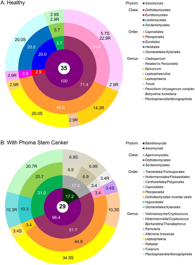
www.nature.com/scientificreports/
Figure 1. General outline of the study and Venn diagram of the isolated culturable endophytes from
symptomless oilseed rape (H, healthy) and oilseed rape showing symptoms of Phoma Stem Canker (D,
diseased).
such as oilseed rape9. Endophytes promote plant growth by various physiological mechanisms such as increasing
nutrient availability for the plant via siderophore production, phosphorus (P) solubilisation and nitrogen (N)
fixation, modulating plant growth and stress responses via phytohormone production, and controlling pathogens
via production of antimicrobial compounds10. Endophytes that inhabit seeds and systemically colonise the grow-
ing plant from there lend themselves to an application as seed t reatment11. Such treatments that remain effective
during a longer period of the OSR growth cycle would remove the need for fine-tuned spray timing, which is a
pivotal and critical issue in chemical control of Phoma stem c anker7 and Sclerotinia stem r ot6.
In OSR, endophyte communities of culturable bacteria12,13 and fungi14 have been characterised, with the
application as plant growth promoters in contaminated soil13, and biocontrol agents12,14–16 in mind. While there
is an extensive focus on the use of endophytes as biocontrol agents, the potential influence of plant pathogens on
endophyte communities has been underexplored so far. Not only can endophytes influence the growth of plant
pathogens, but also plant pathogens may change the environment for endophytes17 and thus have the potential
to alter their community structure. Cultivar resistance against Verticillium wilt affected the community structure
of bacterial endophytes in oilseed rape12. However, to our knowledge, no comparable studies exist for the two
other major diseases in OSR, Sclerotinia and Phoma stem canker.
With the work presented here, we start to close this gap. The two major aims of our study were to assess the
effect of Phoma stem canker infection on bacterial and fungal endophyte communities in oilseed rape and to
explore their potential for plant growth promotion and biocontrol. Similar to the previous studies cited above,
we focused on culturable endophytes for potential industrial production as beneficial inocula. Isolates were
characterised in vitro for their plant growth promoting properties. A selection of bacterial and fungal isolates
representing the major genera was tested in planta for plant growth promotion of OSR under nutrient limit-
ing conditions and for control of the two major pathogens Phoma stem canker and Sclerotinia stem rot. We
hypothesised that both bacterial and fungal communities in plants with symptoms of Phoma stem canker would
differ from communities in healthy plants. We also expected that microbes from the endophyte community
in symptomless plants in particular would provide protection to OSR plants from Phoma stem canker and/or
Sclerotinia white rot.
Results
Bacterial endophyte communities. Culturable bacterial endophyte communities of healthy and dis-
eased OSR plants were distinct at the species level (Venn diagram in Fig. 1) but relatively similar at the genus
level (Fig. 2A,B). Out of several diversity indices calculated in the study Shannon–Wiener, Gini–Simpson and
Pielou evenness indices showed no difference in richness and evenness of bacterial communities isolated from
healthy and diseased plants, but Margalef diversity index and Total taxonomic distinctness clearly indicated
a higher diversity and total taxonomic breadth of an assemblage in diseased plants (Table 1). This probably
arose from several genera (Achromobacter, Pectobacterium and Sphingobacterium) being isolated only from
diseased plants, albeit in very low numbers. In both healthy and diseased plants, Gammaproteobacteria and
Scientific Reports | (2021) 11:3810 | https://doi.org/10.1038/s41598-021-81937-7 2
Vol:.(1234567890)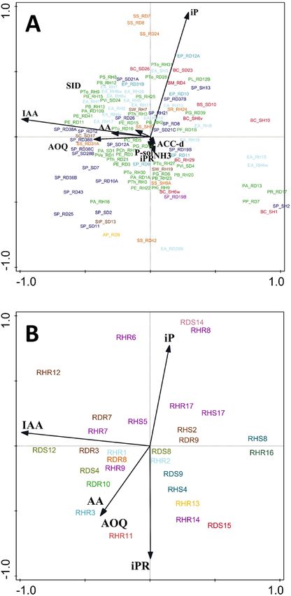
www.nature.com/scientificreports/
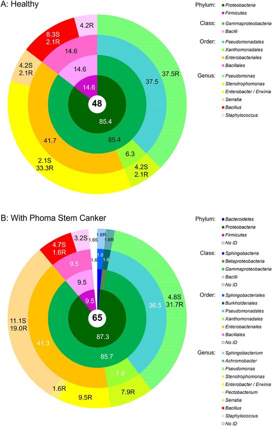
Figure 2. Community composition of the cultivable bacterial endophytic community associated with
symptomless oilseed rape (A) and oilseed rape showing symptoms of Phoma Stem Canker (B). The total
number of isolates in each group is shown in the central circle. The chart shows percentages by phyla, classes,
orders, and genera progressively from the central to the outermost annuli. N.D. not determined. On the genus
level, the abundances of isolates from roots and shoots are presented separately, with the additional letters “R”
for roots, and “S” for shoots. Systematic classification according to49.
Bacilli dominated, with the former contributing more than 85% of all isolates (Fig. 2a,b). With a share around
37%, Pseudomonas was the prevailing genus in both healthy and diseased OSR. The second largest group were
Enterobacteriales represented by the genera Enterobacter and Serratia (Fig. 2b). Xanthomonadales with the genus
Scientific Reports | (2021) 11:3810 | https://doi.org/10.1038/s41598-021-81937-7 3
Vol.:(0123456789)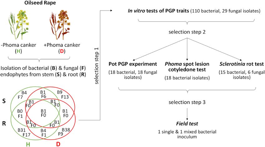
www.nature.com/scientificreports/
OSR Diversity indices
Shannon–Wiener diversity Gini–Simpson diversity Margalef diversity
Endophytes plants index index Pielou Evenness index coefficient Total taxonomy distinctness
Diseased 1.70 0.75 0.77 4.45 373
Bacterial
Healthy 1.45 0.71 0.81 2.97 236
Diseased 1.88 0.80 0.86 5.47 378
Fungal
Healthy 1.85 0.81 0.84 5.18 303
Table 1. Diversity indices of culturable bacterial and fungal communities in healthy (symptomless) and
diseased (with symptoms of Phoma stem canker) OSR assessed at the genus level. Total taxonomy distinctness
was calculated according to Clarke and Warwick68.
Stenotrophomonas were also present in a greater proportion. Bacilli, which made up 9.5–15% of isolates, were
largely represented by the genus Bacillus, and to a lesser extent by the genus Staphylococcus (Fig. 2a,b). While the
majority of Pseudomonas isolates originated from roots, a larger proportion of the Serratia and Bacillus isolates
were isolated from stems (Fig. 2a,b).
With MALDI-TOF scores mostly between 2.0 and 2.3, the genus ID of most isolates was certain, whereas the
species ID was probable, especially in Pseudomonas isolates (Supplementary Table S1). The largest proportion of
pseudomonads (around 85% in diseased plants and 50% in healthy plants) belonged to the P. fluorescens clade
(P. antarctica, P. extremorientalis, P. fluorescens, P. grimontii, P. libanensis, P. rhodesiae, P. synxantha, P. tolaasi,
P. veronii) within the wider P. fluorescens complex18. Among members of the P. fluorescens clade, P. tolaasi was
prevalent in the roots of healthy plants, whereas the other species prevailed in diseased plants (Supplementary
Table S1). In healthy plants, the P. corrugata clade (P. brassicacearum, P. kilonensis, P. thivervalensis) contributed
50% of Pseudomonas isolates. Single isolates of the P. koreenenis clade, the P. pyrolytica clade and of P. viridiflava
were isolated from diseased plants (Supplementary Table S1). Remarkably, P. viridiflava was only isolated from
shoots. Nearly all Bacillus isolates were identified as B. cereus. Only one Bacillus isolate from the roots of diseased
plants was identified as B. mycoides, another one from diseased shoots as B. subtilis. Within other genera, 100%
of respective isolates were identified as Enterobacter amnigenus, Erwinia percinia, and Serratia proteamaculans,
respectively (Supplementary Table S1).
Fungal endophyte communities. Diversity indices of culturable fungal communities were similar in
healthy and diseased plants (i.e. showing comparable genera richness and evenness) except for the Total taxo-
nomic distinctness index which clearly indicated a broader taxonomic span of endophytic fungal genera in
Phoma symptomatic plants (Table 1). Most genera had distinct morphotypes and only few (Cladosporium,
Leptosphaeria, Plectosphaerella) were morphologically variable (Supplementary MS Excel file 1). Nearly all fun-
gal isolates had 99–100% sequence similarity to the closest identified relatives, allowing their taxonomic resolu-
tion to the species level (Genbank accession numbers are listed in the Supplementary MS Excel file 1). Contrary
to bacterial endophyte communities, fungal endophyte communities were profoundly and qualitatively different
in healthy plants and plants affected by Phoma stem canker, and taxonomic groups detected in healthy and dis-
eased OSR were mostly represented by different genera (Fig. 3). Only Ascomycetes were recovered from healthy
plants. In the fungal community isolated from diseased plants, this phylum largely prevailed as well, but 17% of
isolates were Basidiomycetes, represented by the genera Vishniacozyma and Holtermaniella (formerly Cryptococ-
cus) isolated from shoots, and Bjerkandera/Thanatephorus (anamorph of the latter: Rhizoctonia) isolated from
roots (Fig. 3a). Among the Ascomycetes, Dothidiomycetes were the prevailing group in both diseased and healthy
plants; unexpectedly, fungi with 99–100% sequence homology to one causal agent of Phoma stem canker (Lepto-
sphaeria biglobosa, L. maculans species complex) could be isolated in large numbers from the shoots of both
diseased and healthy plants (20% and 34.5% of isolates, respectively). The only other genus that could be found
in both healthy and diseased plants was Plectosphaerella (Sordariomycetes, Glomerellales). Isolates of this genus
were morphologically diverse (see Supplementary MS Excel File 1). While the large majority of closely related
sequences in GenBank is classified as Plectosphaerella, identification by intergenic spacer sequencing does not
allow a clear differentiation from Monographella (Sordariomycetes, Xylariales, 99–100% sequence homology to
members of both genera). All other fungal genera were exclusively isolated from either healthy or diseased
plants. In healthy plants, Cladosporium (Capnodiales) was a dominating Dothideomycete (29% of fungal isola-
tions) that was isolated from shoots as well as from roots (Fig. 3a). Isolates of this genus also exhibited significant
morphological diversity (Supplementary MS Excel file 1). Epicoccum was also isolated in relatively large numbers
in healthy plants; other species of Dothideomycetes detected were Leptosphaerulina, Torula and isolates related
to Periconiella (94% sequence homology). In diseased plants, isolated Dothideomycetes belonged to the genera
Alternaria, Ramularia and Peltaster (Fig. 3b). Among other Ascomycete classes, Botrytis (Helotiales) was only
found in healthy plants whereas Fusarium (Sordariomycetes) was only isolated from diseased plants. While they
cannot be distinguished from Sclerotinia by their intergenic spacer region sequence, all Botrytis isolates could be
identified morphologically by their typical sporulation.
Plant growth promoting traits of bacterial and fungal endophytes determined in vitro. Sidero-
phore production and the ability to solubilise P was particularly widespread among Serratia isolates and Pseu-
domonas, especially those of the P. fluorescens-clade, but less frequent among isolates of the genera Steno-
Scientific Reports | (2021) 11:3810 | https://doi.org/10.1038/s41598-021-81937-7 4
Vol:.(1234567890)www.nature.com/scientificreports/
Figure 3. Community composition of the cultivable fungal endophytic community associated with healthy
oilseed rape (a) and oilseed rape showing symptoms of Phoma Stem Canker (b). The total number of isolates
in each group is shown in the central circle. The chart shows percentages by phyla, classes, orders, and genera
progressively from the central to the outermost annuli. On the genus level, the abundances of isolates from
roots and shoots are presented separately, with the additional letters “R” for roots and “S” for shoots. Systematic
classification according NCBI Genebank Database.
trophomonas, Enterobacter/Erwinia and Bacillus (Supplementary Table S1). All tested bacteria and fungi had
antioxidative activity (Supplementary Tables S1 and S2). Phytohormone production was also common among
bacterial and fungal endophytes. Production of the auxin IAA was generally higher than production of cyto-
kinins (iP, IPR) in both bacteria and fungi. IAA-production of fungi was generally at least one order of magni-
tude higher than that of bacteria. Bacteria mostly produced iP but no iPR (Supplementary Table S1), whereas iPR
was prevailing in fungi among the cytokinin compounds measured (Supplementary Table S2). High cytokinin
production was particularly found among isolates of the genera Stenotrophomonas, Bacillus and Cladosporium.
Only two of all tested fungal isolates (Penicillium chrysogenum RHR13 and Epicoccum sp. RHR15) produced
very high levels of the mycotoxins roquefortin C and meleagrin. While P. chrysogenum is well known to produce
both of these mycotoxins69, the production of either roquefortine C or meleagrin by Epicoccum sp. has yet not
been reported to our knowledge.
Scientific Reports | (2021) 11:3810 | https://doi.org/10.1038/s41598-021-81937-7 5
Vol.:(0123456789)www.nature.com/scientificreports/
Figure 4. Ordination PCA diagram of bacterial (A) and fungal (B) isolates. First two axes explained 66% (A)
and 79% (B) variation. Colours indicate taxonomic grouping (See Table S1 and S2). The two characters behind
the underscore (bacteria) or initial letter “R” (fungi) in the isolates code are related to the origin of the isolate: R,
root; S, shoot/stem; H, healthy = without Phoma stem canker symptoms; D, diseased = with symptoms of Phoma
stem canker.
Neither PCA analysis (Fig. 4) nor CCA cluster analysis (Supplementary Fig. S2) based on PGP trait profiles
separated genera of endophytic bacteria (Fig. 4A, Fig. S2A) or fungi (Fig. 4B, Fig. S2B) into distinctive clusters.
The origin of isolates (shoot vs. root, diseased vs. healthy) had no influence on PGP trait profiles of endophytic
bacteria and fungi at all (Fig. 4, Supplementary Fig. S2).
Representatives of the bacterial genera Achromobacter, Enterobacter, Pseudomonas, Serratia and Stenotropho-
monas, exhibiting all or most plant growth promoting traits (importance weighed in the following order: ACC-
deaminase > siderophore production = P-solubilisation > IAA > iP = iPR > antioxidative activity AA = AOQ > NH3
Scientific Reports | (2021) 11:3810 | https://doi.org/10.1038/s41598-021-81937-7 6
Vol:.(1234567890)www.nature.com/scientificreports/
production) and preferentially isolated from Phoma symptomless oilseed rape, were selected for pot experiments
with OSR. For the selection of fungal isolates a similar approach was used: weighed importance of the traits:
IAA > iP = iPR > antioxidative activity AA = AOQ, preferential origin from Phoma symptomless plants, ease of
spore production based on our observation and literature records. Representatives of the genera Cladosporium,
Epicoccum, Leptosphaeria, Penicillium, Periconiella, Plectosphaerella, Ramularia, Torula and few isolates from
Tremellales (related to Cryptococcus) were selected for pot experiments with OSR (in the case of Sclerotinia rot
test only Cladosporium isolates were tested).
Growth promotion of oilseed rape in pot experiments. Although the large majority of selected bac-
terial strains solubilised P in vitro, none of them promoted the growth of oilseed rape in a P-limited soil signifi-
cantly (Supplementary Table S3). Red leaf discolouration in all treatments after 102 days of growth except for the
control Cp with normal P-fertilisation and a significant increase of growth and pod yield in this control indicated
that insufficient P-supply was limiting the growth. Plants were fertilised after 123 days of growth in order to
alleviate P-limitation and to detect growth promotion caused by mechanisms other than P-solubilisation, but no
significant effects could be found at the end of the experiment. Only tendencies to increase plant dry weight were
observed in plants treated with Achromobacter piechaudii AP_RD9, Pseudomonas chloraphis/veronii PCh_RH1,
Stenotrophomonas sp. SS_RD24, Enterobacter amnigenus EA_RH18 and Serratia proteamaculans SP_RH21.
Also, cumulative height tended to be increased in plants treated with Pseudomonas sp. PG_RD39 (P. fluorescens
clade), and E. amnigenus EA_RH18 (Supplementary Table S3). Isolates A. piechaudii AP_RD9, P. chloraphis/
veronii PCh_RH1, Pseudomonas sp. PG_RD39 and Stenotrophomonas sp. SS_RD24 were selected to be tested
as a growth promoting combined inoculum (IG) in a field experiment (see sub-chapter below); E. amnigenus
EA_RH18 was not included as it showed a tendency to increase Phoma severity (Fig. 5). The strains finally
included the combined growth promoting inoculum IG did not show any mutual antagonism in vitro (Fig. S3).
None of the tested fungal endophytes increased growth and pod yield of OSR (Supplementary Table S4).
Control of Phoma lingam in planta (cotyledon assay). In the cotyledon assay, some plants did not
emerge or showed non-specific stress symptoms (yellowing and wilting of leaves) and thus could not be included
in the analysis. Due to this reduction in the number of replicates (minimum N = 3 as compared to N ≥ 5 in other
experiments) and due to a large variation in lesion sizes, only trends were visible in the cotyledon assay. None of
the tested bacteria was comparable to the fungicide treatment (Fig. 5). Lesion size tended even to be increased
after treatment with some endophytic isolates, most notably with E. amnigenus EA_RH18 or S. proteamaculans
SP_SH2. For S. proteamaculans SP_SH2 and some other isolates, this trend was apparent not only in the directly
sprayed leaf but also in the non-treated second cotyledon that could have been influenced only by a systemic
effect of the seed treatment. Seedlings treated with Pseudomonas sp. PA_RD1A (P. fluorescens clade) had the
lowest lesion score (3.4 vs 5.7 of the non-treated control; Table 1) but only when the leaves were directly sprayed.
Generally, there were no positive systemic effects of endophytes when these were applied as seed treatment
(Fig. 5).
Control of Sclerotinia sclerotiorum in a pot experiment. Amended to soil, S. sclerotiorum caused
damping off in OSR seedlings. First symptoms were a girdled and discoloured hypocotyl, followed by wilting and
finally death of the seedling; symptom development was often accompanied by visible outgrowth of typical white
mycelium. The isolates Pseudomonas sp. PA_RD1A (P. fluorescens clade) and Serratia. proteamaculans SP_RH21
significantly reduced incidence (P = 0.001 and P = 0.005, respectively; Fig. 6) after 27 days of growth. A. piechaudi
AP_RD9 and Stenotrophomonas sp. SS_RD24 showed a weaker but still significant effect (P = 0.014 and P = 0.028,
respectively). Only S. proteamaculans SP_RH21 was able to control Sclerotinia significantly (P = 0.002) over a
longer period of 68 days (Fig. 6). None of the tested Cladosporium isolates had any significant control effect,
whether applied as spores (RHR7), or as mycelial inoculum. Pseudomonas sp. PA_RD1A had a negative effect on
OSR seedlings emergence even before visible symptoms of Sclerotinia infection became apparent (14 d growth;
P = 0,009; Table S5). The most effective isolate S. proteamaculans SP_RH21 was selected to be tested as a protec-
tive inoculum (IP) in a field experiment.
Effect of seed treatments with bacterial endophytes on OSR yield, growth and disease inci-
dence under commercial field conditions (Poříčí, Czech Republic). Except for a highly significant
(P = 0.00002 and P = 0.00033, respectively) reduction of stress symptoms (red discolouration of leaf margins) by
both IP and IG inocula in early spring (March 2017; Table 2) none of the seed treatments with bacterial endo-
phytes affected growth, yield and diseases of OSR significantly under commercial field conditions. Yet, some
trends were observable. Despite its efficacy against Sclerotinia in the pot experiment S. proteamaculans SP_RH21
(treatment IP) failed to control the disease in the field (Table 2). Albeit not statistically significant, there was a
trend towards increased yield (+ 15%) and reduced severity of Phoma stem canker (-42%) after seed treatment
with the IG consortium consisting of A. piechaudii AP_RD9, Pseudomonas sp. PCh_RH1, and PG_RD39, and
Stenotrophomonas sp. SS_RD24. Fungicide treatment significantly lowered disease incidence of Sclerotinia stem
rot and Alternaria on stems (Table 2).
Discussion
In most studies on microbial communities that rely on c ultivation12,14 the number of isolated strains is limited
due to the time effort needed for isolation and isolate characterisation. The small sample size of 48/65 bacterial
isolates and 35/29 fungal isolates from healthy/diseased plants, respectively, in this study means that the relative
abundance of bacterial species needs to exceed 5–6% and the relative abundance of fungal species needs to be
Scientific Reports | (2021) 11:3810 | https://doi.org/10.1038/s41598-021-81937-7 7
Vol.:(0123456789)www.nature.com/scientificreports/
Figure 5. Effect of selected bacterial endophytes on Phoma disease (Leptosphaeria maculans, anamorph Phoma
lingam) in a cotyledon assay on oilseed rape seedlings. Data are presented as means ± SE; AP = Achromobacter
piechaudii, PS = Pseudomonas sp., EA = Enterobacter amnigenus, SP = Serratia proteamaculans, SS
= Stenotrophomonas sp., C = negative (healthy) control without Phoma inoculation and without bacterial
inoculation, C + = positive (diseased) control with Phoma inoculation and without bacterial inoculation,
CF = fungicide treated control, Polyversum = commercial preparation of Pythium oligandrum. There was a
significant treatment effect on Phoma severity in the directly treated cotyledon (leaf A, P = 0.0003) and on
Phoma symptoms after seed treatment only (leaf B, P < 0.0005). Treatments with the same letters are not
significantly different according to Unequal N HSD test. Where Dunnettʼs test showed a significant treatment
effect compared to the control C + the significance of this treatment effect is shown as asterisks in superscript.
greater than 8–10% to be detected with 95% probability19. Thus, only dominant genera were securely detected in
our study. Small sample size means a further risk that observed differences, especially quantitative shifts, might be
based on stochastic e vents13. This was especially apparent for bacteria, where the considerable difference in Mar-
galef index and Total taxonomic distinctness may be due to genera detected in very low numbers (Achromobacter,
Pectobacter and Sphingobacterium). In addition, differences in bacterial communities manifested themselves
mainly at the species level, where identification via MALDI-TOP was uncertain but only probable for most iso-
lates. On the contrary, fungal communities in healthy and diseases plants differed not only quantitatively but also
qualitatively in their dominant groups. Such profound differences, as also observed in poplar under contrasting
environmental conditions20, appear unlikely to be due to stochastic variation alone, but indicate a strong effect
of plant disease on endophyte communities. Though leaves were sampled in one of the symptomless plants next
to stems while only stems were sampled in diseased plants it is improbable that this difference had a significant
influence on the composition of microbial communities in this study. Neither bacterial nor fungal communities
isolated from the aboveground parts of healthy plants were richer in isolated taxa compared to diseased plants
as would be expected from the inclusion of another organ (leaves). Furthermore, the differences in fungal genera
between diseased and healthy plants were also profound in the roots and not only in the aboveground plant parts.
Unexpectedly, isolates with 99–100% sequence homology to the plant pathogen Leptoshaeria biglobosa could
be isolated in high frequency from both apparently healthy plants as well as plants with blackleg symptoms. As a
causal agent of Phoma stem canker, L. biglobosa has been found to prevail over L. maculans in Eastern European
countries21 and causes major damage in this region, especially in later growth stages during summer (also the
time of our sampling)22. Previously it has been considered less aggressive than L. maculans, albeit also depend-
ent on external c onditions23. Its isolation from apparently healthy OSR plants is an indication that it also can be
present asymptotically in OSR. Some isolates of the species may even promote OSR growth14. Moreover, strong
interspecies sequence homology makes secure identification of the species not possible based on intergenic spacer
sequences alone. There is also strong possibility that several Leptosphaeria species were present in our samples as
Scientific Reports | (2021) 11:3810 | https://doi.org/10.1038/s41598-021-81937-7 8
Vol:.(1234567890)www.nature.com/scientificreports/
Figure 6. Effect of seed treatment with endophytes on incidence of Sclerotinia disease in oilseed rape seedlings.
Data are presented as means ± SE; AP, Achromobacter piechaudii; Ps., Pseudomonas sp.; EA, Enterobacter
amnigenus; SP, Serratia proteamaculans; SS, Stenotrophomonas sp.; Cs_HR7s, Cladosporium sp. HR7 applied as
spores; Cs_HR7, Cladosporium sp. HR7 applied as mycelium; bC−, negative (healthy) control without Sclerotinia
inoculation and without bacterial inoculation; bC+, positive (pathogen-inoculated) control in Sclerotinia
infested soil without bacterial inoculation; fC−, fungal negative (healthy) control without Sclerotinia inoculation
and fC+, fungal positive control (with pathogen). bC− and bC+ are the corresponding controls for all bacterial
treatments and Cs-HR7s, while fC− and fC+ are the corresponding controls for all fungal endophyte treatments
applied as mycelium (Cs_). There was a significant treatment effect on incidence of Sclerotinia after 27 days
and 68 days growth (P < 0.0001). Treatments with the same letters are not significantly different according to
Unequal N HSD test (inoculation with cell or spore suspension) or according to Kruskal–Wallis Test. Where
Dunnettʼs test showed a significant treatment effect compared to the control C + the significance of this
treatment effect is shown as asterisks (* P < 0.05, ** P < 0.01, ***P < 0.001) in superscript.
Yield Disease incidence Aug 2017, BBCH81–89 (%) Overwintering March 2017
Plants with Phoma Red discolouration
Var Yield (t/ha) Yield in-crease (%) Phoma stem canker Sclero-tinia stem rot Alter-naria on stems Plants per m
−2 lesions (%) of leaves (%)
C 2.5 ± 0.23 a 100.0 26 ± 3 a 42 ± 4 a 42 ± 4 a 29 ± 1.4 a 9 ± 3.6 a 18 ± 3.7 b
IP 2.7 ± 0.29 a 105.4 24 ± 3 a 48 ± 4 a 49 ± 4 a 31 ± 1.8 a 6 ± 2.2 a 8 ± 2.0 a(P=0.00002)
IG 2.9 ± 0.28 a 115.3 15 ± 3 a 57 ± 4 a 57 ± 4 a 33 ± 1.8 a 9 ± 2.7 a 11 ± 2.2 a(P=0.0033)
−F 2.5 ± 0.20 a 100.0 22 ± 2 a 56 ± 3 b 55 ± 3 b – – –
+F 3.0 ± 0.15 a 117.2 21 ± 3 a 41 ± 3 a 42 ± 3 a – – –
Table 2. Effect of bacterial endophytes applied as a seed coat on winter oilseed rape yield, Disease incidence
of Phoma stem canker (Phoma lingam), Sclerotinia stem rot (Sclerotinia sclerotiorum), and Alternaria stem
infection and on oilseed rape overwintering under commercial field conditions (Poříčí, Czech Republic). Data
are presented as means ± SE. C = Control; IP = Serratia proteamaculans SP_RH21; IG = combined inoculum
of Achromobacter piechaudii AP_RD9, Pseudomonas chloraphis/veronii PCh_RH1, P. grimontii/fluorescens
PG_RD39 (P. fluorescens clade), Stenotrophomonas sp. SS_RD24; − F = no fungicide treatment + F = fungicide
treatment in spring. Where Dunnett test showed a significant treatment effect compared to the control C + the
significance of this treatment effect is shown as P values in superscript brackets. Different letters indicate
significant differences in nested factorial ANOVA (fungicide treatment) or Tukey HSD test nested within strip
(bacterial seed treatment).
Scientific Reports | (2021) 11:3810 | https://doi.org/10.1038/s41598-021-81937-7 9
Vol.:(0123456789)www.nature.com/scientificreports/
Leptoshaeria isolates recovered in this study showed considerable morphological diversity when grown on MEA
Agar (Supplementary Excel file 1). Interestingly, two other sequences with 99–100% sequence homology to our
isolates were described as stemming from Pithomyces, a Phoma isolate causing leaf spot in Japanese horseradish
(KR998327.1 and KR998328.1, unpublished).
The other fungal group that could be isolated from both asymptotic and symptom-bearing plants plants
in this study, Plectosphaerella/Monographella, was previously isolated from the rhizosphere of OSR16,24. While
a majority of closest related sequences in Genbank were characterised as Plectosphaerella, a large number is
described as Monographella25. Although these two genera belong to different orders of Ascomycetes (Glomerellales
vs. Xylariales), intergenic spacer and ribosomal sequences show nearly 100% homology, indicating that their
taxonomy still needs to be resolved.
Of the fungal isolates exclusively originating from either plants with blackleg symptoms or symptomless
plants, a larger proportion were (potentially) plant pathogenic species, i.e. Botrytis, Fusarium and Alternaria.
The observation that Botrytis (as well as Cladosporium and Epicoccum) were isolated in larger numbers, but
only from symptomless plants indicates their potential suppression in diseased plants. Leptosphaeria maculans
increases the level of some glucosinolates in OSR17. Even if this does not affect the pathogen itself, it may inhibit
other fungi. Other examples of mutual suppression have been found among the endophytic fungal community
of OSR where, among other (potential) plant pathogens, L. biglobosa inhibited S. sclerotiorum, an ascomycete
closely related to Botrytis cinerea 14. Conversely, Alternaria and Fusarium, (as well as Basidiomycetes of the for-
mer Cryptococcus group), which were only present in symptom-bearing plants here, might have profited from
a change in the micro-environment, such as the release of nutrients in necrotic tissue. Alternaria, Epicoccum,
Fusarium and Penicillium were also isolated from the endosphere of OSR in China whereas other less frequent
species were not14. On the other hand, we could not detect Chaetomium, Clonostachys and Periconia which were
dominant in their s tudy14.
Studies on the culturable endophytic bacterial community of O SR12,13,26, including this study, also differed in
their outcome, with different genera being predominantly isolated. These differences in outcome may be due to
different growth stages (young plants vs. flowering vs. post-flowering stage), cultivar choice and environmental
conditions (geographical region) and due to differences in sampling size. Not only diseases, as observed here,
but also disease resistance (against Verticillium wilt) changed bacterial endophyte communities in OSR12. We
found the endophyte community of OSR being largely comprised of the genera Pseudomonas, Stenotrophomonas,
Enterobacteriales (Serratia and Enterobacter) and Bacillus which were also frequently isolated as endophytes of
Brassicaceae in numerous other studies9. This spectrum of bacterial genera is remarkably similar to that of the
bacterial community antagonistic to the plant pathogenic fungus Verticillium dahliae in the OSR r hizosphere15,27,
with only small differences. Such similarity of the community of antifungal strains in the rhizosphere and the
endophytic flora of OSR might suggest that these genera colonise both compartments and comprise a large
proportion of the culturable endophytic community in OSR, but that their antifungal traits are not necessarily
expressed in planta, as they were equally abundant in healthy plants and in plants displaying blackleg symp-
toms. Nevertheless, it has to be borne in mind that cultivation-based methods of community profiling favour
these three groups over others and cultivation-independent approaches reveal much more diverse endophytic
community28. This was also the case in OSR, where cultivation independent methods unveiled a much wider
range of systematic groups among root-associated b acteria29,30 than studies confined to culturable bacteria but
other factors come into play here as well, as these authors did not differentiate between the rhizoplane and the
endosphere.
Strong P solubilisation and other plant growth promoting traits in vitro did not translate into plant growth
promotion in planta under P-limiting conditions. Especially the assay with tricalcium phosphate (Pikovskaya’s
agar31) employed here tends to overestimate the P-solubilising capability of isolates, since most P-complexes
in soil are harder to d issolve32,33. It is notable, however, that isolates with strong siderophore production and
P-solubilising capability detected in vitro showed the strongest trend to increase growth in the pot experiment
under P-limiting conditions. They also alleviated stress symptoms in the field and tended to increase yield in
the field when applied as combined inoculum. Consortia of plant growth promoting microorganisms have been
shown to be more effective than the single strains34 probably due to complementarity of their traits and their
different ecological optima. This was confirmed for OSR in another study, where growth promotion by single
strains of Pseudomonas, Serratia and Enterobacter isolates was only significant when the strains were applied
together as one combined inoculum in the fi eld35. In a similar way, our consortium consisting of Achromobacter,
Pseudomonas and Serratia strains showed a tendency to decrease disease incidence of Phoma in the field while
the single isolates were not effective against Phoma stem canker in the cotyledon assay.
Unexpectedly, isolates arising from symptomless plants were not more effective against fungal pathogens
than bacteria isolated from plants with Phoma stem canker, and some of the most antagonistic bacteria against
Phoma and Sclerotinia diseases in our in planta assays were isolated from diseased plants. Hardly any isolates
effective against P. lingam could be detected in the cotyledon assay, possibly because the employed method of
mycelial inoculation is more invasive and creates greater disease pressure than inoculation with pycnidiospores
used by other authors23,65,66. We used in planta tests with seedlings rather than in vitro assays for the screening
of antagonist activity as the latter are not always correlated with in planta performance36, and do not detect the
mechanism of resistance induction, which is particularly common in the genera Pseudomonas 37 and Serratia38.
Bacteria of both genera have been shown to be effective against Sclerotinia in OSR p reviously39,40. An apoplastic
isolate of P. viridiflava effectively supressed growth of three OSR pathogens (L. maculans, S. sclerotiniorum and
Xanthomonas campestris) in dual culture-assays70. The same isolate also restricted propagation of X. campestris
and reduced necrotic lesions caused by S. sclerotiniorum in OSR leaves and promoted the growth of OSR. The
protective effect of the endophyte was probably due to the induction of host resistance via salicylic acid and
jasmonic acid signalling pathways and/or production of antimicrobial c ompounds70. Several other bacteria
Scientific Reports | (2021) 11:3810 | https://doi.org/10.1038/s41598-021-81937-7 10
Vol:.(1234567890)www.nature.com/scientificreports/
controlled S. sclerotiniorum with most of them belonging to the genera Bacillus and Pseudomonas. Protection
mechanisms comprised increased levels of hydrolytic enzymes such as chitinase and β-1,3-glucanase, increased
expression of pathogenesis-related proteins and production of antifungal c ompounds71,72,74. While effective plant
growth promoters have been identified within the genus Achromobacter42–44, fewer reports exists on successful
biocontrol using this g enus45,46. In OSR, Achromobacter has been found to promote plant growth by improving
nutrient uptake47. Next to the above mentioned bacteria Serratia plymuthica HRO-48 was successfully developed
as seed treatment for OSR against Verticillium dahlia 11 and Phoma lingam41. To our knowledge, the species
Serratia proteamaculans has not yet been reported as a biocontrol agent in OSR, but an isolate of this species
controlled Alternaria in t omato38.
Our strain of S. proteamaculans that was most effective against Sclerotinia in the seedling assay failed in the
field, possibly because the seedling assay does not mimic natural infection conditions in OSR closely, where the
spread via ascospores germinating on senescent petals is the prevailing infection p athway6. Systemic colonisation
and a long time of survival in the plant is needed if strains applied as seed treatments are to prevent infection via
this pathway, where infection occurs in the flowering stage. Alternatively, foliar application is an efficient way
of introducing bacteria providing biological control of fungal diseases in oilseed rape but in such a case exact
timing of the application within the time window of petal infection by S. sclerotiniorum ascospores is c rucial72.
In addition, the imbalanced design of the fungicide application in our field experiment (two out of three control
plots but only one out of three IG and IP plots were treated with fungicide) might have putatively lowered the
difference in yield and disease incidence between the control and the bacterial treatments IP or IG.
In contrast to a large number of bacterial endophytes providing biological control against OSR diseases the
number of efficient fungal strains mentioned in literature is limited to a few, corresponding also to the output of
this study where several bacterial strains but no fungal strain exhibited fungal disease suppression. Among fungi,
Gliocladium catenulatum and Acremonium alternatum protected OSR from clubroot caused by Plasmodiophora
brassicae73,75 and Ulocladium atrum and Coniothyrium minitans reduced incidence and severity of Sclerotinia
stem rot76. However, the profound differences in fungal communities in Phoma-symptomless and symptomatic
plants found here raise hope that more fungal endophytes antagonistic to fungal diseases might be found.
In conclusion, we have shown considerable differences in fungal and, to a lesser extent, also in bacterial
endophyte communities in P. lingam-symptomatic and symptomless OSR plants. While the scope of the study
could provide only an introductory characterisation of their plant-growth promoting and biocontrol properties,
it yielded bacterial strains that are worthy of further study (Achromobacter piechaudii AP_RD9, Pseudomonas
chloraphis/veronii PCh_RH1, P. grimontii/fluorescens PG_RD39, and Stenotrophomonas sp. SS_RD24). In future
experiments, it would be desirable to improve the formulations of the s trains48, to optimise their application
technique and to monitor their long-term survival in the plant.
Methods
Isolation of bacterial and fungal endophytes. Endophytes of OSR were isolated by a modified dilution-
to-extinction method50. OSR plants (cv. ‘Viking’ and ’Catonic’) at growth stage of seed ripening (BBCH > 8151)
were harvested in mid July 2013 from a research site of SELGEN a.s. near Chlumec n. Cidlinou, Czech Republic
(N50:7.7552, E 15:29.3410, 218 m above sea level) at experimental plots not treated with pesticides. In the plots
many plants had developed symptoms of Phoma stem canker, Verticillium wilt, Alternaria blight and powdery
mildew (Erysiphe cruciferarum) while only few plants with Sclerotinia stem rot and light leaf spots caused by
Cylindrosporium sp. were present. We harvested plants with no visible disease symptoms (except for slight pres-
ence of Alternaria sp. that was ubiquitous) and separately those with symptoms of Phoma stem canker. Three
plants per variant (symptomless /healthy/, symptomatic /diseased/) were pooled and separated into roots and
shoots (shoots consisted of stems only as leaves already deteriorated at this growth stage except of one plant from
the symptomless variant where few leaves were present as well). Any adhering soil and debris was removed by
washing with tap water. Subsequent isolation of endophytic bacteria and fungi from plant material (shoots and
roots separately) was done as previously d escribed20,52. Briefly, plant material was surface sterilised for 7 min
(2.2% active chlorine with addition of 0.1% Tween 20), transferred to a 70% ethanol solution for 2 min, rinsed
four times with sterile deionised water, and cut to small pieces (< 0.5 cm). After thorough mixing of the cut mate-
rial under sterile conditions approximately 2 g (fresh weight) were homogenised (Ultra-Turrax IKA-T10, max
speed, 1 min) in 20 mL of sterile 12.5 mM potassium phosphate buffer (pH 7.1). The homogenate was filtered
(polyamide sieve Uhelon 13S, porosity 609 μm), diluted (5 to 50 times for fungi), and plated on 96-well plates
with agar media (25 μl medium per well). Fungi were isolated on both synthetic nutrient poor agar (SNA) and
nutrient rich malt extract agar (MEA: 2% malt extract, 0.2% meat peptone), each amended with the mixture of
antibiotics (50 mg L−1 tetracycline, 80 mg L−1 streptomycin sulfate, and 60 mg L−1 penicillin) to suppress bacterial
growth. Twenty-five microliters of the filtered homogenate was applied per well and six plates per given sample
and growth medium were used (576 wells in total) to isolate the endophytic fungi. In the case of bacteria, the
homogenate was diluted 10−1 to 10−4 and plated on Petri dishes (diameter 9 cm) with tryptic soy agar (5% TSA,
50% TSA) amended with cycloheximide (100 mg L−1). Bacteria were incubated at 20 °C for 48 h while fungi were
grown in dark at 20 °C up to 4 weeks. Fungi displaying different morphologies were re-streaked on new plates to
obtain clean axenic cultures. Isolated fungi were maintained on SNA and 1:10 strength MEA.
Identification of bacteria and fungi. Bacterial isolates were identified by MALDI-TOF MS53,54 using
an Autoflex Speed MALDI-TOF/TOF mass spectrometer and MALDI Biotyper 3.1 software (Bruker Daltonik,
Germany). Fungal isolates were morphotyped according to their colony appearance on malt extract agar (MEA;
see Supplementary MS Excel file 1). DNA of at least two representatives of each morphotype was i solated55, and
PCR-amplified for molecular identification based on their ribosomal DNA r egion56,57. Molecular identification
Scientific Reports | (2021) 11:3810 | https://doi.org/10.1038/s41598-021-81937-7 11
Vol.:(0123456789)www.nature.com/scientificreports/
and classification was based on the NCBI GenBank database58; a similarity of > 97% was used as a threshold for
species identification. Higher taxonomic units were selected in the case of lower scoring. When the identity of
the two selected representatives differed, all members of the morphogroup were molecularly identified. Gen-
Bank accession numbers of submitted fungal sequences are MN275840-MN275887.
Testing of in‑vitro traits of bacteria and fungi. Phosphorus (P) solubilisation, siderophore excretion,
ACC-deaminase, antioxidant activity (AA and AOQ = total antioxidative activity expressed in % of quenching
activity) and NH3 production on peptone were detected as previously described31,59–62,77. For the measurement
of phytohormones and antioxidants excreted into the culture medium, bacteria were grown in 20 ml LB in 50 ml
Falcon tubes for 7 days at 28 °C; fungi were grown in 50 ml Falcon tubes in 15 ml malt extract broth (MEB; 2%
malt extract; 0.2% meat peptone; pH 5.5) with 0.5% tryptophan for 10 days at room temperature on a shaker.
Auxin indole-3-acetic acid (IAA), the cytokinins N6-(Δ2-isopentenyl) adenine (iP) and N6-(Δ2-isopentenyl)
adenosine (iPR) in cultural filtrates were quantified by ultra-high performance liquid chromatography (Acquity
UPLC HSS T3 column; 1.8 μm, 2.1 × 100 mm; Waters, Acquity, coupled with tandem mass spectrometry AB
Sciex, Qtrap 5500; electrospray ionisation in positive mode). Blank sterile medium served as control63. The
performance of the endophytic isolates in the in vitro tests was used for the selection step 2 (Fig. 1). The selec-
tion was based on following criteria: (1) Number of traits where the particular strain scored high; importance
of the traits was weighed according the following order: ACC-deaminase > siderophore production = P-solu-
bilization > IAA > iP = iPR > AA = AOQ > NH3 production for bacteria, IAA > iP = iPR > AA = AOQ for fungi, (2)
Origin (we preferred isolates from symptomless plants over isolates from symptomatic plants), (3) Taxonomic
placement of the isolates (we aimed at covering a wide taxonomic diversity in order to arrive to a broad group of
strains putatively complementing each other in function and ecophysiological demands).
Mycotoxin assessment. Fungal isolates were grown on MEA enriched with 2% oilseed rape extract at
21 °C. Mycelial growth was quantitatively transferred into 250 ml Erlenmeyer flasks, mixed with 40 ml extraction
solvent (acetonitrile : 1% formic acid in water, 50:50), homogenised using ultraturrax and shaken for 60 min. The
supernatant was filtered into a 50 ml volumetric flask and filled up to the line with the extraction solvent (same as
specified above). This extract was then analysed according the methodology previously published for quantita-
tive determination of 57 mycotoxins (including Fusarium and Alternaria mycotoxins, ergot alkaloids, aflatoxins,
fumonisins, and others)64. For separation and detection of the analytes, combination of ultra-high performance
liquid chromatography (machine equipped with an Acquity UPLC HSS T3 column; 1.8 μm, 2.1 × 100 mm;
Waters, Acquity), coupled with tandem mass spectrometry (AB Sciex, Qtrap 5500; electrospray ionisation in
positive and negative mode) was used. For the quantification purposes, matrix-matched calibration standards
of the mycotoxins (blanks, 0.05–1000 ng ml−1) were prepared using blank medium extracted by the procedure
described for the samples.
In planta testing for plant growth promotion. Pots were filled with 1.2 L of autoclaved substrate
(7:3 autoclaved zeolite:sand mixture v:v; bulk density 1.17 g ml−1) amended with 11 g L −1 apatite resulting in
a dose of 1200 mg insoluble P per pot. Fertiliser (Osmocote 11:10:18 nitrogen (N):phosphorus (P):potassium
(K); 5–6 month release; Everris, The Netherlands) was added to the substrate (1.2 g per pot) resulting in a
concentration of 50 mg available P, 130 mg N and 190 mg K per pot; a high P treatment received 5 g Osmocote
per pot (260 mg P per pot). Pre-selected bacterial endophytic isolates based on the in vitro test performance
were grown in Luria Bertani (LB) broth for 2 days. Seeds of oilseed rape ʻSherlockʼ were surface sterilised (30 s
in 70% Ethanol, 15 min in 4.7 NaOCl, 3 × washing) and soaked in cell suspensions of the endophytic bacteria
(re-suspended in 0.01 M MgSO4, target OD600nm = 0.5 resulting in 108–109 CFU ml−1); seeds for controls were
soaked in sterile MgSO4. Four treated seeds were sown per pot and thinned to one plant per pot after emergence.
Pre-selected fungal endophyte isolates based on the in vitro test performance were grown on filter paper laid on
MEA and seeds were allowed to germinate on the mycelial mat and then planted into pots; seeds for controls
were germinated on sterile filter paper laid on MEA. After 123 days of growth, plants were fertilised with a mix-
ture of Kristalon fertiliser (AGRO CS a.s., Czech Republic) for tomatoes (N:P2O5:K2O 7.5:12:36 + 4.5 MgO + 10
SO3 + Trace elements (B Cu, Fe, Mn, Mo, Zn) and KH2PO4 dissolved in water (1.67 g Kristalon + 0.17 g KH2PO4
per L, pH 6.1). Four-hundred ml of this solution were added per pot, resulting in a final concentration of 0.05 g
P, 0.05 g N and 0.22 g K (ratio 1:1:4.4), 0.018 g Mg and 0.027 g S per pot. Plants were harvested after approx.
190 days of growth and a number of growth parameters assessed (number of shoots and pods, maximum shoot
height, cumulative shoot height, shoot, pot and root dry weight/all dry weights at 65 °C/together with the phe-
nological phase/BBCH scale/).
In planta test for activity against Phoma leaf spot (Leptosphaeria maculans). Antagonistic
activity of the pre-selected bacterial endophytes against Phoma leaf spot was tested with a cotyledon assay on
OSR seedlings23,65. Twelve mycelial plugs of Leptosphaeria maculans isolate ICBN3 (provided by Dr. Koopmann,
University of Göttingen, Germany) grown on V8-agar were used to inoculate 100 ml liquid cultures. Culture
medium was prepared as follows: 20 g L−1 oilseed rape leaves and roots were shredded in a mixer, boiled for
5 min, filtered, amended with 2% malt extract and 0.2% meat peptone, portioned into 300 ml flasks, amended
with 3 mm filter paper pieces, and autoclaved; fungal cultures were grown for 3 weeks at 20 °C at 200 rpm
before the overgrown filter paper pieces were retrieved for inoculation. Surface sterilised seeds of oilseed rape
ʻSherlockʼ were treated with cell suspensions of the bacterial endophytes as described above and sown in 50 ml
plastic falcon tubes filled with an autoclaved vermiculite:zeolite mixture amended with 0.15 g osmocote (NPK
12-11-17 + 2 MgO + trace elements fast release within 6 weeks); 2 ml of bacterial cell suspension were added to
Scientific Reports | (2021) 11:3810 | https://doi.org/10.1038/s41598-021-81937-7 12
Vol:.(1234567890)www.nature.com/scientificreports/
the substrate. After emergence, plants were thinned to 1–2 seedlings per tube and secondary leaves of plants
were regularly removed. After full development of cotyledons (19 days plant age), one cotyledon was punctured
with a needle on each half and the wounds treated with 10 µl of bacterial endophyte cell suspensions (resus-
pended in 0.01 M M gSO4, 108—109 CFU ml−1) to test a direct protective effect. The other cotyledon was left
untreated to test for the systemic effect of endophytes (introduced via seed treatment). Additional controls were
set up with a commercial fungicide (Horizon 250EW, Bayer Crop Science, active ingredient tebuconazole) and a
commercial biocontrol agent (Polyversum; Biopreparáty Spol. s r. o., Czech Republic; active ingredient Pythium
oligandrum) which were prepared according to manufacturer’s instructions and applied to wounded cotyledons.
Two days later (21 days plant age) all cotyledons were punctured on each side with a needle and covered with
filter papers overgrown with mycelium of Leptosphaeria maculans (non-inoculated controls C- were covered
with sterile filter papers). Filter papers were attached to leaves with a drop of wax. Inoculated plants were kept in
a moist chamber at 20 °C for two days and then grown in a growth chamber (16 h/8 h light/dark cycle, 21–23 °C)
for further 14 days before lesion sizes were assessed (scale according Chèvre et al.66, modified: 0 = no lesion,
1 = lesion size 1.5 mm, 2 = lesion size 1.5 mm and chlorosis, 3 = lesion size 1.5–3 mm, 4 = lesion size 1.5–3 mm,
chlorosis, 5 = lesion size 3.1–5 mm, 6 = lesion size 3.1–5 mm, chlorosis, 7 = lesion size 3.1–5 mm, severe chlorosis,
8 = lesion size > 5 mm, 9 = lesion size > 5 mm, chlorosis, 10 = lesion size > 5 cm, severe chlorosis, 11 10 = lesion
size > 5 cm, severe chlorosis, withered or sporulation).
In planta testing for activity against Sclerotinia. Sclerotinia sclerotiorum strain DBM 4244 (Collec-
tion of Yeasts and Industrial Microorganisms, Institute of Chemical Technology, Prague, Czech Republic) was
inoculated onto OSR and re-isolated from diseased plants prior use. The experiment was set up in 200 ml pots
filled with an autoclaved mixture of horticultural, peat-based substrate and sand (80:20, v:v) This autoclaved
substrate was inoculated with S. sclerotiorum grown for 4 weeks on autoclaved millet (0.75% m:v) and pre-incu-
bated in tightly closed plastic bags for further 4 weeks according El-Tarabily et al.67, modified). Bacterial endo-
phytes were applied on surface-sterilised seeds as described above (~ 108 cfu ml−1 in 0.1 M M gSO4). The fungal
strain Cladosporium strain RHR7 was applied twice: once as a spore suspension (107 spores ml−1 in 0.1 M M gSO4
with 10 µl Tween20) and the other time as a mycelial mat on which OSR seed germinated (analogously to other
Cladosporium isolates). The reason behind was to compare the two methods of introducing the fungus. Seeds of
OSR ‘Sherlock’ were soaked for 1–2 h in bacterial or fungal spore suspensions; seeds for controls were soaked
in sterile 0.1 M M gSO4. Cladosporium isolates (RHR7, RHR8, RHR9, RHR14, RHS1, RHS5) were applied by
allowing surface-sterilised seeds to germinate on the mycelial mat of an overgrown filter paper placed on MEA,
as described above. Nine treated seeds were sown per pot; pots were placed in a warm greenhouse (20–30 °C).
Emergence was assessed after 14 days. Additional millet inoculum grown for 14 days at 20 °C (1.6 g per pot) and
inoculum grown on autoclaved oilseed rape leaves for 7 days at 20 °C (0.8 g overgrown leaves per pot) was dis-
tributed between the plants after 21 days of growth. Concomitantly, pots were moved to a cold greenhouse ( Tmin
8 °C). The percentage of diseased and dead plants (girdled hypocotyl, white mycelial outgrowth at the hypocotyl,
wilting) was assessed after 27 and 68 days of growth.
In vitro test for mutual inhibition of plant growth promising strains. To exclude the risk that
PGPR strains (A. piechaudii AP_RD9, Pseudomonas spp. PCh_RH1, and PG_RD39, and Stenotrophomonas sp.
SS_RD24) that we planned to apply in a combined inoculum in the field (see below) inhibited each other, poten-
tial mutual antagonism was tested in an in-vitro assay as follows. Cells from liquid cultures (grown overnight
on tryptic soy medium at 24 °C) were centrifuged and washed once in phosphate buffered saline (PBS). Each of
the strains was then diluted in PBS to 0.5 McF (150.106 CFU/ml, OD 0.125 for 550 nm) and evenly spread with
a sterile cotton swab on tryptic soy agar (three replicate Petri dishes per strain) and allowed to dry. The other
strains were then applied as droplets (5 µl) at a cell density of 5 McF (1500.106 CFU/ml, OD 1.25 for 550 nm)
suspended in PBS onto this seeded lawn (3 replicate drops per Petri dish, 9 in total). After a growth period of
72 h at 24 °C, it was checked whether the outgrowing colonies produced clear inhibition zones in the surround-
ing bacterial lawn.
Field experiment in Poříčí, Czech Republic. The strains Achromobacter piechaudii AP_RD9, Pseu-
domonas chloraphis/veronii (PCh_RH1), P. grimontii/fluorescens (PG_RD39, P. fluorescens clade), and Steno-
trophomonas sp. SS_RD24 were applied to OSR seeds as a combined inoculum (IG; for plant growth promotion)
and the strain Serratia proteamaculans SP_RH21 was applied as a single inoculum (IP; for plant protection) as
follows. Cultures growing in LB for 2 days were centrifuged and the cell pellet re-suspended in Ringer solution.
For the combined inoculum, optical density at 600 nm of the cell suspensions was measured and pooled in
proportions adjusted to a theoretical value of 2.5 for each isolate within the pooled suspension. Cell suspensions
were lyophilised (Eco Fuel Laboratories, Prague, Czech Republic) leading to a final concentration of 8 × 10⁸ cells
g−1 lyophilisate for the combined inoculum IG, and 5 × 10⁸ cells for the single inoculum IP. For incrustation
of OSR seeds, these lyophilisates were mixed into an apatite – bentonite carrier (1:1 apatite : bentonite + 1.7%
lyophilisate of IG or 0.9% lyophilisate of IP, respectively; all w:w). This incrustation mixture was added to seeds
at the ratio 0.05:1 (w:w). The treatment resulted in 4 × 10⁴ CFU per seed (4.26 µg lyophilisate per seed) for the
inoculum IG, and 2 × 10⁴ CFU per seed (2.24 µg lyophilisate per seed) for inoculum IP. Treated seeds were sown
in a commercial oilseed rape field in Poříčí, Czech Republic (49.8464633 N, 14.6767358 E) in late August 2016
after winter wheat as a pre-crop. An imbalanced, factorial design with the factors bacterial treatment (controls,
IP and IG) and fungicide treatment (+ F, − F) was used (Fig. 1). The field was fertilised with 30 t ha-1 cattle
manure in autumn and with 300 kg ha−1 YaraBela Sulfan fertiliser (24% N + 6% S + 7% CaO) and 210 l ha−1 DAM
390 in spring. In March 2017, the number of plants per m2 and the percentage of plants with Phoma leaf spots
Scientific Reports | (2021) 11:3810 | https://doi.org/10.1038/s41598-021-81937-7 13
Vol.:(0123456789)You can also read