Nonreciprocal and Conditional Cooperativity Directs the Pioneer Activity of Pluripotency Transcription Factors
←
→
Page content transcription
If your browser does not render page correctly, please read the page content below
bioRxiv preprint first posted online May. 9, 2019; doi: http://dx.doi.org/10.1101/633826. The copyright holder for this preprint (which
       was not peer-reviewed) is the author/funder, who has granted bioRxiv a license to display the preprint in perpetuity.
                                      All rights reserved. No reuse allowed without permission.

            Nonreciprocal and Conditional Cooperativity Directs the Pioneer
                    Activity of Pluripotency Transcription Factors

                                       Sai Li1, Eric Bo Zheng2, Li Zhao2, Shixin Liu1,*
      1
       Laboratory of Nanoscale Biophysics and Biochemistry, The Rockefeller University,
      New York, NY 10065, USA
      2
       Laboratory of Evolutionary Genetics and Genomics, The Rockefeller University, New
      York, NY 10065, USA
      *Correspondence: shixinliu@rockefeller.edu

      ABSTRACT
      Cooperative binding of transcription factors (TFs) to chromatin orchestrates gene
      expression programming and cell fate specification. However the biophysical principles
      of TF cooperativity remain incompletely understood. Here we use single-molecule
      fluorescence microscopy to study the partnership between Sox2 and Oct4, two core
      members of the pluripotency gene regulatory network. We find that Sox2’s pioneer
      activity (the ability to target DNA inside nucleosomes) is strongly affected by the
      translational and rotational positioning of its binding motif, while Oct4 can access
      nucleosomal sites with equal capacities. Furthermore, the Sox2-Oct4 pair displays
      nonreciprocal cooperativity, with Oct4 modulating Sox2’s binding to the nucleosome but
      not vice versa. Such cooperativity is conditional upon the composite motif residing at
      specific nucleosomal locations. These results reveal that pioneer factors possess
      distinct properties of nucleosome targeting and suggest that the same set of TFs may
      differentially regulate transcriptional activity in a gene-specific manner on the basis of
      their motif positioning in the nucleosomal context.

      INTRODUCTION
      Transcription factors (TFs) access and interpret the genome by recognizing specific
      DNA sequences and regulating the transcriptional activity of selected sets of genes
      (Lambert et al., 2018; Ptashne and Gann, 2002). In eukaryotic nuclei, genomic DNA is
      wrapped around histone octamers, forming nucleosome building blocks and higher-
      order chromatin structures (Luger et al., 2012; McGinty and Tan, 2015). The
      compaction of DNA into chromatin often occludes TFs from their cognate binding motifs,
      thus constituting an important regulatory layer of gene expression control and cell
      identity determination (Li et al., 2007; Segal and Widom, 2009). A subset of TFs, known
      as pioneer factors (PFs), possesses the ability to access nucleosomal DNA and closed
      chromatin, which further recruits other chromatin-binding proteins and transcription
      machinery to their target sites and initiate transcriptional reprogramming and cell fate
      transitions (Zaret and Mango, 2016).
          TF pairs can exhibit cooperative binding behavior, i.e., binding of one factor to DNA
      facilitates targeting of the other (Morgunova and Taipale, 2017). Such cooperativity can
      be mediated by direct TF-TF interaction or through the DNA substrate (Jolma et al.,
      2015; Kim et al., 2013). Alternatively, due to the competition between TFs and
      nucleosomes for DNA binding, TF-TF cooperativity can be manifested indirectly and,
      often nonspecifically, in the context of chromatin (Adams and Workman, 1995; Mirny,
bioRxiv preprint first posted online May. 9, 2019; doi: http://dx.doi.org/10.1101/633826. The copyright holder for this preprint (which
       was not peer-reviewed) is the author/funder, who has granted bioRxiv a license to display the preprint in perpetuity.
                                      All rights reserved. No reuse allowed without permission.

      2010; Polach and Widom, 1996; Vashee et al., 1998). In this scenario, PFs are the first
      to engage and open up closed chromatin, making it more accessible to other TFs
      (Sartorelli and Puri, 2018; Zaret and Carroll, 2011).
          One of the most prominent examples of TFs shaping gene expression pattern is the
      “Yamanaka” factors (Sox2, Oct4, Klf4, and c-Myc), which can convert mammalian
      somatic cells into induced pluripotent stem cells (Takahashi and Yamanaka, 2006). It
      was shown that Sox2, Oct4, and Klf4, but not c-Myc, can function as PFs by binding to
      nucleosomes in vitro and silent, DNase-resistant chromatin in vivo (Soufi et al., 2012;
      Soufi et al., 2015). During reprogramming towards pluripotency, these factors
      cooperatively target selected enhancers and activate or repress the expression of
      distinct genes in a stage-specific manner (Chronis et al., 2017; Soufi et al., 2012).
          Among these reprogramming factors, Sox2 and Oct4 are also core members of the
      transcriptional regulatory network that governs embryogenesis and the maintenance of
      embryonic stem cells (Li and Belmonte, 2017; Rizzino and Wuebben, 2016). Sox2
      contains an HMG domain that binds to the minor groove of the DNA helix, whereas
      Oct4 harbors a bipartite POU domain that interacts with the DNA major groove, allowing
      the formation of Sox2:Oct4:DNA ternary complexes with the TF pair binding to adjacent
      motifs (Remenyi et al., 2003; Williams et al., 2004). ChIP-seq experiments revealed that
      Sox2 and Oct4 co-occupy the cis-regulatory elements of a large number of target genes
      (Boyer et al., 2005; Chen et al., 2008; Whyte et al., 2013), suggesting that this TF pair
      works synergistically to regulate gene expression. Indeed, juxtaposed HMG:POU
      composite motifs are found upstream of many pluripotency-associated genes
      (Ambrosetti et al., 1997; Nishimoto et al., 1999; Okumura-Nakanishi et al., 2005; Rodda
      et al., 2005; Tomioka et al., 2002).
          Despite extensive research, the capacity of and mutual relationship between Sox2
      and Oct4, and PFs in general, in targeting nucleosomal DNA remains a matter of
      debate (Chronis et al., 2017; Franco et al., 2015; Iwafuchi-Doi et al., 2016; Soufi et al.,
      2015; Swinstead et al., 2016). Recent data suggested that the pioneer activity of a given
      TF may be conditional and dependent on the local chromatin environment (King and
      Klose, 2017; Liu and Kraus, 2017; Soufi et al., 2015; Swinstead et al., 2016). For
      example, it was shown that the binding of Sox2 to a subset of genomic loci is dependent
      on another chromatin-binding protein PARP-1, and that such dependence is correlated
      to the rotational positioning of the Sox2 motif in the nucleosome (Liu and Kraus, 2017).
      Popular methods of choice for studying TF-chromatin interaction such as genome-wide
      binding and bulk biochemical assays lack sufficient temporal resolution to inform the
      time order of binding events (usually occurring on the order of seconds) by multiple TFs.
      Single-particle tracking experiments in living cells can capture the dynamic nature of TF
      binding (Chen et al., 2014; Lam et al., 2012; White et al., 2016), but the DNA sequence
      and chromatin state of the binding targets in these assays are usually not well defined.
      By contrast, in vitro single-molecule measurements allow for precise control of the
      substrates and have been employed to provide quantitative information on the
      binding/dissociation kinetics of TFs and chromatin regulators (Choi et al., 2017; Gibson
      et al., 2017; Kilic et al., 2015; Luo et al., 2014).
          In this work, we used single-molecule fluorescence microscopy to measure the
      binding dynamics of Sox2 and Oct4—both individually and in combination—on a variety
      of DNA and nucleosome substrates. We found that, although both classified as PFs,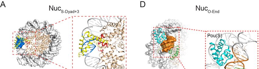
bioRxiv preprint first posted online May. 9, 2019; doi: http://dx.doi.org/10.1101/633826. The copyright holder for this preprint (which
       was not peer-reviewed) is the author/funder, who has granted bioRxiv a license to display the preprint in perpetuity.
                                      All rights reserved. No reuse allowed without permission.

      Sox2 and Oct4 exhibit markedly distinct properties of nucleosome targeting. Sox2
      strongly favors nucleosome-dyad-positioned sites over end-positioned ones, whereas
      Oct4 indiscriminately binds to targets at all positions. Oct4 and Sox2 are hierarchically
      recruited to composite nucleosomal motifs, with Oct4 predominantly engaging first. We
      further demonstrated that the Sox2-Oct4 pair displays nonreciprocal cooperativity, and
      that such cooperativity is dependent on the positioning of the composite motif with
      respect to the nucleosome. Consistent with these in vitro results, our analyses of the
      genomic data showed that the DNA binding sites of Sox2, but not Oct4, are
      preferentially located toward the center of the nucleosome. These results help to clarify
      the biophysical rules governing the Sox2-Oct4 partnership and suggest that genes may
      be differentially regulated by the same set of TFs on the basis of their motif positioning
      in the nucleosomal context.

      RESULTS
      Single-Molecule Analysis of Sox2 Binding to DNA
      We labeled the purified full-length human Sox2 with a Cy5 fluorophore near its C-
      terminus (Figure S1). We then constructed a DNA template containing a 147-
      basepair(bp)-long 601 nucleosome positioning sequence (NPS) with a canonical Sox2
      binding motif (CTTTGTT) located at its end (nucleotide #1-7), which was termed as
      DNAS-End (Figure 1A). Cy3-labeled DNA templates were immobilized on a glass
      coverslip and their locations visualized by total-internal-reflection fluorescence (TIRF)
      microscopy (Figure 1B). Cy5-Sox2 was injected into the flow chamber at a
      concentration of ~ 2 nM. Binding and dissociation of individual Sox2 molecules at the
      DNA loci were monitored in real time. Single-molecule fluorescence trajectories allowed
      us to measure the residence time (tbound) of Sox2 on DNA (Figure 1C). The cumulative
      distribution of tbound built from many binding events can be well-fit by a single-
      exponential function (Figure 1D). After correcting for dye photobleaching (Figure S2),
      we determined the characteristic lifetime of Sox2 binding (τ)—governed by the
      corresponding dissociation rate constant—to be 19.7 ± 2.3 s (Table S1).

      Sox2 Fails to Stably Bind to End-Positioned Nucleosomal DNA
      Sox2 is considered as a PF, capable of targeting binding sites within nucleosome-
      occupied genomic regions (Zaret and Mango, 2016). To directly observe the behavior of
      Sox2 on nucleosomal DNA, we created a mononucleosome substrate, NucS-End, which
      was assembled with DNAS-End and Cy3-labeled histone H2B as well as unlabeled H2A,
      H3, and H4 (Figure 1A). Fluorescence signal from Cy3-H2B was used to locate
      individual nucleosome substrates on the surface (Figure 1E). We used DNase I
      digestion followed by sequencing to show that NucS-End shares an identical digestion
      pattern with nucleosomes assembled with the unmodified 601 NPS (Figures S3A-S3B),
      suggesting that nucleosome positioning is not perturbed by the engineered Sox2
      binding motif.
          The residence time distribution for Sox2 on NucS-End cannot be described by a
      single-exponential model, but is well-fit by two exponential components: a predominant,
      shorter-lived population (τ1 = 1.3 s; fraction A1 = 85%) and a rare, longer-lived one (τ2 =
      14.1 s; A2 = 15%) (Figures 1F-1H; Table S1). The fast component likely corresponds to
      the nonspecific sampling of Sox2 on chromatin substrates as reported before (Chen et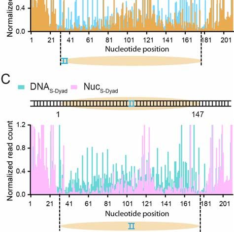
bioRxiv preprint first posted online May. 9, 2019; doi: http://dx.doi.org/10.1101/633826. The copyright holder for this preprint (which
       was not peer-reviewed) is the author/funder, who has granted bioRxiv a license to display the preprint in perpetuity.
                                      All rights reserved. No reuse allowed without permission.

      al., 2014). The slow component, on the other hand, likely represents the specific
      interaction between Sox2 and its cognate DNA sequence. Notably, the specific binding
      events only constitute a minor fraction of all binding events and feature a shorter lifetime
      than those on bare DNA (14.1 s vs. 19.7 s), indicating that nucleosome packing makes
      the DNA motif less accessible to Sox2. Indeed, when we superimposed the Sox2-DNA
      structure with the nucleosome structure, we observed a significant steric clash of the
      Sox2 HMG domain against the histone H3 and the neighboring DNA gyre (Figure 1I,
      NucS-End). Thus, single-molecule measurements led to the unexpected finding that
      nucleosome wrapping can significantly affect DNA targeting by Sox2.
           Next we moved the Sox2 binding motif inward (toward the nucleosome dyad) by 7
      bp and created a new nucleosome substrate NucS-End+7. This change shifts the phasing
      of the minor groove face of the DNA motif—which is recognized by Sox2—relative to
      the histone octamer, such that the steric hindrance between Sox2 and the nucleosome
      is much reduced (Figure 1I, NucS-End+7). Accordingly, we observed a significantly larger
      fraction of long-lived binding events compared to NucS-End (A2 = 62% for NucS-End+7 vs.
      15% for NucS-End; Figure 1H), supporting the notion that these events correspond to the
      specific Sox2-nucleosome interaction mode.

      Sox2 Stably Engages with Binding Sites near the Nucleosome Dyad
      A recent study showed that the Sox family TFs exhibit preferred binding around the
      nucleosome dyad region (Zhu et al., 2018). To dissect the single-molecule binding
      kinetics of Sox2 at the dyad, we placed the Sox2 binding motif at the center of the 601
      NPS (nucleotide #72-78) and assembled a nucleosome substrate NucS-Dyad (Figure 2A).
      Again, such sequence engineering did not change the overall nucleosomal organization
      according to the DNase digestion pattern (Figure S3C). In stark contrast to its behavior
      on NucS-End, Sox2 exhibited more prolonged binding to NucS-Dyad (an average residence
      time of 22.2 s for NucS-Dyad vs. 4.6 s for NucS-End). The residence time distribution for
      Sox2 on NucS-Dyad is also characterized by a double-exponential function (τ1 = 2.1 s; τ2 =
      36.8 s). Importantly, specific Sox2 binding events on NucS-Dyad are both longer-lived (τ2)
      and more prevalent (A2) than those on NucS-End (Figures 2B-2C, compare End and Dyad
      columns), consistent with the idea that the dyad position presents an optimal
      environment for Sox2 engagement. This is further supported by structural superposition
      that shows minimal interference imposed by histones and nucleosomal DNA on Sox2
      (Figure 2D).
          To evaluate whether the dyad region is in general favored by Sox2, we generated
      three more nucleosome substrates by shifting the Sox2 motif away from the dyad axis,
      either to the right by 3 bp or 6 bp, or to the left by 2 bp (NucS-Dyad+3, NucS-Dyad+6, NucS-
      Dyad-2, respectively; Figures 2A). We found that all dyad-positioned sites can
      accommodate extended Sox2 binding compared to the end-positioned site, but to
      different degrees (Figures 2B-2C). NucS-Dyad and NucS-Dyad+3, in which the minor groove
      of the Sox2 binding motif is mostly outward facing, have the highest fractions of long-
      lived binding. On the other hand, the minor groove is mostly inward facing for NucS-
      Dyad+6 and NucS-Dyad-2, which also feature lower fractions of specific Sox2 interaction
      (Figures 2D-2E, S4A-S4C). Thus, even though Sox2 generally prefers the dyad region,
      the likelihood and lifetime of its specific nucleosome-binding mode are nonetheless
      influenced by the rotational phasing of the DNA motif.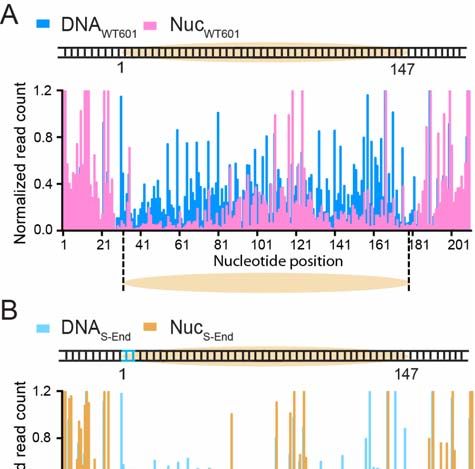
bioRxiv preprint first posted online May. 9, 2019; doi: http://dx.doi.org/10.1101/633826. The copyright holder for this preprint (which
       was not peer-reviewed) is the author/funder, who has granted bioRxiv a license to display the preprint in perpetuity.
                                      All rights reserved. No reuse allowed without permission.

         The results above collectively demonstrate that Sox2’s pioneer activity is sensitively
      modulated by the translational and rotational positioning of its cognate DNA motif within
      the nucleosome.

      Oct4 Binds Equally to End- and Dyad-Positioned Nucleosomal DNA Motifs
      Next we examined the behavior of the other core pluripotency TF, Oct4, on DNA and
      nucleosome substrates. We purified and fluorescently labeled full-length human Oct4
      with Cy5 (Figure S1), incorporated an 8-bp-long Oct4 binding motif into the 601 NPS at
      either the end or the dyad position (DNAO-End and DNAO-Dyad), and assembled
      nucleosomes with these DNA templates (NucO-End and NucO-Dyad; Figure 3A). We then
      conducted single-molecule TIRF experiments to measure the interaction between Oct4
      and these substrates (Figure 3B). The residence time distribution of Oct4 on each
      substrate can be well described by single-exponential kinetics (Figure 3C). Interestingly,
      unlike Sox2, the lifetimes of Oct4 on these substrates are statistically identical among
      each other (Figure 3D and Table S1), suggesting that Oct4 displays no discrimination
      between bare DNA and nucleosome substrates, or between end-positioned and dyad-
      positioned nucleosomal motifs (Figures S4D-S4E). We note that in all of the Sox2/Oct4
      binding experiments described above, the density of fluorescent nucleosomes on the
      surface did not decrease after addition of the TF (Figure S5), suggesting that Sox2/Oct4
      binding did not eject histones from the DNA.

      Nonreciprocal Regulation Between Sox2 and Oct4 in Nucleosome Binding
      Having characterized the individual behaviors of Sox2 and Oct4, next we set out to
      interrogate the cooperativity between this TF pair in nucleosome targeting. First we
      engineered a composite Sox2:Oct4 binding motif into the 601 NPS at the end position
      (nucleotide #1-15) and assembled the nucleosome substrate termed NucSO-End (Figure
      4A). We then examined the effect of Oct4 on Sox2 binding by complementing Cy5-Sox2
      with unlabeled Oct4 in the single-molecule experiments. We found that Oct4
      dramatically prolongs the average residence time of Sox2 on NucSO-End (Figure 4B).
      Notably, the stimulatory effect of Oct4 on Sox2 is restricted to the nucleosome substrate,
      as Oct4 has no effect on Sox binding to the bare DNA substrate DNASO-End (Figure 4B).
      A detailed kinetic analysis revealed that the lengthened dwell time of Sox2 on NucSO-End
      by the presence of Oct4 is mainly attributed to an increased weight of the specific mode
      of Sox2 binding events (A2 = 13% without Oct4 vs. 36% with Oct4; Figures 4C-4D;
      Table S1). Therefore, Oct4 appears to enhance the affinity of Sox2 to an end-positioned
      nucleosomal target. This result was corroborated by the bulk electrophoretic mobility
      shift assay (EMSA), which showed that the dissociation constant (KD) for Sox2-NucSO-
      End interaction is smaller in the presence of Oct4 than in its absence (Figures 4E-4F,
      S6A).
          Besides the canonical composite motif in which Sox2 and Oct4 binding sites are
      immediately juxtaposed with each other, a variant motif composed of Sox2 and Oct4
      sites separated by 3 bp is also found in some cis-regulatory elements such as the Fgf4
      enhancer (Ambrosetti et al., 1997). We generated a nucleosome substrate that contains
      such a gapped composite motif at its end position (NucSO+3-End; Figure 4A) and found
      that Oct4 exerts a similar, albeit somewhat weaker, positive effect on Sox2 engagement
      (compare NucSO+3-End and NucSO-End, Figures 4B-4D).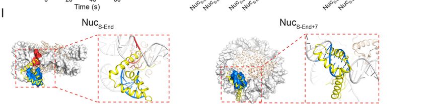
bioRxiv preprint first posted online May. 9, 2019; doi: http://dx.doi.org/10.1101/633826. The copyright holder for this preprint (which
       was not peer-reviewed) is the author/funder, who has granted bioRxiv a license to display the preprint in perpetuity.
                                      All rights reserved. No reuse allowed without permission.

         We then conducted the converse experiments by using Cy5-Oct4 and unlabeled
      Sox2 to check the influence of Sox2 on Oct4’s binding activity. Neither DNA nor
      nucleosome targeting by Oct4 was significantly affected by Sox2 (Figure 4G). Therefore,
      despite both being classified as PFs, Oct4 can help Sox2 access nucleosomal DNA
      ends but not vice versa. In other words, the regulation of Sox2 binding by Oct4 is not
      reciprocal.

      Hierarchically Ordered Targeting of Oct4 and Sox2 to Nucleosomes
      To directly follow the order of binding by Sox2 and Oct4 to the same nucleosome target,
      we labeled the TF pair with distinct fluorophores—Oct4 with Cy5 and Sox2 with
      AlexaFluor488—and used an alternating laser excitation scheme to simultaneously
      monitor their behaviors (Figure 4H). We found that the vast majority of overlapping
      Sox2/Oct4 binding events feature Oct4 binding first followed by Sox2 arrival (Figures 4I-
      4J). This result, together with the single-color residence time measurements described
      above, suggests that Oct4 behaves as a pioneer factor that recruits Sox2 and stabilizes
      its interaction with target sites located at the nucleosome end. Consistently, in the dual-
      color experiment, the Sox2 binding events that overlapped with an Oct4 binding event
      are significantly longer than those that did not overlap (Figure 4K).

      Effect of Oct4 on Sox2’s Nucleosome Binding Activity Is Position-Dependent
      We then asked whether the enhanced binding of Sox2 in the presence of Oct4 could be
      observed at other nucleosomal positions besides the end. In particular, we were curious
      as to whether binding to the nucleosome dyad, which is already preferred by Sox2,
      could be further stimulated by Oct4. To answer this question, we placed a canonical
      Sox2:Oct4 composite motif at the dyad position (nucleotide #72-86) of the 601 NPS
      (NucSO-Dyad; Figure 5A). Surprisingly, this construct produced a markedly different
      picture than NucSO-End: Oct4 has a negligible impact on the average residence time of
      Sox2 on NucSO-Dyad (Figure 5B); it moderately reduces the lifetime of specific Sox2
      binding (Figure 5C), but does not affect its relative population (Figure 5D). Moreover,
      EMSA results showed that KD for the Sox2-NucSO-Dyad interaction was increased by the
      presence of Oct4 (Figures 5E-5F, S6B). These results suggest that, instead of
      promoting Sox2 binding as observed at end-positioned composite motifs, Oct4 weakly
      diminishes the affinity of Sox2 to the nucleosome dyad. We speculated that such
      inhibitory effect might be caused by the geometrical interference between the two TFs.
      Indeed, when a 3-bp gap was inserted between Sox2 and Oct4 binding sites at the dyad
      position (NucSO+3-Dyad; Figure 5A), the negative effect of Oct4 on the specific Sox2
      binding mode was attenuated (Figure 5C). On the other hand, Sox2 exerted minimal
      influence on Oct4 binding to dyad-positioned composite motifs (Figure 5G), similar to
      the results obtained with end-positioned motifs (Figure 4G).
          Overall, these results show that the nonreciprocal cooperativity bestowed by Oct4
      upon Sox2 could be positive or negative depending on the position and configuration of
      the composite motif in the nucleosome context.

      Cooperativity between Pluripotency TFs at a Native Genomic Locus
      Next we explored the binding behavior of Sox2 and Oct4 at a natural genomic site. We
      chose the human LIN28B locus, which encodes a key protein regulating cell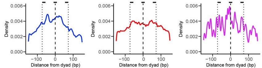
bioRxiv preprint first posted online May. 9, 2019; doi: http://dx.doi.org/10.1101/633826. The copyright holder for this preprint (which
       was not peer-reviewed) is the author/funder, who has granted bioRxiv a license to display the preprint in perpetuity.
                                      All rights reserved. No reuse allowed without permission.

      pluripotency and reprogramming (Shyh-Chang and Daley, 2013). We cloned a 162-bp-
      long DNA segment from this region, which is occupied by a well-positioned nucleosome
      and targeted by both Sox2 and Oct4 (Soufi et al., 2015). We then used this DNA
      template to reconstitute nucleosomes, termed NucLIN28B, and conducted single-molecule
      binding experiments using fluorescently labeled Sox2 or Oct4. The predicted cognate
      Sox2 binding site is located between the end and dyad of the LIN28B nucleosome
      (Figure 6A), as suggested by DNase I footprinting results (Soufi et al., 2015).
      Accordingly, the average residence time of Sox2 on NucLIN28B is longer than that on
      NucS-End but shorter than that on NucS-Dyad (Figure 6B). In contrast, Oct4, with its
      predicted cognate site also located between the end and dyad of the LIN28B
      nucleosome, exhibited a binding lifetime on NucLIN28B indistinguishable from those on
      NucO-End and NucO-Dyad (Figure 6C). These results lend further support to our conclusion
      that Sox2’s pioneer activity is position-dependent, while Oct4’s is not.
          We then examined Sox2-Oct4 partnership on NucLIN28B. The Sox2 and Oct4 binding
      sites within the LIN28B nucleosome are separated by 3 bp and oriented in opposite
      directions (Figure 6A), unlike all aforementioned composite motifs used in this study that
      feature a co-directional arrangement. We did not observe significant changes in the
      Sox2 binding kinetics caused by the addition of Oct4, nor vice versa (Figures 6D-6E).
      Therefore, besides the position and separation of Sox2/Oct4 binding motifs, their
      relative direction is also important for cooperativity (Chang et al., 2017).
          The LIN28B locus is also bound by c-Myc, another member of the Yamanaka factor
      set that, unlike Oct4 and Sox2, is thought to lack the pioneer activity and preferentially
      binds to open chromatin by itself (Soufi et al., 2015). We purified and fluorescently
      labeled c-Myc together with its heterodimeric partner Max (Figure S1), and tested its
      ability to bind DNA and nucleosome substrates at the single-molecule level (Figure 6F).
      We found that c-Myc binds to DNALIN28B much more transiently than Sox2 and Oct4.
      The same trend was observed on the nucleosome substrate NucLIN28B (Figure 6G).
      Thus, c-Myc possesses an inherent, albeit weak, ability to target nucleosomes. The
      binding lifetime of c-Myc on DNA and nucleosome substrates is prolonged by the
      presence of Sox2 and, to a lesser extent, Oct4 (Table S1). Similar to Sox2, c-Myc’s
      residence time distribution on DNALIN28B is described by two exponential components
      (Figure 6H). Interestingly, the long-lived binding component vanished when c-Myc was
      interacting with NucLIN28B (Figure 6I), and was restored by the addition of Sox2 but not
      Oct4 (Figures 6J-6K). Notably, no Sox2 cognate sequence is found near the specific c-
      Myc binding site. Hence the stabilizing effect of Sox2 on c-Myc is probably mediated by
      nonspecific Sox2-nucleosome interaction or by direct Sox2:c-Myc contacts.

      Genome-Wide Binding Preference of Sox2 and Oct4 With Respect to Nucleosome
      Positioning
      The in vitro single-molecule data described above revealed the differential positional
      preference of Sox2 and Oct4 for nucleosomal DNA. To investigate whether such a
      difference can be recapitulated on a genomic scale, we mined published nucleosome
      mapping and Sox2/Oct4 ChIP-seq data and interrogated the distributions of Sox2 and
      Oct4 binding sites relative to nucleosome locations (Figure 7A). We first calculated the
      aggregate nucleosome-positioning score surrounding Sox2/Oct4 binding sites in mouse
      embryonic stem cells (mESCs) (Teif et al., 2012) and epiblast stem cells (mEpiSCs)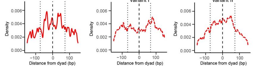
bioRxiv preprint first posted online May. 9, 2019; doi: http://dx.doi.org/10.1101/633826. The copyright holder for this preprint (which
       was not peer-reviewed) is the author/funder, who has granted bioRxiv a license to display the preprint in perpetuity.
                                      All rights reserved. No reuse allowed without permission.

      (Matsuda et al., 2017). In both cell types, the average nucleosome occupancy across all
      Sox2 binding sites is much greater than that for all Oct4 binding sites (Figure 7B),
      indicating that Sox2 binding sites are more enriched in nucleosome-occupied regions
      than Oct4 sites.
          We then plotted the distribution of Sox2/Oct4 binding sites in mEpiSCs averaged
      over all nucleosome-bound regions aligned by their dyad positions. Within the 147-bp
      window corresponding to the nucleosome, Sox2 binding sites exhibit a strong
      preference for the dyad region over the edges of the nucleosome (Figure 7C). In
      contrast, the distribution of Oct4 binding sites within the nucleosome shows no
      significant difference between nucleosome dyad and ends (Figure 7D). The preference
      for dyad diminishes in the case of Sox2:Oct4 composite sites as compared to Sox2-
      alone sites (Figure 7E), suggesting that the strong positional bias of Sox2 binding to
      nucleosomal DNA is alleviated by the cooperative interaction of Oct4, in line with the
      single-molecule results. Similar patterns were observed in mouse and human ESCs
      (Figure S7). Intriguingly, the distributions of noncanonical Sox2 and Oct4 motifs in the
      human genome, which are enriched in nucleosome-occupied regions as previously
      reported (Soufi et al., 2015), show distinct patterns from those of canonical motifs. For
      example, the tendency of dyad targeting vanishes for the noncanonical Sox2 motif that
      lacks a predominant “G” at the sixth nucleotide position (Figure S7E), which has been
      suggested to better accommodate nucleosome binding by abolishing DNA distortion
      (Soufi et al., 2015). These results imply that the genome sequence contains subtle and
      diverse codes that regulate differential modes of TF targeting.

      DISCUSSION
      TF binding positions are usually classified into nucleosome-depleted and nucleosome-
      enriched regions. In this work, we went beyond such general co-localization analysis by
      dissecting the dynamical binding pattern of a TF within a nucleosome. Compared to
      bulk biochemical and genome-wide binding assays, our single-molecule platform affords
      higher temporal resolution and unique kinetic information, which enabled us to
      quantitatively determine the cooperativity between pluripotency TFs in the nucleosome
      context and discover an unexpectedly intricate Sox2-Oct4 partnership. These results
      are corroborated by analyses of the in vivo genomic data, suggesting that the
      biophysical principles revealed by the in vitro reconstituted system may indeed be
      exploited by the cell to achieve differential gene regulation.

      Distinct Nucleosome Targeting Properties of PFs
      TFs generally prefer binding to nucleosome-depleted regions in the genome (Wang et
      al., 2012), with the exception of PFs that are able to access closed chromatin (Slattery
      et al., 2014; Zaret and Mango, 2016). The ensuing question then is: does a PF target all
      nucleosomal DNA sites with equivalent kinetics? We show that, at least for some PFs,
      the answer is no. In accordance with earlier studies (Liu and Kraus, 2017; Zhu et al.,
      2018), our results demonstrate that Sox2’s pioneer activity is regulated by both
      translational and rotational positioning of its cognate motif within the nucleosome. Dyad-
      positioned sites support prolonged Sox2 binding compared to end-positioned sites. This
      may be due to the fact that the dyad region, where only one DNA gyre is wound, can
      better accommodate DNA bending and minor groove widening that are essential for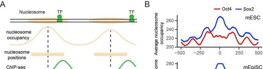
bioRxiv preprint first posted online May. 9, 2019; doi: http://dx.doi.org/10.1101/633826. The copyright holder for this preprint (which
       was not peer-reviewed) is the author/funder, who has granted bioRxiv a license to display the preprint in perpetuity.
                                      All rights reserved. No reuse allowed without permission.

      recognition by the HMG domain of Sox2 (Scaffidi and Bianchi, 2001). Moreover, the
      specific Sox2 binding mode at the dyad region is even more stable than Sox2
      interaction with bare DNA (Table S1), indicating additional contacts between the
      histones and Sox2. As to the rotational setting of the Sox2 binding motif, a more
      accessible minor groove (facing away from the histone octamer) generally corresponds
      to a higher affinity (Figures 1I, 2D, S4A-S4C). Similar preference for rotational phasing
      of binding motifs has been suggested for other TFs such as p53 (Cui and Zhurkin,
      2014).
          In contrast, we found no such positional preference for Oct4, which indiscriminately
      targets nucleosomal DNA at different locations. This could be rationalized by the fact
      that Oct4 contains two DNA-binding domains (POUS and POUHD), each recognizing a 4-
      bp half-motif locating at opposite sides of the DNA helix (Esch et al., 2013). Regardless
      of the nucleosomal DNA position, one of these half-motifs remains solvent-exposed
      (Figures S4D-S4E), which is apparently sufficient for stable Oct4-nucleosome
      interaction. In support of this idea, partial motifs are prevalently found in Oct4 target
      sites located in nucleosome-enriched genomic regions (Soufi et al., 2015). Therefore,
      although both regarded as PFs, Sox2 and Oct4 exhibit drastically different nucleosome
      binding profiles. We note that these interpretations are based on the superposition
      between the available structures of TF:DNA complexes and unbound nucleosomes. It is
      also conceivable that the nucleosome structure may be remodeled upon TF
      engagement.
          We found that c-Myc—previously thought not to have an intrinsic nucleosome
      targeting capability—can nonetheless bind to nucleosomal DNA, albeit transiently. A
      fully folded bHLHZ domain—the DNA binding domain of c-Myc—occupies a major
      fraction of the DNA circumference as seen in the c-Myc:DNA structure (Nair and Burley,
      2003), incompatible with nucleosome binding. Therefore, the c-Myc binding events
      observed on nucleosome substrates likely represent a nonspecific mode of interaction
      that does not require folding of the basic helix 1 in the bHLHZ domain (Soufi et al.,
      2015), consistent with our kinetic analysis.
          With more sensitive methods such as single-molecule imaging being deployed to
      interrogate TF-chromatin interaction, the list of nucleosome-binding TFs is expected to
      continue to grow. Our data further suggest that the pioneer activity of these TFs,
      governed by the structural characteristics of their respective DNA binding domains, is
      not a binary property but rather falls on a continuous spectrum.

      Nonreciprocal and Conditional TF-TF Cooperativity
      Clustered binding of TFs is a hallmark of cis-regulatory elements, such as promoters
      and enhancers, which integrate multiple TF inputs to direct gene expression. High-
      throughput methods have been developed to systematically determine the binding
      patterns of TF pairs on DNA (Chang et al., 2017; Jolma et al., 2015; Siggers et al.,
      2011; Slattery et al., 2011). However, the biophysical basis for cooperative TF binding in
      the nucleosome context remains underexplored. In particular, the relationship of
      chromatin targeting between a PF and a non-pioneer factor, and between a pair of PFs,
      is still under debate. Using Sox2-Oct4 as a model system, our study sheds new light on
      this issue. First of all, cooperativity can be unilateral. Oct4 stabilizes the binding of Sox2
      to an end-positioned motif, perhaps by opening up a stretch of nucleosomal DNA and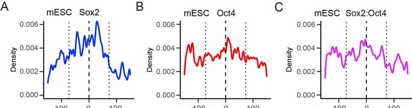
bioRxiv preprint first posted online May. 9, 2019; doi: http://dx.doi.org/10.1101/633826. The copyright holder for this preprint (which
       was not peer-reviewed) is the author/funder, who has granted bioRxiv a license to display the preprint in perpetuity.
                                      All rights reserved. No reuse allowed without permission.

      generating a local environment permissive to Sox2 binding. Conversely, Sox2 has no
      effect on Oct4’s behavior on the nucleosome. Using multi-color imaging, we directly
      followed the order of TF engagement with the nucleosome and found that Oct4
      preceding Sox2 is the predominant scenario. This is notably different from live-cell
      results, which concluded that Sox2 is the lead TF that guides Oct4 to its target sites
      (Chen et al., 2014). Such discrepancy could be due to the heterogeneous chromatin
      states inside the cell, which may complicate data interpretation. In any case, the pioneer
      activity of a TF appears to be hierarchical, reinforcing the aforementioned notion that it
      should not be considered as an all-or-none trait.
          Secondly, we showed that the Sox2-Oct4 cooperativity is strongly dependent on the
      geometry of the composite motif in the nucleosome context. Contrary to the positive
      effect exerted at nucleosomal-end positions, Oct4 has a negative impact on Sox2’s
      access to the dyad region, where Sox2 exhibits a robust pioneer activity by itself. We
      speculate that, without the benefit of creating extra free DNA surface for Sox2, steric
      hindrance between the two proteins may become the deciding factor around the dyad
      region. Therefore, when two TFs are invading the same nucleosome, multiple
      mechanisms can contribute to their interplay, yielding synergistic or antagonistic binding
      depending on the specific motif arrangement. The structural determinants of Sox2-Oct4
      cooperativity have been studied in depth with DNA substrates (Jauch et al., 2011;
      Merino et al., 2014; Remenyi et al., 2003; Tapia et al., 2015). Future work tackling the
      structures of Sox2:Oct4:nucleosome ternary complexes will help illuminate the
      mechanism by which the nonreciprocal and conditional cooperativity is accomplished.

      Implications for Combinatorial Gene Regulation by TFs
      Combinatorial control of gene expression by specific sets of TFs underlies the operation
      of diverse gene regulatory networks (Thompson et al., 2015). Our single-molecule
      results, complemented with genomic data analyses, illustrate that the regulatory
      capacity of TF circuits can be further expanded when integrated into the chromatin
      context. One could envision that, depending on the location, spacing, and orientation of
      individual binding motifs within the nucleosome, the same group of TFs can display
      different nature and strength of cooperativity, thereby activating one set of genes—to
      varying extents—while at the same time repressing another set of genes. Importantly,
      TFs need not to directly interact with each other in order to achieve nucleosome-
      mediated cooperativity, which greatly broadens the potential scope of this mechanism in
      gene regulation. In this work we used pluripotency TFs as a model system. It will be
      interesting to examine the differential cooperativity for other TF circuits in diverse cell
      types.
          In conclusion, our study suggests that, besides the chemical space provided by the
      nucleosome—in the form of post-translational modifications of histones—that is well
      known to instruct gene expression, the physical organization of chromatin can also be
      exploited to encode transcriptional logic. Further research along this direction will bring
      us closer to a complete understanding of how TF-chromatin association and its variation
      underpins normal cell physiology and disease (Deplancke et al., 2016), and how
      dynamic and stochastic molecular interactions lead to deterministic and precise gene
      expression programs.
bioRxiv preprint first posted online May. 9, 2019; doi: http://dx.doi.org/10.1101/633826. The copyright holder for this preprint (which
was not peer-reviewed) is the author/funder, who has granted bioRxiv a license to display the preprint in perpetuity.
All rights reserved. No reuse allowed without permission.
ACKNOWLEDGMENTS
We thank Bryan Harada and Rachel Leicher for help with sample preparation, Michael
Wasserman for single-molecule data analysis, Xiangwu Ju for the DNase footprinting
assay, and other members of the Liu laboratory for discussions. We also thank
Abdenour Soufi for sharing the human Sox2/Oct4 ChIP-seq dataset. S.Li was supported
by a Tri-Institutional Starr Stem Cell Scholars Fellowship. E.B.Z. was supported by a
Medical Scientist Training Program grant from the NIH (T32GM007739) to the Weill
Cornell/Rockefeller/Sloan Kettering Tri-Institutional MD-PhD Program. L.Z. was
supported by the Robertson Foundation, a Monique Weill-Caulier Career Scientist
Award, and an Alfred P. Sloan Research Fellowship (FG-2018-10627). S.Liu was
supported by the Robertson Foundation, the Quadrivium Foundation, a Monique Weill-
Caulier Career Scientist Award, a March of Dimes Basil O’Connor Starter Scholar
Award (#5-FY17-61), a Kimmel Scholar Award, a Sinsheimer Scholar Award, an NIH
Pathway to Independence Award (R00GM107365), and an NIH Director’s New
Innovator Award (DP2HG010510).bioRxiv preprint first posted online May. 9, 2019; doi: http://dx.doi.org/10.1101/633826. The copyright holder for this preprint (which
was not peer-reviewed) is the author/funder, who has granted bioRxiv a license to display the preprint in perpetuity.
All rights reserved. No reuse allowed without permission.
FIGURES
Figure 1. Sox2 Displays Differential Binding Kinetics on DNA and Nucleosome
Substrates
(A) Diagrams of DNA and nucleosome substrates containing a Sox2 binding motif (blue)
located near the end of a 601 nucleosome positioning sequence (orange).
(B) Schematic of the single-molecule TF binding assay using a total-internal-reflection
fluorescence microscope.
(C) A representative fluorescence-time trajectory showing Cy5-labeled Sox2 binding to
a Cy3-labeled DNAS-End substrate. A 532-nm laser was first turned on briefly to locate
the surface-immobilized substrates. Then a 640-nm laser was switched on to monitor
Sox2 binding and dissociation.bioRxiv preprint first posted online May. 9, 2019; doi: http://dx.doi.org/10.1101/633826. The copyright holder for this preprint (which
was not peer-reviewed) is the author/funder, who has granted bioRxiv a license to display the preprint in perpetuity.
All rights reserved. No reuse allowed without permission.
(D) Cumulative distribution (open circles) of the Sox2 residence time on DNAS-End [tbound
as shown in (C)] and its fit to a single-exponential function y(t) = A × exp(-t/τ) + y0 (red
curve).
(E) A representative fluorescence-time trajectory showing Cy5-labeled Sox2 binding to
a Cy3-labeled NucS-End substrate. The two photobleaching steps under 532-nm
excitation confirm the existence of two Cy3-labeled H2B, suggesting an intact
nucleosome.
(F) Cumulative distribution (filled circles) of the Sox2 residence time on NucS-End and its
fit to a double-exponential function y(t) = A1 × exp(-t/τ1) + A2 × exp(-t/τ2) + y0 (solid blue
curve). The dashed blue curve displays a poor fit to a single-exponential function.
(G) Time constants for the two exponential components (τ1 and τ2) from the double-
exponential fit shown in F (blue bars for NucS-End; red bars for NucS-End+7).
(H) Relative weights of the fast (A1) and slow (A2) exponential components for NucS-End
(blue) and NucS-End+7 (red).
(I) The Sox2HMG:DNA structure (PDB: 1GT0; yellow) superimposed on the nucleosome
structure (PDB: 3LZ0; grey) aligned by the DNA motif (blue), which spans nucleotide
#1-7 for NucS-End (left) and #8-14 for NucS-End+7 (right). Steric clash between Sox2 and
the nucleosome is highlighted in red.
Data are represented as mean ± SD.bioRxiv preprint first posted online May. 9, 2019; doi: http://dx.doi.org/10.1101/633826. The copyright holder for this preprint (which
was not peer-reviewed) is the author/funder, who has granted bioRxiv a license to display the preprint in perpetuity.
All rights reserved. No reuse allowed without permission.
Figure 2. Sox2’s Pioneer Activity Near the Nucleosome Dyad Is Regulated by the
Rotational Phasing of the DNA Motif
(A) Diagrams of nucleosome substrates harboring a Sox2 binding motif around the
nucleosome dyad axis.
(B) Time constants for the fast and slow exponential components (τ1 and τ2) that
describe Sox2’s residence time on different nucleosome substrates.
(C) Fractions of long-lived, specific Sox2 binding events (A2) for different nucleosome
substrates.
(D) Structural superposition illustrating the putative binding configuration of Sox2 on the
NucS-Dyad substrate. The Sox2 HMG domain and the DNA motif are shown in yellow and
blue, respectively.
(E) Zoomed-in view of the nucleosome dyad region displaying the orientation of the
DNA minor groove. The midpoints of the Sox2 binding motif placed at different positions
(Dyad-2, Dyad, Dyad+3, and Dyad+6) are indicated in blue.
Data are represented as mean ± SD.bioRxiv preprint first posted online May. 9, 2019; doi: http://dx.doi.org/10.1101/633826. The copyright holder for this preprint (which
was not peer-reviewed) is the author/funder, who has granted bioRxiv a license to display the preprint in perpetuity.
All rights reserved. No reuse allowed without permission.
Figure 3. Oct4’s Nucleosome Targeting Activity Is Insensitive to Its Motif Position
(A) Diagrams of DNA and nucleosome substrates containing an Oct4 binding motif
located at the end or dyad of the 601 nucleosome positioning sequence.
(B) Representative single-molecule fluorescence trajectories showing Cy5-labeled Oct4
binding to different DNA and nucleosome substrates.
(C) Cumulative distributions of the Oct4 residence time on different substrates and their
respective single-exponential fit.
(D) A comparison of the average Oct4 residence time on different substrates.
Data are represented as mean ± SD.bioRxiv preprint first posted online May. 9, 2019; doi: http://dx.doi.org/10.1101/633826. The copyright holder for this preprint (which
was not peer-reviewed) is the author/funder, who has granted bioRxiv a license to display the preprint in perpetuity.
All rights reserved. No reuse allowed without permission.
Figure 4. Oct4 and Sox2 Display Nonreciprocal Cooperativity and Hierarchical
Engagement in Nucleosome Targeting
(A) Diagrams of nucleosome substrates containing an end-positioned Sox2:Oct4
composite motif, either with no gap or with a 3-bp gap between Sox2 and Oct4 binding
sites.
(B) Average residence times of Sox2 on different DNA and nucleosome substrates
containing a composite motif in the absence and presence of Oct4.
(C) Time constants for the long-lived, specific Sox2 binding mode (τ2) on nucleosome
substrates with a non-gapped or gapped composite motif in the absence and presence
of Oct4.bioRxiv preprint first posted online May. 9, 2019; doi: http://dx.doi.org/10.1101/633826. The copyright holder for this preprint (which
was not peer-reviewed) is the author/funder, who has granted bioRxiv a license to display the preprint in perpetuity.
All rights reserved. No reuse allowed without permission.
(D) Relative populations of specific Sox2 binding events (A2) for different nucleosome
substrates in the absence and presence of Oct4.
(E) A representative EMSA gel showing the formation of Sox2:NucSO-End complexes, or
the formation of Sox2:Oct4:NucSO-End ternary complexes when Oct4 is present, at
different Sox2 concentrations.
(F) Dissociation constants (KD) for the Sox2:NucSO-End interaction in the absence and
presence of Oct4 determined from the EMSA results.
(G) Average dwell times for Oct4 binding to different DNA and nucleosome substrates
that contain a composite motif in the absence and presence of Sox2.
(H) Schematic of the three-color TIRF assay that simultaneously monitors Sox2 and
Oct4 binding. Histone H2B, Oct4 and Sox2 are labeled with Cy3, Cy5 and
AlexaFluor488, respectively.
(I) Representative fluorescence-time trajectories showing overlapping Sox2 and Oct4
binding events on NucSO-End, which reveal the order of TF engagement.
(J) Pie chart showing the distribution of different scenarios regarding the order of
Sox2/Oct4 targeting to nucleosome substrates.
(K) Cumulative distributions of the lifetime of Sox2 binding events that overlapped with
an Oct4 binding event (filled circles) and those that did not overlap (open circles) (P =
3.3 × 10⁻¹¹, two-sided Kolmogorov-Smirnov test).
Data are represented as mean ± SD.bioRxiv preprint first posted online May. 9, 2019; doi: http://dx.doi.org/10.1101/633826. The copyright holder for this preprint (which
was not peer-reviewed) is the author/funder, who has granted bioRxiv a license to display the preprint in perpetuity.
All rights reserved. No reuse allowed without permission.
Figure 5. Oct4 Negatively Influences Sox2 Binding to the Nucleosome Dyad
(A) Diagrams of nucleosome substrates containing a dyad-positioned Sox2:Oct4
composite motif, either with no gap or with a 3-bp gap between the Sox2 and Oct4
motifs.
(B) Average residence times of Sox2 on different DNA and nucleosome substrates
containing a dyad-positioned composite motif in the absence and presence of Oct4.
(C) Time constants for the specific Sox2 binding mode (τ2) on NucSO-Dyad and NucSO+3-
Dyad in the absence and presence of Oct4.
(D) Relative populations of specific Sox2 binding events (A2) for NucSO-Dyad and NucSO+3-
Dyad in the absence and presence of Oct4.
(E) A representative EMSA gel showing the formation of Sox2:NucSO-Dyad complexes at
different Sox2 concentrations in the absence and presence of Oct4.
(F) Dissociation constants (KD) for the Sox2:NucSO-Dyad interaction in the absence and
presence of Oct4 determined from the EMSA results.bioRxiv preprint first posted online May. 9, 2019; doi: http://dx.doi.org/10.1101/633826. The copyright holder for this preprint (which
was not peer-reviewed) is the author/funder, who has granted bioRxiv a license to display the preprint in perpetuity.
All rights reserved. No reuse allowed without permission.
(G) Average dwell times for Oct4 binding to different DNA and nucleosome substrates
that contain a dyad-positioned composite motif in the absence and presence of Sox2.
Data are represented as mean ± SD.bioRxiv preprint first posted online May. 9, 2019; doi: http://dx.doi.org/10.1101/633826. The copyright holder for this preprint (which
was not peer-reviewed) is the author/funder, who has granted bioRxiv a license to display the preprint in perpetuity.
All rights reserved. No reuse allowed without permission.
Figure 6. Pluripotency TFs Exhibit Distinct Binding Behaviors at a Native
Genomic Locus
(A) Diagram of the LIN28B genomic locus. Positions of the predicted Sox2, Oct4, and c-
Myc binding sites are indicated.
(B) Comparison of the average residence time of Sox2 on nucleosome substrates with
differentially positioned Sox2 binding motifs.
(C) Comparison of the average residence time of Oct4 on nucleosome substrates with
differentially positioned Oct4 binding motifs.
(D) Time constants (Left) and relative populations (Right) of the specific Sox2 binding
mode on NucLIN28B in the absence and presence of Oct4.
(E) Time constants for Oct4 binding to NucLIN28B in the absence and presence of Sox2.bioRxiv preprint first posted online May. 9, 2019; doi: http://dx.doi.org/10.1101/633826. The copyright holder for this preprint (which
was not peer-reviewed) is the author/funder, who has granted bioRxiv a license to display the preprint in perpetuity.
All rights reserved. No reuse allowed without permission.
(F) Representative fluorescence-time trajectories showing Cy5-labeled c-Myc:Max
binding to Cy3-labeled DNALIN28B and NucLIN28B.
(G) Comparison of the average residence time of each pluripotency TF on DNALIN28B
(Left) and NucLIN28B (Right).
(H) Cumulative distribution (open circles) of the c-Myc residence time on DNALIN28B and
its fit to a single-exponential (dashed curve) or double-exponential function (solid curve).
(I) Cumulative distribution of the c-Myc residence time on NucLIN28B and its fit to a single-
exponential function.
(J) Cumulative distribution of the c-Myc residence time on NucLIN28B in the presence of
Sox2 and its fit to a single-exponential or double-exponential function.
(K) Fractions of long-lived c-Myc binding events (A2) for c-Myc by itself, in the presence
of Oct4, or in the presence Sox2 on DNALIN28B and NucLIN28B.
Data are represented as mean ± SD.bioRxiv preprint first posted online May. 9, 2019; doi: http://dx.doi.org/10.1101/633826. The copyright holder for this preprint (which
was not peer-reviewed) is the author/funder, who has granted bioRxiv a license to display the preprint in perpetuity.
All rights reserved. No reuse allowed without permission.
Figure 7. Genome-wide Analyses of the Positional Preference of Sox2 and Oct4
Binding Relative to Nucleosome Positioning
(A) Diagram illustrating the procedure of determining genome-wide nucleosome
occupancy and TF binding sites. Nucleosome positions were derived from MNase-seq
data. TF binding sites were identified by searching for a cognate sequence motif near a
ChIP-seq peak for the given TF.
(B) Nucleosome occupancy scores within a 1,000-bp window surrounding a Sox2 (blue)
or Oct4 (red) binding site averaged over all sites identified in mouse embryonic stem
cells (top) or epiblast stem cells (bottom). Position 0 corresponds to the center of a
given TF binding motif.
(C) Distribution of the distance between the center of a Sox2 binding site and the
nearest nucleosome dyad smoothed with a 3-bp filter. Position 0 (dashed line)
corresponds to the dyad; the dotted lines approximate the edges of the nucleosome. t-
tests were conducted between a 15-bp window centered at the dyad and a 15-bp
window inside the nucleosome edge.
(D) Same as (C), except for analyzing the distribution of Oct4 binding sites with respect
to a nucleosome.
(E) Same as (C), except for analyzing the distribution of Sox2:Oct4 composite sites with
respect to a nucleosome.bioRxiv preprint first posted online May. 9, 2019; doi: http://dx.doi.org/10.1101/633826. The copyright holder for this preprint (which
was not peer-reviewed) is the author/funder, who has granted bioRxiv a license to display the preprint in perpetuity.
All rights reserved. No reuse allowed without permission.
SUPPLEMENTARY FIGURES AND TABLES
Figure S1. Purification and Site-Specific Labeling of Pluripotency TFs
(A) Schematic of the labeling strategies for full-length human Sox2, Oct4, c-Myc, and
Max proteins. The DNA-binding domains and their positions are indicated.
(B) SDS-PAGE analysis of Cy5-labeled Sox2, Oct4, and c-Myc:Max heterodimer.bioRxiv preprint first posted online May. 9, 2019; doi: http://dx.doi.org/10.1101/633826. The copyright holder for this preprint (which
was not peer-reviewed) is the author/funder, who has granted bioRxiv a license to display the preprint in perpetuity.
All rights reserved. No reuse allowed without permission.
Figure S2. Measuring the Photobleaching Kinetics of Fluorescently Labeled TFs
(A) Cumulative distributions of the observed Cy5-labeled Sox2 residence time on DNA
measured at different levels of laser power.
(B) Same analysis for Cy5-labeled Oct4.
The photobleaching rate (kbleach) is assumed to be linearly dependent on the laser power
at non-saturating conditions. As such, kbleach can be calculated by solving koff,obs = kbleach
+ koff at multiple laser powers, koff,obs and koff represent the observed TF dissociation rate
constant and true dissociation rate constant, respectively. At 30% power, the time
constant for Cy5-Sox2 photobleaching is 75 s; the time constant for Cy5-Oct4
photobleaching is 42 s.bioRxiv preprint first posted online May. 9, 2019; doi: http://dx.doi.org/10.1101/633826. The copyright holder for this preprint (which
was not peer-reviewed) is the author/funder, who has granted bioRxiv a license to display the preprint in perpetuity.
All rights reserved. No reuse allowed without permission.
Figure S3. Evaluating Nucleosome Positioning Using a DNase Footprinting Assay
(A) DNase I footprinting patterns for a DNA substrate containing a wildtype 601 NPS
(DNAWT601, blue) and a mononucleosome substrate reconstituted from the same DNA
template (NucWT601, pink). The DNase-protected region in the NucWT601 data reports the
position of the nucleosome.
(B) Same as (A), except with a DNA template containing a 7-bp-long Sox2 binding motif
placed at the end of the 601 NPS (DNAS-End, light blue; NucS-End, brown).
(C) Same as (A), except with a DNA template containing a Sox2 binding motif placed at
the dyad of the 601 NPS (DNAS-Dyad, light green; NucS-Dyad; light pink).
All three nucleosome constructs share an identical protection pattern, suggesting that
nucleosome positioning is not perturbed by the engineered DNA sequences.bioRxiv preprint first posted online May. 9, 2019; doi: http://dx.doi.org/10.1101/633826. The copyright holder for this preprint (which
was not peer-reviewed) is the author/funder, who has granted bioRxiv a license to display the preprint in perpetuity.
All rights reserved. No reuse allowed without permission.
Figure S4. Additional Structural Modeling for Binding Configurations of Sox2 and
Oct4 on Nucleosome Substrates
(A-C) The Sox2HMG:DNA structure (PDB: 1GT0) superimposed on the 601 nucleosome
structure (PDB: 3LZ0) aligned by the DNA motif (blue) located at the dyad+3 (A),
dyad+6 (B), and dyad-2 (C) positions. Steric clash between Sox2 and the nucleosome
is highlighted in red.
(D-E) Superposition between the Oct4POU:DNA structure (PDB: 1GT0) and the 601
nucleosome structure (PDB: 3LZ0) aligned by the DNA motif (orange) located at the
end (D) and dyad (E) positions. The Oct4 POUHD and POUS domains are shown in
green and cyan, respectively.bioRxiv preprint first posted online May. 9, 2019; doi: http://dx.doi.org/10.1101/633826. The copyright holder for this preprint (which
was not peer-reviewed) is the author/funder, who has granted bioRxiv a license to display the preprint in perpetuity.
All rights reserved. No reuse allowed without permission.
Figure S5. Sox2/Oct4 Binding Does Not Cause Nucleosome Disassembly
(A) Average number of surface-immobilized fluorescent nucleosomes containing Cy3-
labeled H2B per field of view before (white bar) and 10 minutes after (red bar) the
addition of 2 nM Sox2.
(B) Average number of fluorescent nucleosomes before (white bar) and 10 minutes
after (green bar) the addition of 2 nM Oct4.
(C) Average number of fluorescent nucleosomes before (white bar) and 10 minutes
after (yellow bar) the addition of both Sox2 and Oct4.
Data are represented as mean ± SD.bioRxiv preprint first posted online May. 9, 2019; doi: http://dx.doi.org/10.1101/633826. The copyright holder for this preprint (which
was not peer-reviewed) is the author/funder, who has granted bioRxiv a license to display the preprint in perpetuity.
All rights reserved. No reuse allowed without permission.
Figure S6. Quantification of Sox2-Nucleosome Interaction from EMSA
(A) Fraction of NucSO-End substrates that are bound to Sox2 as a function of Sox2
concentration in the absence (blue circles) or presence (black circles) of 10 nM Oct4. KD
values were determined by fitting the data to a Hill function (blue and black curves).
(B) Same as (A), except for analyzing the Sox2:NucSO-Dyad interaction.
Data are represented as mean ± SD from three experimental replicates.You can also read