LCM-seq reveals unique transcriptional adaptation mechanisms of resistant neurons and identifies protective pathways in spinal muscular atrophy ...
←
→
Page content transcription
If your browser does not render page correctly, please read the page content below
Downloaded from genome.cshlp.org on November 23, 2020 - Published by Cold Spring Harbor Laboratory Press

Research

LCM-seq reveals unique transcriptional adaptation
mechanisms of resistant neurons and identifies
protective pathways in spinal muscular atrophy
Susanne Nichterwitz,1 Jik Nijssen,1,4 Helena Storvall,2,3,4 Christoph Schweingruber,1
Laura Helen Comley,1 Ilary Allodi,1 Mirjam van der Lee,1 Qiaolin Deng,3,5
Rickard Sandberg,2,3 and Eva Hedlund1,6
1
 Department of Neuroscience, 2Department of Cell and Molecular Biology, Karolinska Institutet, 171 77 Stockholm, Sweden; 3Ludwig
Institute for Cancer Research, Karolinska Institutet, 171 77 Stockholm, Sweden

        Somatic motor neurons are selectively vulnerable in spinal muscular atrophy (SMA), which is caused by a deficiency of the
        ubiquitously expressed survival of motor neuron protein. However, some motor neuron groups, including oculomotor and
        trochlear (ocular), which innervate eye muscles, are for unknown reasons spared. To reveal mechanisms of vulnerability and
        resistance in SMA, we investigate the transcriptional dynamics in discrete neuronal populations using laser capture micro-
        dissection coupled with RNA sequencing (LCM-seq). Using gene correlation network analysis, we reveal a TRP53-mediated
        stress response that is intrinsic to all somatic motor neurons independent of their vulnerability, but absent in relatively re-
        sistant red nucleus and visceral motor neurons. However, the temporal and spatial expression analysis across neuron types
        shows that the majority of SMA-induced modulations are cell type–specific. Using Gene Ontology and protein network
        analyses, we show that ocular motor neurons present unique disease-adaptation mechanisms that could explain their resil-
        ience. Specifically, ocular motor neurons up-regulate (1) Syt1, Syt5, and Cplx2, which modulate neurotransmitter release;
        (2) the neuronal survival factors Gdf15, Chl1, and Lif; (3) Aldh4, that protects cells from oxidative stress; and (4) the caspase
        inhibitor Pak4. Finally, we show that GDF15 can rescue vulnerable human spinal motor neurons from degeneration. This
        confirms that adaptation mechanisms identified in resilient neurons can be used to reduce susceptibility of vulnerable neu-
        rons. In conclusion, this in-depth longitudinal transcriptomics analysis in SMA reveals novel cell type–specific changes that,
        alone and combined, present compelling targets, including Gdf15, for future gene therapy studies aimed toward preserving
        vulnerable motor neurons.
        [Supplemental material is available for this article.]

Spinal muscular atrophy (SMA) is an autosomal recessive disease                     2010). Recently, the first drug treatment for SMA, based on increas-
characterized by the progressive degeneration of somatic motor                      ing full-length SMN through SMN2 exon 7 inclusion, was ap-
neurons in spinal cord and lower brainstem. SMA is caused by                        proved. This presents a very promising therapeutic strategy for
the loss of functional survival motor neuron (SMN) protein caused                   the broad treatment of SMA, with positive outcomes in several
by loss of or mutations in the telomeric gene SMN1. SMA displays a                  clinical studies (Finkel et al. 2017; Pechmann et al. 2018).
wide clinical spectrum and is classified based on age of onset and                  However, the timing of the initiation of treatment appears crucial
severity of the disease. An increased copy number of the centro-                    for the outcome, and patients will very likely benefit from addi-
meric SMN2 gene is the main predictor of disease severity                           tional treatments that aim to preserve or improve motor function.
(Lorson et al. 1999; Vitali et al. 1999; Wirth 2000; Feldkötter                           The best characterized function of SMN is its role in the as-
et al. 2002). SMN1 and SMN2 differ by five nucleotides only and                     sembly of small nuclear ribonucleoproteins (snRNPs), which are
would encode identical proteins. However, a C to T nucleotide                       major components of the pre-mRNA splicing machinery (Fischer
transition in exon 7 of the SMN2 gene disrupts an exonic splicing                   et al. 1997). SMN can be found in nuclear gems and in the cyto-
enhancer and leads to alternative splicing, in which a majority of                  plasm but in neurons it is also located in axons and growth cones
SMN2 transcripts lack exon 7 (SMNΔ7) (Lorson et al. 1999; Monani                    (Pagliardini et al. 2000; Jablonka et al. 2001; Giavazzi et al. 2006). A
et al. 1999). Although SMNΔ7 appears to be a functional SMN pro-                    more widespread role for SMN in RNP assembly is now accepted
tein, it is highly unstable and quickly degraded (Cho and Dreyfuss                  owing to the disruption of axonal mRNA localization and transla-
                                                                                    tion in an SMN-deficient context (Donlin-Asp et al. 2016). There is
                                                                                    also strong evidence that SMN can prevent DNA damage and apo-
4
  These authors contributed equally to this work.                                   ptosis (Vyas et al. 2002; Sareen et al. 2012; Zhao et al. 2016). As of
Present addresses: 5Department of Physiology and Pharmacology,                      now, it remains unclear which of these functions, when disrupted,
Karolinska Institutet, 171 77 Stockholm, Sweden; 6Department of                     lead to SMA.
Biochemistry and Biophysics, Stockholm University, 106 91
Stockholm, Sweden
Corresponding authors: eva.hedlund@ki.se, eva.hedlund@dbb.su.se,
susanne.nichterwitz@gmail.com
Article published online before print. Article, supplemental material, and publi-   © 2020 Nichterwitz et al. This article, published in Genome Research, is avail-
cation date are at http://www.genome.org/cgi/doi/10.1101/gr.265017.120.             able under a Creative Commons License (Attribution 4.0 International), as de-
Freely available online through the Genome Research Open Access option.             scribed at http://creativecommons.org/licenses/by/4.0/.

30:1–14 Published by Cold Spring Harbor Laboratory Press; ISSN 1088-9051/20; www.genome.org                                       Genome Research               1
                                                                                                                                     www.genome.org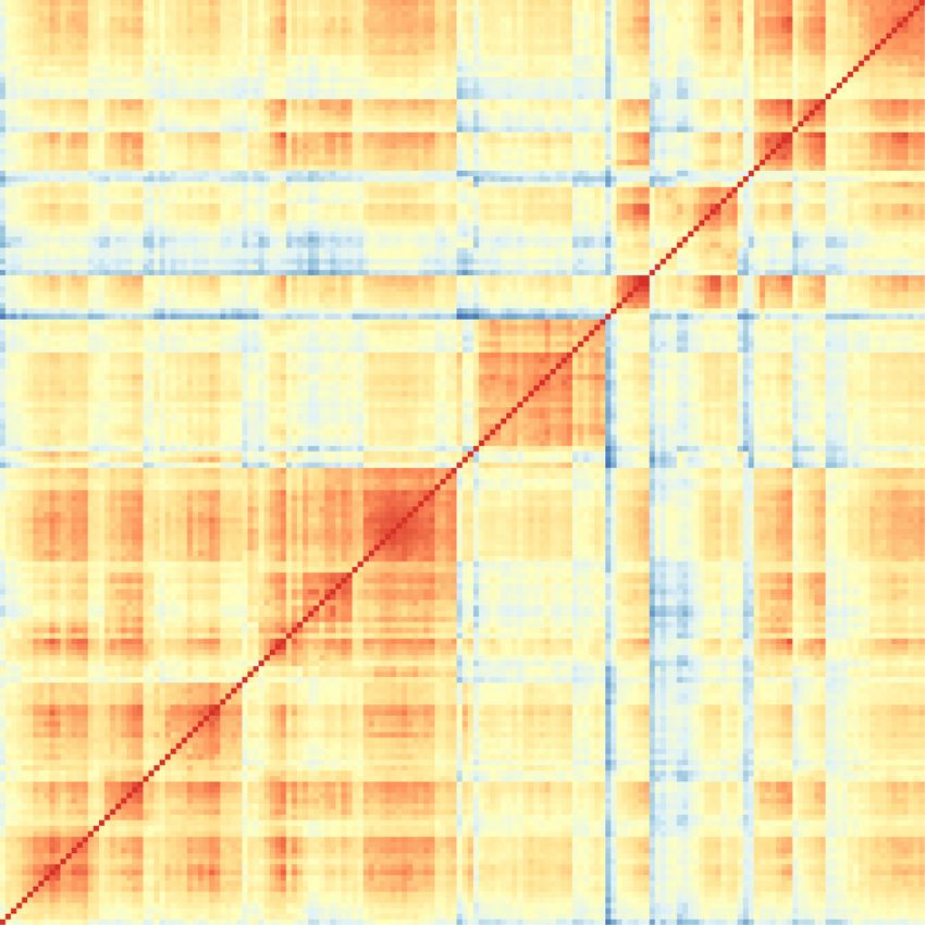
Downloaded from genome.cshlp.org on November 23, 2020 - Published by Cold Spring Harbor Laboratory Press

Nichterwitz et al.

      SMN is ubiquitously expressed and its complete depletion           sion, we analyzed several disease stages. We included a presymp-
leads to early embryonic lethality (Schrank et al. 1997). In SMA,        tomatic stage (P2) and an early symptomatic stage (P5) when
somatic motor neurons are for unknown reasons selectively vul-           motor neuron loss in this model is restricted to discrete regions
nerable to the lower level of SMN protein. However, different            of the spinal cord (toward rostral and medial levels) (Mentis et al.
somatic motor neuron groups show varying degrees of susceptibil-         2011). We also included a symptomatic stage (P10) when the
ity to degeneration. Spinal motor neurons are the primarily affect-      Smn-deficient mice have clear motor dysfunction and show a
ed cell type in disease. Facial motor neurons and hypoglossal            more widespread loss of spinal motor neurons (Fig. 1A; Le et al.
motor neurons that innervate the tongue are to some extent affect-       2005; Mentis et al. 2011). We applied laser capture microdissection
ed in severe cases of the human disease (Rudnik-Schöneborn et al.        (LCM) to isolate neurons from different regions of the brainstem
2009; Petit et al. 2011; Harding et al. 2015). Neuromuscular junc-       and spinal cord (Fig. 1A; Supplemental Fig. S1) coupled with
tions (NMJs), the specialized synapses between motor neurons and         poly(A)-enriched RNA sequencing (LCM-seq) (Nichterwitz et al.
muscle, of facial motor neurons present pathology in mouse mod-          2016, 2018). We collected somatic motor neurons from the oculo-
els of SMA (Murray et al. 2008) while hypoglossal NMJs remain un-        motor and trochlear nuclei [cranial nuclei 3 and 4 (CN3/4)]
affected (Comley et al. 2016). Ocular motor neurons, which               (Supplemental Fig. S1D–F) and the hypoglossal nucleus [cranial
innervate extraocular muscles and thus control movement of the           nucleus 12 (CN12)] (Supplemental Fig. S1M–O) that are resistant
eyes, appear consistently resistant in SMA. This is evidenced by         to degeneration in this SMA model. We also isolated relatively vul-
the use of ocular tracking devices as a communication tool for pa-       nerable somatic motor neurons from the facial nucleus [cranial nu-
tients (Kubota et al. 2000) and that NMJs are preserved in extraoc-      cleus 7 (CN7)] (Supplemental Fig. S1G–I) and along the lumbar
ular muscles of end-stage SMA mice (Comley et al. 2016).                 spinal cord (SC) (Supplemental Figs. S1P–R, S2A,B). To ensure
      Genes active within specific neuronal types define their           the inclusion of vulnerable neurons within motor neuron popula-
unique identities and functions in health as well as their suscept-      tions with mixed vulnerabilities over time, for example, CN7 neu-
ibility to specific neurodegenerative diseases. Data from SMA ani-       rons innervating the rostral versus the caudal band of the levator
mal models and SMA patient motor neurons derived from                    auris longus muscle (Murray et al. 2008) or neurons in the medial
induced pluripotent stem cells (iPSCs) indicate that factors intrin-     and lateral motor columns in the spinal cord (Mentis et al. 2011),
sic to motor neurons are important for degeneration (Park et al.         we collected cells across the entire motor neuron nuclei.
2010; Corti et al. 2012; Van Hoecke et al. 2012). Thus, investigat-            Furthermore, we collected visceral motor neurons from the
ing cell intrinsic pathways that are differentially activated within     dorsal motor nucleus of the vagus nerve (vagus motor neurons)
resistant and vulnerable motor neurons could reveal mechanisms           [cranial nucleus 10 (CN10)] (Supplemental Fig. S1J–L) to deduct
of selective neuronal degeneration and lead to therapies prevent-        events occurring within all cholinergic motor neurons versus
ing progressive motor neuron loss (Hedlund et al. 2010; Kaplan           those specific for somatic motor neurons. Visceral motor neurons
et al. 2014; Comley et al. 2015; Murray et al. 2015; Allodi et al.       are generally considered more resilient to SMA disease processes
2016; Nijssen et al. 2017; Allodi et al. 2019).                          than somatic motor neurons and were thus also included as a rel-
      Previous transcriptome studies in SMA have compared vul-           atively resistant neuronal population. We also isolated red nucleus
nerable patient-derived motor neurons (Ng et al. 2015) or whole          neurons (RN) (Supplemental Fig. S1A–C), which are noncholiner-
spinal cords isolated from SMA mice (Zhang et al. 2008; Bäumer           gic neurons involved in motor coordination that are resilient to
et al. 2009; Murray et al. 2010; Staropoli et al. 2015) with healthy     degeneration in SMA, to elucidate disease-induced transcriptional
controls. These studies have improved our understanding of motor         regulation selective to cholinergic neurons versus more broad reg-
neuron disease mechanisms but could not explain how the loss of          ulation across neuronal populations. We thus acquired an exten-
a ubiquitously expressed protein can induce degeneration in a se-        sive library of six neuronal populations at three different time
lect cell type. Zhang et al. (2013) included unaffected cells from the   points throughout disease progression in health and SMA.
white matter in their analysis and a more recent study by Murray               After conducting LCM-seq, we performed a quality control,
et al. (2015) investigated transcriptional changes in differentially     using only samples in which we achieved a gene detection level
affected motor neuron pools. However, these studies were restrict-       of greater than 11,000 expressed genes (Supplemental Fig. S3A),
ed to a single presymptomatic stage, limiting the scope of the find-     which left a total of 168 samples for further analysis
ings. To unravel temporal mechanisms of neuronal resilience and          (Supplemental Table S1). To evaluate the purity of the LCM-seq
susceptibility, we have conducted a comprehensive longitudinal           samples, we analyzed the level of neuronal and glial markers and
analysis of resistant and vulnerable neuron groups from a pre-           compared these to a published RNA sequencing data set of neu-
symptomatic stage to early and late symptomatic stages. We               rons, astrocytes, oligodendrocytes, and microglia (NCBI Gene
used laser capture microdissection coupled with RNA sequencing           Expression Omnibus [GEO] accession number GSE52564)
(LCM-seq) (Nichterwitz et al. 2016, 2018) to profile discrete neuro-     (Zhang et al. 2014). The neuronal markers neurofilament heavy
nal populations in SMA mice and littermate controls over time.           chain (Nefh) and peripherin (Prph) were highly expressed in all
                                                                         LCM-seq samples. The motor neuron markers choline acetyl trans-
                                                                         ferase (Chat) and Islet-1/2 were readily detected in all motor neu-
Results                                                                  ron groups, whereas Hb9 (Mnx1) expression was largely restricted
                                                                         to SC and CN12 motor neurons. Glial markers, Gfap, Mfge8,
Transcriptional profiling of neurons with differential                   Sox10, Pdgfrb, Enpp6, and Mog were detectable in all neuron sam-
susceptibility to degeneration reveals cell type–specific gene           ples, but at much lower levels than in glial populations, whereas
expression                                                               the macrophage/microglia markers Itgam (CD11b) and Ccl3 were
To investigate the transcriptional dynamics of neuronal popula-          absent from our neuronal samples (Supplemental Fig. S3C). This
tions with differential vulnerability in SMA, we used the widely         cross-comparison showed that the LCM-seq samples were highly
studied “delta7” mouse model (Smn −/−/SMN2 +/+/SMNΔ7 +/+).               enriched in neuronal transcripts and only included minor con-
Because we were interested in longitudinal changes in gene expres-       taminations of glial transcripts. We analyzed the sustained

2    Genome Research
       www.genome.org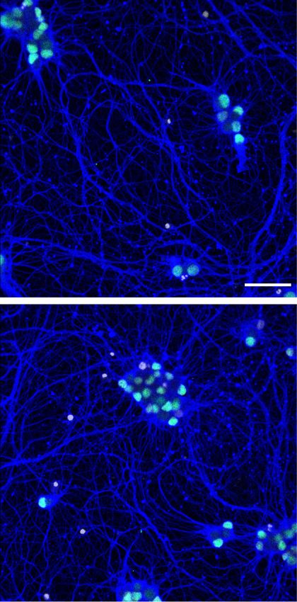
Downloaded from genome.cshlp.org on November 23, 2020 - Published by Cold Spring Harbor Laboratory Press

                                                                                        Transcriptome of neuronal resilience in SMA

         A

         B                                                                  C

Figure 1. LCM-seq strategy to reveal cell intrinsic mechanisms of motor neuron vulnerability and resistance in SMA. (A) We used “delta 7” mice
(Smn −/−/SMN2 +/+/SMNΔ7 +/+) as a model for severe SMA and littermates homozygous for murine Smn as controls (Smn +/+/SMN2 +/+/SMNΔ7 +/+).
(B) Pairwise Spearman’s correlation on log10-transformed data of all samples per cell type, genotype, and age. (C) Principal component analysis on
the whole gene expression data set. (P) Postnatal day; (CNS) central nervous system; (LCM-seq) laser capture microdissection coupled with RNA se-
quencing; (SC) spinal cord; (CN) cranial nerve; (CN12) hypoglossal nucleus; (CN10) dorsal motor nucleus of the vagus nerve; (CN7) facial nucleus;
(CN3/4) oculomotor and trochlear nuclei; (RN) red nucleus; asterisks in A indicate vulnerable cell types in this mouse model; (WGCNA) weighted
gene correlation network analysis; (GO) Gene Ontology.

homeobox transcription factor (Hox) gene profiles of collected             CN3/4 motor neurons clustered closely to RN neurons consistent
neurons (Hedlund et al. 2010; Nichterwitz et al. 2016), which con-         with their specification in the ventral midbrain (Deng et al.
firmed their anterior-posterior positions along the body axis. RN          2011). CN7, CN12, and SC motor neurons formed a dense cluster,
and CN3/4 neurons, which are located in the midbrain were, as ex-          whereas visceral CN10 motor neurons clustered distinct from all
pected, devoid of Hox gene expression (Supplemental Fig. S3D).             the other cell populations. Altogether, we could show that biolog-
Together, these data confirmed the identity, purity, and high qual-        ical replicates show high reproducibility and that neuronal groups
ity of the LCM-seq neuronal samples.                                       form distinct clusters indicative of a high sensitivity.
      To investigate reproducibility among biological replicate sam-             In-depth gene expression analysis across neuron types con-
ples and to determine correlations between different neuron types,         firmed known marker gene expression and identified several novel
we conducted pairwise Spearman’s correlation. This analysis                cell type–specific transcripts. We confirmed their mRNA localiza-
showed a high correlation between samples originating from the             tion in the adult central nervous system using the Allen Mouse
same neuron type and revealed a close relationship between SC,             Brain Atlas (available at https://portal.brain-map.org). We could
CN7, and CN12 motor neurons (Fig. 1B). After unsupervised hier-            show that Cxcl13 was restricted to RN neurons (Supplemental
archical clustering, midbrain neurons (RN and CN3/4) were dis-             Fig. S4A). The transcription factor Lhx4 was preferentially ex-
tinct from other brainstem and SC motor neurons, and visceral              pressed in CN3/4 motor neurons (Supplemental Fig. S4B), but
CN10 motor neurons clustered separately within this group (Fig.            Shox2 was present in CN7 motor neurons (Supplemental Fig.
1B). Principal component analysis (PCA) on the entire gene ex-             S4C). The peptide hormone Nppb distinguished visceral CN10 mo-
pression data set revealed clustering of cell types with a strong in-      tor neurons from the other cell types (Supplemental Fig. S4D),
fluence of their developmental origin (Fig. 1C). Consequently,             whereas the proteoglycan Dcn was a marker for CN12 motor

                                                                                                                     Genome Research            3
                                                                                                                        www.genome.org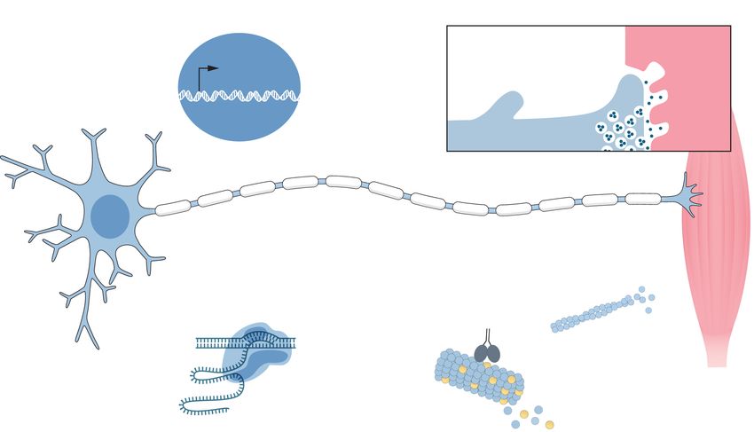
Downloaded from genome.cshlp.org on November 23, 2020 - Published by Cold Spring Harbor Laboratory Press

Nichterwitz et al.

neurons (Supplemental Fig. S4E). We could thus validate the cell            the early (P2) and late (P10) time points and changed over time
type–specific expression from the RNA-seq analysis with available           (from a negative to a positive correlation or vice versa) (Supplemen-
in situ data and identify unique cell type–specific markers.                tal Fig. S6A,B; Supplemental Table S2). PCA with a total of 5843
      Spinal motor neurons display inefficient SMN2 exon 7 inclu-           genes in these modules confirmed the separation of somatic motor
sion compared to other cells in the spinal cord, thus rendering             neurons based on age along the first principal component (PC1)
these cells low in full-length SMN (Ruggiu et al. 2012). To investi-        (Supplemental Fig. S6C), supporting a change in expression of
gate if SMN2 splicing differences could account for the differential        these genes during normal postnatal development. Gene Ontology
susceptibility among the neuron types investigated here, we first           (GO) analysis revealed enrichment of several terms related to devel-
specifically examined the expression levels of full-length SMN2             opment (Supplemental Fig. S6D), and we used the 1341 genes be-
mRNA by qPCR (Supplemental Fig. S5A). In many of the samples                longing to these terms (Supplemental Table S2) to perform PCA
we could not detect any exon 7 inclusion, and where we did, the             of control and SMA samples. As expected, the PCA showed a clear
signal was close to the detection threshold (Supplemental Fig.              age component between P2 and P10 samples. However, SMA sam-
S5B). We also analyzed the RNA-seq data for exon 7 inclusion.               ples did not appear to cluster differently on the age axis (PC1) com-
By pooling aligned reads from several samples within each neuron            pared to control samples (Fig. 2A). To better visualize the position
group, we could obtain enough read coverage of SMN2 to make a               of the SMA samples along the “age component,” we plotted each
quantitative statement. Although we cannot exclude contribu-                cell type separately along PC1. We did not observe any difference
tions from the near-identical SMNΔ7 expression construct in this            in the age component in P2 and P5 samples but P10 SMA samples
analysis, sashimi plots of SMN2 showed that there was no differ-            shifted toward the younger age (PC1 negative) relative to control
ence in splicing of SMN2 across the different neuron groups (Sup-           samples (Fig. 2B). Because this shift occurred only at a time when
plemental Fig. S5C), supporting the qPCR analysis. The known                SMA mice are visibly affected by disease, it likely reflects a disease
SMN splicing target Uspl1 served as a positive control for the detec-       process rather than a general developmental delay. In support of
tion of splicing differences in the LCM-seq data. Thus, the differen-       this, only 10.1% (136 genes) of all developmentally regulated genes
tial vulnerability of the neuron types investigated is not explained        were also significantly differentially expressed in disease (Fig. 2C;
by differences in exon 7 splicing efficiency. The RNA-seq data              Supplemental Table S2), confirming that the majority of age-regu-
therefore warrant further investigation to identify cell intrinsic          lated genes are not affected in SMA. Further, if there was indeed a
mechanisms of selective neuronal vulnerability in SMA.                      developmental delay in SMA motor neurons, we would expect to
                                                                            find genes regulated in the opposite directions in development
                                                                            and disease. However, of the 71 genes with opposite regulation,
SMA mice do not present a general developmental                             76% (54 genes) were only regulated at the latest time point in dis-
delay as determined by gene expression and neuromuscular                    ease, supporting our findings from the PCA (Fig. 2A,B). Altogether,
junction analyses                                                           our data challenge the notion that SMA motor neuron somas dis-
It has been described that SMA patients and transgenic mouse                play a general developmental delay.
models display a developmental delay in their neuromuscular sys-                  To exclude any possible peripheral developmental phenotype
tems (for review, see Hausmanowa-Petrusewicz and Vrbová 2005).              in the neuromuscular system in SMA mice, we quantified the levels
Differences in the developmental state of SMA and control motor             of poly-innervation and endplate perforations in target muscle
neurons that may not reflect a pathological mechanism per se                groups. These measures are excellent indicators of early postnatal
could hamper the identification of disease-relevant transcriptional         developmental stages until P14 in mice. Our analysis of extraocular
changes. We thus addressed this issue here using the gene expres-           muscles (EOM; innervated by CN3/4 motor neurons), tongue mus-
sion data. We first used weighted gene correlation network analysis         cles (innervated by CN12 motor neurons), and lumbrical muscles
(WGCNA) to identify gene sets that were regulated during early              from the hind limbs (innervated by lumbar SC motor neurons)
postnatal development of control somatic motor neurons. We                  showed that the level of poly-innervation was equal in SMA mice
chose the three modules that were most highly correlated with               and control littermates (multiple two-tailed t-tests) (Supplemental

          A                                                      B                                              C

Figure 2. Evaluation of the developmental state of SMA somatic motor neuron somas. (A) PCA with development-related genes (DGs, 1341 genes) of all
somatic motor neurons in control and SMA. (B) Plot of PC1 alone to better visualize the “age component.” (C) Venn diagram depicting overlap between
DGs and all differentially expressed genes between control and SMA (DEGs, no fold-change cutoff, Padj < 0.05) per cell type and time point.

4    Genome Research
       www.genome.org
Downloaded from genome.cshlp.org on November 23, 2020 - Published by Cold Spring Harbor Laboratory Press

                                                                                         Transcriptome of neuronal resilience in SMA

Fig. S7A; Supplemental Table S3). We could also show that perfora-           positively correlated with disease and negatively correlated with
tions were completely unaffected by disease in tongue muscle, and            control samples (Supplemental Table S8), which we will refer to
only very slightly affected in extraocular muscles at end-stage of           as “disease module.” As shown by the mean eigengene values for
disease (P14, multiple two-tailed t-tests, Padj < 0.05). As expected,        the module within sample replicates, there was a clear genotype
lumbrical muscle endplates were severely affected at late stages of          separation with disease progression in all somatic populations in-
disease, lacking in perforations compared to control muscles (mul-           dependent of their vulnerability (Fig. 3C). We plotted all genes of
tiple two-tailed t-tests, Padj (P10) < 0.05, Padj (P14) < 0.0001) (Supple-   the module in a heatmap, which confirmed the similar expression
mental Fig. S7B; Supplemental Table S4). Collectively, the NMJ               levels across all somatic motor neurons (Supplemental Fig. S8E).
poly-innervation and endplate perforation data show that there               Conversely, the relatively unaffected RN and CN10 neurons did
was no obvious developmental delay in the maturation of neuro-               not show separation based on disease and displayed unique re-
muscular synapses in agreement with the transcriptome data. We               sponses to disease (Supplemental Fig. S8F). Besides Smn, only
therefore conclude that there is no major developmental delay in             one gene of the disease module, Plek2, was differentially expressed
the motor system of the SMA mouse model, but that it is affected             in RN, and four genes, Lars2, Olig2, Ptgds, and Ubap1l, were regulat-
as disease progresses. Consequently, the same ages in control and            ed in CN10 motor neurons with disease (Supplemental Fig. S8G).
SMA mice can be compared to distinguish disease-induced tran-                Furthermore, 45% of the genes (113 genes) in the module were
scriptional changes without developmental processes obscuring                identified as significantly differentially expressed with disease in
the data sets.                                                               one or more somatic populations using DESeq2 (Fig. 3D), includ-
                                                                             ing 14 of the 19 DEGs that are common to all somatic motor neu-
                                                                             rons. We thus identified a disease signature specific to somatic
SMA-induced gene expression changes imply a common                           motor neurons independent of their vulnerability. GO analysis
TRP53-mediated stress response in vulnerable and resistant                   for biological processes resulted in the significant (adjusted P-val-
somatic motor neurons                                                        ue < 0.05) enrichment of 23 GO terms in total (Fig. 3E;
Toward our main goal of elucidating mechanisms of neuronal re-               Supplemental Table S9). The pathways we identified as regulated
silience and susceptibility, we investigated the transcriptional dy-         included, for example, apoptotic signaling, signal transduction in-
namics in resistant and vulnerable neurons in SMA. Using DESeq2              duced by p53 class mediator, positive regulation of intracellular
(Love et al. 2014), we performed pairwise differential expression            signal transduction, spliceosomal tri-snRNP complex assembly,
analyses between control and SMA per cell type and time point.               and several terms suggesting changes in metabolism (proteolysis,
We found the strongest early transcriptional response in resistant           hydrolase activity) as well as cellular component organization
CN3/4 and CN12 motor neurons with 134 and 211 differentially                 (e.g., organelle organization, supramolecular fiber organization,
expressed genes (DEGs; no fold-change cutoff, adjusted P-value               cellular component biogenesis). To better visualize the timing of
Downloaded from genome.cshlp.org on November 23, 2020 - Published by Cold Spring Harbor Laboratory Press
Nichterwitz et al.
A B
C D
F
E
Figure 3. Analysis of disease-induced gene expression changes in SMA somatic motor neurons. (A) Number of significant genes from pairwise differential
expression analysis per cell type and time point (DEGs, no fold-change cutoff, Padj < 0.05). Numbers on bars represent total numbers of DEGs. (B) Venn
diagram depicting the overlap in gene expression changes between somatic motor neurons (number of up-/down-regulated genes in SMA, all time points
combined). (C) Mean eigengene values (first principal component of the disease module) within replicates. (D) Venn diagram depicting the overlap be-
tween genes in the disease module and DEGs. (E) GO term analysis for biological processes of the 251 genes in the disease module. Shown are selected GO
terms; a complete list of enriched terms can be found in Supplemental Table S9. Numbers indicate the number of genes in a given term, color scale is the
adjusted P-value. Asterisks indicate gene sets that are plotted in F. (F ) Expression heatmap of genes that belong to GO terms related to apoptosis and TRP53
signaling. Expression values were log2-transformed and mean centered.
responses (Fig. 4A; Supplemental Table S10). There was no overlap ron type. Specifically, at P5, three genes were commonly up-regu-
between CN3/4 and SC motor neurons in their early response to lated in CN3/4 and SC motor neurons; at P10, 56 genes were
loss of Smn (Fig. 4B; Supplemental Table S11). Forty-three percent commonly up-regulated and 11 down-regulated (Fig. 4B). Thus,
of all CN3/4-regulated genes at P2 belong to the GO term “nucle- 100% of the genes regulated at P2 were unique to CN3/4 motor
us,” including several genes involved in RNA processing and tran- neurons, 97% at P5, and 80% at P10.
scriptional activation/repression such as Cnot9, Ube2b, Wtap, GO term analysis of DEGs in CN3/4 neurons at P10 pinpoint-
Cbx6, Hdac6, Ino80, and Jmjd1c (Supplemental Table S9), suggest- ed a number of fundamental processes that were activated in re-
ing an early fine-tuning of gene expression. As disease progressed, sponse to disease. These pathways included neurogenesis,
more genes were jointly regulated across the neuron groups but the nervous system development, positive regulation of neuron projec-
majority of transcriptional changes were still unique to each neu- tion development, regulation of cell communication, regulation of
6 Genome Research
www.genome.orgDownloaded from genome.cshlp.org on November 23, 2020 - Published by Cold Spring Harbor Laboratory Press
Transcriptome of neuronal resilience in SMA
A B scriptional response. The respective net-
work for DEGs in SC motor neurons
consisted of only 61 genes (25.5% of all
DEGs) and a second major network in-
cluded 22 genes (8.9% of all DEGs)
(Supplemental Fig. S12). TRP53 and 15
of the directly interacting proteins were
up-regulated in both neuron types
(Supplemental Figs. S11, S12, gray out-
lines), in line with the GO term analysis.
Up-regulated genes involved in DNA
damage repair, were Polk (shared), Tnks2
and Mgmt (CN3/4-specific), and Rad51d
and Timeless (SC-specific). CN3/4-specif-
ic down-regulation included pro-apopto-
tic factors like Itpr1 and Dffa, which was
accompanied by the up-regulation of
anti-apoptotic and survival factors such
as Pak4, Pak6, Chl1, Tmbim4, Aldh4a1,
and Gdf15. Neurotransmitter release is
impaired in motor nerve terminals in
SMA (Ruiz and Tabares 2014). It is there-
C fore compelling to see the CN3/4-specific
regulation of genes involved in neuro-
transmitter release, including the up-
regulation of Syt1, Syt5, and Cplx2, sug-
gesting a compensatory mechanism in
the disease-resistant cells. Among the
many DEGs that are implicated in cyto-
skeletal reorganization, we found CN3/
4-specific regulation of genes that are im-
portant for neurite outgrowth including
Gap43, Chl1, Syt1, Cald1, and Serpine2.
(Fig. 5A,C; Supplemental Fig. S11).
In contrast, in vulnerable SC motor
neurons, we found increased levels of
Inf2, which can disassemble actin fila-
ments (Supplemental Table S5), and the
tubulin isoform Tubb6, which is associat-
ed with decreased microtubule stability
(Bhattacharya and Cabral 2004; Salinas
et al. 2014). We also found a significant
decrease in mRNA levels of several motor
proteins (Dnahc, Kif3a, Kif5a) including
genes that function in the anterograde
transport of a variety of cargos to the
Figure 4. Comparison of gene expression changes in ocular and spinal motor neurons. (A) Venn dia-
grams depicting shared and time point–specific DEGs between control and SMA motor neurons in CN3/ cell periphery including the synapse
4 (top) and SC (bottom). (B) Venn diagrams illustrating the overlap of DEGs between CN3/4 and SC at (Fig. 5B,C; Supplemental Fig. S12).
each time point. (C) GO term analysis of DEGs in CN3/4 and SC per time point. Shown are selected Thus, we identified multiple tran-
terms; a complete list of enriched terms can be found in Supplemental Table S9. Numbers indicate scriptional programs specifically activat-
the number of genes in a given term, and the color scale shows the adjusted P-value. Terms belong to
ed in ocular motor neurons in SMA that
the domain biological processes unless specified otherwise: (CC) cellular compartment; (MF) molecular
function. could confer protection against detri-
mental disease processes.
apoptotic processes, and cell death (Fig. 4C; Supplemental Table The oculomotor-regulated factor GDF15 confers protection
S9). Among the enriched cellular compartments were neuron pro- onto human spinal motor neurons
jection and synapse. To visualize CN3/4-restricted pathways that
could be protective, we used the STRING database to retrieve pro- Next, we wanted to confirm our hypothesis that adaptation mech-
tein–protein interaction networks from all DEGs at P10. We ob- anisms identified in resilient neurons can be used to reduce sus-
tained a highly interconnected protein network that consisted of ceptibility of vulnerable motor neurons. We thus analyzed the
158 genes, corresponding to 46.7% of all DEGs in CN3/4 at P10 effect of adding GDF15, which was highly up-regulated uniquely
(Supplemental Fig. S11), thus indicating a highly coordinated tran- in resilient CN3/4 motor neurons in SMA, onto vulnerable spinal
Genome Research 7
www.genome.orgDownloaded from genome.cshlp.org on November 23, 2020 - Published by Cold Spring Harbor Laboratory Press
Nichterwitz et al.
A B
C
Figure 5. Common and cell type–specific disease mechanisms in SMA. Expression changes in key genes between SMA and wild type in CN3/4 (A) and SC
motor neurons (B). Colors indicate significance levels. (C) Somatic motor neurons display transcriptional changes caused by the loss of full-length SMN
protein that are distinct from red nucleus and vagus (CN10) neurons. For example, prominent changes in expression levels of genes that function in
RNA processing are restricted to somatic motor neurons. These neurons are furthermore exposed to cellular stress, including oxidative stress and DNA dam-
age, and DNA repair genes are induced. TRP53- and cell death signaling pathways are activated in all somatic motor neurons independent of their suscep-
tibility to degeneration in SMA. Vulnerable spinal (SC) motor neurons show signs of axon degeneration and axonal transport deficits. Resistant ocular (CN3/
4) motor neurons selectively up-regulate the expression of genes that counteract apoptosis and promote cell survival. Increased levels of genes functioning
in neurite outgrowth, axon regeneration, and neurotransmission, which support the maintenance of a functional neuromuscular synapse, are also seen in
ocular motor neurons in disease.
motor neurons. For this purpose, we generated human spinal mo- up-regulated in CN3/4 motor neurons with disease and could pro-
tor neurons from induced pluripotent stem cells (iPSCs) according tect vulnerable spinal motor neurons from degeneration when
to established protocols (Fig. 6A; Guo et al. 2017; Nijssen et al. added to these.
2019). We first showed that these human neurons degenerate
over time in culture in a growth factor–deprivation assay (Fig.
6B; Lamas et al. 2014). We then showed that addition of GDF15
Discussion
to these vulnerable spinal motor neurons significantly improved In this study, we conducted a longitudinal analysis of the tran-
their survival in a dose-dependent manner (Fig. 6C–F). The effect scriptional dynamics in CN3/4 (ocular), CN7 (facial), CN10 (va-
was evident when the cell death assay was not too harsh, days gus), CN12 (hypoglossal), SC (spinal), and RN (red nucleus)
35–42 in vitro (Fig. 6C,D,F). This is clear confirmation that targets neuron groups, which show differential vulnerabilities to degener-
identified in resilient neurons using our approach can be used to ation during disease progression in SMA mice. We found that, in-
reduce susceptibility of vulnerable motor neurons. dependent of their vulnerability, somatic motor neurons activate
Collectively, our analysis shows that resistant neurons re- TRP53 signaling pathways that are associated with DNA damage
spond early and uniquely to the loss of Smn. Arguably this re- and cell death. This up-regulation was absent in visceral vagus mo-
sponse involves the regulation of bona fide neuroprotective tor neurons and red nucleus neurons. This suggests that the activa-
factors and processes, as exemplified by Gdf15 that was uniquely tion of the TRP53 pathway is a stress response specific to somatic
8 Genome Research
www.genome.orgDownloaded from genome.cshlp.org on November 23, 2020 - Published by Cold Spring Harbor Laboratory Press
Transcriptome of neuronal resilience in SMA
A et al. 2017; Simon et al. 2017). Cell death
signaling was not restricted to vulnerable
populations in our study, but was also ev-
ident in resistant neurons, predominant-
ly at a late stage of disease (P10).
Consistently, more resistant spinal mo-
tor neurons of the lateral motor column
show increased TRP53 protein levels
B with progression of disease (Simon et al.
2017). TRP53 itself can activate a number
of targets that exert anti-apoptotic ef-
fects, such as Gtse1, Dcxr, Gpx, and
Cdkn1a (for review, see Jänicke et al.
2008), which were also up-regulated in
all the somatic populations. Specifically,
the cyclin-dependent kinase inhibitor
1A (P21) (Cdkn1a) plays a crucial role in
cell cycle regulation and response to
DNA damage. Cdkn1a can also prevent
C D E F apoptosis, for instance, by transcription-
al repression of pro-apoptotic genes or in-
hibiting caspases (for review, see Gartel
and Tyner 2002). Thus, the up-regulation
of Cdkn1a could be part of a protective re-
sponse in somatic motor neurons, which
is not sufficient to protect the most vul-
nerable cells. Apparent shared protective
responses may also in part stem from the
enrichment of more resilient neurons
within the vulnerable populations with
time, for example, enrichment of the
more resilient lateral spinal motor neu-
rons versus more medial motor neurons
(Mentis et al. 2011) and CN7 neurons in-
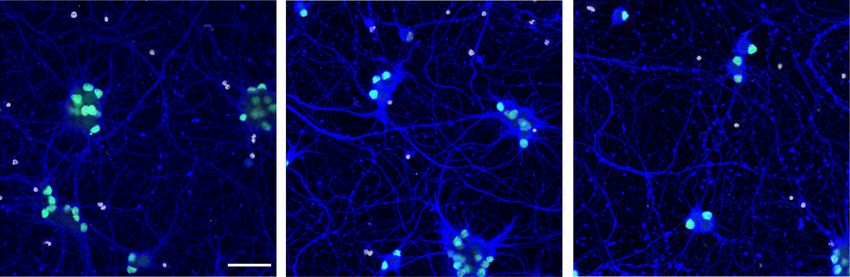
Figure 6. The oculomotor-enriched factor GDF15 protects human spinal motor neurons. (A) nervating the rostral versus caudal band
Schematic of the differentiation protocol for human iPSCs into motor neurons and the treatment with
of the levator auris longus muscle (Mur-
GDF15. (B) Human spinal motor neurons degenerate over time in culture when grown without growth
factors. Representative images show immunostaining with antibodies against ISLET-1/2 (ISL1/2) and ßIII- ray et al. 2008). Back-labeling tech-
tubulin (TUJ1). (C ) At day 35 in vitro (DIV35), addition of 50 or 200 ng/mL GDF15 improves survival of niques, as used by Murray et al. (2015),
motor neurons compared to control, but 10 ng/mL had no effect. (D) At DIV 42, 50 and 200 ng/mL or single-cell RNA sequencing (Nichter-
GDF15 protects motor neurons. (E) At DIV 49, there is no significant effect of GDF15 at concentrations
witz et al. 2016; Hedlund and Deng
10–200 ng/mL. (F) Representative images showing motor neurons treated with 10 ng/mL or 50 ng/mL
GDF15 at DIV42. The scale bar in B and F is 50 µM. Student’s t-test: (∗∗∗ ) P < 0.001; (∗∗ ) P < 0.01; (∗ ) P < 2018) could further aid in the distinction
0.05; (^) P < 0.1. of vulnerable and resistant cells within
neuronal populations to further dissect
motor neurons in SMA but that it does not necessarily lead to protective from detrimental pathways. However, even at P10, so-
degeneration as ocular motor neurons persist. We also show that mas of vulnerable motor neurons that show peripheral pathology
the majority of gene expression changes induced by the loss of (Comley et al. 2016) are still present in the spinal cord. Thus, al-
Smn are cell type–specific and reveal several pathways that are re- though we cannot exclude that some detrimental pathways were
stricted to resistant ocular motor neurons. Such bona fide protec- masked by the inclusion of more resilient neurons, we are confi-
tive pathways include increased levels of survival factors, and dent that we have included vulnerable cells at all time points.
pro-apoptotic genes are selectively down-regulated (Fig. 5A–C). With our comprehensive comparison of resistant ocular and
We also observed ocular motor neuron-restricted transcriptional vulnerable spinal motor neurons using Gene Ontology and pro-
regulation of genes involved in neurotransmission and neurite tein network analyses, we were able to pinpoint several protective
outgrowth that may aid in the maintenance of a functional neuro- pathways that are selectively regulated in the resilient motor neu-
muscular synapse and thus contribute to the selective resistance of ron group and thus likely counteract commonly activated stress re-
these motor neurons in SMA. sponses. These are pathways that appear particularly compelling to
TRP53, which was up-regulated across all somatic motor neu- modulate in susceptible neurons to make these more resilient to
ron groups in SMA, is a master regulator in response to several cel- degeneration. Prosurvival factors with increased levels in ocular
lular stressors such as oxidative stress or DNA damage. SMN motor neurons in SMA were, for instance, (1) the growth differen-
directly interacts with TRP53 (Young et al. 2002), and transcrip- tiation factor 15 (Gdf15), which can protect dopamine neurons
tional activation of Trp53 or its target genes has been previously from Parkinson-like 6-hydroxydopamine-induced degeneration
shown in different models of SMA (Zhang et al. 2008, 2013; (Strelau et al. 2000). GDF15-deficient mice show a progressive
Bäumer et al. 2009; Murray et al. 2015; Staropoli et al. 2015; Jangi loss of motor neurons in spinal cord and several brain stem nuclei,
Genome Research 9
www.genome.orgDownloaded from genome.cshlp.org on November 23, 2020 - Published by Cold Spring Harbor Laboratory Press
Nichterwitz et al.
suggesting that it is a genuine trophic factor for motor neurons help these to reconnect to muscle targets if disconnected during
(Strelau et al. 2009). (2) Leukemia inhibitory factor (LIF) promotes disease.
survival of primary rat and mouse motor neurons grown in vitro Furthermore, our functional analysis supports the idea that
(Martinou et al. 1992; Arce et al. 1999). Lif knockout mice display an up-regulation of prosurvival factors could in part account for
loss of distal motor axons and decrease in motor endplate size, in- neuronal resilience as the oculomotor-regulated factor GDF15 pro-
dicating that it also has an important role for maintaining motor tected vulnerable human spinal motor neurons from degenera-
neuron connectivity with muscle (Holtmann et al. 2005). (3) tion. The protective effect was most prominent during the first
The mitochondrial aldehyde dehydrogenase ALDH4A1 can safe- 21 d of the growth-deprivation assay (up until day 42).
guard cells from oxidative stress (Yoon et al. 2004). There is recent Thereafter, the assay becomes very harsh with a 58% drop in motor
evidence for increased oxidative stress linked to mitochondrial neurons within a 7-d period, and a rescue is thus much more diffi-
dysfunction in primary motor neuron cultures from SMA mice cult to orchestrate. The neuroprotection seen is very encouraging
(Miller et al. 2016). (4) CDKN1A activated kinases PAK6 and because it shows that factors up-regulated within ocular motor
PAK4; PAK4 prevents caspase activation and thus apoptosis neurons with disease are protective and that using the response
(Gnesutta et al. 2001; Gnesutta and Minden 2003) and its neuro- of resilient neurons to disease can identify pathways that can be
protective function was recently shown in a rat model of used to protect also vulnerable neurons. Although GDF15 alone
Parkinson’s disease (Won et al. 2016). (5) TMBIM4, also known can support motor neuron survival for an extended amount of
as Lifeguard4, shows anti-apoptotic functions likely through mod- time, we believe that treatment with a combination of factors
ulation of intracellular Ca2+ (for review, see Carrara et al. 2017). up-regulated within ocular motor neurons will be even more ben-
Consistently, the Ca2+ channel Itpr1 (IP3 Receptor), which plays eficial for motor neuron survival. Future studies will therefore aim
a role in endoplasmic reticulum (ER) stress-induced apoptosis, to determine the most optimal cocktail of ocular motor-regulated
was selectively down-regulated in ocular motor neurons. The acti- factors through gain- and loss-of-function studies.
vation of ER stress pathways in SMA was shown in a transcriptom- In summary, the transcriptional regulation of genes related to
ics study using iPSC-derived motor neurons from SMA patients (Ng neuroprotection, neurotransmission, and neurite outgrowth pre-
et al. 2015). Thus, resistant ocular motor neurons appear to, by ne- sents compelling bona fide protective mechanisms that are activat-
cessity, regulate pathways that counteract the detrimental process- ed during disease progression selectively in these resistant motor
es that are activated with disease in all somatic motor neurons. neurons.
Other resistant neurons groups, including red nucleus and visceral In conclusion, this study provides important insights into
motor neurons showed no such compensatory gene regulation, mechanisms of selective resistance and vulnerability in SMA. We
but also lacked DNA damage pathway and TRP53 activation. show that all somatic motor neurons, irrespective of their vulner-
As SMN also functions locally in the axon, including in nerve ability in SMA, present stress responses owing to SMN deficiency.
terminals, preventing apoptosis is unlikely to fully rescue motor However, resistant ocular motor neurons selectively activate sur-
neuron function. We therefore believe that genes involved in neu- vival pathways, including Gdf15, Lif, and Chl1, which could pro-
rotransmission and neurite outgrowth, that would affect neigh- tect vulnerable spinal motor neurons from degeneration, and
boring neurons and muscle in addition to motor neurons show transcriptional regulation of genes that are important for
themselves, are important candidates for neuroprotection. Excit- the maintenance and/or regeneration of a functional neuromuscu-
ing candidates that were specifically up-regulated in ocular motor lar synapse. The modulation of such mechanisms presents a prom-
neurons with disease were synaptotagmin 1 and 5 (Syt1 and Syt5) ising strategy, not only for the additional treatment of SMA
(Fig. 5A,C). SYT1 functions in the release of synaptic vesicles and patients in which splicing correction of SMN2 is not sufficient
has recently been associated with differential vulnerability in but also other motor neuron diseases like ALS. We thus revealed
SMA (Tejero et al. 2016). By counteracting the impaired neuro- novel targets that will be exciting to investigate further, both alone
transmitter release that has been observed in SMA motor neurons and in combinations, in the context of motor neuron disease.
(Kariya et al. 2008; Ruiz and Tabares 2014), ocular motor neurons
may be able to maintain their connection to target muscles and en-
sure their functionality. In support of this, we also found com- Methods
plexin II (Cplx2) up-regulated in ocular motor neurons, which
Ethics statement and animal model
also modulates synaptic vesicle release (Ono et al. 1998). The ge-
netic depletion of Cplx2 in mice results in locomotor deficits All work was carried out in accordance with the Code of Ethics of
(Glynn et al. 2003), suggesting an important function in motor the World Medical Association (Declaration of Helsinki) and with
neurons. We also identified caldesmon 1 (Cald1), the growth asso- national legislation and institutional guidelines. Animal proce-
dures were approved by the Swedish animal ethics review board
ciated protein 43 (Gap43), and the L1 cell adhesion molecule ho-
(Stockholm Norra Djurförsöksetiska Nämnd). Ethical approval
molog Chl1 to be selectively up-regulated in ocular motor
for the use of human iPSCs was obtained from the regional ethical
neurons. CALD1 is a regulator of neurite outgrowth (Morita et al.
review board in Stockholm, Sweden (Regionala Etikprövnings-
2012), and GAP43 is crucial for developmental axon outgrowth
nämnden, Stockholm, EPN).
and regeneration. Likewise, CHL1 levels increase in regenerating
Animals were housed under standard conditions with a 12-h
motor neurons after sciatic nerve injury (Zhang et al. 2000); it is dark/light cycle and had access to food and water ad libitum.
a survival factor for primary rat motor neurons, acting via the Neonatal pups were used as a model of SMA (Smn −/−/SMN2 +/+/
PI3K/AKT pathway (Nishimune et al. 2005). It was recently shown SMNΔ7 +/+), and age matched littermates that were homozygously
that Gap43 mRNA and protein levels were reduced in axons and wild type for murine Smn (Smn +/+/SMN2 +/+/SMNΔ7 +/+) were used
growth cones of primary spinal motor neurons isolated from a as controls (Le et al. 2005) (Jackson Laboratory stock number
severe mouse model of SMA (Fallini et al. 2016), predisposing these 005025). For transcriptomics, we used 2-, 5-, and 10-d old pups
to a lower degree of reconnectivity. Thus, the SMA-induced in- (P2, P5, and P10), whereas neuromuscular junction analysis was
crease in Cald1, Gap43, and Chl1 in ocular motor neurons could performed on 5-, 10-, and 14-d old pups (P5, P10, and P14).
10 Genome Research
www.genome.orgDownloaded from genome.cshlp.org on November 23, 2020 - Published by Cold Spring Harbor Laboratory Press
Transcriptome of neuronal resilience in SMA
P2 and P5 pups were sacrificed by decapitation, and P10 and P14 were plated in clear bottom, black 96-well plates (CLS3603,
pups were anesthetized with a lethal dose of avertin (2,2,2- Corning), coated with laminin (Sigma-Aldrich), Fibronectin
Tribromoethanol in 2-Methylbutanol, Sigma-Aldrich) before (Sigma-Aldrich), and poly-L-ornithine (Thermo Fisher Scientific),
decapitation. at 17,000 cells/well in Neurobasal supplemented with B27, 10
ng/mL of both brain-derived neurotrophic factor (BDNF,
Laser capture microdissection of distinct neuronal populations Peprotech) and glial cell-derived neurotrophic factor (GDNF,
Peprotech), 200 nM retinoic acid (Sigma-Aldrich) for 1 d, and 10
Six neuronal populations (CN3/4, RN, CN7, CN10, CN12, spinal
μM DAPT (Tocris) for 4 d. After day 14, the media consequently
motor neurons) were collected using laser capture microdissection,
contained only BDNF, GDNF, and ascorbic acid; media was
as previously described (Nichterwitz et al. 2016, 2018). Brain and
changed every other day. For the growth factor deprivation,
spinal cord tissues were dissected and immediately snap frozen.
BDNF and GDNF were removed from the media at day 21. From
Coronal cryosections (12 µm) were prepared and placed onto
this time point and onward, the motor neurons were treated
PEN membrane glass slides (Zeiss). Immediately before LCM, a
with GDF15 (Peprotech) at the indicated concentrations with me-
quick histological staining was performed to visualize cells
dia changes every other day.
(Histogene, Arcturus). After 100–200 cells were collected into the
dry cap of a PCR tube, 5 µL of lysis buffer was added and samples
were snap frozen on dry ice. For a more detailed description, see Immunocytochemistry and image analysis of iPSC-derived
Supplemental Methods. motor neurons
Fixed motor neuron cultures were stained with mouse anti-ISL1/2
cDNA and sequencing library preparation (DSHB, 39.4D5) at 1:50 and rabbit anti-Tuj1 (beta-3 tubulin,
Library preparation was performed with a modified version of the Biolegend, 802001), combined with Alexa-fluor conjugated sec-
Smart-seq2 protocol (Picelli et al. 2013, 2014) and was previously ondary antibodies (Thermo Fisher Scientific). Nuclei were counter-
described in detail (Nichterwitz et al. 2016, 2018; see also stained with Hoechst 33342. Two to five replicate wells were
Supplemental Methods). Equal amounts of cDNA from up to 30 imaged per condition per experiment (12 images per well). Cells
samples were pooled per lane of a flow cell. Then, 43-bp single- were counted using the “analyze particles” function in Fiji
end sequencing was performed on an Illumina HiSeq 2000 se- (ImageJ) after thresholding. The number of (Islet-positive) cells
quencing platform resulting in an average read depth of 14.1 ± was then aggregated per well, and all data are presented as data
0.4M (mean ± SEM) reads per sample. points per well. Statistical analysis was performed using R software
for statistical computing (R Core Team 2020). For the analysis of
control samples, values are expressed as percent of motor neurons
RNA-seq data analysis
at DIV 35 and two-sample t-tests (unpaired, one-sided) were used
The RNA-seq reads were mapped simultaneously to the mm10 to compare time points. For the analysis of GDF15 treatment, val-
mouse genome assembly and the genomic sequence of human ues are expressed as percent of motor neurons in control samples
SMN2 (including introns) to the hg19 assembly using STAR (ver- within each time point and experiment. One-sample t-tests were
sion 2.4.1c) (Dobin et al. 2013). The genomic sequence of SMN2 performed for each condition (GDF15 dose) against the control
is identical between the hg19 and hg38 human genome versions; within each time point (mu = 100, one-sided). A detailed descrip-
thus, aligning the reads to either genome would give identical re- tion can be found in Supplemental Methods.
sults. Quality control was performed with rrnaseq (https://github
.com/edsgard) to ensure sufficient sequencing depth and mapping
ratios appropriate for this poly(A)-enriched sequencing strategy. Data access
We used uniquely mapped reads (69.1 ± 0.39% mean ± SEM)
All raw and processed sequencing data generated in this study have
(Supplemental Fig. S3B) for further analyses. Expression levels
been submitted to the NCBI Gene Expression Omnibus (GEO;
were determined using the rpkmforgenes.py software (http
https://www.ncbi.nlm.nih.gov/geo/) under accession number
://sandberg.cmb.ki.se/rnaseq) with the Ensembl gene annotation.
GSE115706.
Only samples with more than 11,000 detected genes (≥1 RPKM)
were included in the analysis. A detailed description of principal
component analyses (PCAs), weighted gene correlation network Competing interest statement
analysis (WGCNA), differential gene expression, GO term, and
STRING analyses is supplied in Supplemental Methods. The authors declare no competing interests.
Use of published data sets
Acknowledgments
Some samples used in this study (control SC P5 samples) were pre-
viously deposited into GEO by our laboratory with the accession We would like to thank Mattias Karlén for his excellent work in cre-
number GSE76514. For the evaluation of the purity of the samples, ating the schematics in Figures 1 and 5. We would also like to
we compared them to a previously published data set (Zhang et al. thank Marta Paterlini for providing excellent technical expertise
2014). Raw data were obtained from the Gene Expression regarding the Leica laser capture microscope. Confocal microscopy
Omnibus (GEO, accession number GSE52564) and processed as was performed in the Biomedicum Imaging Core Facility with sup-
described for our own samples. port of the Karolinska Institutet. This work was supported by
grants from the Swedish Research Council (2016-02112) to E.H.,
European Union Joint Programme for Neurodegenerative Disease
Growth factor–deprivation assay on human iPSC-derived motor (JPND) (529-2014-7500) to E.H. and R.S., and the Karolinska
neurons Institutet to E.H. C.S. was supported by a postdoctoral fellowship
For derivation of motor neurons, an established differentiation from the Swiss National Science Foundation.
protocol was used (Guo et al. 2017; Nijssen et al. 2019). After dis- Author contributions: E.H. conceived the study, and E.H. and
sociation of embryoid bodies at day 10, motor neuron progenitors R.S. supervised the project. S.N., H.S., R.S., and E.H. designed
Genome Research 11
www.genome.orgDownloaded from genome.cshlp.org on November 23, 2020 - Published by Cold Spring Harbor Laboratory Press
Nichterwitz et al.
experiments. S.N., J.N., L.H.C., I.A., M.v.d.L., C.S., and Q.D. ac- Giavazzi A, Setola V, Simonati A, Battaglia G. 2006. Neuronal-specific roles
quired data. S.N., H.S., J.N., C.S., R.S., and E.H. analyzed data. of the survival motor neuron protein. J Neuropathol Exp Neurol 65: 267–
277. doi:10.1097/01.jnen.0000205144.54457.a3
S.N. and E.H. wrote the manuscript with the help of H.S., J.N., Glynn D, Bortnick RA, Morton AJ. 2003. Complexin II is essential for nor-
C.S., and R.S. All authors edited and gave critical input on the mal neurological function in mice. Hum Mol Genet 12: 2431–2448.
manuscript. doi:10.1093/hmg/ddg249
Gnesutta N, Minden A. 2003. Death receptor-induced activation of initiator
caspase 8 is antagonized by serine/threonine kinase PAK4. Mol Cell Biol
23: 7838–7848. doi:10.1128/MCB.23.21.7838-7848.2003
Gnesutta N, Qu J, Minden A. 2001. The serine/threonine kinase PAK4 pre-
References vents caspase activation and protects cells from apoptosis. J Biol Chem
276: 14414–14419. doi:10.1074/jbc.M011046200
Allodi I, Comley L, Nichterwitz S, Nizzardo M, Simone C, Aguila Benitez J, Guo W, Naujock M, Fumagalli L, Vandoorne T, Baatsen P, Boon R, Ordovás
Cao M, Corti S, Hedlund E. 2016. Differential neuronal vulnerability L, Patel A, Welters M, Vanwelden T, et al. 2017. HDAC6 inhibition re-
identifies IGF-2 as a protective factor in ALS. Sci Rep 6: 25960. doi:10 verses axonal transport defects in motor neurons derived from FUS-
.1038/srep25960 ALS patients. Nature Commun 8: 861. doi:10.1038/s41467-017-00911-y
Allodi I, Nijssen J, Aguila Benitez J, Schweingruber C, Fuchs A, Bonvicini G, Harding BN, Kariya S, Monani UR, Chung WK, Benton M, Yum SW,
Cao M, Kiehn O, Hedlund E. 2019. Modeling motor neuron resilience in Tennekoon G, Finkel RS. 2015. Spectrum of neuropathophysiology in
ALS using stem cells. Stem Cell Reports 12: 1329–1341. doi:10.1016/j spinal muscular atrophy type I. J Neuropathol Exp Neurol 74: 15–24.
.stemcr.2019.04.009 doi:10.1097/NEN.0000000000000144
Arce V, Garces A, de Bovis B, Filippi P, Henderson C, Pettmann B, Hausmanowa-Petrusewicz I, Vrbová G. 2005. Spinal muscular atrophy: a de-
deLapeyrière O. 1999. Cardiotrophin-1 requires LIFRβ to promote sur- layed development hypothesis. Neuroreport 16: 657–661. doi:10.1097/
vival of mouse motoneurons purified by a novel technique. J Neurosci 00001756-200505120-00001
Res 55: 119–126. doi:10.1002/(SICI)1097-4547(19990101)55:13.0.CO;2-6 ments and biological applications. Mol Aspects Med 59: 36–46. doi:10
Bäumer D, Lee S, Nicholson G, Davies JL, Parkinson NJ, Murray LM, .1016/j.mam.2017.07.003
Gillingwater TH, Ansorge O, Davies KE, Talbot K. 2009. Alternative Hedlund E, Karlsson M, Osborn T, Ludwig W, Isacson O. 2010. Global gene
splicing events are a late feature of pathology in a mouse model of spinal expression profiling of somatic motor neuron populations with differ-
muscular atrophy. PLoS Genet 5: e1000773. doi:10.1371/journal.pgen ent vulnerability identify molecules and pathways of degeneration
.1000773 and protection. Brain 133: 2313–2330. doi:10.1093/brain/awq167
Bhattacharya R, Cabral F. 2004. A ubiquitous β-tubulin disrupts microtubule Holtmann B, Wiese S, Samsam M, Grohmann K, Pennica D, Martini R,
assembly and inhibits cell proliferation. Mol Biol Cell 15: 3123–3131. Sendtner M. 2005. Triple knock-out of CNTF, LIF, and CT-1 defines co-
doi:10.1091/mbc.e04-01-0060 operative and distinct roles of these neurotrophic factors for motoneu-
Carrara G, Parsons M, Saraiva N, Smith GL. 2017. Golgi anti-apoptotic pro- ron maintenance and function. J Neurosci 25: 1778–1787. doi:10
tein: a tale of camels, calcium, channels and cancer. Open Biol 7: 170045. .1523/JNEUROSCI.4249-04.2005
doi:10.1098/rsob.170045 Jablonka S, Bandilla M, Wiese S, Bühler D, Wirth B, Sendtner M, Fischer U.
Cho S, Dreyfuss G. 2010. A degron created by SMN2 exon 7 skipping is a 2001. Co-regulation of survival of motor neuron (SMN) protein and its
principal contributor to spinal muscular atrophy severity. Genes Dev interactor SIP1 during development and in spinal muscular atrophy.
24: 438–442. doi:10.1101/gad.1884910 Hum Mol Genet 10: 497–505. doi:10.1093/hmg/10.5.497
Comley L, Allodi I, Nichterwitz S, Nizzardo M, Simone C, Corti S, Hedlund Jangi M, Fleet C, Cullen P, Gupta S V, Mekhoubad S, Chiao E, Allaire N,
E. 2015. Motor neurons with differential vulnerability to degeneration Bennett CF, Rigo F, Krainer AR, et al. 2017. SMN deficiency in severe
show distinct protein signatures in health and ALS. Neuroscience 291:
models of spinal muscular atrophy causes widespread intron retention
216–229. doi:10.1016/j.neuroscience.2015.02.013
and DNA damage. Proc Natl Acad Sci 114: E2347–E2356. doi:10.1073/
Comley LH, Nijssen J, Frost-Nylen J, Hedlund E. 2016. Cross-disease com-
pnas.1613181114
parison of amyotrophic lateral sclerosis and spinal muscular atrophy re-
Jänicke RU, Sohn D, Schulze-Osthoff K. 2008. The dark side of a tumor sup-
veals conservation of selective vulnerability but differential
pressor: anti-apoptotic p53. Cell Death Differ 15: 959–976. doi:10.1038/
neuromuscular junction pathology. J Comp Neurol 524: 1424–1442.
cdd.2008.33
doi:10.1002/cne.23917
Kaplan A, Spiller KJ, Towne C, Kanning KC, Choe GT, Geber A, Akay T,
Corti S, Nizzardo M, Simone C, Falcone M, Nardini M, Ronchi D, Donadoni
Aebischer P, Henderson CE. 2014. Neuronal matrix metalloproteinase-
C, Salani S, Riboldi G, Magri F, et al. 2012. Genetic correction of human
9 is a determinant of selective neurodegeneration. Neuron 81: 333–
induced pluripotent stem cells from patients with spinal muscular atro-
phy. Sci Transl Med 4: 165ra162. doi:10.1126/scitranslmed.3004108 348. doi:10.1016/j.neuron.2013.12.009
Deng Q, Andersson E, Hedlund E, Alekseenko Z, Coppola E, Panman L, Kariya S, Park GH, Maeno-Hikichi Y, Leykekhman O, Lutz C, Arkovitz MS,
Millonig JH, Brunet JF, Ericson J, Perlmann T. 2011. Specific and inte- Landmesser LT, Monani UR. 2008. Reduced SMN protein impairs matu-
grated roles of Lmx1a, Lmx1b and Phox2a in ventral midbrain develop- ration of the neuromuscular junctions in mouse models of spinal mus-
ment. Development 138: 3399–3408. doi:10.1242/dev.065482 cular atrophy. Hum Mol Genet 17: 2552–2569. doi:10.1093/hmg/
Dobin A, Davis CA, Schlesinger F, Drenkow J, Zaleski C, Jha S, Batut P, ddn156
Chaisson M, Gingeras TR. 2013. STAR: ultrafast universal RNA-seq align- Kubota M, Sakakihara Y, Uchiyama Y, Nara A, Nagata T, Nitta H, Ishimoto K,
er. Bioinformatics 29: 15–21. doi:10.1093/bioinformatics/bts635 Oka A, Horio K, Yanagisawa M. 2000. New ocular movement detector
Donlin-Asp PG, Bassell GJ, Rossoll W. 2016. A role for the survival of motor system as a communication tool in ventilator-assisted Werdnig-
neuron protein in mRNP assembly and transport. Curr Opin Neurobiol Hoffmann disease. Dev Med Child Neurol 42: 61–64. doi:10.1017/
39: 53–61. doi:10.1016/j.conb.2016.04.004 S0012162200000116
Fallini C, Donlin-Asp PG, Rouanet JP, Bassell GJ, Rossoll W. 2016. Lamas NJ, Johnson-Kerner B, Roybon L, Kim YA, Garcia-Diaz A, Wichterle
Deficiency of the survival of motor neuron protein impairs mRNA local- H, Henderson CE. 2014. Neurotrophic requirements of human motor
ization and local translation in the growth cone of motor neurons. J neurons defined using amplified and purified stem cell-derived cultures.
Neurosci 36: 3811–3820. doi:10.1523/JNEUROSCI.2396-15.2016 PLoS One 9: e110324. doi:10.1371/journal.pone.0110324
Feldkötter M, Schwarzer V, Wirth R, Wienker TF, Wirth B. 2002. Le TT, Pham LT, Butchbach MER, Zhang HL, Monani UR, Coovert DD,
Quantitative analyses of SMN1 and SMN2 based on real-time Gavrilina TO, Xing L, Bassell GJ, Burghes AHM. 2005. SMNΔ7, the major
LightCycler PCR: fast and highly reliable carrier testing and prediction product of the centromeric survival motor neuron (SMN2) gene, extends
of severity of spinal muscular atrophy. Am J Hum Genet 70: 358–368. survival in mice with spinal muscular atrophy and associates with full-
doi:10.1086/338627 length SMN. Hum Mol Genet 14: 845–857. doi:10.1093/hmg/ddi078
Finkel RS, Chiriboga CA, Vajsar J, Day JW, Montes J, De Vivo DC, Yamashita Lorson CL, Hahnen E, Androphy EJ, Wirth B. 1999. A single nucleotide
M, Rigo F, Hung G, Schneider E, et al. 2017. Treatment of infantile-onset in the SMN gene regulates splicing and is responsible for spinal
spinal muscular atrophy with nusinersen: a phase 2, open-label, dose- muscular atrophy. Proc Natl Acad Sci 96: 6307–6311. doi:10.1073/pnas
escalation study. Lancet 388: 3017–3026. doi:10.1016/S0140-6736(16) .96.11.6307
31408-8 Love MI, Huber W, Anders S. 2014. Moderated estimation of fold change
Fischer U, Liu Q, Dreyfuss G. 1997. The SMN–SIP1 complex has an essential and dispersion for RNA-seq data with DESeq2. Genome Biol 15: 550.
role in spliceosomal snRNP biogenesis. Cell 90: 1023–1029. doi:10 doi:10.1186/s13059-014-0550-8
.1016/S0092-8674(00)80368-2 Martinou JC, Martinou I, Kato AC. 1992. Cholinergic differentiation factor
Gartel AL, Tyner AL. 2002. The role of the cyclin-dependent kinase inhibitor (CDF/LIF) promotes survival of isolated rat embryonic motoneurons in
p21 in apoptosis. Mol Cancer Ther 1: 639–649. vitro. Neuron 8: 737–744. doi:10.1016/0896-6273(92)90094-T
12 Genome Research
www.genome.orgYou can also read