SHP2 blockade enhances anti tumor immunity via tumor cell intrinsic and extrinsic mechanisms - Nature
←
→
Page content transcription
If your browser does not render page correctly, please read the page content below
www.nature.com/scientificreports
OPEN SHP2 blockade enhances
anti‑tumor immunity via tumor cell
intrinsic and extrinsic mechanisms
Ye Wang1, Morvarid Mohseni1, Angelo Grauel2, Javier Estrada Diez1, Wei Guan2,
Simon Liang1, Jiyoung Elizabeth Choi2, Minying Pu1, Dongshu Chen1, Tyler Laszewski2,
Stephanie Schwartz2, Jane Gu1, Leandra Mansur3, Tyler Burks3, Lauren Brodeur1,
Roberto Velazquez1, Steve Kovats1, Bhavesh Pant1, Giri Buruzula2, Emily Deng2,
Julie T. Chen1, Farid Sari‑Sarraf4, Christina Dornelas4, Malini Varadarajan1, Haiyan Yu1,
Chen Liu1, Joanne Lim2, Huai‑Xiang Hao1, Xiaomo Jiang2, Anthony Malamas1,
Matthew J. LaMarche5, Felipe Correa Geyer1, Margaret McLaughlin1, Carlotta Costa6,
Joel Wagner1, David Ruddy1, Pushpa Jayaraman2, Nathaniel D. Kirkpatrick4,
Pu Zhang2, Oleg Iartchouk3, Kimberly Aardalen1, Viviana Cremasco2, Glenn Dranoff2,
Jeffrey A. Engelman1, Serena Silver1, Hongyun Wang1, William D. Hastings2,7* &
Silvia Goldoni1,7*
SHP2 is a ubiquitous tyrosine phosphatase involved in regulating both tumor and immune cell
signaling. In this study, we discovered a novel immune modulatory function of SHP2. Targeting
this protein with allosteric SHP2 inhibitors promoted anti-tumor immunity, including enhancing T
cell cytotoxic function and immune-mediated tumor regression. Knockout of SHP2 using CRISPR/
Cas9 gene editing showed that targeting SHP2 in cancer cells contributes to this immune response.
Inhibition of SHP2 activity augmented tumor intrinsic IFNγ signaling resulting in enhanced
chemoattractant cytokine release and cytotoxic T cell recruitment, as well as increased expression
of MHC Class I and PD-L1 on the cancer cell surface. Furthermore, SHP2 inhibition diminished
the differentiation and inhibitory function of immune suppressive myeloid cells in the tumor
microenvironment. SHP2 inhibition enhanced responses to anti-PD-1 blockade in syngeneic mouse
models. Overall, our study reveals novel functions of SHP2 in tumor immunity and proposes that
targeting SHP2 is a promising strategy for cancer immunotherapy.
Immune checkpoint inhibitors and CAR-T cell therapies have emerged as highly effective approaches for treat-
ing cancer1,2. Yet, the majority of cancer patients do not respond to immunotherapy, creating a need for novel
approaches3. Experimental and clinical exploration has led to the initiation of numerous clinical trials combining
immune checkpoint blockade with targeted and conventional therapies such as radiation and c hemotherapy4.
Targeted therapies, including cell cycle inhibitors targeting CDK4/65,6, MAP kinase signaling inhibitors7 and
chromatin-modifying enzyme inhibitors targeting EZH2, HDAC and D NMT8–11, have been shown to favor anti-
tumor immune responses and the effectiveness of immunotherapy in pre-clinical cancer models. Investigating
the effect of targeted therapies on anti-tumor immunity is critical to rationally advance the design of combination
therapies to improve patient outcomes.
The Src homology-2 domain-containing phosphatase 2 (SHP2), encoded by the gene PTPN11, is a ubiq-
uitously expressed non-receptor tyrosine phosphatase which plays a regulatory role in signal transduction
downstream of multiple receptor tyrosine kinases (RTKs) such as EGFR, FGFR and MET12,13. SHP2 promotes
1
Oncology Disease Area, Novartis Institutes for BioMedical Research, 250 Massachusetts Avenue, Cambridge,
MA 02139, USA. 2Exploratory Immuno‑Oncology, Novartis Institutes for BioMedical Research, 250 Massachusetts
Avenue, Cambridge, MA 02139, USA. 3Chemical Biology & Therapeutics, Novartis Institutes for BioMedical
Research, Cambridge, USA. 4Analytical Sciences & Imaging, Novartis Institutes for BioMedical Research,
Cambridge, USA. 5Global Discovery Chemistry, Novartis Institutes for BioMedical Research, Cambridge,
USA. 6Oncology Disease Area, Novartis Institutes for BioMedical Research, Basel, Switzerland. 7These authors
contributed equally: William D. Hastings and Silvia Goldoni. *email: bill.hastings@novartis.com; s.goldoni22@
gmail.com
Scientific Reports | (2021) 11:1399 | https://doi.org/10.1038/s41598-021-80999-x 1
Vol.:(0123456789)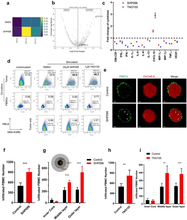
www.nature.com/scientificreports/

          Scientific Reports |   (2021) 11:1399 |   https://doi.org/10.1038/s41598-021-80999-x   2

Vol:.(1234567890)
www.nature.com/scientificreports/
◂Figure 1. SHP2 inhibition in cancer cells enhances immune cells-mediated tumor killing. (a) Absolute
tumor cell counts from each well of 384-well Elplasia plate after 6 days co-culture of OVCAR-8 spheroids with
human PBMCs (2 donors). Relative percentage of absolute tumor counts (SHP099 treated over DMSO group)
is labeled. (b) Absolute tumor cell counts of paired DMSO and SHP099 (20 μM) treated co-culture groups
of OVCAR-8 spheroids with PBMCs from multiple replicates with different donors. (c) Absolute tumor cell
counts of paired DMSO and SHP099 (20 μM) treated tumor only groups from multiple replicates. (d) Absolute
tumor cell counts from each well of 384-well Elplasia plate after 6 days co-culture of OVCAR-8 spheroids with
human PBMCs (3 donors). Relative percentage of absolute tumor counts (TNO155 treated over DMSO group)
is labeled. (e) Absolute tumor cell counts of paired DMSO and TNO155 (1 μM) treated co-culture groups of
OVCAR-8 spheroids with PBMCs from multiple replicates with different donors. (f) Absolute tumor cell counts
of paired DMSO and TNO155 (1 μM) treated tumor only groups from multiple replicates. (g) Absolute tumor
cell counts from each well of 384-well Elplasia plate after 6 days co-culture of OVCAR-8-CAS9-sgPTPN11-1
or OVCAR-8-CAS9-sgPTPN11-2 spheroids with human PBMCs (3 donors). OVCAR-8 cells were treated with
or without doxycycline (100 ng/ml) for 5 days before co-culture. Relative percentage of absolute tumor counts
(Dox+ over Dox− group) is labeled. (h) Absolute tumor cell counts from each well of 384-well Elplasia plate after
6 days co-culture of OVCAR-8-SHP2-WT or OVCAR-8-SHP2-T253M/Q257L spheroids with human PBMCs.
OVCAR-8 cells were treated with or without doxycycline (100 ng/ml) for 5 days before co-culture. Relative
percentage of absolute tumor counts (SHP099 treated over DMSO group) is labeled.
proliferation and survival of cancer cells by promoting GTP loading of R AS14, thereby activating RAS-MAPK
signaling. SHP2 inhibition by the selective allosteric inhibitor SHP099 exhibits promising therapeutic potential
in RTK/KRAS-driven cancers15,16. In addition to its oncogenic role in cancer cells, SHP2 is involved in multiple
signaling pathways in immune cells. In T lymphocytes, SHP2 is recruited to the cytoplasmic tails of PD-1 and
CTLA-4 and suppresses T cell activation by dephosphorylating TCRζ chains, ZAP70 and the costimulatory
receptor CD2817–20. Beyond these molecular mechanisms, the role of SHP2 in the tumor microenvironment is
largely unknown and its role in anti-tumor immunity remains to be explored. T cell-restricted ablation of SHP2
in a murine colon xenograft model increases anti-tumor immune responses by enhancing the function of CD8
cytotoxic T cells21. However, selective deletion of SHP2 in CD4 and CD8 T cells in a different study ultimately
leads to melanoma progression and m etastasis22. SHP2 is also downstream of CSF-1 signaling promoting mac-
rophage proliferation and M2 polarization, suggesting another mechanism by which SHP2 inhibition could
enhance anti-tumor immunity23–25. Given the complexity of the tumor microenvironment and the important
role of SHP2 in cancer cell signaling, it is necessary to study the effect of inhibiting SHP2 activity in both tumor
and immune cells to gain a better understanding of its therapeutic potential.
In the present study, we uncovered an immune modulatory role of SHP2 in the context of tumor-immune
cell interactions and discovered that inhibiting SHP2 function triggers favorable changes in the tumor micro-
environment and control of cancer progression. In cancer cells, SHP2 inhibition augmented interferon γ (IFNγ)
signaling, which resulted in increased expression of its downstream targets, including chemoattractant cytokines
and antigen presenting machinery. SHP2 inhibition also promoted T cell proliferation and function through its
effect in cancer cells and immune suppressive myeloid cells. Furthermore, the combination of SHP2 inhibitors
with anti-PD1 antibody resulted in significant regressions in tumor growth in syngeneic mouse models.
Our study describes the immune phenotypes associated with inhibiting SHP2 in cancer cells and the tumor
microenvironment, supporting the promise of therapeutically inhibiting SHP2 activity in patients with molecu-
larly and phenotypically diverse malignancies.
Results
SHP2 inhibition in cancer cells enhances immune cell‑mediated tumor killing. To explore the
immune modulatory function of SHP2 in vitro, we optimized a tumor spheroid-PBMC co-culture (Supplemen-
tary Fig. 1a). The tumor spheroid format retains some physiologically relevant features of the tumor microenvi-
ronment such as oxygen and cellular proliferation g radients26, 27. In addition, in the context of co-cultures with
activated T cells, it facilitates the study of immune cell infiltration. Initially, we tested SHP099 in the human
ovarian carcinoma cell line OVCAR-8 co-cultured with freshly isolated human peripheral blood mononuclear
cells (PBMCs) from healthy donors. No tumor killing was observed in co-culture in the absence of PBMC stimu-
lation (Supplementary Fig. 1b). We activated PBMCs with anti-CD3/CD28 beads in order to simulate activation
of T cells with tumor antigen. Following activation, CD3-positive T cells expanded and became the predomi-
nant immune population over 6 days (Supplementary Fig. 1c,d). When activated PBMCs were incubated with
OVCAR-8 spheroids, there was substantial tumor cell killing (Fig. 1a). To assess the role of SHP2 in this process,
we treated co-cultured spheroids with SHP099 and found that it decreased tumor cell numbers after 6 days,
exclusively in the presence of activated T cells, in a dose-dependent manner (Fig. 1a). OVCAR-8 cells were not
sensitive to SHP2 inhibition despite effective target engagement and suppression of signaling (Fig. 1a,c and Sup-
plementary Fig. 1e). Thus, the phenotype observed in co-culture is attributed to immune cell-mediated tumor
killing. The same phenotype was observed with treatment by TNO155, an allosteric SHP2 inhibitor currently in
clinical development (Fig. 1d–f). Although we observed variation in baseline killing by T cells across donors, the
phenotype observed following treatment with SHP2 inhibitors was reproducible (Fig. 1b,e) and consistent across
a panel of cancer cell lines (Supplementary Fig. 2a–d). Ablation of antigen presenting machinery by knocking
out B2M did not affect immune-mediated tumor killing (Supplementary Fig. 2e), indicating the absence of an
allogeneic response.
Scientific Reports | (2021) 11:1399 | https://doi.org/10.1038/s41598-021-80999-x 3
Vol.:(0123456789)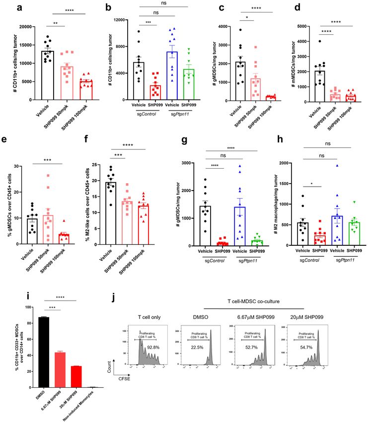
www.nature.com/scientificreports/

          Scientific Reports |   (2021) 11:1399 |   https://doi.org/10.1038/s41598-021-80999-x   4

Vol:.(1234567890)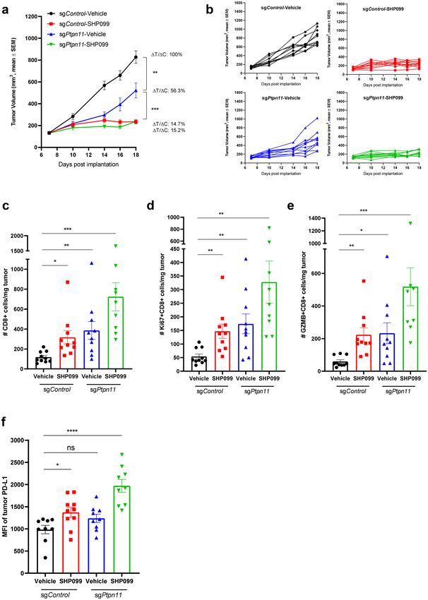
www.nature.com/scientificreports/
◂Figure 2. SHP2 inhibition in cancer cells promotes T cell proliferation/function. (a) Flow cytometry analysis
of T cell proliferation in OVCAR-8-PBMC co-culture with or without SHP099 treatment. T cells with diluted
CFSE signal were gated. Percentage of T cell population with diluted CFSE signal was labeled. (b) Percentage of
proliferating T cell from paired DMSO and SHP099 (20 μM) treated co-culture groups of OVCAR-8 spheroids
with PBMCs from multiple replicates with different donors. (c) Percentage of proliferating T cell from paired
DMSO and TNO155 (1 μM) treated co-culture groups of OVCAR-8 spheroids with PBMCs from multiple
replicates with different donors. (d) Flow cytometry analysis of T cell proliferation in co-culture of OVCAR-
8-CAS9-sgPTPN11-1 or OVCAR-8-CAS9-sgPTPN11-2 spheroids with PBMCs. OVCAR-8 cells were treated
with or without doxycycline (100 ng/ml) for 5 days before co-culture. T cells with diluted CFSE signal were
gated. Percentage of T cell population with diluted CFSE signal was labeled. (e) Flow cytometry analysis of T
cell proliferation in co-culture of OVCAR-8-SHP2-WT or OVCAR-8-SHP2-T253M/Q257L spheroids with
PBMCs. OVCAR-8 cells were treated with or without doxycycline (100 ng/ml) for 5 days before co-culture. T
cells with diluted CFSE signal were gated. Percentage of T cell population with diluted CFSE signal was labeled.
(f) Flow cytometry analysis of intracellular staining of Granzyme B in OVCAR-8-PBMC co-culture with or
without SHP099 treatment. Granzyme B-positive immune cells were gated. Percentage of Granzyme B-positive
population was labeled. Flow cytometry data was analyzed and processed with FlowJo (Version 10.7.1, https://
www.flowjo.com/solutions/flowjo/downloads/previous-versions).
To explore the specific contribution of inhibiting SHP2 in the cancer cells to the immune-mediated killing, we
adopted a doxycycline-inducible SHP2 knockout strategy. Two single guide RNAs (sgRNAs) targeting PTPN11
were transduced into OVCAR-8 cells constitutively expressing CAS9 protein. OVCAR-8-CAS9-sgPTPN11-1 and
2 cell pools showed substantial knockout efficiency (Supplementary Fig. 2f). Pools were then co-cultured with
PBMCs in the presence or absence of doxycycline. Enhanced immune-mediated tumor killing was observed
in doxycycline-treated co-cultures, utilizing PBMCs from 3 separate donors, indicating that SHP2 depletion
in cancer cells sensitizes them to immune-mediated killing (Fig. 1g). To interrogate the effect of inhibiting
SHP2 specifically in the immune cell compartment, we exogenously expressed a doxycycline-inducible SHP099-
untargetable SHP2 mutant, SHP2-T253M/Q257L, in OVCAR-8 and performed co-culture with PBMCs. As
cancer cells overexpressing SHP2-T253M/Q257L, but not SHP2-WT, were still able to maintain activation of
MAPK signaling in the presence of SHP099, we concluded that the effect of SHP099 in this context was mostly
in immune cells (Supplementary Fig. 2g). Expression of the SHP2 mutant in cancer cells dramatically attenu-
ated immune-mediated tumor killing upon SHP099 treatment (Fig. 1h), suggesting that inhibition of SHP2 in
the cancer cell is critical for SHP099 to enhance tumor cell killing by T cells. However, it should be noted that
overexpression of the SHP2 mutant could not completely rescue the phenotype, suggesting that inhibition of
SHP2 in immune cells may also contribute to tumor cell killing in vitro.
SHP2 inhibition in cancer cells promotes T cell proliferation/function. We next assessed the effect
of SHP2 inhibition on T cell proliferation in the presence or absence of OVCAR-8 cells. PBMCs were pre-labeled
with carboxyfluorescein succinimidyl ester (CFSE) and analyzed by FACS to assess cell divisions. In the pres-
ence of OVCAR-8 cells, SHP2 inhibition significantly enhanced T cell proliferation in co-culture using cells
from multiple donors (Fig. 2a–c). We also observed enhanced expression of Granzyme B on CD8 T cells in
the SHP099 treated co-culture (Fig. 2f and Supplementary Fig. 3g). This further suggests that SHP2 inhibition
enhances the tumor killing capability of cytotoxic T cells.
In the absence of tumor cells, T cell proliferation was either slightly diminished by SHP2 inhibition (in
the case of SHP099) or not affected (in the case of TNO155) without affecting cell viability. (Supplementary
Fig. 3a–d). It has been reported that SHP2 is a downstream mediator of immune checkpoint signaling such as
PD-1 and CTLA-4, acting by dephosphorylating CD28 and ZAP70 and thus preventing TCR-mediated MAPK
signaling activation17–20. This suggests that SHP2 inhibition might relieve the immune inhibitory effect of check-
point signaling, activate MAPK pathway and promote T cell proliferation. However, MAPK signaling in T cells
was not obviously enhanced by SHP099 treatment (Supplementary Fig. 3e), suggesting that SHP2 inhibition
did not augment T cell proliferation through the MAPK pathway. We observed enhanced T cell proliferation
upon SHP2 inhibition exclusively in the presence of tumor cells, indicating that the effect of SHP2 on T cells is
context-dependent.
We hypothesized that inhibiting SHP2 activity in tumor cells in co-culture mediates the T cell phenotype.
Indeed, doxycycline-induced SHP2 depletion in tumor cells boosted T cell proliferation in co-culture (Fig. 2d
and Supplementary Fig. 3f). On the contrary, T cell proliferation was unaffected by SHP099 in co-culture with
OVCAR-8 expressing SHP2 mutant (Fig. 2e).
Taken together, these data demonstrate that targeting SHP2 in tumor cells promotes T cell proliferation and
killing of tumor cells, suggesting that SHP2 might have an important role in anti-tumor immunity.
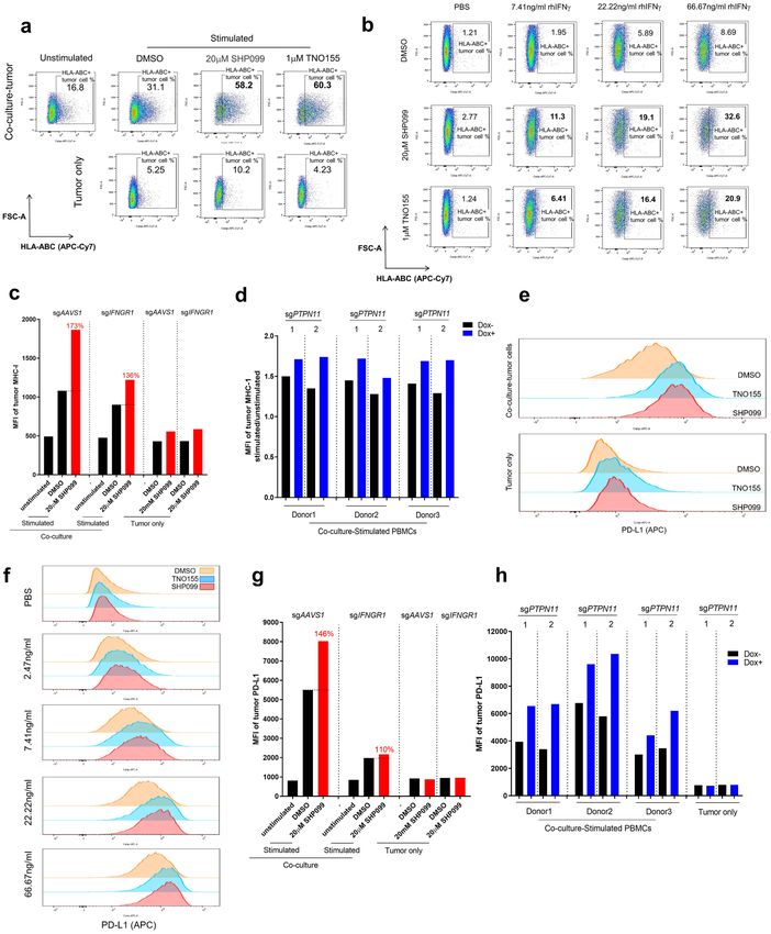
SHP2 inhibition upregulates expression of CXCR3 ligands and promotes immune infiltration
in vitro. To define mechanisms of anti-tumor immunity elicited by SHP2 inhibition, we conducted single cell
RNA sequencing (scRNAseq) of OVCAR-8 cancer cells co-cultured with PBMCs in the absence or presence of
SHP099 (Supplementary Fig. 4a,b). We found that treatment with SHP099 led to increased expression of three
CXCR3 ligands, the chemoattractant cytokines CXCL9, CXCL10 and CXCL11 specifically in tumor cells in
co-culture, with CXCL10 showing the most significant upregulation and magnitude of expression (Fig. 3a,b).
We confirmed scRNAseq data at the protein level via Luminex cytokine analysis of conditioned media from
co-culture. CXCL10 secretion was enhanced by SHP099/TNO155 treatment of the co-culture (Fig. 3c). Follow-
Scientific Reports | (2021) 11:1399 | https://doi.org/10.1038/s41598-021-80999-x 5
Vol.:(0123456789)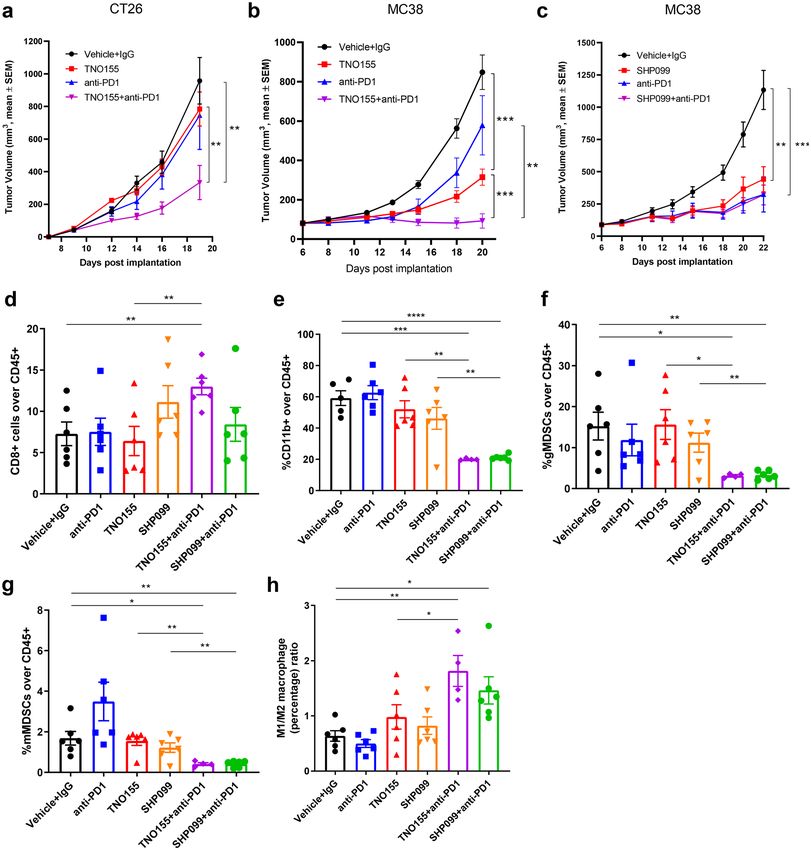
www.nature.com/scientificreports/
Figure 3. SHP2 inhibition upregulates expression of CXCR3 ligands and promotes immune cell infiltration.
(a) Heatmap of transcriptional expression level of CXCR3 ligand genes CXCL9, CXCL10, CXCL11 in OVCAR-8
tumor cells in co-culture from scRNAseq data. (b) Volcano plot of transcriptional expression level of CXCR3
ligand genes CXCL9, CXCL10, CXCL11 in OVCAR-8 tumor cells in co-culture from scRNAseq data. (c) Luminex
analysis of TNO155 and SHP099-induced fold change of a panel of cytokines (normalized to control group)
in supernatants collected from co-culture of OVCAR-8 spheroids with PBMCs. (d) Flow cytometry analysis of
intracellular staining of CXCL10 from 2 days of co-culture and tumor only. CXCL10-positive cells are gated.
Percentage of CXCL10-positive population is labeled. (e) Light sheet microscopy imaging of infiltrated PBMCs
(Green) inside OVCAR-8 tumor spheroids (Red) after 24 h of co-culture. Scale bar: 50 μm. (f, h) Histogram of
number of total infiltrated PBMCs inside tumor spheroids. (g, i) Histogram of number of infiltrated PBMCs in
different layers of tumor spheroids. Flow cytometry data was analyzed and processed with FlowJo (Version 10.7.1,
https://www.flowjo.com/solutions/flowjo/downloads/previous-versions).
Scientific Reports | (2021) 11:1399 | https://doi.org/10.1038/s41598-021-80999-x 6
Vol:.(1234567890)www.nature.com/scientificreports/
up ELISA analysis confirmed this observation (Supplementary Fig. 5a). In addition, SHP2 inhibition increased
expression of CXCR3 ligands, CXCL9 and CXCL10, in MIA PaCa-2-PBMCs co-culture, suggesting that upregu-
lation of chemoattractant cytokines by inhibition of SHP2 may be a common phenotype in the tumor microen-
vironment (Supplementary Fig. 5b).
To identify the cellular origin of CXCL10, we performed intracellular CXCL10 staining followed by FACS
analysis. CXCL10 upregulation by SHP099/TNO155 treatment was observed exclusively in CD45-negative tumor
cells in co-culture but not in immune cells, consistent with scRNAseq data (Fig. 3d).
CXCL9, CXCL10 and CXCL11 are chemoattractant cytokines for anti-tumor leukocytes that express CXCR3,
such as effector T c ells28. We explored the effect of inhibiting SHP2 on immune cell migration in vitro by imaging
immune cell infiltration in tumor spheroids with light sheet fluorescence microscopy. Volume of tumor spheroids
shrank over time during co-culture presumably due to immune cell-mediated killing (Supplementary Fig. 5c).
Consistent with data in Fig. 1a, tumor spheroids were smaller upon SHP099 treatment compared to controls, on
Day 3 and Day 6 of co-culture (Supplementary Fig. 5c,d). To avoid the confounding effect of changes in tumor
spheroid size on the quantification of immune infiltration, we measured the effect of SHP099 treatment after
24 h. As most immune cells were still surrounding the surface of tumor spheroids, it was apparent that SHP099
treatment promoted infiltration of immune cells into the tumor mass (Fig. 3e, Supplementary Movie 1 and
Supplementary Movie 2), with nearly twice as many infiltrated immune cells in spheroids treated with SHP099
compared to control (Fig. 3f). Further analysis dissected tumor spheroids into three regions across the radius
where over 90% of infiltrated immune cells were located in the outer and middle layer of spheroids, as expected
after 24 h of co-culture. SHP099 treatment augmented immune cell infiltration in both layers (Fig. 3g). The same
phenotype was observed in TNO155-treated tumor spheroids (Fig. 3h,i).
SHP2 mediates anti‑tumor immunity via interferon γ signaling. From the scRNAseq data set, we
compared the tumor cells from co-culture group and tumor only group based on their transcriptional profile. As
Supplementary Fig. 4c showed, tumor cells from each group displayed their own clustering feature (Co-culture
group tumor cells in cluster 0, 2, 4, 5, 6, 7; Tumor only group tumor cells in cluster 1, 3). Further analysis on
tumor cells from co-culture revealed that SHP099-treated tumor cells specifically clustered in cluster 6 (Sup-
plementary Fig. 4c, right). Pathway signature analysis of cluster 6 tumor cells illustrated that cytokine-mediated
signaling pathway and lymphocyte proliferation/activation pathways are among the top 10 upregulated path-
ways, which is consistent with our results from Figs. 2 and 3 (Supplementary Fig. 4d). In addition, interferon
signaling showed up multiple times as upregulated signaling in SHP099-treated tumor cells (Supplementary
Fig. 4d), suggesting the involvement of interferon pathway in SHP2 inhibition-mediated immune response.
CXCL10 and other chemoattractant cytokines are transcriptional targets of the IFNγ pathway and their
expression in cancer often correlates with clinical response to immune checkpoint b lockade29–31. We characterized
gene expression changes in the IFNγ signaling pathway of tumor cells from scRNAseq data (Fig. 4a). Strikingly,
a large proportion of the IFNγ signature genes were upregulated by SHP2 inhibition specifically in tumor cells
in co-culture, including cytokines (CXCL9, CXCL10, CXCL11 and CCL5) and antigen presenting machinery
(HLA-A, HLA-B and B2M) (Fig. 4a). Next, we quantified IFNγ concentration in co-culture conditioned media.
As expected, IFNγ was detectable only upon T cell stimulation, and the overall amount of IFNγ was signifi-
cantly higher in co-cultures than in cultures with only PBMCs comparably stimulated (Supplementary Fig. 6a).
SHP099 treatment did not enhance IFNγ secretion from immune cells in either co-culture or PBMCs alone
(Supplementary Fig. 6a,b). We hypothesized that SHP2 blockade in cancer cells could augment their response
to IFNγ and tested the response of OVCAR-8 tumor spheroids to recombinant human IFNγ (rhIFNγ) in the
presence of SHP099 or TNO155. Both SHP099 and TNO155 upregulated CXCL10 secretion upon rhIFNγ treat-
ment (Fig. 4b and Supplementary Fig. 6c); in addition, SHP2 blockade lowered the dose of rhIFNγ required to
affect OVCAR-8 cell viability, indicating that SHP2 inhibition sensitized tumor spheroids to IFNγ (Fig. 4c and
supplementary Fig. 6d).
Downstream signaling of IFNγ is mediated through the JAK-STAT p athway32. Given that SHP2 negatively
regulates the JAK-STAT pathway by dephosphorylating S TAT133,34, we performed western blots to assess STAT1
phosphorylation in OVCAR-8 cells treated with rhIFNγ in the absence or presence of SHP099. We found that
treatment with SHP099 upregulated rhIFNγ-induced phosphorylation of STAT1 while blocking p-ERK activa-
tion (Fig. 4d).
Furthermore, we knocked out IFNGR1 in OVCAR-8. Almost 75% of IFNGR1 protein was depleted in the
cell pool (Supplementary Fig. 6e). In the context of co-cultures, tumor cell killing in the presence of SHP099 was
considerably attenuated with IFNGR1 knockout OVCAR-8 (Fig. 4e). This supports the hypothesis that SHP2
may enhance resistance of tumor cells to immune-mediated killing via negatively regulating IFNγ signaling, and
that SHP2 blockade may function to release this inhibition.
SHP2 inhibition enhances major histocompatibility complex (MHC) class I and programmed
death‑ligand 1 (PD‑L1) protein expression in cancer cells through IFNγ signaling. ScRNAseq
data revealed that antigen processing and presentation genes were upregulated by SHP099 treatment in tumor
cells in co-culture (Fig. 4a). This was also true at the protein level as measured by FACS (Fig. 5a). In the context
of rhIFNγ treatment of OVCAR-8 spheroids, HLA-ABC proteins were upregulated in a dose-dependent man-
ner, and SHP2 inhibition substantially augmented their expression in cancer cells (Fig. 5b). Knockout of IFNGR1
in OVCAR-8 cells mitigated the upregulation of MHC class I in co-culture upon SHP099 treatment (Fig. 5c
and Supplementary Fig. 7a). Furthermore, genetic knockout of PTPN11 in tumor cells enhanced MHC class I
expression in co-culture, while the exogenous expression of the drug-resistant SHP2 mutant in tumor cells failed
to upregulate tumor MHC class I levels upon SHP099 treatment (Fig. 5d and Supplementary Fig. 7b), thereby
Scientific Reports | (2021) 11:1399 | https://doi.org/10.1038/s41598-021-80999-x 7
Vol.:(0123456789)www.nature.com/scientificreports/
Figure 4. SHP2 mediates anti-tumor immunity via IFNγ signaling. (a) scRNAseq data showing IFNγ signaling signature
genes expression level change induced by SHP099. (b) ELISA analysis of CXCL10 level in supernatant collected from
tumor spheroids treated with IFNγ of gradient concentration in the absence or presence of SHP099 for 6 days. (c) Dose
response curve of OVCAR-8 tumor spheroids to IFNγ treatment in the absence or presence of SHP099 after 6 days. (d)
Immunoblotting of p-STAT1 and p-ERK in OVCAR-8 tumor spheroids after different treatments. (e) Absolute tumor cell
counts from each well of 384-well Elplasia plate after 6 days co-culture of OVCAR-8-sgAAVS1 or OVCAR-8-sgIFNGR1
spheroids with PBMCs. Relative fold change of absolute tumor counts (SHP099 treated over DMSO group) is labeled.
Scientific Reports | (2021) 11:1399 | https://doi.org/10.1038/s41598-021-80999-x 8
Vol:.(1234567890)www.nature.com/scientificreports/
confirming that the enhanced IFNγ signaling in the cancer cells by SHP099 is due to on-target inhibition of
SHP2. Defects in antigen presentation are associated with resistance to T cell-mediated tumor k illing35–37, hence,
upregulation of MHC class I expression on tumor cells by SHP2 blockade provides a rationale for combining
SHP2 inhibition with immunotherapy in cancer patients.
Cancer cells exploit the immune inhibitory function of PD-L1 to evade the host immune system38,39. PD-L1
expression in cancer cells is positively regulated by T cell-derived IFNγ through the JAK-STAT signaling
pathway40. We observed that PD-L1 expression was upregulated in tumor cells in co-culture with activated
PBMCs, and that SHP2 inhibition further enhanced expression of PD-L1 (Fig. 5e and Supplementary Fig. 7c).
SHP2 inhibition also boosted rhIFNγ-induced PD-L1 upregulation in OVCAR-8 spheroids (Fig. 5f and Supple-
mentary Fig. 7d). The involvement of IFNγ signaling was confirmed by IFNGR1 knockout in OVCAR-8 (Fig. 5g
and Supplementary Fig. 7e). Similarly, knockout of PTPN11 in tumor cells could increase PD-L1 expression in
co-culture (Fig. 5h); in contrast, protecting tumor cells by overexpression of the SHP2 mutant impaired SHP099-
induced PD-L1 upregulation (Supplementary Fig. 7f).
SHP2 inhibition decreases tumor load in the 4T1 syngeneic mouse model. To examine the effect
of SHP2 inhibition on tumor immunity in vivo, we utilized the murine syngeneic 4T1 breast cancer model. Mice
treated with SHP099 showed significantly attenuated tumor growth in a dose-dependent fashion (Fig. 6a,b).
To understand if an intact immune system is necessary for this effect, we implanted 4T1 cells into immune-
compromised NSG mice and treated with SHP099. SHP099 treatment led to a mild dose-dependent effect on
4T1 growth, indicating that SHP2 inhibition slows 4T1 tumor growth also in immune-compromised mice (Sup-
plementary Fig. 8a, b). When SHP099 treatment data are normalized within each model by using the ΔT/ΔC
formula, the higher dose of SHP099 treatment (100 mg/kg) in the 4T1 syngeneic model shows a considerably
smaller ΔT/ΔC (26.83% vs. 50.01%), highlighting enhanced efficacy of SHP099 in the immune-competent 4T1
syngeneic model (Fig. 6a and Supplementary Fig. 8a). In summary, slower tumor growth in the 4T1 model
caused by SHP099 treatment is likely to be at least partially attributable to enhanced anti-tumor immunity.
To investigate mechanisms of anti-tumor immunity triggered by SHP2 inhibition in vivo, we collected tumor
tissue and performed immunohistochemistry and FACS analyses to profile immune phenotypes. We observed
increased CD8 T cells in SHP099-treated tumors, which is consistent with SHP099-enhanced immune cell
infiltration in co-culture (Fig. 6c,d). The percentage and number of CD4 T cells were unchanged by treatment
(Supplementary Fig. 8c,d). In addition, SHP099 treatment significantly enhanced CD8 T cell proliferation and
activation, also in line with in vitro data (Fig. 6e,f). IHC staining confirmed significant infiltration of CD8 T cells
in 4T1 tumor tissues upon SHP099 treatment (Fig. 6g). To explore if IFNγ signaling was augmented in SHP099
treated tumors in vivo, we measured MHC Class I and PD-L1 expression. We observed an increased trend of
expression at the protein level (Fig. 6h,i). At the transcriptional level, we observed significantly increased expres-
sion in SHP099 treated tumors at the lower dose (Supplementary Fig. 8e–g). Moreover, CXCR3 ligands Cxcl9 and
Cxcl11 also showed a trend of enhanced mRNA expression in SHP099 treated groups (Supplementary Fig. 8h,i).
To further illustrate the regulation of IFNγ signaling by SHP2 blockade in vivo, we utilized adoptive transfer
of human bone Ewing’s sarcoma cells RD-ES and human PBMCs in NSG mice. SHP099 treatment upregulated
expression of IFNγ pathway signature genes in RD-ES tumors specifically in the presence of human immune
cells (Supplementary Fig. 8k–r).
Taken together, our in vitro and in vivo data demonstrates that SHP2 blockade enhances IFNγ signaling in
tumors and triggers anti-tumor immunity via cytotoxic T cell recruitment and activation.
SHP2 inhibition in cancer cells promotes T cell function in 4T1 syngeneic mouse model. To
understand the specific contribution of inhibiting SHP2 in the cancer cells to anti-tumor immunity in vivo, we
knocked out SHP2 in 4T1 cells by direct electroporation of CAS9 protein and sgRNA targeting Ptpn11 into 4T1
cells and established syngeneic model with 4T1-CAS9-sgPtpn11 cell pool which showed substantial knockout
efficiency (Supplementary Fig. 9a). SHP2 knockout in cancer cells significantly slowed tumor growth, but not as
efficiently as SHP099 treatment (100 mg/kg) (Fig. 7a,b). The ΔT/ΔC analysis indicated that targeting SHP2 in
cancer cells induced less anti-tumor immunity than SHP099 (Fig. 7a), suggesting SHP099 mediated anti-tumor
effects may also be mediated by SHP2 inhibition in immune or stromal cells.
Immune profiling analysis of tumor tissues illustrated that targeting SHP2 in 4T1 cells displayed increased
CD8 T cell in tumors, which phenocopied the effect of SHP099 treatment (Fig. 7c). Consistent with in vitro data,
SHP2 knockout in 4T1 cells promoted CD8 T cell proliferation and activation in 4T1 syngeneic model (Fig. 7d,e).
Moreover, we observed an increased trend of PD-L1 expression in 4T1 cells when SHP2 gene was knocked out
(Fig. 7f), suggesting that IFNγ signaling was enhanced by SHP2 depletion in tumor cells. The combination of
tumor cell SHP2 knockout with SHP099 treatment exhibited more significant effect on regulation of T cell func-
tion and IFNγ signaling (Fig. 7c–f).
Overall, our in vivo genetic data confirmed our observation using pharmacological inhibition of SHP2, and
further suggest that SHP2 inhibition in tumor cells can contribute to anti-tumor immunity. However, other inhib-
itory effect from SHP099 is required to decrease tumor load more efficiently in addition to targeting cancer cells.
SHP2 blockade impairs the inhibitory effect of immunosuppressive myeloid cells. Differ-
entiation of immature myeloid cells is often dysregulated in cancer, leading to the accumulation of immuno-
suppressive myeloid cells that promote cancer progression41. It has been reported that SHP2 inhibition pro-
duced a marked shift in polarized macrophage populations in tumor microenvironment in favor of anti-tumor
immunity42. We observed a dose-dependent decrease of total CD45 + immune cells in SHP099-treated groups
in the 4T1 syngeneic mouse model (Supplementary Fig. 9b). Further analysis indicated that much of this effect
Scientific Reports | (2021) 11:1399 | https://doi.org/10.1038/s41598-021-80999-x 9
Vol.:(0123456789)www.nature.com/scientificreports/
Scientific Reports | (2021) 11:1399 | https://doi.org/10.1038/s41598-021-80999-x 10
Vol:.(1234567890)www.nature.com/scientificreports/
◂ Figure 5. SHP2 inhibition enhances MHC class I and PD-L1 expression in cancer cells through IFNγ signaling.
(a) Flow cytometry analysis of cell surface staining of MHC class I in tumor cells after 6 days of co-culture of
OVCAR-8 spheroids with PBMCs or tumor only. MHC class I-positive cells were gated. Percentage of MHC
class I-positive population was labeled. (b) Flow cytometry analysis of cell surface staining of MHC class I
in tumor spheroids treated with IFNγ of gradient concentration in the absence or presence of SHP099 and
TNO155 for 6 days. MHC class I-positive cells were gated. Percentage of MHC class I-positive population was
labeled. (c) MFI of MHC class I in tumor cells after 6 days co-culture of OVCAR-8-sgAAVS1 or OVCAR-8-
sgIFNGR1 spheroids with PBMCs. Relative percentage of MFI of tumor MHC I (SHP099 treated over DMSO
group) was labeled. (d) MFI fold change (stimulated/unstimulated) of MHC class I in tumor cells after 6 days
co-culture of OVCAR-8-CAS9-sgPTPN11-1 or OVCAR-8-CAS9-sgPTPN11-2 spheroids with PBMCs (3
donors). OVCAR-8 cells were treated with or without doxycycline (100 ng/ml) for 5 days before co-culture.
(e) Histogram of tumor cell surface PD-L1 level from 6 days of co-culture and tumor only. (f) Histogram of
cell surface PD-L1 level in tumor spheroids treated with IFNγ of gradient concentration in the absence or
presence of SHP099 (20 μM) and TNO155 (1 μM) for 6 days. (g) MFI of PD-L1 in tumor cells after 6 days
co-culture of OVCAR-8-sgAAVS1 or OVCAR-8-sgIFNGR1 spheroids with PBMCs. Relative percentage of
MFI of tumor PD-L1 (SHP099 treated over DMSO group) was labeled. (h) MFI of PD-L1 in tumor cells after
6 days co-culture of OVCAR-8-CAS9-sgPTPN11-1 or OVCAR-8-CAS9-sgPTPN11-2 spheroids with PBMCs
(3 donors). OVCAR-8 cells were treated with or without doxycycline (100 ng/ml) for 5 days before co-culture.
Flow cytometry data was analyzed and processed with FlowJo (Version 10.7.1, https://www.flowjo.com/solut
ions/flowjo/downloads/previous-versions).
could be attributed to decreased CD11b + myeloid cells (Fig. 8a). Total numbers of CD3 + lymphoid cells were
not affected by SHP099 treatment (Supplementary Fig. 9c). By using 4T1-sgPtpn11 syngeneic mouse model, we
confirmed that the effect of SHP2 blockade on decreasing CD11b + myeloid cells was through direct targeting
of myeloid lineage cells because SHP2 inhibition in tumor cells did not affect myeloid cells (Fig. 8b and Sup-
plementary Fig. 9e,f).
We analyzed which subpopulations of tumor-infiltrated myeloid cells were affected by SHP099 treatment.
Myeloid-derived suppressor cells (MDSC) are a heterogeneous group of immune cells that include monocytic
(mMDSC) and granulocytic (gMDSC) subsets. Both of these cell populations accumulate in tumor-bearing
mice and cancer patients and contribute to the immunosuppressive tumor m icroenvironment43. We observed
a significant decrease in total numbers of gMDSCs and mMDSCs in SHP099-treated tumors, and a decrease
in the percentage of gMDSCs in tumors treated with the higher dose of SHP099 (Fig. 8c–e and Supplementary
Fig. 9f), suggesting that SHP2 inhibition affects the differentiation of MDSCs. Tumor-associated macrophages
(TAMs) exist in the cancer microenvironment and influence tumor formation, growth, and metastasis44. Under
diverse stimuli, macrophages can polarize and differentiate into cancer-inhibiting M1 and cancer-promoting
M2 populations45. We discovered that SHP099 treatment significantly reduced the percentage of M2-like mac-
rophages without affecting M1-macrophages (Fig. 8f and Supplementary Fig. 9g). Similarly, specifically knock-
ing out SHP2 in tumor cells could not recapitulate SHP099-induced decrease of immunosuppressive gMDSCs
and M2-like macrophages, implying a direct inhibitory effect of SHP099 on myeloid populations (Fig. 8g,h and
Supplementary Fig. 9h).
To better understand the effect of SHP2 inhibition on MDSCs, we isolated CD14 + monocytes from whole
blood of healthy donors and induced them to differentiate to immunosuppressive MDSCs by GM-CSF and IL-6
stimulation41; monocytes were treated with DMSO or SHP099 during differentiation. GM-CSF and IL-6 induced
nearly 90% of cells to differentiate to an MDSC phenotype and SHP099 treatment dramatically inhibited differ-
entiation in a dose-dependent way, suggesting that inhibiting SHP2 has a direct effect on myeloid differentiation
(Fig. 8i). After 6 days, MDSCs were collected and co-cultured with CFSE-labeled activated T cells from the same
donor at a ratio of 1:2 (MDSCs:T cells). T cell proliferation was measured by FACS after 5 days of co-culture.
In control co-cultures, T cell proliferation was inhibited by MDSCs. This effect was partially reversed by treat-
ment with SHP099 (Fig. 8j). These results indicate that SHP2 inhibition may enhance anti-tumor immunity by
inhibiting the ability of monocytes to differentiate into MDSCs as well as by impairing the immune suppressive
function of MDSCs.
SHP2 inhibition displays combination benefit with PD1 blockade. Clinically, high tumor expres-
sion of PD-L1 is associated with tumor immune escape and poor prognosis46–48. Immune checkpoint inhibition,
particularly PD1 blockade, has revolutionized the clinical oncology p ractice49–51. We showed upregulated tumor
PD-L1 expression following SHP2 inhibition both in vitro and in vivo, which could be a potential barrier to the
efficacy of this treatment. Combination of anti-human PD1 antibody with SHP2 inhibitors showed combinato-
rial benefit on enhancing immune cells-mediated tumor killing in co-culture of OVCAR-8 spheroids with acti-
vated PBMCs, while PD1 antibody did not show any single-agent effect (Supplementary Fig. 10a,b). Blockade
of SHP2 in combination with anti-PD1 synergistically attenuated tumor growth in the murine syngeneic CT26
colon carcinoma model (Fig. 9a). Combination benefit was also observed in the MC38 colorectal cancer model
(Fig. 9b), although, there is variation of the single-agent anti-tumor effect of anti-PD1 (Fig. 9c and Supplemen-
tary Fig. 10d). SHP2 blockade displayed single-agent effect on tumor growth inhibition since MC38 syngeneic
mice treated with SHP2 inhibitors (TNO155 or SHP099) had significantly smaller tumors in comparison to
control groups (Fig. 9b,c and Supplementary Fig. 10d). MC38 tumor cells are insensitive to direct inhibition
of SHP2 as they carry the G503V mutation of Ptpn11 gene. We confirmed this by treating MC38 tumors with
SHP099 and measuring expression of Dusp6, a gene positively regulated by SHP2 (Supplementary Fig. 10c).
Scientific Reports | (2021) 11:1399 | https://doi.org/10.1038/s41598-021-80999-x 11
Vol.:(0123456789)www.nature.com/scientificreports/
Figure 6. SHP2 inhibition decreases tumor load in the 4T1 syngeneic mice model. (a) Tumor growth curve
(expressed as tumor volume) of subcutaneous 4T1 tumors in 4T1 syngeneic mice with different treatments.
ΔT/ΔC of last time point was labeled. (b) Spider plot of tumor growth curve (expressed as tumor volume) of
subcutaneous 4T1 tumors in 4T1 syngeneic mice with different treatments. (c) Flow cytometry analysis of
the percentage of CD8+ T cells over CD45+ cells in tumor tissues with different treatments in 4T1 syngeneic
model. (d) Flow cytometry analysis of the absolute number of CD8+ T cells per milligram tumor with different
treatments in 4T1 syngeneic model. (e) Flow cytometry analysis of the percentage of Ki67+ CD8+ T cells over
CD8+ cells in tumor tissues with different treatments in 4T1 syngeneic model. (f) Flow cytometry analysis of the
percentage of GZMB+ CD8+ T cells over CD8+ cells in tumor tissues with different treatments in 4T1 syngeneic
model. (g) IHC analysis of CD8 T cells in tumor tissues with different treatments from 4T1 syngeneic model. Y
axis means the percentage of CD8+ cells over all the cells in the same slide. (h) MFI of the tumor surface MHC
Class I with different treatments in 4T1 syngeneic model. (i) MFI of the tumor surface PD-L1 with different
treatments in 4T1 syngeneic model. Flow cytometry data was analyzed and processed with FlowJo (Version
10.7.1, https://www.flowjo.com/solutions/flowjo/downloads/previous-versions).
Scientific Reports | (2021) 11:1399 | https://doi.org/10.1038/s41598-021-80999-x 12
Vol:.(1234567890)www.nature.com/scientificreports/
Indeed, SHP099/TNO155 treatment did not significantly inhibit MC38 xenograft growth in immune-compro-
mised NSG mice (Supplementary Fig. 10e,f), suggesting that SHP2 inhibition in immune cells contributed to
decreasing the tumor load in MC38 syngeneic mouse model.
Next, we profiled tumor-infiltrated immune cells from MC38 tumors. Similarly to what was observed in the
4T1 model, tumor-infiltrated total CD45 + cells significantly decreased after SHP099/TNO155 treatment, while
combination with PD1 antibody further decreased the percentage (Supplementary Fig. 10g). We also observed
that these treatments did not affect the number of CD4 + T cells but increased the percentage of CD8 + T cells
significantly only in the TNO155 plus anti-PD1 combination arm (Fig. 9d and Supplementary Fig. 10h). Unlike
in the 4T1 model, SHP2 inhibition alone did not enhance tumor infiltrated CD8+ T cells in MC38 syngeneic
model (Fig. 9d), which suggests that targeting tumor intrinsic signaling contributed to CD8+ T cell recruitment
to the tumor microenvironment, in consideration that MC38 is SHP2 inhibitor-insensitive. Blockade of SHP2
and PD1 showed combinatorial benefit on decreasing CD11b+ myeloid cells (Fig. 9e), which might contribute
to decreased CD45+ immune cells in tumors. Percentages of both immunosuppressive myeloid-derived gMD-
SCs and mMDSCs showed a strong decrease after SHP099/TNO155 treatment in combination with immune
checkpoint blockade (Fig. 9f,g). The combination also effectively increased the ratio of immune-promoting M1
macrophages over immunosuppressive M2 macrophages (Fig. 9h).
Taken together, these data indicate that SHP2 inhibition in combination with immune checkpoint blockade
boosts anti-tumor immunity through targeting immunosuppressive cells of the myeloid lineage in the tumor
microenvironment, potentially leading to enhanced T cell-mediated tumor cell killing.
Discussion
Inhibition of the phosphatase SHP2 exhibits therapeutic potential in cancers dependent on receptor tyrosine
kinases and mutated KRAS signaling, as SHP2 plays a critical role in mediating MAPK signaling in cancer
cells15,16. In this study, by utilizing both in vitro co-cultures of tumor spheroids and human immune cells and
in vivo syngeneic mouse models, we discovered that inhibiting SHP2 activity alters the cellular composition of
the tumor microenvironment through its effect on both tumor and immune cell populations to promote anti-
tumor immunity.
Specifically, SHP2 inhibition enhanced T cell-mediated tumor killing and T cell proliferation/activation
in vitro. Mechanistically, SHP099/TNO155 induced these effects through targeting SHP2 in cancer cells and
augmenting cancer cell IFNγ signaling in the context of tumor-immune cell crosstalk. In vivo, SHP2 inhibition
also increased tumor IFNγ signaling and displayed anti-tumor activity by promoting cytotoxic T cell function
and inhibiting immune suppressive myeloid cells.
In cancer cells, SHP2 inhibition potentiated the response to IFNγ by enhancing JAK-STAT signaling. Con-
sequently, IFNγ pathway targets including CXCL9, -10, and -11, MHC Class I, and PD-L1, were upregulated at
both the transcriptional and protein levels. Defects in IFNγ signaling cause resistance to T cell-mediated tumor
killing in both pre-clinical models and cancer p atients35,52,53. In this study, we demonstrated the importance
of IFNγ signaling in our tumor model by knocking out the IFNγ receptor, IFNGR1; we observed impaired T
cell-mediated tumor killing as well as attenuated MHC class I expression in co-culture. PD-L1 expressed on
the surface of cancer cells serves as a negative feedback mechanism to anti-tumor immunity. Clinically, higher
PD-L1 expression in the tumor microenvironment has been positively correlated with response to immune
checkpoint blockade with PD1 a ntagonists38,39. Since SHP2 inhibition augmented PD-L1 expression in cancer
cells in co-culture with T cells, we combined SHP2 blockade with immune checkpoint inhibition and the combo
showed combinatorial benefit on boosting anti-tumor immunity and slowing down tumor growth. This raises
the possibility that therapeutic inhibition of SHP2 could be effectively combined with checkpoint blockade to
enhance treatment efficacy in clinic.
Our in vitro MDSC differentiation and T cell-MDSC co-culture experiments showed that SHP2 inhibition
could directly affect myeloid cells by inhibiting their immunosuppressive function. We also observed modulation
of myeloid cell subsets by SHP2 inhibition in both the 4T1 and MC38 syngeneic mouse models. The presence
of immunosuppressive M2 macrophages and MDSCs in the tumor microenvironment was decreased by SHP2
inhibition. From these observations we hypothesize that modulation of myeloid cells may contribute to tumor
growth inhibition by SHP099/TNO155 treatment in vivo.
Taken together, our data show that SHP2 inhibition is effective in controlling tumor growth by enhancing
immune surveillance and cancer cell killing. We propose that this is achieved by: (a) amplifying IFNγ signaling
in cancer cells54; (b) enhancing chemoattractant cytokine secretion by cancer cells which may recruit anti-tumor
lymphocytes to the tumor; (c) increasing antigen presentation by cancer cells; (d) positively regulating CD8 T cell
proliferation and function; (e) inhibiting the function of immunosuppressive myeloid cells on anti-tumor T cells.
In light of published work and the data presented in this study, we propose that inhibition of SHP2 activity
in cancer patients has the potential to be therapeutically beneficial by both directly inhibiting cancer cell growth
and promoting anti-tumor immunity.
Materials and methods
Cell lines and compound. Cell lines were obtained from Novartis’ CCLE c ollection55 and were tested to
be free of mycoplasma. OVCAR-8, OV-90, RD-ES, DMS-273 and 4T1 cells were cultured in RPMI1640 media
(Gibco, Thermo Fisher Scientific) containing 10% fetal bovine serum (FBS, GE Life Sciences). HEK293T, MIA
PaCa-2 and MC-38 cells were cultured in DMEM media (Gibco, Thermo Fisher Scientific) containing 10% FBS.
All cells were maintained in a humidified incubator at 37 °C with 5% CO2. For co-culture with immune cells,
media of cells cultured in DMEM were changed to RPMI1640 media before seeding into Elplasia plates. SHP2
inhibitor SHP099 and TNO155 was synthesized and structurally verified by NMR/LC–MS at Novartis Institutes
Scientific Reports | (2021) 11:1399 | https://doi.org/10.1038/s41598-021-80999-x 13
Vol.:(0123456789)www.nature.com/scientificreports/
Scientific Reports | (2021) 11:1399 | https://doi.org/10.1038/s41598-021-80999-x 14
Vol:.(1234567890)www.nature.com/scientificreports/
◂ Figure 7. SHP2 inhibition in cancer cells promotes T cell function in 4T1 syngeneic mouse model. (a) Tumor
growth curve (expressed as tumor volume) of subcutaneous 4T1-sgControl and 4T1-sgPtpn11 tumors in
4T1 syngeneic mice with different treatments. ΔT/ΔC of last time point was labeled. (b) Spider plot of tumor
growth curve (expressed as tumor volume) of subcutaneous 4T1-sgControl and 4T1-sgPtpn11 tumors in 4T1
syngeneic mice with different treatments. (c) Flow cytometry analysis of the absolute number of CD8+ T cells
per milligram tumor with different treatments in 4T1 syngeneic model. (d) Flow cytometry analysis of the
absolute number of Ki67+ CD8+ T cells per milligram tumor with different treatments in 4T1-sgControl and
4T1-sgPtpn11 syngeneic model. (e) Flow cytometry analysis of the absolute number of GZMB+ CD8+ T cells
per milligram tumor with different treatments in 4T1-sgControl and 4T1-sgPtpn11 syngeneic model. (f) MFI of
the tumor surface PD-L1 with different treatments in 4T1-sgControl and 4T1-sgPtpn11 syngeneic model. Flow
cytometry data was analyzed and processed with FlowJo (Version 10.7.1, https://www.flowjo.com/solutions/
flowjo/downloads/previous-versions).
for BioMedical Research. Recombinant human IFNγ (#285-IF-100) was purchased from R&D Systems. Murine
anti-PD1 clone 332.1D2 is a kind gift of Prof. Gordon Freeman at DFCI. Human anti-PD1 is produced and veri-
fied by Novartis Institutes for BioMedical Research.
Co‑culture of tumor spheroids and PBMCs. Elplasia square type 384-well-plates (SQ 200 100 384,
Kuraray), which have 225 100 × 200 μm micro-spaces divided by wall in each single well, were coated with poly-
2-hydroxyethyl methacrylate (poly-HEMA) (192066, Sigma-Aldrich). As Supplementary Fig. 1a shows, 2 × 10⁴
tumor cells were seeded per well on Day-1 and cultured overnight to form round shaped spheroids. Freshly
isolated human PBMCs from healthy donor blood were seeded on top of the spheroids at E-to-T ratio of 1:1 on
the following day (Day0). PBMCs were either unstimulated or stimulated with Dynabeads CD3/CD28 Human
T-Activator (11132D, Thermo Fisher Scientific) at bead-to-cell ratio of 1:8. The co-culture system was treated
with compounds on Day0 as well after PBMCs seeding and then underwent co-culture for days. Multiple donors
were used in co-culture with unknown HLA (human leukocyte antigen) matching status. RPMI1640 media con-
taining 10% FBS was used for co-culture. Cells with doxycycline-inducible sgRNA or SHP2-WT/Mutant were
treated with 100 ng/ml doxycycline for 5 days before co-culture. ACCUMAX cell detachment solution (#7921,
STEMCELL Technologies) was used to dissociate tumor spheroids into single cells for flow cytometry analysis.
Human peripheral blood mononuclear cells (PBMCs) were isolated by spinning CPT tubes (BD Bioscience
#362761) containing exsanguinated whole blood at 1800 rpm for 20 min and slowing down without brake. All
studies with human blood were performed under ethical approval by the WCG Clinical Western Institutional
Review Board (WIRB)-Copernicus Group and in compliance with the guideline from Standard Operating Pro-
cedure for Obtaining Venous Blood and Other Non-Invasive Biological Specimens for the Novartis Institutes
for Biomedical Research, Inc. All the participants provided informed consent prior the study.
Light sheet fluorescence microscopy. 5000 OVCAR-8 cells constitutively expressing mCherry were
seeded in each well of a 384 ULA round bottom well plate and let form spheroids overnight. CFSE-labeled
PBMCs were seeded on the following day for co-culture and treated with SHP099. After 24 h of treatment,
spheroids were imaged using a Zeiss Lighsheet Z.1 microscope. Each spheroid was embedded in 2% agarose in
1 × HBSS and drawn into a 1 mm glass capillary tube. The capillary tube was placed into the imaging chamber
that was filled with PBS. The imaging objective was a 20X, 1.0 NA water immersion with 10X illumination
objectives. The spheroids were centered in the field of view (584X584 μm) and a z-stack (0.5 μm steps) with
two fluorescence channels was collected with channel 1 settings 488 nm laser (1.5% power), emission filter
505–545 nm (15 ms exposure) and channel 2 settings 561 nm laser (0.5% poser), emission filter 575–615 nm
(15 ms exposure). The illumination was set to dual side to increase depth of imaging and dual side image data
was fused using maximum intensity fusion.
Single‑cell RNA sequencing. Single cell libraries were prepared according to 10X Genomics specifications
(Chromium Single Cell V(D)J User Guide PN-1000006, and the v2 Reagent Kit (PN-10000075, PN-1000153).
The quality of the cDNA was assessed using an Agilent TapeStation 4200, High Sensitivity D5000 Kit (5067–
5592, 5067–5593) and the quality of the libraries were assessed using the High Sensitivity D1000 Kit (5067–5584,
5067–5585). Libraries were diluted to 10 nM and clustered using Illumina’s MiSeq, on a 150-cycle v3 paired-end
read flow cell and sequenced for 26 cycles on R1 (10X barcode and the UMIs), followed by 8 cycles of I7 Index
(sample Index), and 98 bases on R2 (transcript), in order to normalize for read depth and cell number. Libraries
were then clustered and sequenced using Illumina’s HiSeq4000 on a paired-end flow cell, achieving a sequencing
depth of around 50,000 reads per cell.
Raw sequencing data were processed using cellranger v3.0 from 10X Genomics to generate sequencing and
alignment QC metrics and the gene by cell unique molecular identifiers (UMI) count matrix. The unfiltered raw
output from Cellranger was loaded with Scanpy56 and filtering was applied to account for cells with a minimum
number of expressed genes (300) and genes expressed in a minimum number of cells. After normalization,
further filtering was applied to keep genes with a minimum average expression of 0.0125 (log10) and dispersion
greater than 0. We performed dimensionality reduction taking into account 40 principal components and clus-
tered the cells using the Louvain a lgorithm57. The population of cells was then divided into two groups: tumor
and immune, based on the CD45 expression of each Louvain cluster (Supplementary Fig. 4a). Those populations
were later analyzed independently to estimate gene expression levels for Figs. 3a and 4a. The immune cells were
divided into 5 major groups based on different gene expression sets of immune populations (Supplementary
Scientific Reports | (2021) 11:1399 | https://doi.org/10.1038/s41598-021-80999-x 15
Vol.:(0123456789)www.nature.com/scientificreports/
Scientific Reports | (2021) 11:1399 | https://doi.org/10.1038/s41598-021-80999-x 16
Vol:.(1234567890)www.nature.com/scientificreports/
◂ Figure 8. SHP2 blockade impairs the inhibitory effect of immunosuppressive myeloid cells. (a) Flow cytometry
analysis of the absolute number of CD11b + myeloid cells per milligram tumor with different treatments in 4T1
syngeneic model. (b) Flow cytometry analysis of the absolute number of CD11b + myeloid cells per milligram
tumor in 4T1-sgControl and 4T1-sgPtpn11 syngeneic model with different treatments. (c) Flow cytometry
analysis of the absolute number of gMDSCs per milligram tumor with different treatments in 4T1 syngeneic
model. (d) Flow cytometry analysis of the absolute number of mMDSCs per milligram tumor with different
treatments in 4T1 syngeneic model. (e) Flow cytometry analysis of the percentage of gMDSCs over CD45+ cells
in tumor tissues with different treatments in 4T1 syngeneic model. (f) Flow cytometry analysis of the percentage
of M2-like macrophages over CD45+ cells in tumor tissues with different treatments in 4T1 syngeneic model.
(g) Flow cytometry analysis of the absolute number of gMDSCs per milligram tumor in 4T1-sgControl and
4T1-sgPtpn11 syngeneic model with different treatments. (h) Flow cytometry analysis of the absolute number of
M2-like macrophages per milligram tumor in 4T1-sgControl and 4T1-sgPtpn11 syngeneic model with different
treatments. (i) Flow cytometry analysis of the percentage of CD11b+ CD33+ MDSCs over CD14+ monocytes
after 5 days of GM-CSF and IL-6 induced MDSCs differentiation from monocytes with or without SHP099
treatment. (j) Flow cytometry analysis of the percentage of proliferating T cells after 6 days co-culture of
differentiated MDSCs (treated with or without SHP099) with activated T cells (MDSCS:T cells = 1:2). T cells
with diluted CFSE signal were gated. Percentage of T cells with diluted CFSE signal was labeled. Flow cytometry
data was analyzed and processed with FlowJo (Version 10.7.1, https://www.flowjo.com/solutions/flowjo/downl
oads/previous-versions).
Fig. 4b). The pathway signature analysis is conducted in https: //metasc ape.org/gp/index. html#/main/step1. IFNγ
signaling pathway signature genes are from GSEA/mSigDB hallmark gene set collection (http://software.broad
institute.org/gsea/msigdb/collections.jsp#H).
In vivo mouse models. All animal studies were performed under approval by the Novartis Institutes for
BioMedical Research Institutional Animal Care and Use Committee and in compliance with the Guide for the
Care and Use of Laboratory Animals. The methods were adapted/adjusted from the methods of another lit-
erature published by Novartis58: before implantation, all cell lines were confirmed as mycoplasma- and rodent
virus-negative. 0.25 × 10⁶ 4T1 cells in Hank’s balanced salt solution (HBSS) were inoculated subcutaneously into
the right flank of BALB/c mice (for immune-competent syngeneic mouse model) or NSG mice (for immune-
compromised xenograft model). 1 × 10⁶ MC-38 cells in HBSS were inoculated subcutaneously into the right
flank of C57BL/6 mice (for immune-competent syngeneic mouse model) or NSG mice (for immune-compro-
mised xenograft model). Tumor bearing mice were randomized into 3 treatment groups once tumor volumes
reached 100 ~ 150 mm3. 0.2 × 10⁶ CT26 cells in HBSS were inoculated subcutaneously into the right flank of
C57BL/6 mice (for immune-competent syngeneic mouse model). Generally on Day 6–9 after inoculation, tumor
bearing mice started to receive treatments. Small molecule compound treatments were dosed via oral gavage.
Anti-PD1 treatment was dosed via intraperitoneal injection. Treatment groups includes: Vehicle (0.5% MC/0.5%
Tween 80, daily), SHP099 (50 mg/kg, daily), SHP099 (100 mg/kg, daily), TNO155 (20 mg/kg, twice per day),
anti-mouse IgG (10 mg/kg, weekly), anti-mouse PD1 (10 mg/kg, weekly). Tumors were measured twice weekly
by calipering in two dimensions. Tumor volume was calculated using a modified ellipsoid formula: tumor vol-
ume = L × W2 × π/6, where L is the longest axis of the tumor and W is perpendicular to L. Anti-tumor activity was
reported as percentage treatment/control (%T/C) values and calculated using the formula %T/C = 100 × (ΔT/
ΔC); here ΔT is the mean tumor volume (mTV) of the SHP099-treated group on day f (final day) minus the mTV
of the SHP099-treated group on day i (initial dosing day), and ΔC is the mTV of the Vehicle group on day f minus
the mTV of the Vehicle group on day i. Syngeneic mice for PD study were euthanized on Day 7 after dosing and
tumor tissues were taken for digestion and immunostaining.
Tumor tissue digestion. For tumors from 4T1 syngeneic mice, the digestion was conducted following
reference59: tumor tissues were minced into fine pieces (approximately 1 m m3) using scissors and razor blades,
transferred into 15 mL conical tubes containing 2 ml of digestion buffer [RPMI1640 (Gibco, Thermo Fisher
Scientific), 2% FBS, 0.2 mg/ml Collagenase P (#11249002001, Roche), 0.2 mg/ml Dispase (#17105-041, Gibco),
and 0.1 mg/ml DNase I (#10104159001, Roche)], and placed into a water bath at 37 °C. The tubes were vortexed
every 5 min for 15 min, the tissue pieces were allowed to settle for 5 min, and then the supernatant containing
freed cells was collected and quenched at 4 °C in 50 mL conical tubes containing 20 ml of cold flow cytometry
buffer (PBS, 2% FBS, and 2 mmol/l EDTA). Next, 2 ml of fresh digestion buffer was added to the remaining
tumor fragments, and tubes were incubated at 37 °C for another 20 min, vortexing every 5 min, prior to collect-
ing the freed cells and adding them to the previously collected fractions on ice. These 20 min digestion cycles
were repeated for a total of 5 to 6 times, with progressively more forceful agitation methods (vortexing, pipetting
1 ml up and down using large orifice tips, then mixing with uncut 1 ml tips), until no tumor fragments larger
than 1 mm remained. The collection tube containing the digested fractions in flow cytometry buffer was kept
on ice until the digestion was complete, and then the contents was filtered through 70 μm mesh, centrifuged
(1500 rpm, 10 min, 4 °C), and the cells were counted. Pellets were resuspended in flow cytometry buffer and
subjected to immunostaining. For Q-PCR, pellets went through CD45, CD90 and CD31 positive enrichment
to remove immune cells and stromal cells by using Easysep Biotin Positive Selection Kit (#18559, STEMCELL
Technologies) following the manufacturer’s instruction. Negative portion (tumor cells) was collected for future
Q-PCR analysis. Antibodies used for positive enrichment are: Biotinylated CD45 (#103104, Biolegend), Bioti-
nylated CD31 (#102504, Biolegend), Biotinylated CD90/Thy1.2 (13-0902-85, Invitrogen).
Scientific Reports | (2021) 11:1399 | https://doi.org/10.1038/s41598-021-80999-x 17
Vol.:(0123456789)You can also read