Polycladida (Platyhelminthes, Rhabditophora) from Cape Verde and related regions of Macaronesia
←
→
Page content transcription
If your browser does not render page correctly, please read the page content below
European Journal of Taxonomy 736: 1–43                                            ISSN 2118-9773
           https://doi.org/10.5852/ejt.2021.736.1249                        www.europeanjournaloftaxonomy.eu
                                                                                    2021 · Cuadrado D. et al.
This work is licensed under a Creative Commons Attribution License (CC BY 4.0).

                                             Research article
                   urn:lsid:zoobank.org:pub:FC9085BE-73C4-4F33-BD9B-6A9F573AB01D

        Polycladida (Platyhelminthes, Rhabditophora) from Cape Verde
                      and related regions of Macaronesia

                    Daniel CUADRADO 1, Jorge RODRÍGUEZ 2, Leopoldo MORO 3,
                             Cristina GRANDE 4 & Carolina NOREÑA 5,*
1,5
      Departmento de Biodiversidad y Biología Evolutiva, Museo Nacional de Ciencias Naturales (CSIC),
                               c/ José Gutiérrez Abascal 2, 28006 Madrid, Spain.
       2
         Marine Invertebrates Department, Australian Museum Research Institute, Australian Museum, 1
                                 William Street, Sydney, NSW 2010, Australia.
      3
        Servicio de Biodiversidad, Gobierno de Canarias, Edif. Usos Múltiples I, Av. Anaga n° 35, Pl. 11,
                                 38071 S/C de Tenerife, Canary Islands, Spain.
             4
               Departamento de Biología, Facultad de Ciencias, Universidad Autónoma de Madrid,
                                       Cantoblanco, 28049 Madrid, Spain.
                                 *
                                  Corresponding author: norena@mncn.csic.es
                                      1
                                        Email: cuadradopm@hotmail.com
                               2
                                 Email: jorge.rodriguezmonter@austmus.gov.au
                                   3
                                     Email: lmoraba@gobernodecanarias.org
                                        4
                                          Email: cristina.grande@uam.es
               1
                 urn:lsid:zoobank.org:author:F0C14D94-9996-4A20-9D56-B02DDA1A78CA
               2
                 urn:lsid:zoobank.org:author:B833502E-CBA4-40CA-AE5A-BAD02F539062
               3
                 urn:lsid:zoobank.org:author:B66DDDE6-98E6-42FD-8E58-A1DF6A386BE5
                 4
                   urn:lsid:zoobank.org:author:C8634A50-D3EC-467A-A868-225C231B40F2
               5
                  urn:lsid:zoobank.org:author:DD03B71F-B45E-402B-BA32-BB30343E0D95

Abstract. The systematics and distribution of the order Polycladida within the Macaronesian archipelagos
are analysed. New species (Marcusia alba sp. nov., Prostheceraeus crisostomum sp. nov., Parviplana
sodade sp. nov., Euplana claridade sp. nov., Stylochus salis sp. nov. and Distylochus fundae sp. nov.),
new variety (Pseudoceros rawlinsonae var. galaxy), new records and records of shared species among
different archipelagos are studied to compare the marine flatworm biodiversity of each island. The
complex of archipelagos known as Macaronesia (including Madeira, Selvagens Islands, Canary Islands,
Azores and Cape Verde) share a volcanic origin and European political influence. The five archipelagos
are located along the eastern coast of the Atlantic Ocean and are subject to similar trade winds, streams
(like the Gulf Stream) and cold currents. The term Macaronesia has suffered several changes throughout
the years and it still is a topic of discussion in present times. The new delimitation of Macaronesia is
mainly based on systematic studies on the invertebrate fauna of the islands. The resulting analyses shed
new light on the differences and similarities among these archipelagos. In addition, molecular analyses
employing 28S nuclear gene sequences are compared to verify relationships among anatomically similar
species of marine polyclads.

                                                             1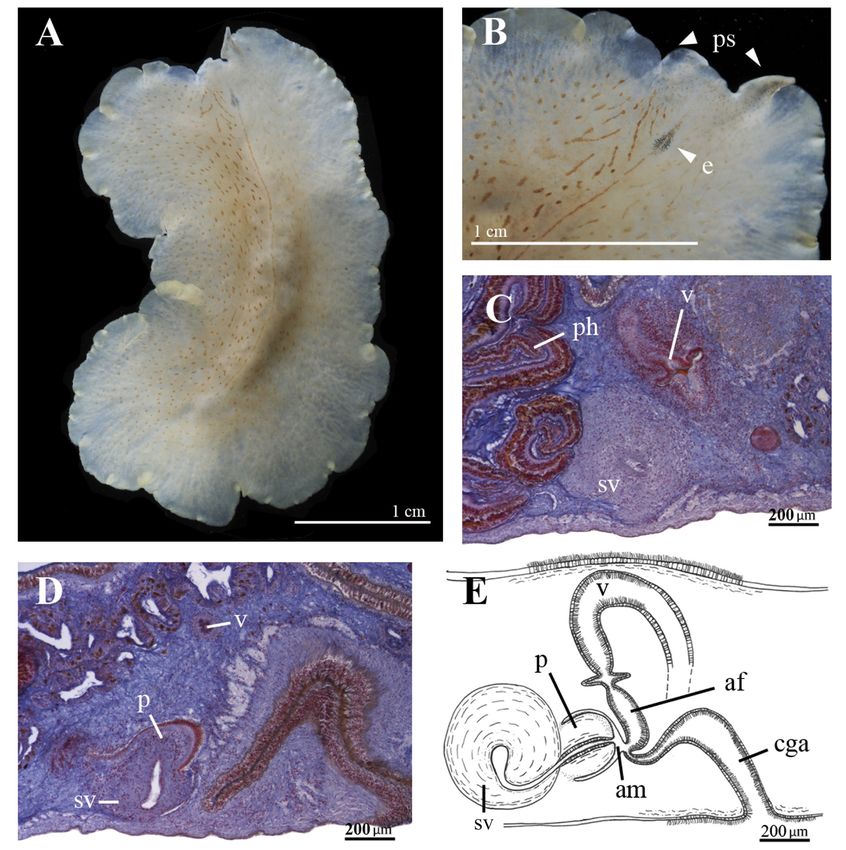
European Journal of Taxonomy 736: 1–43 (2021)

Keywords. Flatworms, distribution, Acotylea, Cotylea, 28S.

Cuadrado D., Rodríguez J., Moro L., Grande C. & Noreña C. 2021. Polycladida (Platyhelminthes, Rhabditophora)
from Cape Verde and related regions of Macaronesia. European Journal of Taxonomy 736: 1–43.
https://doi.org/10.5852/ejt.2021.736.1249

Introduction
From a political and biogeographic point of view, the complex of archipelagos known as Macaronesia
is a mosaic of ecological and political factors, with two common denominators. One is that they are of
volcanic origin and the other is that they are mostly European. Azores, Madeira and Selvagens Islands are
Portuguese, the Canary Islands Spanish and Cape Verde independent, but with great European influence.

The five archipelagos are located along the eastern region of the Atlantic Ocean between latitude 15° N
(Cape Verde) and 40° N (Azores) (Fig. 1) and are influenced by trade winds, the eastern branch of the
Gulf Stream and the cold currents of the Canary Islands. Nonetheless, the biogeographic limits and
definition of Macaronesia have undergone several changes over the years. Sunding (1979) included
as part of Macaronesia a land strip ranging from Agadir (Morocco) to South Western Sahara (northern
coast of the African continent), the so-called Macaronesian continental enclave. Sunding’s inclusion of
North Africa was the last addition to Macaronesia.

According to De Nicolas et al. (1989), Cape Verde belongs to Macaronesia due to its volcanic origin, but
the presence of coral barriers, a tropical climate and specific vegetation make this archipelago notably
different. Nonetheless, Kunkel (1993) subdivided Macaronesia into three subregions including Cape
Verde: 1. Great Macaronesia (“Gross-Makaronesien”), which comprises the five archipelagos and the
African and Iberian continental enclaves; 2. Central Macaronesia with Madeira, Selvagens Islands and
Canary Islands and 3. Lauri-Macaronesia including Madeira, Selvagens Island, Canary Islands, Azores
and the southwest region of Portugal (Algarve) (Fig. 1D), where some subregions overlap.

Médail & Quézel (1999), after an exhaustive study on the vegetation of southwestern Morocco and
the Canary Islands, concluded that North Africa should not be part of Macaronesia and included it in
the Mediterranean region as the Agadirense province (Galán de Mera et al. 2003) or Agadiro-Ifniense
region (Rivas-Martínez 2009; Salas & Naranjo 2015).

Macaronesia should be included in the Mediterranean hotspot as both biogeographical regions share
similar fauna, flora and climatology (Myers et al. 2000). In recent studies (Spalding et al. 2007), two
marine ecoregions were established in relation to Macaronesia, namely the Lusitanian-Macaronesia
region, formed by the archipelagos of Azores, Madeira, Selvagens Islands and Canary Islands, and the
West African Transition region, which only includes Cape Verde. These considerations are shared also
by Freitas et al. (2019) who proposes that Cape Verde must be considered a unique biogeographic region
not included in the Macaronesia complex.

Today, most scientists accept that the term ‘Macaronesia s. str.’ is no longer valid as a biogeographical
concept (Salas & Naranjo 2015), but some archipelagos have conserved the Macaronesian idea: Madeira,
Selvagens Islands and Canary Islands are considered ‘Eumacaronesia’ (Fig. 1) within the Canary-
Madeira sub-region (Rivas-Martínez 2009) or ‘Webbnesia’ after Freitas et al. (2019).

In the past as well as today, the interest on these archipelagos remains due to their ecological conditions
and its strategic position along marine routes. However, some islands are much better studied and
explored than others. The best known, geologically and biologically, are the Canary Islands, Cape Verde
and Madeira, followed by the Azores. Selvagens Islands are practically unknown.

                                                     2
CUADRADO D. et al., New species and records from Macaronesia and Cape Verde

In the present study, the systematics and distribution of the Polycladida Lang, 1881 order within
this archipelagos complex are analysed. New species, new records and records of shared species are
considered to study the biodiversity between the archipelagos. The Macaronesian polyclad species
are mainly known from the detailed studies conducted by Plehn (1896) and Laidlaw (1903, 1906) in
Cape Verde and the Azores. According to Laidlaw (1906), the species of Cape Verde have a markedly
Mediterranean character, but it should be noted that not all the regions and coasts bathed by the Atlantic
waters are known.

Additionally, the morphological results were studied alongside the analyses of the 28S nuclear gene to
verify relationships among anatomically similar and/or similarly distributed species (Table 1).

In summary, new concepts of the Macaronesia are based mainly in systematic studies on the invertebrate
fauna (echinoderms, molluscs, decapod crustaceans, or polychaete annelids), fishes and algae of the
islands. The present study on the systematics and distribution of the order Polycladida sheds new light

Fig. 1. Location of sampling points, distribution of the Macaronesian archipelagos and limitation of
regions: Lauri-Macaronesia or Lusitanian Macaronesia (white); Central Macaronesia or Eumacaronesia
(bordeaux) and Great Macaronesia (green). Abbreviations: A1 = San Miguel Island, Azores; C1 = Pasito
Blanco, Gran Canaria, Canary Islands; C2 = Tenerife, Canary Islands; C3 = Garachico, Tenerife, Canary
Islands; C4 = Amarilla Golf, Tenerife, Canary Islands; CV1 = Astillero Mindelo, São Vicente Island,
Cape Verde; CV2 = Bahía das Gatas, São Vicente Island, Cape Verde; CV3 = Calheta Funda, Sal Island,
Cape Verde; M1 = Ifni, South Morocco; M2 = Agadir, South Morocco.

                                                    3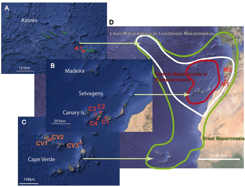
European Journal of Taxonomy 736: 1–43 (2021)

Table 1 (continued on next three pages). Species, localities and accession numbers of the material
studied in the present work. Species sequenced for this study are highlighted in blue.

                  Species                               Localities        GenBank code
Outgroup          Prorhynchus stagnalis                                   KC869866
ACOTYLEA          Callioplana marginata                 Japan             LC100082
                  Imogine zebra                         Massachusetts     AF342800
                  Imogine refertus                      Brazil            KY263694
                  Stylochus sp.                         Peru              KY263743
                  Paraplanocera oligoglena              Hawaii            KC869849
                  Imogine ijimai                        Japan             LC100079
                  Leptostylochus gracilis               Japan             LC100078
                  Latocestus plehni                     Cape Verde Is.    MK299376
                                                        Canary Is.        MK299377
                  Hoploplana villosa                    Japan             LC100076
                  Hoploplana californica                California        KC869850
                  Hoploplana divae                      Brazil            KY263692
                                                        Brazil            KY263693
                  Paraplanocera sp.                     Greece            KY263699
                  Planocera multitentaculata            Japan             LC100081
                  Planocera pellucida                   Canary Is.        MK299355
                                                        Cape Verde Is.    MK299356
                                                        Canary Is.        MK299363
                  Amemiyaia pacifica                    Japan             LC100077
                  Adenoplana evelinae                   Brazil            KY263647
                  Phaenocelis medvedica                 Brazil            KY263701
                                                        Brazil            KY263702
                  Discocelis tigrina                    Canary Is.        MK299370
                                                        Barcelona         AF022744
                  Ilyella gigas                         Japan             LC100080
                  Leptoplana tremellaris                Spain             KY263695
                                                        Spain             KY263696
                  Armatoplana leptalea                  Brazil            KY263648
                                                        Brazil            KY263649
                  Notoplana sp.                         Brazil            KY263651
                                                        Brazil            KY263650
                  Notoplana delicata                    Japan             LC100088
                  Koinostylochus elongatus              Japan             LC100083
                  Pseudostylochus obscurus              Japan             LC100084
                  Notoplana australis                   Australia         AY157153
                  Notocomplana humilis                  Japan             LC100085
                  Notocomplana sp.                      Japan             LC100089

                                                4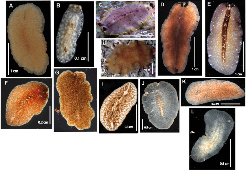
CUADRADO D. et al., New species and records from Macaronesia and Cape Verde

Table 1 (continued).

                  Species                             Localities       GenBank code
                                                      Spain            KY263689
                  Cycloporus gabriellae               Brazil           KY263656
                                                      Brazil           KY263658
                  Eurylepta cornuta var. melobesiarum Cape Verde       MK299350
                  Eurylepta sp.                       Martinica Is.    MK299372
                  Pseudoceros harrisi                 Panama           EF514802
                  Pseudoceros astrorum                Brazil           KY263737
                  Pseudoceros atropurpureus           Japan            LC100098
                  Pseudoceros hancockanus             Japan            LC100100
                  Pseudoceros velutinus               Greece           KY263740
                                                      Spain            KY263741
                                                      Japan            LC100095
                                                      Japan            LC100095
                  Pseudoceros velutinus               Cape Verde Is.   MK299360
                                                      Cape Verde Is.   MK299380
                                                      Canary Is.       MK299381
                  Pseudoceros rawlinsonae             Brazil           KY263731
                                                      Brazil           KY263733
                                                      Florida          GQ398102
                                                      Bahamas          GQ398101
                                                      Virgin Is.       EF514803
                  Pseudoceros rawlinsonae var. galaxy Cape Verde Is.   MK299357
                                                      Cape Verde Is.   MK299358
                  Pseudoceros nipponicus              Japan            LC100096
                  Pseudoceros bicolor                 Brazil           KY263730
                                                      Panama           GQ398096
                                                      Florida          GQ398097
                                                      Jamaica          GQ398100
                  Pseudoceros contrarius              Papua NG         KY263728
                  Yungia sp.                          Florida          HQ659018
                  Yungia aurantiaca                   Cádiz (Spain)    MK299386
                  Pseudobiceros nigromarginatus       Japan            LC100097
                  Pseudobiceros pardalis              Brazil           KY263723
                                                      Panama           EF514807
                                                      Panama           EF514808
                  Pseudobiceros wirtzi                Senegal          KY263725
                  Pseudobiceros bedfordi              Papua NG         KY263715
                  Maiazoon orsakii                    Papua NG         KY263697
                  Phrikoceros mopsus                  Brazil           KY263707

                                               5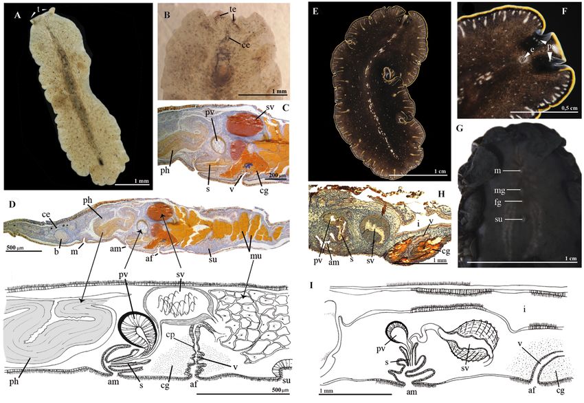
European Journal of Taxonomy 736: 1–43 (2021)

Table 1 (continued).

                  Species                           Localities     GenBank code
                  Notocomplana japonica             Japan          LC100087
                  Notocomplana koreana              Japan          LC100086
COTYLEA           Cestoplana rubrocinta             Cyprus         MK299367
                                                    Cyprus         MK299368
                                                    Cyprus         MK299369
                  Cestoplana salar                  Brazil         KY263653
                  Cestoplana techa                  Brazil         KY263652
                                                    Brazil         KY263654
                                                    Brazil         KY263655
                  Pericelis sp.                     Cape Verde     MK299353
                                                    Cape Verde     MK299354
                  Pericelis byerleyana              Martinica      MK299374
                  Pericelis cata                    Brazil         KY263700
                                                    Canary Is.     MK299351
                                                    Cape Verde     MK299352
                                                    Cape Verde     MK299373
                  Boninia divae                     Panama         KC869846
                  Chromyella sp.                    Panama         KC869848
                  Theama sp.                        Panama         KC869845
                  Anonymus ruber                    Canary Is.     MK299347
                                                    Cape Verde     MK299348
                                                    Cape Verde     MK299364
                                                    Cape Verde     MK299365
                                                    Cape Verde     MK299366
                  Enchiridium evelinae              Brazil         KY263682
                                                    Brazil         KY263683
                  Enchiridium magec                 Canary Is.     MK299349
                                                    Canary Is.     MK299371
                  Prosthiostomum grande             Japan          LC100090
                  Prosthiostomum vulgare            Japan          LC100091
                  Cycloporus variegatus             Brazil         KY263657
                                                    Spain          KY263659
                  Cycloporus japonicus              Japan          LC100092
                  Maritigrella fuscopunctata        Indo-Pacific   KU674837
                                                    Indo-Pacific   KU674838
                  Prostheceraeus vittatus           Sweden         AJ315647
                  Maritigrella crozieri             Florida        KY263686
                                                    Florida        KY263687
                  Prostheceraeus roseus             Spain          KY263688

                                                6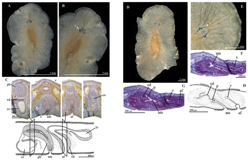
CUADRADO D. et al., New species and records from Macaronesia and Cape Verde

Table 1 (continued).

                   Species                                Localities         GenBank code
                                                          Brazil             KY263709
                                                          Brazil             KY263711
                   Monobiceros sp.                        Cape Verde         MK299359
                   Monobiceros langi                      Spain              KY263710
                                                          Greece             KY263738
                                                          Spain              KY263713
                   Thysanozoon alagoensis                 Brazil,            KY263747
                                                          Martinica Is.      MK299383
                                                          Martinica Is.      MK299384
                                                          Martinica Is.      MK299385
                   Pseudobiceros sp.                      Santa Helena Is.   KY263724
                   Pseudobiceros caribbensis              Florida            EF514805
                                                          Jamaica            EF514806
                                                          Martinica Is.      MK299378
                                                          Martinica Is.      MK299379
                                                          Cadiz (Spain)      MK299387
                   Pseudobiceros flavomarginatus          Japan              LC100099
                   Pseudobiceros evelinae                 Brazil             KY263716
                                                          Brazil             KY263717
                   Pseudobiceros splendidus               Florida            HQ659016
                   Thysanozoon brocchii                   Brazil             KY263744
                                                          Sicily             KY263745
                                                          Australia          HQ659017
                                                          Japan              LC100093
                                                          Cape Verde         MK299361
                                                          Canary Is.         MK299382
                   Thysanozoon japonicum                  Japan              LC100094
                   Acanthozoon aranfaibo                  Canary Is.         MK299362
                   Thysanozoon raphaeli                   Panama             EF514809
                                                          Belize             EF514810

on the differences and similarities among these archipelagos and provides new evidence to reach key
conclusions about their biogeography and fauna.

Material and methods
Localities, sampling sites and material processed
    Azores (Fig. 1A)
The Azores is the northernmost archipelago (37°44′ N, 25°40′ W) and includes nine islands: São Miguel,
Pico, Terceira, São Jorge, Faial, Flores, Santa Maria, Graciosa and Corvo.

                                                   7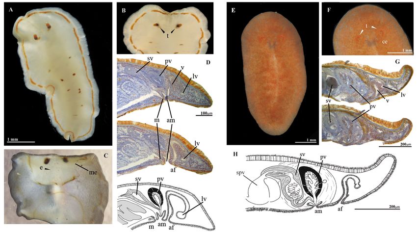
European Journal of Taxonomy 736: 1–43 (2021)

    Canary Islands (Fig. 1B)
The Canary archipelago (28°06′ N, 15°04′ W) includes two island complexes or provinces: Santa Cruz
de Tenerife with four large islands (El Hierro, La Gomera, La Palma and Tenerife) and Las Palmas
with three main islands and some small ones (Fuerteventura, Gran Canaria, Lanzarote and Isla Lobos).
The Chinijo archipelago is part of Las Palmas and includes five small islands (La Graciosa, Alegranza,
Montaña Clara, Roque del Este and Roque del Oeste).

   Madeira (Fig. 1B)
The Madeira archipelago (32°45′ N, 17°00′ W) consists of five islands; two inhabited, Madeira and Porto
Santo, and three small, uninhabited isles, the Desertas. Madeira enjoys a Mediterranean climate, like the
Canary and Selvagens Islands, which is characterized by 3 months of dryness and high temperatures.

    Cape Verde (Fig. 1C)
Cape Verde (15°07′ N, 23°37′ W) is the southernmost archipelago of Macaronesia and is divided into
two groups of islands, Ilhas de Barlovento and Ilhas de Sotavento. Cape Verde comprises ten large
islands: Santo Antão, São Vicente, Santa Luzia, São Nicolau, Sal, Boa Vista (Barlovento Island) and
Maio, Santiago, Fogo, and Brava (Sotavento Island) as well as five minor isles Raso, Branco, Grande,
Cima and Carneiro.

    Southwestern Morocco (Fig. 1B)
Although actually northern Morocco may not be part of Macaronesia, we include the species found in
Agadir due to the similar ecological conditions and geographical proximity to the Canary Islands.

The study material from the Canary Islands, Cape Verde, Azores, Agadir and Ifni (Morocco) was
collected by hand while scuba diving from rocks, the sea bottom, cave surfaces and macro-algae samples.
Additional specimens were collected by taking rock and algae samples from the field to the laboratory
and waiting for oxygen to be depleted, forcing animals to come out of hiding.

Exhaustive information about the external features was carefully recorded with notes, photographs and
drawings. Information about pigmentation, color patterns, movement, size, and presence or absence of
tentacles or eyes was gathered. Dorsal structures like papillae, warts or any type of epithelial or dermal
formations were compiled.

Most of the photographs were taken on a black background with transmitted light using a Nikon D300
camera fitted with a Micro Nikon 60 mm lens, a Kenko extension tube and two wireless R1 speed lights.

Histological processing
For fixation, the individuals were previously anesthetized with seawater/magnesium chloride (7%).
A small piece of tissue was removed for molecular analysis and the whole individual was fixed in Bouin
solution (0.8 gr of picric acid in 80 ml, 20 ml of formaldehyde and 2 ml of acetic acid) for histological
studies. Histological sagittal series from 6 to 12 micrometres thick were stained with AZAN trichrome.
The histological preparations of the studied specimens were deposited in the collections of the Nacional
Museum of Natural Sciences (MNCN), Madrid.

For the definitive identification of genus/species internal anatomic reconstructions, particularly of the
reproduction apparatus, were made using a Zeiss Axio Scope A1 microscope.

DNA extraction, amplification and sequencing
Tissues for molecular studies were fixed in absolute ethanol. Total genomic DNA was extracted from
each sample following the phenol-chloroform protocol (Chen et al. 2010). DNA concentration and

                                                    8
CUADRADO D. et al., New species and records from Macaronesia and Cape Verde
purity of the extraction was measured using a NanoDrop Fluorospectrometer (Thermo Fisher Scientific).
Sequences of the ribosomal gene 28S of the investigated Polycladida species were studied. All PCRs
were performed using Taq DNA polymerase of Mastermix (Invitrogen, Carlsbad, CA) following the
manufacturer’s protocol in a total volume of 25 μl.
Sequences of approximately 1100 bp of the 28S gene were amplified with degenerated primers designed
de novo by the first two authors: forward primer (5´-AGCCCAGCACCGAATCCT3-´) and reverse
(5´-GCAAACCAAGTAGGGTGTCGC-3´). The PCR consisted in an initial denaturation step at 95°C
(3 min), followed a pre-cycle of 5 cycles of denaturation at 96°C (30 sec), annealing at 55°C (30 sec)
and extension at 72°C (1 min), followed by 40 cycles of denaturation at 95°C (30 sec), annealing at 59°C
(30 sec) and extension at 72°C (1 min), with a final extension of 10 min at 72°C.
Finally, the sequences obtained from forward and reverse primers were combined using the program
Geneious R6 (ver. 6.1.5) (http://www.geneious.com; Kearse et al. 2012).
Sequence alignment and phylogenetic analyses
A comparative analysis with both newly obtained sequences and those obtained from the NCBI
GenBank database was carried out. A total of 147 sequences were aligned and edited using Geneious R6
(ver. 6.1.5). Forty-one of them were new sequences (NCBI accession numbers in Table 1).
The newly obtained sequences of 1100 bps were adapted to the length of those gathered from GenBank.
The alignment was generated using the program MAFFT ver. 7 (Katoh & Standley 2013). Ambiguously
aligned and variable regions were recognized and excluded using the program Gblocks ver. 0.91b
(Castresana 2000) with relaxed parameters (smaller final blocks, gap positions within the final blocks,
and less strict flanking positions allowed). Thus, a matrix of 744 bp was obtained.
Maximum likelihood (ML) was implemented through IQ-TREE (Trifinopoulos et al. 2016), using the
evolutionary model BIC: TIM2+I+G4. The consensus tree of 1000 bootstrap pseudoreplicates was
selected and edited with iTOL ver. 3.1.1 (Letunic & Bork 2016).
Bayesian inference (BI) analyses were carried out using MrBayes ver. 3.2.2 (Ronquist et al. 2012)
(-mset option). Two independent runs of 1 000 000 generations and four chains (one cold, three heated)
were run. Trees were sampled every 1000 generations. Convergence of chains was diagnosed using a
deviation of standard frequencies below 0.05 and of the 1001 sampled trees, 250 trees were discarded
as burn-in. A majority-rule consensus tree was constructed from the remaining 751 trees to approximate
posterior probabilities.
Abbreviations used in the figures
af = female atrium
am = male atrium
b = brain
ce = cerebral eyes
cg = cement glands
cga = common genital atrium
cp = cement glands pouch
e = eyes
ed = ejaculatory duct
fg = female gonopore
i = intestine
lv = Lang’s vesicle
9European Journal of Taxonomy 736: 1–43 (2021)
m = mouth
me = marginal eyes
mg = male gonopore
mu = multiple uterine vesicles
p = penis papilla
pb = penis bulb
ph = pharynx
ps = pseudotentacles
pt = pseudotentacles
pv = prostatic vesicle
s = stylet
su = sucker
sv = seminal vesicle
t = tentacles
te = tentacular eyes
u = uteri
v = vagina
Institutional acronyms for collections
MNCN = Museo Nacional de Ciencias Naturales, Madrid, Spain
RCCN = Research Collection of Carolina Noreña, Museo Nacional de Ciencias Naturales, Madrid,
Spain
Results
Table 2 lists the complete register of the Polycladida from the Macaronesian Archipelagos. New species
and records are described as follows.
AZORES (Fig. 1A). Currently, only six species are recorded for the Azores: Enchiridium cf. magec
Cuadrado, Moro & Noreña, 2017, Thysanozoon cf. brocchii (Risso, 1818), Prostheceraeus giesbrechtii
Lang, 1884, Prostheceraeus cf. roseus Lang, 1884 and the new records Prostheceraeus moseleyi Lang,
1884 and Stylochus sp. (Table 2) (Wirtz 1994; Wirtz & Debelius 2003).
CANARY ISLANDS (Fig. 1B). The species of the Canary Islands were listed and described recently in
Cuadrado et al. (2017); therefore, only new records are described in this study: Discocelis tigrina
(Blanchard, 1847) and Gnesioceros sargassicola (Mertens, 1833).
MADEIRA (Fig. 1B). Pseudoceros wirtzi (Bahia & Schrödl, 2016), Pseudoceros cf. maximus Lang, 1884
and Prostheceraeus giesbrechtii (Wirtz & Debelius 2003; Bahia & Schrödl 2016) are the three polyclad
species currently recorded for Madeira. The new record Planocera pellucida (Mertens, 1833) is recorded
in this study.
CAPE VERDE (Fig. 1C). 31 species are described for Cape Verde. The new records described in this study
include Pericelis cata Marcus & Marcus, 1968, Anonymus ruber Cuadrado, Moro & Noreña, 2017,
Eurylepta cornuta var. melobesiarum Lang, 1884, Cycloporus gabriellae Marcus, 1950, Pseudoceros
velutinus (Blanchard, 1847), Pseudoceros mororum Cuadrado, Moro & Noreña, 2017, Monobiceros
langi Faubel, 1984 and Stylochus pillidium (Götte, 1881). Additionally, a new variety Pseudoceros
rawlinsonae var. galaxy and six new species Marcusia alba sp. nov., Prostheceraeus crisostomus
sp. nov., Stylochus salis sp. nov., Distylochus fundae sp. nov., Euplana claridade sp. nov. and Parviplana
sodade sp. nov. are described.
10Table 2 (continued on next two pages). Species of the insular complex of Macaronesia s. lat. Locations and bibliographical references. Species
analyzed and described in this study are highlighted in blue.
Species Azores Madeira Canary Islands Cape Verde Morocco References
Cestoplana rubrocincta Tenerife Boa Vista; Laidlaw 1906;
São Vicente Cuadrado et al. 2017;
This study
Pericelis cata Tenerife São Vicente Cuadrado et al. 2017;
This study
Traunfelsia elongata Cape Verde Laidlaw 1906
Marcusia alba sp. nov. São Vicente This study
Anonymus virilis Tenerife São Vicente Laidlaw 1906;
Cuadrado et al. 2017
Anonymus ruber Tenerife São Vicente Cuadrado et al. 2017;
11
This study
Enchiridium magec Faial Tenerife Wirtz & Debelius 2003;
Cuadrado et al. 2017
Prosthiostomum dohrnii São Vicente Laidlaw 1906
Eurylepta cornuta São Vicente, Sal This study
var. melobesiarum
Eurylepta guayota El Hierro Cuadrado et al. 2017
Cycloporus papillosus Santiago Laidlaw 1906
Cycloporus gabriellae Fuerteventura São Vicente This study
Oligocladus sanguinolentus São Vicente Laidlaw 1906
Prostheceraeus giesbrechtii Faial Porto Santo La Gomera Wirtz & Debelius 2003;
Cuadrado et al. 2017
Prostheceraeus moseleyi São Miguel This study
Prostheceraeus roseus Lanzarote Cuadrado et al. 2017
CUADRADO D. et al., New species and records from Macaronesia and Cape VerdeTable 2 (continued).
Species Azores Madeira Canary Islands Cape Verde Morocco References
Prostheceraeus rubropunctatus Cape Verde Laidlaw 1906
Prostheceraeus crisostomum sp. nov. Sal This study
Phrikoceros mopsus Canary Islands Cuadrado et al. 2017
Pseudoceros maximus Madeira Fuerteventura Wirtz & Debelius, 2003;
Cuadrado et al. 2017
Pseudoceros velutinus Lanzarote, Tenerife São Vicente Cuadrado et al. 2017;
This study
Pseudoceros mororum Gran Canaria São Vicente Cuadrado et al. 2017;
This study
Pseudoceros rawlinsonae São Vicente This study
var. galaxy
European Journal of Taxonomy 736: 1–43 (2021)
Pseudoceros sp. La Gomera Cuadrado et al. 2017
12
Pseudobiceros wirtzi Madeira Fuerteventura Santo Antão Bahia & Schrödl 2016;
Cuadrado et al. 2017
Monobiceros langi São Vicente This study
Thysanozoon brocchii Faial Tenerife São Vicente Laidlaw 1906;
Wirtz & Debelius 2003;
This study
Acanthozoon aranfaibo El Hierro Cuadrado et al. 2017
Yungia aurantiaca Fuerteventura Cuadrado et al. 2017
Multisepta fengari El Hierro Cuadrado et al. 2017
Zygantroplana verrilli São Vicente Laidlaw 1906;
This study
Cryptocelis loveni Agadir Beauchamp 1951Table 2 (continued).
Species Azores Madeira Canary Islands Cape Verde Morocco References
Planocera pellucida Madeira El Hierro Boa Vista; Laidlaw 1906;
São Vicente Cuadrado et al. 2017;
Faubel 1984; This study
Stylochus alexandrinus Agadir Prudhoe 1989
Stylochus neapolitanus Cape Verde Ifni Laidlaw 1906,
Stylochus sp. São Miguel This study
Stylochus salis sp. nov. Sal This study
Stylochus pilidium São Vicente Agadir This study
Imogine mediterranea Temara Prudhoe 1989
Distylochus fundae sp. nov. Sal This study
Stylochoplana graffi Deep Sampling, Laidlaw 1906
13
Boa Vista
Latocestus plehni Tenerife São Vicente Laidlaw 1906;
This study
Polyphalloplana bocki Agadir Beauchamp 1951
Gnesioceros sargassicola Gran Canaria; This study
Tenerife
Discocelis tigrina Gran Canaria This study
Notoplana alcinoi São Vicente Laidlaw 1906
Emprosthopharynx pallida Boa Vista Laidlaw 1906
Euplana claridade sp. nov. São Vicente This study
Parviplana sodade sp. nov. São Vicente This study
CUADRADO D. et al., New species and records from Macaronesia and Cape VerdeEuropean Journal of Taxonomy 736: 1–43 (2021)
SOUTHWESTERN MOROCCO (Fig. 1B). Four Polycladida species are known from the south coast near
Agadir (Morocco), three species recorded by Beauchamp (1951): Polyphalloplana bocki Beauchamp,
1951, Stylochus alexandrinus Steinböck, 1937 and Cryptocelis loveni Bergendal, 1890, and Stylochus
mediterraneus Galleni, 1976 by Prudhoe (1989). In the present study, two new records are described
from Morocco, Stylochus neapolitanus (Delle Chiaje, 1841-1844) and Stylochus pilidium (Götte, 1881)
(Table 2).
New species and varieties
Order Polycladida Lang, 1881
Suborder Cotylea Lang, 1884
Family Diposthidae Woodworth, 1898 sensu Litvaitis et al., 2019
Genus Marcusia Hyman, 1953
Marcusia alba sp. nov.
urn:lsid:zoobank.org:act:DE4AF1C8-D784-4AB7-B536-A1E88524115B
Figs 1C, 2
Etymology
The name of the new species, Marcusia alba, comes from the Latin ‘albus’ (white), and refers to the
ivory white coloration this species shows.
Material examined (3 specs)
Holotype
CAPE VERDE • São Vicente Island, Mindelo; 16°53′46.54″ N, 24°59′32.93″ W (Fig. 1C V1); 6 May
2017; Leopoldo Moro leg.; MNCN 4.01/2620 to 2683 (64 slides). One sagittally sectioned specimen
stained with AZAN.
Additional material
CAPE VERDE • 2 specs; São Vicente Island, Baía das Gatas; 16°54′09.33″ N, 24°54′25.25″ W (Fig. 1C
V2); 6 May 2017; Leopoldo Moro leg.; RCCN.
Description
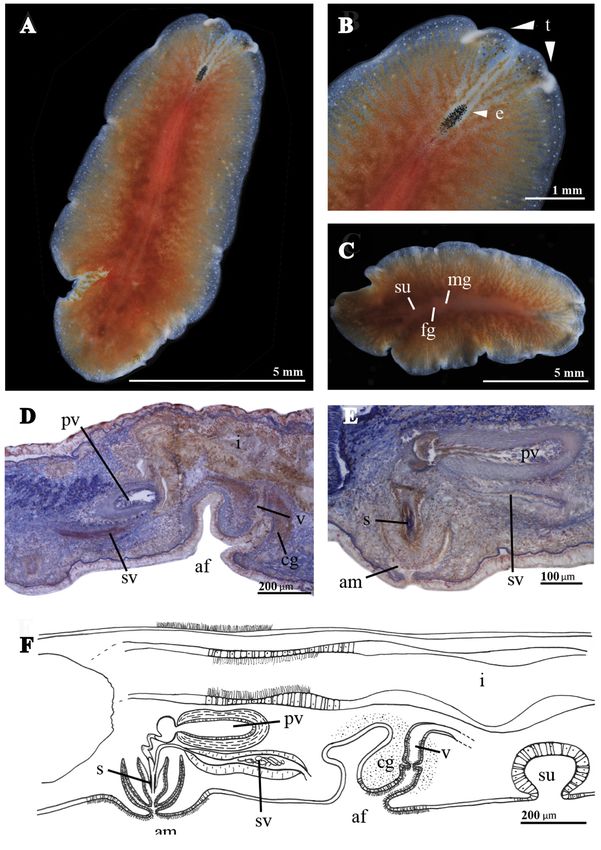
BODY. Shape oval. Length 2.6 cm. Smooth dorsal surface. Background pigmentation ivory white, darker
along the middle dorsal region of the body. Amber dots and thin brushstroke-like lines garnish the dorsal
surface. A thin, dark stripe, sometimes interrupted, extends along the central dorsal line (Fig. 2A). Two
delicate marginal folds, separate and pointed, could be interpreted as pseudotentacles. Tentacular eyes
scarce and widely dispersed over the tentacles. Cerebral eyes anterior drop-shaped and crossed by the
central midline. Marginal eyes only at the anterior part (Fig. 2B). Ruffled pharynx, in the middle of the
body with a central oral pore. Ventral sucker at the posterior part of the body. Male and female genital
pore lead in a common genital atrium (Fig. 2D–E) that opens in the posterior body region after the
pharynx (Fig. 2C).
MALE REPRODUCTIVE SYSTEM. Male copulatory organ backwards oriented, with a muscular penis papilla,
very muscular seminal vesicle (Fig. 2C, E) and without a prostatic vesicle, instead a simple glandular
epithelium leads into the penis papilla (Fig. 2D–E). Seminal vesicle rounded, frontal oriented and with
thick muscular walls, opening into the ejaculatory duct. Short ejaculatory duct opens into the penis
papillae. The male atrium is small and thin, connected to the common atrium.
14CUADRADO D. et al., New species and records from Macaronesia and Cape Verde
FEMALE REPRODUCTIVE SYSTEM (Fig. 2C, E). The vagina runs from the common genital atrium and
continues dorsally into a narrowed duct that widens into a chamber, the cement pouch. The vagina
continues dorsally, then curves posteriorly and ventrally and ends with the entry of the oviducts.
Remarks
Marcusia alba sp. nov. belongs to the genus Marcusia due to the presence of cerebral, frontal and
marginal eyes, male copulatory organ enclosed in a muscular bulb, the absence of prostatic vesicle and
the common male and female atrium genital, as well as the common gonopore.
Fig. 2. Marcusia alba sp. nov. (MNCN 4.01/2620 to 2683). A. Whole live animal, dorsal view. B. Anterior
region with cerebral eyes cluster and pseudotentacles (white arrows). C. Histological section of the male
copulatory organ stained with AZAN. D. Histological section of the female copulatory organ stained
with AZAN. E. Sagittal reconstruction of the reproduction system. Abbreviations: see Material and
methods.
15European Journal of Taxonomy 736: 1–43 (2021)
The genus Marcusia contained only one species, Marcusia ernesti Hyman, 1953, known from the
coast of the Gulf of California (Hyman 1953). Marcusia ernesti and M. alba sp. nov. can be easily
distinguished by their coloration patterns. Marcusia ernesti is black or grey with darker splotches and
dotted with white spots, only visible in preserved individuals after Hyman (1953), M. alba sp. nov. is
ivory white with brownish dots and stripes. The penis papilla is spherical in M. alba and elongated in M.
ernesti, with the male atrium being tube-like and longer in the Californian species.
Another difference lies in the eyes’ presence and distribution. Marcusia ernesti presents marginal,
frontal and cerebral eyes as well as two characteristic eye clusters with diagnostic value (Hyman 1953).
Marcusia alba sp. nov., on the other hand, has cerebral, marginal and tentacular eyes, but not frontal
eyes or eye clusters.
The differences listed are enough to consider M. ernesti and M. alba sp. nov. as two different species
of the same genus. Furthermore, the molecular analyses show the genus Marcusia (represented in this
case by Marcusia alba sp. nov.) as a genus closely related to Pericelis Laidlaw, 1902 within the family
Anonymidae Lang, 1884, but as a clearly independent genus.
Family Euryleptidae Lang, 1884
Genus Prostheceraeus Schmarda, 1859
Prostheceraeus crisostomum sp. nov.
urn:lsid:zoobank.org:act:EC7A7E2B-99FD-447D-A189-676144875AC2
Figs 1C, 3A–D
Etymology
The name of the new species, Prostheceraeus crisostomum, is dedicated to the little cat, Crisostomo,
roommate during the description of this species.
Material examined
Holotype
CAPE VERDE • Sal Island, Calheta Funda; 16°39′03.34″ N, 22°56′42,94″ W (Fig. 1C V3); 8 Jul. 2018;
Leopoldo Moro leg.; MNCN 4.01/2684 to 2698 (15 slides). One sagittally sectioned specimen stained
with AZAN.
Description
BODY. Shape elongated. Length 0.5 cm. Smooth dorsal surface; background pigmentation ivory white,
darker along the middle dorsal region of the body between the cerebral eyes and the posterior end of the
body. In the posterior middle end it shows a large conspicuous black spot. Small black dots on the entire
dorsal surface (Fig. 3E). Two marginal tentacles, separate. Tentacular eyes scarce and widely dispersed
between the tentacles. Cerebral eyes arrow-shaped located by the central midline (Fig. 3F). Bell-shaped
pharynx located in the first body half. Ventral sucker in the middle of the body. Male and female genital
pores well separated and located after the pharynx (Fig. 3G–H).
MALE REPRODUCTIVE SYSTEM. Male copulatory organ oriented forward. The muscular prominent penis
papilla houses a conical, elongated stylet of pseudosclerotized nature. The rounded and well developed
prostatic vesicle joins transversally with the sperm duct and lies over the penis papilla (Fig. 3H).
Muscular seminal vesicle oval, dorsally located and caudo-frontally oriented. The vasa deferentia
join at the ventro-caudal region of the vesicle and the sperm duct open ventro-frontally. Seminal and
prostatic vesicles open together into the proximal region of the developed penis papillae. The male
atrium surrounded the penis papillae and opens near the posterior end of the pharynx.
16CUADRADO D. et al., New species and records from Macaronesia and Cape Verde
FEMALE REPRODUCTIVE SYSTEM. Atrium elongated and highly ciliated, continues dorsally into the long but
not ciliated vagina externa. The vagina externa narrows into a non-ciliated small cavity that continues
in the vagina interna. It presents a widened epithelium and ends with the entry of the oviducts (Fig. 3H).
Cement and shell glands lie around the female atrium, vagina externa and distal region of the vagina
interna, but opens into the small cavity (pouch) between both vaginas.
Remarks
Prostheceraeus crisostomum sp. nov. belongs to the genus Prostheceraeus due to the presence of
cerebral, frontal and marginal eyes, true anterior tentacles, bell-shaped pharynx, the male copulatory
system with prostatic vesicle, penis armed whit stylet and the presence of multiple uterine vesicles.
The genus Prostheceraeus comprises 10 species, mainly characterized by coloration pattern, with colorful
pigmentations and dorsal longitudinal lines of different widths, as in P. fuscolineatus Dixit, Raghunathan
& Chandra, 2017, P. roseus, P. pseudolimax Lang, 1884, P. giesbrechtii, P. vittatus (Montagu, 1815) and
P. zebra (Hyman, 1955) or with fine, transversal lines as in P. crozieri (Hyman, 1939).
Fig. 3. A–D. Prostheceraeus crisostomum sp. nov. (MNCN 4.01/2684 to 2698). A. Whole live animal,
dorsal view. B. Anterior region with cerebral eyes cluster and tentacles (black arrows). C. Histological
section of the reproductive system stained with AZAN. D. Histological section of the whole animal
stained with AZAN and sagittal reconstruction. – E–I. Pseudoceros rawlinsonae var. galaxy (MNCN
4.01/2729 to 2798). E. Whole live animal, dorsal view. F. Anterior region with eyes cluster and tentacles
(white arrows). G. Whole live animal, ventral view. H. Histological sagittal section of male and female
copulatory organ. I. Sagittal reconstruction of the reproductive system. Abbreviations: see Material and
methods.
17European Journal of Taxonomy 736: 1–43 (2021)
Three other species of Prostheceraeus show a color pattern free of lines or bands: P. albocinctus Lang,
1883, P. moseleyi and P. rubropunctatus Lang, 1884. These three species, together with P. crisostomum
sp. nov., have a dotted pattern, but the background colors are different in the four species: caramel brown
background with white or whitish spots and white marginal line in P. albocinctus, blue-gray or cream
background with black dots and yellow marginal band in P. moseleyi and finally P. rubropunctatus
with a pink to reddish background color, white dots and without marginal band. The base coloration
of P. crisostomum is similar to P. albocinctus, but much clearer and almost ivory; the dorsal points
are black like in P. moseleyi and lacks a marginal line or band similar to P. rubropunctatus. All these
differences delimit P. crisostomum sp. nov. as a new species of the genus Prostheceraeus.
Family Pseudocerotidae Lang, 1884
Genus Pseudoceros Lang, 1884
Pseudoceros rawlinsonae var. galaxy var. nov.
Figs 1C, 3E–I
Etymology
The name ‘galaxy’ comes from the pattern of the small white spots on the dorsal surface, which resemble
a star galaxy.
Material examined (2 specs)
Holotype
CAPE VERDE • São Vicente Island, Baía das Gatas; 16°54′09.33″ N, 24°54′25.25″ W (Fig. 1C
V2); 5 May 2017; Leopoldo Moro leg.; MNCN 4.01/2729 to 2798 (70 slides). One sagittally
sectioned specimen stained with AZAN.
Additional material
CAPE VERDE • 1 spec.; same collection data as for holotype; RCCN.
Description
BODY. Shape oval. Length 2.3 cm. Smooth dorsal surface. Background color dark to velvety brown with
white dots and spots that draw a longitudinal line along the body axis. One thin bright yellow external
line and another internal and black line surround the entire body margin. The yellow one is interrupted
at the pseudotentacles level (Fig. 3E–F). Marginal body edges lined with transversal whitish elongated
drops (Fig. 3F). Ventral coloration dark grey to black. Ventral sucker in the middle of the body (Fig. 3G).
Pseudotentacles constitute two simple folds that present each of them a small cluster of tentacular eyes
in their margin. Round cluster of cerebral eyes present and surrounded by a spot of white pigment.
Pharynx ruffled, butterfly-shaped and located at the anterior third of the body. Oral pore, female and
male gonopore close to each other and located at the anterior end (Fig. 3G). Male and female genital
pores located after the pharynx in the anterior half of the body (Fig. 3H–I).
MALE REPRODUCTIVE SYSTEM. Male genital pore between the posterior lobes of the ruffled pharynx. Male
copulatory organ dorso-ventrally orientated consists in a prostatic vesicle and a very muscular seminal
vesicle, as well as a penis papilla armed with a stylet (Fig. 3H–I). Vasa deferentia open separately into
the seminal vesicle. Seminal vesicle rounded, frontally oriented and lined with a thick muscular wall.
Prostatic vesicle rounded, muscular and smaller than the seminal vesicle. Sperm duct muscular and
long, extends frontally to join the prostatic duct inside the proximal end of the conical stylet. The short
ejaculatory duct appears surrounded by the stylet cone and the penis sheath. The male atrium is wide and
tetra-folding (fork-like) as characteristic of the genus (Fig. 3I).
18CUADRADO D. et al., New species and records from Macaronesia and Cape Verde
FEMALE REPRODUCTIVE SYSTEM. With a short muscular vagina, backwards oriented and surrounded by
cement glands.
Remarks
The genus Pseudoceros comprises approximately 89 species with similar copulatory organs, but bright
and unique coloration patterns. However, within these patterns some taxa share evident similarities.
Pseudoceros rawlinsonae var. galaxy shares with P. bicolor Verril, 1902, P. mororum and P. rawlinsonae
Bolaños, Quiroga & Litvaitis, 2007 the brown background and one whitish, broad marginal band, but
in P. bicolor the marginal band is wide with inner waves (Litvaitis et al. 2010: fig. 4a–i); P. rawlinsonae
shows, in addition to the wide band, a thin orange line (Litvaitis et al. 2010: fig. 4j–p); in P. mororum
the whitish band is interrupted and drop-shaped and additionally, two orange marginal stripes border the
entire body (Cuadrado et al. 2017: fig. 6a–b); finally, the Cape Verdean species shows, together with the
drop-shaped white band, two black and orange thin lines (Fig. 3E–F).
Although the four previously mentioned species can be clearly differentiated due to their coloration,
this is not the case in the molecular analysis (Fig. 8). In both the Bayesian and Maximum Likelihood
analyses, individuals from Cape Verde appear closely related to P. rawlinsonae, so much so that the
separation of both populations (the Cape Verdean population and the Caribbean population) is only
possible at the level of variety, not of species. Therefore, we determined the individuals from Cape Verde
as a variety within the species P. rawlinsonae.
Nonetheless, we want to emphasize that the decision to maintain this population (organisms) as a
‘variety’ of the species P. rawlinsonae is the sole and exclusive responsibility of the authors. We are
aware that ‘variety’ is not a taxonomic category (according to ICZN) and that therefore it will remain a
non-existent species until molecular analyses allow us to consider it as such.
Suborder Acotylea Lang, 1884
Superfamily Leptoplanoidea Faubel, 1984
Family Leptoplanidae Stimpson, 1857
Genus Parviplana Hyman, 1953
Parviplana sodade sp. nov.
urn:lsid:zoobank.org:act:10171C70-7431-41EF-B39D-AC3982D24143
Figs 1C, 4A–C
Etymology
The name of the new species, Parviplana sodade, comes from ‘sodade’ the Cape Verdean expression for
saudade and regional song with rhythms of ‘coladeira’.
Material examined
Holotype
CAPE VERDE • São Vicente Island, Mindelo; 16°53′46.54″ N, 24°59′32.93″ W (Fig. 1C V1); 24 Nov.
2017; Leopoldo Moro leg.; MNCN 4.01/2699 to 2708 (10 slides). One sagittally sectioned specimen
stained with AZAN.
Description
BODY. Shape oval elongated. Length 0.8 cm. Smooth dorsal surface. Background pigmentation light
white, transparent where the intestinal braches can be appreciated (Fig. 4A–B). Four clusters of cerebral
eyes, two anterior with few eyes and more elongated than the posterior two. In sum around 50 cerebral
eyes (Fig. 4B). Ruffled pharynx. Male and female genital pores located in the posterior half of the body.
19European Journal of Taxonomy 736: 1–43 (2021)
MALE REPRODUCTIVE SYSTEM. Directed backwards and with a dorso-ventrally oriented penis papilla.
With elongated prostatic vesicle, tall granular lining included in the muscular penis bulb (Fig. 4C). The
vasa deferentia enter the seminal vesicle separately. Seminal vesicle rounded, below the penis bulb and
connected with a sort seminal duct to the prostatic vesicle (Fig. 4C). The male atrium is small and thin,
with an internal fold that surrounds the distal part of the penis bulb like a penis sheath (Fig. 4C).
FEMALE REPRODUCTIVE SYSTEM. With a vagina bulbosa (Fig. 4C) and backwards oriented. Cement and
shell glands open in a pouch located within the vaginal complex. Lang’s vesicle present.
Remarks
Parviplana sodade sp. nov belongs to the genus Parviplana due to the absence of tentacles, presence
of seminal vesicle, and prostatic vesicle with a tall granular lining with prostatic functions. Female
apparatus with vagina bulbosa and Lang´s vesicle.
Parviplana comprises 3 species, P. hymani Faubel, 1983, P. jeronimoi Pérez-García, Noreña & Cervera,
2018 and P. lynca (Du Bois-Reymond Marcus, 1958). Parviplana lynca can be easy and clearly
distinguished from the other two species by the presence of nuchal tentacles, exclusive of this species.
Parviplana hymani can be distinguished from P. sodade sp. nov., by the vas deferens which opens
together into the seminal vesicle, and the prostate vesicle not included into de penis bulb.
Parviplana sodade sp. nov. possesses more similarities with P. jeronimoi. Both species share the penis
sheath and more than 25 cerebral eyes, but clear differences separate them. Parviplana jeronimoi has a
fleshy appearance and amber pigmentation. The size is also noticeable different, P. jeronimoi can reach
lengths of 20 mm, while P. sodade in full mature state does not reach 8 mm. Parviplana jeronimoi also
presents vasa deferentia joined in a single vas deferens, a small female atrium and a corrugated surface
between the two genital pores, characteristics not present in P. sodade.
Family Euplanidae Marcus & Marcus, 1966
Genus Euplana Girard, 1893
Euplana claridade sp. nov.
urn:lsid:zoobank.org:act:C7E211FD-32AF-4FA4-A3EB-ACA1737DF2EE
Figs 1C, 4D–H
Etymology
The name of the new species, Euplana claridade, comes from “Claridade”, a journal of literary review
that revolutionized Cape Verdean culture during the first half of the twentieth century.
Material examined
Holotype
CAPE VERDE • São Vicente Island, Mindelo; 16°53′46.54″ N, 24°59′32.93″ W (Fig. 1C V1); 23 Nov.
2017; Leopoldo Moro leg.; MNCN 4.01/2709 to 2718 (10 slides). One sagittally sectioned specimen
stained with AZAN.
Description
BODY. Shape oval. Length 1.1 cm. Smooth dorsal surface. Background pigmentation ivory white, denser
along the middle dorsal region of the body and in the intestinal braches (Fig. 4D). Two clusters of 16
cerebral eyes each (Fig. 4E). Ruffled pharynx. Male and female genital pores located in the second half
of the body close behind the posterior end of the pharynx.
20CUADRADO D. et al., New species and records from Macaronesia and Cape Verde
MALE REPRODUCTIVE SYSTEM. Male copulatory organ backwards oriented, englobed in a muscular bulb
with a small penis papilla. Vas deferens opens proximally into the ejaculatory duct (Fig. 4F–H). Without
prostatic or seminal vesicle. Male atrium deep and thickened in the point of union with the penis
(Fig. 4F–H).
FEMALE REPRODUCTIVE SYSTEM. Rounded female atrium (Fig. 4F–H). Vagina backwards oriented,
surrounded by muscular fibres. Without Lang´s vesicle.
Remarks
Euplana claridade sp. nov. belongs to the genus Euplana due to the absence of tentacles, prostatic
vesicle and Lang´s vesicle, and presence of a true seminal vesicle and elongated coiling ejaculatory duct.
The genus Euplana encompasses 3 species, E. carolinensis Hyman, 1940, E. gracilis Girard, 1853
and E. hymanae Marcus, 1947. The three species can be differentiated through the eyes number and
disposition of them. E. gracilis and E. carolinensis present four cluster of eyes, two cerebral and two
tentacular; E. hymanae and E. claridade sp. nov. only show two groups of cerebral eyes.
Fig. 4. A–C. Parviplana sodade sp. nov. (MNCN 4.01/2699 to 2708). A. Whole live animal, dorsal
view. B. Anterior region with eyes cluster and tentacles (white arrows). C. Histological cross sections
of male and female copulatory organ stained with AZAN and sagittal reconstruction of the reproductive
system. – D–H. Euplana claridade sp. nov. (MNCN 4.01/2709 to 2718). D. Whole live animal, dorsal
view. E. Anterior region with cerebral eyes cluster and tentacles (white arrows). F. Histological section
of female copulatory organ stained with AZAN. G. Histological section of male copulatory organ
stained with AZAN. H. Sagittal reconstruction of reproductive system. Abbreviations: see Material and
methods.
21European Journal of Taxonomy 736: 1–43 (2021)
Within the reproductive system another differences can have founded, E. gracilis and E. hymanae
present a small, short male atrium and a backwards oriented vagina externa, while E. carolinensis and
E. claridade sp. nov. share a long tubular male atriumand a forward directed vagina externa.
Both species differ mainly in the male copulatory organ. The ejaculatory duct in E. carolinensis is wide
and straight and by E. claridade sp. nov. is narrow and sinuous. Furthermore, the penis papilla is very
short in E. carolinensis, while in E. claridade sp. nov. it is longer in comparison. On the other hand,
the seminal vesicle is practically non-existent in E. claridade sp. nov., represented by the confluence of
the two vasa deferentia, while in E. carolinensis it is a well-developed seminal vesicle, surrounded by
circular muscles and in which the vas deferens empties proximally.
Superfamily Stylochoidea Poche, 1926
Family Stylochidae Stimpson, 1857
Genus Stylochus Ehrenberg, 1831
Stylochus salis sp. nov.
urn:lsid:zoobank.org:act:8A640FC8-D74C-4617-9766-F0FB9806BD4B
Figs 1C, 5A–D
Etymology
The name of the new species, Stylochus salis refers to the type locality, Sal, a Cape Verdean Island.
Material examined (2 specs)
Holotype
CAPE VERDE • Sal Island, Calheta Funda; 16°39′03.34″ N, 22°56′42.94″ W (Fig. 1C V3); 12 Nov.
2018; Leopoldo Moro leg.; MNCN 4.01/2719 to 2723 (5 slides). One sagittal sectioned specimen stained
with AZAN.
Additional material
CAPE VERDE • 1 spec.; Sao Vicente Island, Mindelo (Fig. 1C V1); 16°53′46.54″ N, 24°59′32.93″ W;
6 May 2017; Leopoldo Moro leg.; RCCN.
Description
BODY. Shape elongated. Length 0.4 cm. Smooth dorsal surface. Background pigmentation cream white
with an orange internal outline, sometimes interrupted, along the body margin and bordered by a white/
creamy outer band (Fig. 5A). Few cerebral and marginal eyes, scattered between the tentacles and
anterior end (Fig. 5C). Two small nuchal tentacles with abundant basal eyes (Fig. 5B). Ruffled pharynx
in the middle of the body and the oral pore in the end of the pharynx pouch and close to the gonopores.
Male and female gonopores located close together in the posterior end of the body.
MALE REPRODUCTIVE SYSTEM. Male copulatory organ backwards oriented and provided with an
inconspicuous unarmed penis papilla. Prostatic vesicle muscular with granular lining (polyglandular-
type after Bulnes et al. 2005) (Fig. 5D). Seminal vesicle elongated, empties at the distal end of the
prostatic vesicle. The short penis papilla and ejaculatory duct emerge in a small male atrium (Fig. 5D).
FEMALE REPRODUCTIVE SYSTEM. Shows the characteristic configuration of the genus. A tubiform canal with
s-shaped ending in a small widening.
22CUADRADO D. et al., New species and records from Macaronesia and Cape Verde
Remarks
Stylochus salis sp. nov. belongs to the genus Stylochus due to the presence of gonopores separate and
arranged in the second body half. With large and much ruffled pharynx. Tentacular, cerebral, marginal,
and often frontal eye-spots present. Male copulatory apparatus with seminal vesicle and papillate penis.
Lang’s vesicle lacking (after Faubel 1983).
Stylochus salis sp. nov. clearly differs from other known species of Stylochus Ehrenberg, 1831 by its
peculiar cream pigmentation bordered with the internal orange outline and the white/creamy outer
band. The color of the eastern Atlantic known species (S. alexandrinus, S. castaneus Palombi, 1939,
S. neapolitanus, S. plessissii Lang, 1884, and S. suesensis Ehrenberg, 1831) varies between brown,
light brown, reddish or beige and spotted as in S. neapolitanus. None of them present a continuous (or
discontinuous) marginal line.
The most conspicuous anatomical feature is the location of the oral pore, very close to the gonopore,
clearly different from the central position of the oral pore in this genus. The peculiar location of the oral
and genital pores distinguishes S. salis sp. nov. from the remaining species. Such a close location of the
pores could only be found in the genus Latocestus Plehn, 1896 (Latocestidae, Stylochoidea).
Fig. 5. A–D. Stylochus salis sp. nov. (MNCN 4.01/2719 to 2723). A. Whole live animal, dorsal view.
B. Anterior region with eyes cluster and tentacles (black arrows). C Whole live animal ventral view.
D. Histological sagittal sections of the reproductive system and sagittal reconstruction. – E–H. Distylochus
fundae sp. nov. (MNCN 4.01/2724 to 2725). E. Whole live animal, dorsal view. F. Anterior region with
cerebral eyes and tentacles (white arrows). G. Histological sagittal section of the reproductive system.
H. Sagittal reconstruction of reproductive system. Abbreviations: see Material and methods.
23European Journal of Taxonomy 736: 1–43 (2021)
Genus Distylochus Faubel, 1983
Distylochus fundae sp. nov.
urn:lsid:zoobank.org:act:27AE0619-D25F-48A0-8345-31D38765F732
Figs 1C, 5E–H
Etymology
The name of the new species, Distylochus fundae sp. nov. refers to the type locality Calheta Funda in
the Island of Sal.
Material examined
Holotype
CAPE VERDE • Sal Island, Calheta Funda; 16°39′03.34″ N, 22°56′42.94″ W (Fig. 1C V3); 14 Nov.
2018; Leopoldo Moro leg.; MNCN 4.01/2724 to 2725 (5 slides). One sagittally sectioned specimen
stained with AZAN.
Description
BODY. Shape elongated. Length 0.4 cm. Smooth dorsal surface. Background pigmentation orange-garnet
(Fig. 5E). Cerebral and tentacular eyes, scattered between the small tentacles (Fig. 5F). Ruffled pharynx,
well developed, extending along ⅔ of the body. Male and female genital pores located in the posterior
half of the body, together, but clearly separated.
MALE REPRODUCTIVE SYSTEM. Male copulatory system backwards oriented, with a small penis papilla.
Prostatic vesicle surrounded by muscular layers and lined with fingered granular lining, most probably
polyglandular (Fig. 5G–H). Seminal vesicle divided into two sections. A muscular and elongated
proximal section, and a more rounded distal section provided with a thin wall. Both regions separated
by muscle narrowing (Fig. 5H). The distal section leads to the seminal duct that opens into the prostatic
duct and forms a short ejaculatory duct. The two vasa deferentia dilated to form spermatic vesicles, open
into the proximal section. Male atrium small, englobing a short penis papilla (Fig. 4H).
FEMALE REPRODUCTIVE SYSTEM. Apparatus simple and backwards oriented. Comprises an elongated tube
without clear differentiation between external and internal vagina and ends in a small widening, without
Lang’s vesicle.
Remarks
The new species belongs to the genus Distylochus due to the presence of few scattered marginal,
tentacular and cerebral eyes. Gonopores together and are located near the posterior end. Male apparatus
with a short papilla and unarmed. Seminal vesicle configured in two regions, following the “double-
vesicle-system” after Faubel (1983) and female apparatus simple, without Lang’s vesicle.
There are currently only three known species for the genus Distylochus: D. pusillus (Bock, 1913)
recorded for Hong Kong, D. martae (Marcus, 1947) in Brazil and D. isifer (Du Bois-Reymond, 1955)
also from Brazil. These species were described on fixed specimens, therefore the colors are unknown,
but apparently and after the original descriptions, they have pale pigmentation that contrast sharply with
the orange-vermilion colors of the new species.
The most conspicuous difference of the new species is the disposition of female and male gonopore.
In Distylochus fundae sp. nov. the gonopores are clearly separated, while in the Brazilian species are
common and in the Chinese species are very close together.
24CUADRADO D. et al., New species and records from Macaronesia and Cape Verde
New records
Following the known species that are captured in the study area. All of them have been studied through
photographs and histological sections, currently in RCCN.
Suborder Cotylea Lang, 1884
Family Euryleptidae Lang, 1884
Genus Eurylepta Ehrenberg, 1831
Eurylepta cornuta var. melobesiarum (Schmidtlein, 1880)
Figs 1C, 6
Material examined
CAPE VERDE • 1 spec.; São Vicente Island, Baía das Gatas; 16°54′09.33″ N, 24°54′25.25″ W (Fig. 1C
V2); May 2017; Leopoldo Moro leg.; RCCN.
Distribution
Ireland (Thompson 1845); Norway (Müller 1776); northwest France (Keferstein 1868); United Kingdom
(Gamble 1893).
New record
São Vicente Island, Cape Verde.
Description
Body shape oval. Length 0.5 cm. Smooth dorsal surface; background color red-orange, with white dots
scattered over the dorsal surface (Fig. 6A–C). Ventral sucker in the posterior half of the body (Fig. 6F).
Small tentacles. Few tentacular eyes distributed frontally and at the base of the tentacles. Cerebral eyes
fused in a single elongated oval cluster (Fig. 6B). Ruffled pharynx located at the anterior third of the
body. Oral pore posterior to the cerebral ganglion.
The reproductive system coincides with the original description, presenting the characteristic fold in
front of the female genital pore mentioned by Lang (1884) for some specimens (Fig. 6D–F).
Genus Cycloporus Lang, 1884
Cycloporus gabriellae Marcus, 1950
Figs 1C, 7B
Material examined
CAPE VERDE • 1 spec.; São Vicente Island, Mindelo; 16°53′46″ N, 24°59′32.93″ W (Fig. 1C V1); May
2017; Leopoldo Moro leg.; RCCN.
Distribution
São Sebastião Isle, Brazil (Marcus 1950); São Paulo, Brazil (Marcus 1952); Antigua and Barbuda;
Curação, Netherlands Antilles; Isla de Aves, Venezuela (Marcus & Marcus 1968).
New record
Mindelo, São Vicente Island, Cape Verde.
25You can also read