Δ133p53α enhances metabolic and cellular fitness of TCR-engineered T cells and promotes superior antitumor immunity
←
→
Page content transcription
If your browser does not render page correctly, please read the page content below
Open access Original research
Δ133p53α enhances metabolic and
J Immunother Cancer: first published as 10.1136/jitc-2020-001846 on 10 June 2021. Downloaded from http://jitc.bmj.com/ on October 11, 2021 by guest. Protected by copyright.
cellular fitness of TCR-engineered T
cells and promotes superior
antitumor immunity
Kevin Jan Legscha,1 Edite Antunes Ferreira,1 Antonios Chamoun,1
Alexander Lang,1 Mohamed Hemaid Sayed Awwad,2 Gigi Nu Hoang Quy Ton,2
Danuta Galetzka,3 Borhane Guezguez,1,4 Michael Hundemer,2
Jean-Christophe Bourdon,5 Markus Munder,1,6 Matthias Theobald,1,4,6
Hakim Echchannaoui 1,4
To cite: Legscha KJ, Antunes ABSTRACT and personalized immunotherapy for chemo-
Ferreira E, Chamoun A, et al. Background Tumor microenvironment-associated T cell refractory leukemia and solid cancer.1 2
Δ133p53α enhances metabolic senescence is a key limiting factor for durable effective
and cellular fitness of TCR-
Encouraging results have been achieved in
cancer immunotherapy. A few studies have demonstrated clinical trials using TCR transduced T cells.
engineered T cells and promotes
the critical role of the tumor suppressor TP53-derived However, further improvements are needed
superior antitumor immunity.
p53 isoforms in cellular senescence process of non-
Journal for ImmunoTherapy to achieve durable responses.3 Potential obsta-
of Cancer 2021;9:e001846. immune cells. However, their role in lymphocytes, in
particular tumor-antigen (TA) specific T cells remain largely
cles of therapeutic efficacy are likely associ-
doi:10.1136/jitc-2020-001846
unexplored. ated with ineffective trafficking and homing
Methods Human T cells from peripheral blood were of T cells to and within the tumor.2 Moreover,
►► Additional online
supplemental material is retrovirally engineered to coexpress a TA-specific T cell multiple immunosuppressive mechanisms
published online only. To view, receptor and the Δ133p53α-isoform, and characterized mediated by, for example, the interaction of
please visit the journal online for their cellular phenotype, metabolic profile and effector programmed cell death ligand 1 (PD-L1) or
(http://dx.doi.org/10.1136/jitc- functions. poliovirus receptor (PVR, CD155) expressed
2020-001846). Results Phenotypic analysis of Δ133p53α-modified on tumor cells with programmed cell death
T cells revealed a marked reduction of the T-cell 1 (PD-1) and T cell immunoreceptor with
Accepted 25 April 2021 inhibitory molecules (ie, CD160 and TIGIT), a lower
Ig and ITIM domains (TIGIT) upregulated
frequency of senescent-like CD57+ and CD160+
CD8+ T cell populations, and an increased number
on T cells can counteract T cell effector
of less differentiated CD28+ T cells. Consistently, we functions.4–6 The development of antibodies
demonstrated changes in the cellular metabolic program blocking these negative regulators (immune
toward a quiescent T cell state. On a functional level, checkpoints) represents a breakthrough in
Δ133p53α-expressing T cells acquired a long-term cancer immunotherapy and has led to durable
proliferative capacity, showed superior cytokine secretion responses in various cancer types.7 8 Ulti-
and enhanced tumor-specific killing in vitro and in mouse mately, the tumor microenvironment (TME)
tumor model. Finally, we demonstrated the capacity of drives T cells to an exhausted or senescent
Δ133p53α to restore the antitumor response of senescent state with terminal differentiation character-
T cells isolated from multiple myeloma patients.
ized by poor proliferation and impaired anti-
Conclusion This study uncovered a broad effect of
Δ133p53α isoform in regulating T lymphocyte function.
tumor responses.9 Engineering T cells that
Enhancing fitness and effector functions of senescent T are less prone to tumor-driven dysfunction is
cells by modulation of p53 isoforms could be exploited therefore fundamental to improve antitumor
for future translational research to improve cancer responses in patients.
immunotherapy and immunosenescence-related diseases. Cellular senescence displays a state of
© Author(s) (or their
employer(s)) 2021. Re-use
permanent proliferation arrest and is classi-
permitted under CC BY. fied as either telomere-dependent (replicative
Published by BMJ. BACKGROUND senescence) due to a limitation of prolifer-
For numbered affiliations see Adoptive cellular therapy using T cells engi- ative capacity10 or telomere- independent
end of article. neered to recognize tumor- associated anti- (premature senescence) due to external
Correspondence to gens or neoantigens following viral transfer stimuli, such as oncogenic stress.11 Increasing
Dr Hakim Echchannaoui; and expression of T cell- receptor (TCR) numbers of senescent T cells are associated
echchann@uni-mainz.de encoding genes has advanced as a promising with many pathological conditions, such as
Legscha KJ, et al. J Immunother Cancer 2021;9:e001846. doi:10.1136/jitc-2020-001846 1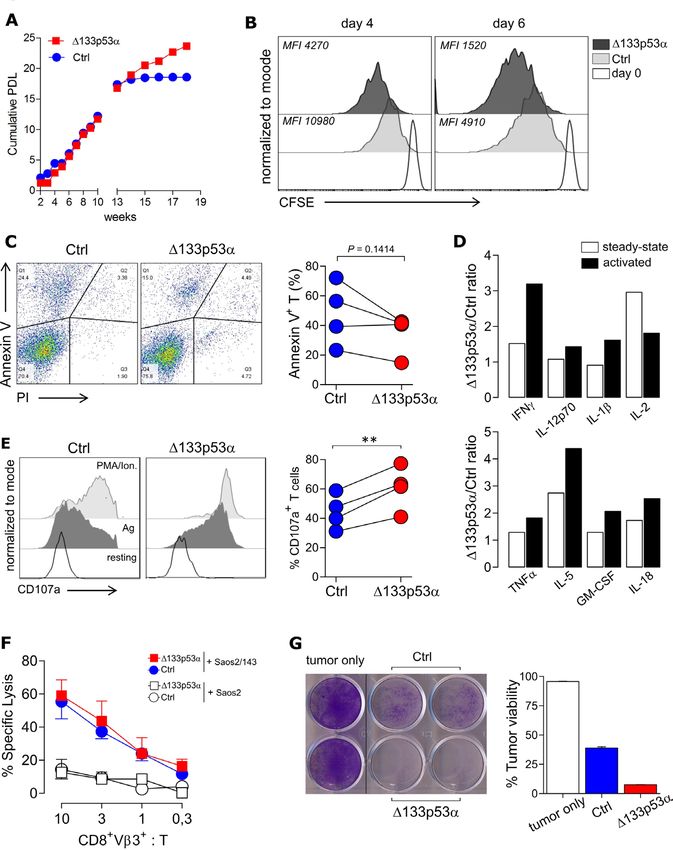
Open access
infection and cancer.12 13 Moreover, senescent T cells accu- promoters to regulate gene transcription.29Δ133p53α
J Immunother Cancer: first published as 10.1136/jitc-2020-001846 on 10 June 2021. Downloaded from http://jitc.bmj.com/ on October 11, 2021 by guest. Protected by copyright.
mulate during the normal lifespan of healthy individuals also acts as a dominant negative inhibitor of senescence
as well. These cells are mainly characterized by downregu- genes.18 30 Overexpression of Δ133p53α in near senes-
lation of the costimulatory molecule CD28 and increased cent human fibroblasts extends the cellular replicative
expression of CD57,13 CD160, KLRG1, and exhibit an lifespan by inhibiting the expression of p21Waf1/Cip1 and
extenuated response to antigen stimulation.14 15 Although other p53 transcriptional target genes, including microR-
the mechanisms which regulate T cell senescence remain NA- 34a. In contrast, overexpression of p53β induces
unclear, the tumor suppressor p53 emerged as a potential cellular senescence by the upregulation of p53 target
key player. TP53 has a crucial role in the maintenance of genes such as p21Waf1/Cip1 via cooperation with full-length
the genetic stability and, thus the prevention of cancer p53.13 Although in T lymphocytes, the role of these two
formation. It induces a number of cellular responses p53 isoforms as potential regulators of cellular senescence
including DNA repair, cell cycle arrest and apoptosis. TP53 has been reported,20 their function in more complex and
regulates the expression of many genes and is involved in tissue-specific context, including immune-related disor-
cellular senescence.16 17 The finding that the human p53 ders or cancer remains unexplored.
gene contains an alternative promotor and transcribes Here, we demonstrated for the first time that gene
multiple splice variants, resulting in the expression of 12 expression of the Δ133p53α isoform in tumor-antigen
different protein isoforms p53 (α, β, γ), Δ40p53 (α, β, γ), (TA) TCR-engineered T cells improves effector functions
Δ133p53 (α, β, γ), and Δ160p53 (α, β, γ)18 19 highlight in vitro assays and in a mouse model of adoptive T cell
the complexity of the p53 network. The human isoforms transfer. Circumventing senescence in antigen receptor-
that are most associated with cellular senescence are redirected T cells by genetic modification with Δ133p53α
the C- terminally and the N- terminally truncated p53β provides a novel strategy to improve robustness and resil-
and Δ133p53α. The p53β protein isoform contains the ience of antitumor responses.
transactivation domains (TAD) and the DNA- binding
domain (DBD) but terminates with 10 additional amino
acids, lacking half of the classic oligomerization domain, METHODS
a nuclear export signal (NES) domain and the negative Animal studies
regulation domain. In contrast, Δ133p53α lacks both of CyA2Kb,31 OT- I and NOD.Cg- PrkdcscidIL2rgtm1Wjl/SzJ
the TAD and part of the second conserved region of the (NSG) mice were obtained from the central animal
DBD but contains the NES. These two naturally occurring facility of the Johannes Gutenberg University Mainz,
p53 isoforms act as physiological regulators of cellular Germany. CyA2Kb and OT-I mice were used as source of
proliferation and senescence in normal human fibro- donor splenocytes and NSG mice as recipient animals for
blasts, in human T lymphocytes,13 20 but also in human tumor models. NSG mice were injected (s.c.) with osteo-
brain astrocytes and in induced pluripotent stem cells.21 22 sarcoma cell line Saos2/143 in the left flank and adop-
In mice, the first report revealing that the p53 gene codes tively transferred (intravenous) with genetically modified
for more than one functional protein was published by human CD3+ T cells as previously published.31 Mice were
Rotter and colleagues back in 1985,23 describing the pres- sacrificed when the tumor size was 1 cm3 (or at termina-
ence of another p53 variant in transformed fibroblasts as tion of the study), and freshly isolated tumors and spleens
a result of alternative splicing of intron 10, named p53AS were dissociated by mincing the tissue with scalpels into
(alternatively spliced). Later, MΔ41p53, the mouse coun- 0.5 mm small pieces. Dissociated tissue was further tritu-
terpart of human Δ40p53α forms was isolated.24 In addi- rated and filtered through a 100 µm cell strainer to obtain
tion, a shorter N-terminal form produced by an internal single-cell suspension and analyzed by flow cytometry.
promoter within the mouse p53 gene, MΔ157p53, Serum was obtained from the peripheral blood at the
equivalent to the human Δ160p53 form was identified. time of sacrifice.
It was also shown that MΔ41p53 and MΔ157p53 can
be expressed as a C-terminal AS variant.25 However, in Blood samples, primary cells and cell lines
contrast to human and high order primates, mice do Buffy Coats from healthy donors were obtained from
not produce Δ133p53 isoforms or beta isoforms. Trans- the Transfusion Center of the University Medical Center
genic mice overexpressing Δ40p53 present an increased Mainz and the Institute for Immunology/IKTZ, Heidel-
cellular senescence, a slower growth rate, and premature berg University, Germany. Primary human T cells were
aging phenotype due to abnormal hyper-activation of the obtained from the peripheral blood of patients with newly
insulin-like growth factor signaling.26 Mechanistically, diagnosed multiple myeloma (MM). Peripheral blood
while human p53β can bind differentially to promoters mononuclear cells were isolated from peripheral blood
and enhance expression of senescence- associated p53 via ficoll density gradient centrifugation. Tumor cell lines
target genes, Δ133p53α modulates gene expression used in this study included the human HLA-A2.1+ p53
and increases DNA- repair efficiency through interac- null osteosarcoma Saos2, its p53-transfectant Saos2/14331
tion with full-length p53 and homologous p53 protein, and the C57BL/6-derived colon carcinoma MC38-OVA
p63 and p73.27–29 Δ133p53α also directly binds DNA on and melanoma B16- OVA. The A2Kb p53mutant MEF
a novel type of p53-responsive element in enhancer and tumor cell line was described earlier.31 Human cell lines
2 Legscha KJ, et al. J Immunother Cancer 2021;9:e001846. doi:10.1136/jitc-2020-001846
Open access
were maintained in RPMI 1640 supplemented with 10% (PMA) + ionomycin (Sigma-Aldrich) were used as control
J Immunother Cancer: first published as 10.1136/jitc-2020-001846 on 10 June 2021. Downloaded from http://jitc.bmj.com/ on October 11, 2021 by guest. Protected by copyright.
heat inactivated fetal calf serum (FCS), 2 mM L-gluta- inducers of CD107a expression. The determination of
mine, 100 U/mL penicillin and 0.1 mg/mL streptomycin. apoptotic cells was performed with a standard Annexin
Murine tumor cell lines were maintained in DMEM V and propidium iodide (PI) (BD Biosciences) staining
(GIBCO) supplemented with 10% FCS, 25 mM HEPES- according to the manufacturer’s instructions. TCR Vβ
buffer, 1% glutamine, and 1% penicillin-streptomycin. repertoire of TCR-engineered T cells in vivo was analyzed
by flow cytometry with antibodies from the Beta Mark
Peptides, antibodies and reagents TCR Vβ Repertoire Kit (Beckman Coulter) directed
HLA-A2.1 restricted p53aa264-272 (LLGRNSFEV) was synthe- against 19 individual TCR/Vβ chains. Flow cytometry
sized by Biosyntan (Berlin, Germany) and dissolved acquisitions were performed on a FACSCanto II and BD
in DMSO. Antibodies used for Western blotting were FACSLyric (BD Biosciences), and data were analyzed with
mouse monoclonal antibodies (mAb) antihuman p53 FlowJo_V10 software (Tree Star).
(DO-2) sc-53394 (Santa Cruz Biotechnology, Heidelberg,
Germany), antihuman p53 (HR231) MA1-12648 (Invit- T cell functional assays
rogen), sheep antihuman p53 isoforms KJC12 (gift from T cell proliferation in vitro was assessed by carboxyfluores-
Bourdon, Dundee, UK), rabbit mAbs anti- tri-
methyl- cein succinimidyl ester (CFSE) dilution assay. T cells were
Histone H3 (Lys4) (C42D8), anti- tri-
methyl-
Histone labeled with 5 µM CFSE for 5 min and subsequently stim-
H3 (Lys9) (D4W1U) (Cell Signaling Technology), anti- ulated with antigen-presenting cells as described before.
HIF1a (Ab92498) (Abcam), rabbit mAb anti-Akt (pan) Cells were analyzed at the indicated time points for CFSE
(C67E7) (Cell Signaling Technology, Cambridge), rabbit dilution by flow cytometry. Standard 5h 51Cr-release assays
mAb anti-p21 (ab109520) (Abcam) and the mouse mAb were performed at the indicated effector-to-target (E:T)
anti-MDM2 (SMP14) sc-965 (Santa Cruz). Bafilomycin A1 ratio in duplicate wells as described earlier.31 Long-term
(InvivoGen) was used as autophagy blocker. killing capacity of TCR- engineered T cells was deter-
mined by colony forming assay (CFA). Briefly, effector
Genetic Modification of T Lymphocytes T cells were cocultured with antigen+ (Saos2/143) or
cDNA encoding for human Δ133p53α and p53β (gift antigen- (Saos2) target tumor cells in 6- or 12-well plates
from Bourdon, Dundee, UK) were cloned into the at 37°C with 5% CO2 at the indicated E:T ratio. After
retroviral vector pMx_Ires_puromycin (RTV-014, Cell 24 hours, T cells as well as non-adherent lysed tumor cells
Biolabs) via BamHI/NotI. As a TA model, we used our were washed out and if necessary, transferred to a second
high-affinity sc p53TCR specific for the broadly expressed round of fresh tumor cells. The remaining adherent
(non-mutant) HLA.A2.1-restricted p53aa264-272 peptide. viable tumor cells were fixed with 4% PFA for 10 min at
The original scTCR scaffold was described earlier,26 room temperature (RT) and subsequently stained with
and the cDNA- encoding sequence was further codon- 1% crystal violet dye (Merck KGaA, Germany) for 15 min
modified (Invitrogen GeneArt, Regensburg, Germany) at RT. Crystal violet was washed off by adding Phosphate
and cloned into one single bicistronic 2A-based retroviral buffered saline (PBS) and the plate scanned for visual eval-
vector.24 We used the pGMP retroviral vector encoding uation of colony counts. For quantitative analysis, the dye
the scTCR31 for gene transfer in murine T cells. To redi- was dissolved by adding 5% SDS and the corresponding
rect human T cells, the scTCR coding sequence was optical density (absorbance) measured at 570 nm using a
cloned into the retroviral vector pBullet_Ires_neomycin32 microplate reader (Dynex MRX, Magellan BioScience).
via NcoI/BamHI. Retroviral transduction, selection and Replicative senescence of T cells in vitro was evaluated by
expansion of human and mouse bulk CD8+/CD4+ T cells calculating the population doubling levels (PDLs) using
(with CD3/CD28-beads or by peptide-specific stimulation the following formula as described in20 : log10(number of
(K562_A2+_CD80+ pulsed with 10 µg p53 peptide) were cells after expansion) - log10(number of cells seeded)/
performed as described earlier.31 33 Separation of human log102. Secreted cytokines/chemokines in vitro culture
CD8+ T cell fractions (TIGIThigh and TIGITlow) was and in serum were determined by Luminex Multiplex
performed with MACS cell separation beads (Miltenyi Assays (MAGPIX) using Human Cytokine & Chemokine
Biotec) according to the manufacturer’s protocol. (34 plex) kit (eBioscience, San Diego, USA) according
to the manufacturer protocol. For in vitro assays, effector
Flow cytometry analysis T cells and target cells were cocultured in 12-well plates
Antibodies used in this study are listed in online supple- before for 24–48 hours as described for CFA and cytokine
mental table S1. Cells were stained according to the concentrations measured in the culture supernatant.
manufacturer’s instructions. To detect the expression of Serum was diluted 1:5 before measurement.
the lysosomal-associated membrane protein 1 (LAMP1/
CD107a) (as a surrogate marker for degranulation) on T cell metabolic assays
the surface of CD8+ T-cells, effector cells were incubated The oxygen consumption rate (OCR) and the extracel-
for 24 hours with target tumor cells in the presence of lular acidification rate (ECAR) were measured using the
monensin (Monensin Solution 1000X, eBioscience) and Seahorse XFp Analyzer (Agilent). Culture Miniplates
the CD107a antibody. Phorbol 12- myristate 13-acetate were coated, using Poly-D-Lysine (Sigma P6407). To each
Legscha KJ, et al. J Immunother Cancer 2021;9:e001846. doi:10.1136/jitc-2020-001846 3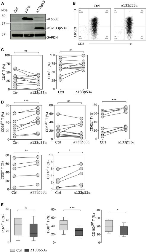
Open access
well, 50 µL of Poly-D-Lysine was added. After an incu- 60 min at 37°C and 5 min at 85°C. Relative expression of
J Immunother Cancer: first published as 10.1136/jitc-2020-001846 on 10 June 2021. Downloaded from http://jitc.bmj.com/ on October 11, 2021 by guest. Protected by copyright.
bation time of 1 hour at RT, the wells were rinsed with transcripts was determined by quantitative real-time-PCR
sterile water. XFp Sensor Cartridge wells were hydrated using the PowerUp SYBR Green master mix (Applied
by adding 200 µl XF calibrant to each well and 400 µL Biosystems) and QuantStudio 3 system (Applied Biosys-
to each outside well. Cartridges were then stored in a tems). The ΔΔCt method was used according to the manu-
non-CO2 incubator at 37°C overnight. XFp Cell Energy facturer’s protocol (Real-time PCR handbook, Applied
Phenotype Test Kit (Agilent) was performed according Biosystems) to determine fold difference in expression
to the manufacturers’ protocol. In detail, 1.0×10⁶ T cells normalized to the reference gene as indicated.
per well were seeded in XF Base Medium (DMEM). The
medium was supplemented with 10 mM glucose, 1 mM Statistical analysis
sodium pyruvate and 2 mM L-Glutamine and the pH was Statistical analysis of differences between groups was
adjusted to 7.4. Using the Cell Energy Phenotype Test Kit, determined by two-tailed Student’s t test or log-rank test
OCR and ECAR were measured under basal steady-state for mouse studies using GraphPad Prism V.5.01.(*P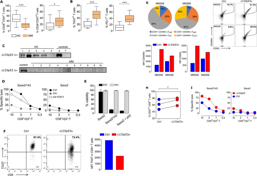
Open access
J Immunother Cancer: first published as 10.1136/jitc-2020-001846 on 10 June 2021. Downloaded from http://jitc.bmj.com/ on October 11, 2021 by guest. Protected by copyright.
Figure 1 Δ133p53α modulates expression of exhaustion markers and inhibitory receptors on T cells. (A) Immunoblot
confirming the overexpression of p53β and Δ133p53α isoforms after retroviral transduction and puromycin selection of human T
cells. Empty vector was used for control cells. (B) Representative data for the cell surface expression of the transduced scTCR
in CD8+ T cells, which was determined by flow cytometry using anti-TCRVβ3 mAb. (C) Percentage of CD8+ and CD4+ T cells for
paired samples of Δ133p53α-modified or control T cells from healthy donors (n=12 biological replicates). (D) Flow cytometry
data for cell surface expression of CD28, CD57, CD62L, CD27 and CCR7 for paired samples of Δ133p53α-modified or control
T cells from different donors, shortly after transduction (n=5–16). Analysis of the cellular phenotype of T cells was carried out
at early stage (1–3 weeks) after transduction. (E) Difference in PD-1 (p=0.1848), TIGIT and CD160 expression between control
and Δ133p53α-transduced cells demonstrated in box plots. Error bars indicate SE of mean (SEM). SP stands for single positive.
*P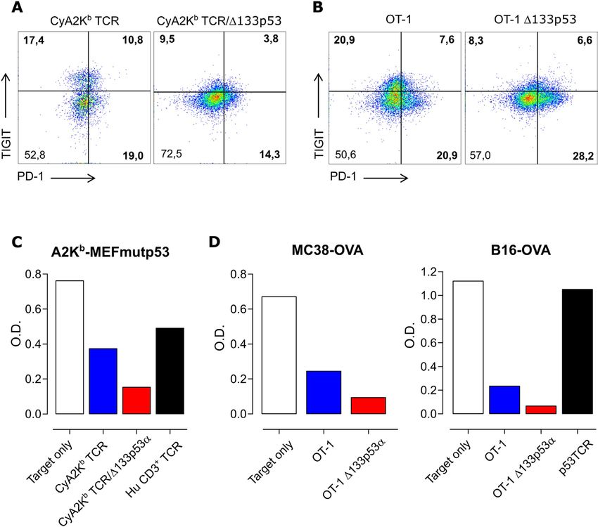
Open access
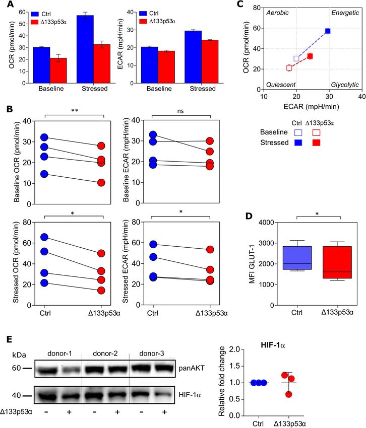
senescence factors and terminally differentiated markers, of in vitro culture in a CFSE-based proliferation assay and
J Immunother Cancer: first published as 10.1136/jitc-2020-001846 on 10 June 2021. Downloaded from http://jitc.bmj.com/ on October 11, 2021 by guest. Protected by copyright.
CD160 and TIGIT were expressed on a lower frequency noticed a higher cell division rate in Δ133p53α-T cells as
in Δ133p53α-modified T cells (figure 1E), suggesting demonstrated by lower mean fluorescence intensity (MFI)
a less senescence phenotype. Of note, although the values of CFSE dye (figure 3B). In addition, the modified
frequency of PD-1+CD8+ T cells was lower on expression T cells exhibited a reduced apoptosis after stimulation,
of Δ133p53α, it did not reach significance (p=0.1848). indicated by a lower (p=0.1414) frequency of Annexin
V+PI- cells (figure 3C). To further, evaluate effector func-
Δ133p53α triggers metabolic reprogramming in T cells tions of the near-senescent T cells, cytokine secretion,
Beside the cellular phenotype and differentiation status, degranulation and cytotoxic capacity were examined.
T cell function relies on the uptake of nutrients and Cytokine profiles were determined under steady- state
adequate energy production. Therefore, the metabolic conditions, and after activation with TA. Secretion levels
program is adapted to meet the metabolic demands of cytokines, with potent effector or stimulatory effect
and functional needs.34 Resting cells, like naïve T cells, were elevated in Δ133p53α-overexpressing T cells (up to
remain in a cellular and metabolic quiescent state.34 35 fourfold) compared with control T cells, particularly after
On antigen-recognition and activation, however, T cells antigen activation (figure 3D). Raw values are depicted
increase aerobic glycolysis and mitochondrial oxidative in online supplemental figure S2. We further evaluated
phosphorylation (OXPHOS) activity for clonal expan- the capacity of modified T cells to mobilize lytic granules,
sion.36 37 To further characterize Δ133p53α-transduced analyzing the expression of the (LAMP1/CD107a). The
T cells, we assessed their dynamic metabolic reprogram- data show a strong degranulation response (indicated by
ming by XFp Extracellular Flux analysis early after trans- an increased expression of CD107a) of Δ133p53α-modi-
duction. Under basal conditions, as well as after OXPHOS fied T cells and control T cells after TA encounter (=Ag)
inhibition and mitochondrial uncoupling (=stressed as compared with ‘residual’ response under steady-state
conditions), Δ133p53α-T cells had a decreased glycolytic (=resting) condition (figure 3E, left plot). Importantly,
activity, measured by the ECAR and a reduced (OCR, the percent of CD107a+ T cells is significantly higher in
indicator of OXPHOS) (figure 2A). Corresponding to Δ133p53α-modified T cells (figure 3E, right plot). To vali-
the less differentiated cellular phenotype, the reduced date these observations, we assessed the cytolytic activity
activity of glycolysis and OXPHOS indicates a quiescent of Δ133p53α-engineered T cells against target tumor
metabolic phenotype of Δ133p53α-T cells under basal cells. Interestingly, the killing capacity was similar in both
conditions compared with control cells (figure 2B). groups in a short-term (4–6 hours) lytic assay (figure 3F),
These data provide evidence that Δ133p53α-overexpres- yet, on repetitive coculture with tumor target cells over
sion does not only lead to changes in cellular phenotype, 24 hours, Δ133p53α-T cells showed a remarkable stronger
but is also associated with bioenergetic shifts toward a elimination of tumor cells compared with control cells
more quiescent metabolic phenotype (figure 2C), with (25% vs 40% of viable tumor colonies, figure 3G). In
the capacity to adapt their metabolic activity to the func- sharp contrast to Δ133p53α, p53β expression in T cells
tional needs. To further, document these metabolic was associated with induction of premature senescence,
differences, we included the analysis of the glucose trans- as documented by shorter lifespan, lower CD28 and
porter 1 (GLUT1), which is also differently express in higher CD57 expression, higher apoptosis levels and
quiescent/naïve vs effector T cells.38 Concordantly, we low cytolytic activity (online supplemental figure S3).
observe a lower expression intensity in Δ133p53α-T cells However, as opposed to Δ133p53α data, the expression of
as compared with controls (figure 2D), which correlate the immune inhibitory molecule TIGIT (and to a lesser
with their ‘quiescent-like’ phenotype. However, expres- extent PD-1) showed a trend toward higher levels.
sion analysis of HIF-1a, as an additional factor in the meta-
bolic transition to glycolysis, did not show a significant TIGIT expression is antigen-dependent and affects TCR-
change (figure 2E). These results suggest the metabolic mediated T cell cytolytic activity
switch in Δ133p53α-T cells is most likely HIF-1a indepen- TIGIT is a central marker of T cell dysfunction and
dent process. has been shown to be upregulated on human tumor-
infiltrating CD8+ T cells (also regulatory T cells and
Overexpression of Δ133p53α invigorates T cell proliferation NK cells) in a variety of cancers. Furthermore, TIGIT
and improves effector functions of TCR-engineered T cells signaling can also alter T cell metabolism, as reported in
In order to estimate the long-term proliferative capacity of patients with cancer.38 We then addressed the relevance
Δ133p53α-T cells, we determined the PDL of aging cells of the reduced TIGIT expression in Δ133p53α-modified
in culture. Although cumulative PDL values were similar T cells in a functional in vitro assay. As a target tumor
in the logarithmic phase, the proliferation index (or life- model, we used the osteosarcoma cell line Saos2/143
span) of control T cells reached a plateau around week 14, that naturally express the ligands for TIGIT (CD155/
indicating a cellular senescence state, while Δ133p53α-T PVR) but also other inhibitory receptors, such as PD-L1
cells remained strongly proliferative (figure 3A). To (figure 4A). scTCR- transduced T cells were separated
confirm this finding, we compared the proliferation ability into two fractions, CD3+TIGIThigh and CD3+TIGITlow
Δ133p53α-modified and control T cells at late timepoints populations (figure 4B), and compared for their cytolytic
6 Legscha KJ, et al. J Immunother Cancer 2021;9:e001846. doi:10.1136/jitc-2020-001846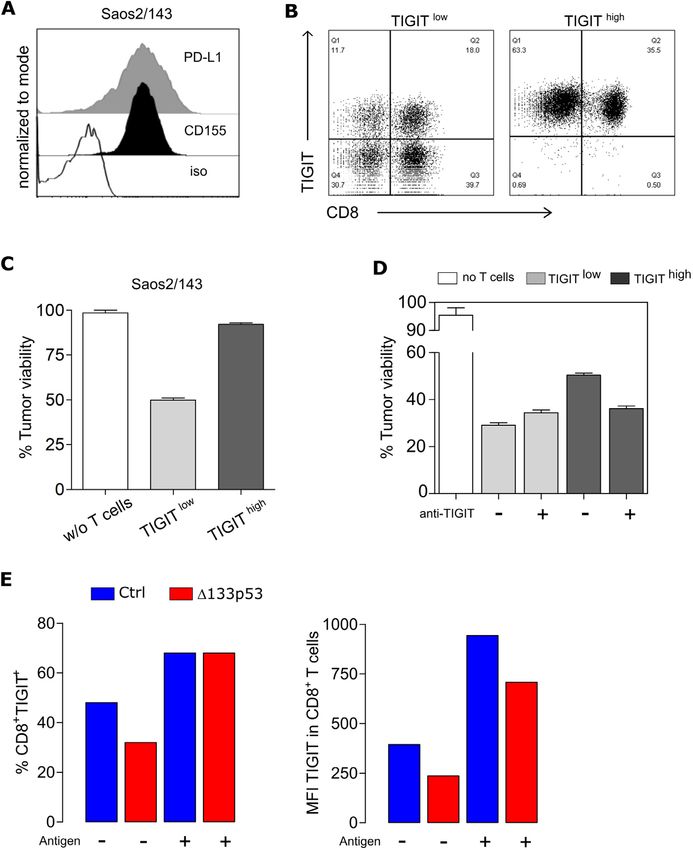
Open access
J Immunother Cancer: first published as 10.1136/jitc-2020-001846 on 10 June 2021. Downloaded from http://jitc.bmj.com/ on October 11, 2021 by guest. Protected by copyright.
Figure 2 Δ133p53α dictates metabolic reprogramming in T cells. (A) Oxygen consumption rate (OCR, left figure) and
extracellular acidification rate (ECAR, right figure) of Δ133p53α-transduced and control cells before (Baseline) and after
(Stressed) addition of oligomycin and FCCP measured with a XFp Extracellular Flux Analyzer. Representative of n=4 biological
replicates, shown as mean±SEM. (B). Dot plots depicting OCR and ECAR measures for paired samples of Δ133p53α-modified
or control T cells from four healthy donors. (C) Energy Phenotype Profile of indicated cells demonstrating the relative utilization
of glycolysis and mitochondrial respiration. (D) Box plots showing the expression level (as mean fluorescence intensity, MFI) of
glucose transporter 1 (GLUT1) in control and Δ133p53α-transduced cells (n≥5). (E) Protein expression of HIF-1a in engineered
CD8+ T cells from three healthy donors. Immunoblot (left) and dot plots (right) showing the fold change expression in Δ133p53α-
modified as compared with control T cells. Analysis of the metabolic activity, including the expression of GLUT1 and HIF-1a
were carried out early stage (1–3 weeks) after transduction. Error bars indicate SE of mean (SEM). *POpen access
J Immunother Cancer: first published as 10.1136/jitc-2020-001846 on 10 June 2021. Downloaded from http://jitc.bmj.com/ on October 11, 2021 by guest. Protected by copyright.
Figure 3 Δ133p53α invigorates T cell proliferation, cytokine response and improves long-term killing potential of antigen TCR-
engineered T cells. (A) Cumulative PDL of control and Δ133p53α-transduced T cells over time from a representative donor. (B)
CFSE proliferation assay of control and Δ133p53α-overexpressing T cells after several weeks of in vitro culture. Y-axis indicates
the reduction of intracellular CFSE on Day 4 (left plot) and Day 6 (right plot) after antigen-specific stimulation (Day 0) and was
normalized to mode. (C) Representative flow plots of the apoptosis marker Annexin V and Propidium Iodide (PI) for control and
Δ133p53α-transduced T cells. Summary results are depicted as paired dot plots for each individual biological replicate (n=4).
(D) Fold change of secreted cytokines from Δ133p53α-overexpressing to control cells under resting and activated conditions
measured by Multiplex Immunoassay at late stage in vitro culture. For activation, T cells were cultured over 24 hours with target
tumor cells Saos2/143 (E:T=1:1) (n=2 biological replicates). (E) Degranulation Assay of control and Δ133p53α-overexpressing
T cells again under steady-state (=resting) and activated conditions (Ag=antigen-specific stimulation) at the same time point.
Stimulation with PMA/Ionomycin was included as positive control. Degranulation is indicated by cell surface expression of
LAMP1/CD107a, assessed by flow cytometry. Dot chart showing the percentage of CD107a+CD8+ T cells for paired samples
of Δ133p53α-modified or control T cells from four healthy donors. (F) 51Cr-release Assay and (G) Tumor Colony-Forming Assay
were used to evaluate short-term and long-term antitumor responses of Δ133p53α-modified compared with control T cells.
For 51Cr-release assay-specific lysis of target (Saos2/143) and control (Saos2p53null) tumor cells is illustrated at the indicated
effector to target ratio (CD8+Vβ3+:T). Tumor colony-forming assays were performed over 24 hours per round with an effector to
target ratio (CD8+Vβ3+:T) of 1:1. Remaining tumor colonies were labeled with crystal violet dye. Quantification was performed by
measuring the optical density (OD) of the residual dye, and values expressed as per cent of tumor viability. One representative
experiment out of four biological replicates is shown. **POpen access
J Immunother Cancer: first published as 10.1136/jitc-2020-001846 on 10 June 2021. Downloaded from http://jitc.bmj.com/ on October 11, 2021 by guest. Protected by copyright.
Figure 4 TIGIT induction is antigen-dependent and affects TCR-mediated T cell cytolytic activity. (A) Expression levels of
PD-L1 and CD155 in the target tumor cell line Saos2/143 determined by flow cytometry. (B) Flow Cytometry Data showing
TIGIT expression of CD8+ T cells after MACS-separation into TIGITlow and TIGIThigh population. (C) Quantified killing capacity of
TIGITlow and TIGIThigh T cells was determined in a tumor colony-forming assay, and is expressed as the percentage of remaining
viable tumor cells after coculture with effector T cells. (D) Quantified killing capacity of TIGITlow and TIGIThigh T cells on TIGIT
blockade as determined in a tumor colony-forming assay. (E) Upregulation of TIGIT on CD8+ T cells before and after antigen
recognition via coculture with Saos2/143. Percentage (left) and MFI (right) was assessed by flow cytometry. Results from one
representative experiment out of three biological replicates are shown. MFI, mean fluorescence intensity; PD-L1, programmed
cell death ligand 1.
cell-free supernatant experiments (online supplemental cells by preventing its degradation by autophagy using the
figure S4B) and p53-pulsed Saos2 cells (online supple- ATPase inhibitor bafilomycin A120. We demonstrated that
mental figure S4C). In an attempt to understand the the level of TIGIT transcripts as well as TIGIT expression
mechanism by which Δ133p53α overexpression correlates was severely reduced in T cells after treatment with bafilo-
with lower TIGIT expression, we examined this pathway mycin A1 (online supplemental figure S5A,B). Addition-
at the transcriptional level. Comparative analysis of TIGIT
ally, we assessed the wide changes in histone modifications
mRNA revealed lower transcripts in Δ133p53α-T cells
in comparison with control cells (online supplemental that might results from the overexpression of Δ133p53α
figure S5A), suggesting a transcriptional regulation. To isoform and affecting transcription at promoter sites for
validate these data in a ‘gene-unmodified’ experimental both H3K4me3 (active marks) and H3K9me3 (repressive
model, we stabilized Δ133p53α expression in control T marks). By testing three different biological T-cell donors,
Legscha KJ, et al. J Immunother Cancer 2021;9:e001846. doi:10.1136/jitc-2020-001846 9Open access
J Immunother Cancer: first published as 10.1136/jitc-2020-001846 on 10 June 2021. Downloaded from http://jitc.bmj.com/ on October 11, 2021 by guest. Protected by copyright.
Figure 5 Expression of Δ133p53α in murine T cells is associated with reduced TIGIT levels and improved T-cell effector
function. (A) Representative, flow cytometric data of TIGIT and PD-1 expression of mouse T cells obtained from CyA2Kb
mice, transduced with scTCR and of T cells obtained from OT-I mice (B). Both were transduced with human Δ133p53α or
empty vector. Quantitative data from a representative tumor colony-forming assay using CyA2Kb TCR mouse T cells and
A2KbMEFmutp53 (C). For OT-I cells, MC38OVA and B16OVA served as target cells (D). The optical density (O.D.) indicates the
amount of remaining viable tumor colonies after incubation with effector T cells. PD-1, programmed cell death 1; TCR, T cell-
receptor.
we observed a slight increase in H3K4me3 without notice- supplemental figure S6) more efficiently than control
able change in H3K9me3 (online supplemental figure cells, consistent with the results of Δ133p53α in human
S5C), suggesting a minor focal increase in promoter acti- T cells.
vation and gene expression in Δ133p53α-overexpressing
T cells. Adoptive transfer of TCR/Δ133p53α-equipped T cells results
in superior antitumor response in vivo
Expression of Δ133p53α in murine T cells is associated with Next, we assessed the antitumor efficacy of Δ133p53α
reduced TIGIT levels and improved T-cell effector function antigen-specific TCR- modified T cells in a xenograft
To further confirm and validate our findings, we tested tumor model. Immunodeficient NSG mice were injected
whether the major effects of Δ133p53α demonstrated in with Saos2/143 tumor cells, and infused with genetically
human CD8+ T cells could be observed in murine T cells. modified-T cells (figure 6A). Δ133p53α-transduced T cells
Therefore, we cotransduced T lymphocytes from CyA2Kb exhibited an improved antitumor response as reflected by
mice with a scTCR31 and the human Δ133p53α isoform a significantly prolonged survival of tumor bearing mice
or a mock control. As in human T cells, Δ133p53α-trans- (figure 6B). A no transfer (PBS) experiment, to exclude
duced CyA2Kb/TCR+ mouse T cells exhibited reduced any unspecific effect of mock-control T cells on tumor
levels of TIGIT compared with control cells (figure 5A). growth showed similar survival outcome as the control
Similar results were obtained in Δ133p53α-modified T group (online supplemental figure S7A). Analysis of the
cells from OT-I mice (figure 5B). Similar to human T cells, starting T cell population showed comparable phenotype
Δ133p53α did not affect PD-1 expression levels in murine (CD4/CD8 ratio, TCRVβ3 and CD45RA/CCR7 expres-
T cells (figure 5A,B). In addition, these models demon- sion levels) in Δ133p53α- and control-T cells at the time
strated that Δ133p53α-modified CyA2Kb (figure 5C) or of infusion (online supplemental figure S7B–D). Of note,
OT-I (figure 5D) murine T cells were able to eradicate superior tumor control in Δ133p53α-treated mice was
target cells expressing high levels of CD155/PVR (online occasionally accompanied with signs of graft-versus-host
10 Legscha KJ, et al. J Immunother Cancer 2021;9:e001846. doi:10.1136/jitc-2020-001846Open access Figure 6 Adoptive transfer of TCR/Δ133p53α-equipped T cells results in superior antitumor response but is associated with J Immunother Cancer: first published as 10.1136/jitc-2020-001846 on 10 June 2021. Downloaded from http://jitc.bmj.com/ on October 11, 2021 by guest. Protected by copyright. cytokine release syndrome. (A) Schematic representation of the experimental model. NOD-scid IL2rgnull (NSG) were injected subcutaneously with Saos2/143 on day 0. T cells and IL-2 were injected intravenous on day 7 as described in the Methods. (B) Survival curves for mice treated with mock-control- (Ctrl), TCR-modified or TCR/Δ133p53α-modified T cells (pooled data of three individual experiments), n=32 mice. Significance between the animal groups was determined by log-rank test (p=0.0199). (C) Frequency of CD8+ and CD4+ T cells in the peripheral blood at days 7, 22 and 34 after adoptive transfer. *P
Open access
CD8+ T cells in TCR/Δ133p53α mice as opposed to TCR study showing that Δ133p53α triggers proinflammatory
J Immunother Cancer: first published as 10.1136/jitc-2020-001846 on 10 June 2021. Downloaded from http://jitc.bmj.com/ on October 11, 2021 by guest. Protected by copyright.
control group (figure 6C, online supplemental figure cytokine response, including interleukin 6 (IL-6), IL-1β
S7E). Concordantly, Δ133p53α-T cells demonstrated a and IL-8 (via inhibition of p53 and induction of NF-κB) in
longer engraftment in vivo as documented by a higher a Helicobacter pylori infection model or cancer model.40 41
frequency of CD8+ and CD4+ T cells in the peripheral Further analysis of the TCR repertoire in spleen-
blood at days 34 after adoptive transfer (figure 6D). The infiltrating Δ133p53α-T cells revealed a polyclonal TCRβ
frequency of antigen- specific TCR(Vβ3)-infiltrating T signature (online supplemental figure S7G), indicating a
cells was also higher, yet not significant in the spleen and diversity of responsive T cells in vivo.
tumor-tissue of TCR/Δ133p53α animals (online supple-
mental figure S7E). As anticipated, while the starting T Reverting T cell senescence of MM patients by Δ133p53α
cell population infused in mice was mainly composed of gene transfer
naïve and effector T cells, persistent T cells in vivo exhib- To further examine the in vivo relevance of Δ133p53α in
ited rather an effector memory phenotype. Comparative the regulation of T cell senescence, peripheral blood T
analysis of sera collected from both animal groups shortly cells from newly diagnosed MM patients were phenotyped.
before sacrifice, revealed markedly increased levels of MM patients had a reduced number of ‘naïve’ CD8+C-
pro-inflammatory and immunomodulatory cytokines D28+CD57- and an increased frequency of senescent-
and chemokines in mice treated with Δ133p53α-T cells like CD8+CD28-CD57+ single positive (SP) T cell subsets
(figure 6E). These data are concordant with a previous (figure 7A), suggesting a senescent state of CD8+ T cells
Figure 7 Reverting senescence in multiple myeloma (MM) patient T cells by Δ133p53α gene transfer. (A) Reduced frequency
of CD8+CD28+CD57- (left, p=0.0014) and increased frequency of CD8+CD28-CD57+ (right, p=0.0461) T cells from MM patients
(n=10) compared with healthy donor T cells (HC, n=15). (B) T cells from MM patients (n=27) revealed high levels of TIGIT and
PD-1 expression compared with healthy donor T cells (HC, n=7–15) (pOpen access
in MM patients. Interestingly, analysis of the fraction of modulate TA-specific T cell functions, in particular in
J Immunother Cancer: first published as 10.1136/jitc-2020-001846 on 10 June 2021. Downloaded from http://jitc.bmj.com/ on October 11, 2021 by guest. Protected by copyright.
total CD28+ and CD57+ in CD8+ T cells showed similar more complex in vivo tumor suppressive environment.
expression profiles (online supplemental figure S8). Here, we demonstrated that gene expression of
Furthermore, we observed a markedly increased expres- Δ133p53α is associated with a metabolic switch and
sion of the T cell-associated senescent markers TIGIT and profound functional changes in TA- specific cytotoxic
PD-1 in MM patients (figure 7B). We then anticipated CD8+ T cells in vitro and in mouse tumor model. Pheno-
a reduced expression of the Δ133p53α isoform in MM typing of T cells revealed a shift toward a less differen-
as compared with healthy individuals. Protein expres- tiated state characterized by an upregulation of CD28,
sion data confirmed high levels of Δ133p53α-isoform CD27 and CD62L and a downregulation of key inhib-
in healthy individuals, while marginal or no expression itory receptors, CD160 and TIGIT. Accordingly, we
was detected in MM patient T cells (figure 7C). Next, observed a prolonged long-term proliferative potential
we evaluated the senescence status of MM CD8+ T cells and improved effector functions as demonstrated by
in functional assays. T cells from two patients (P37 and enhanced tumor-specific cytolytic activity and a superior
P41) with distinct TIGIT, CD28SP and CD57SP expres- cytokine response in vitro. In contrast, p53β expression
sion profiles were selected. P41 and P37, characterized as in T cells drives premature senescence phenotype, char-
TIGIThighCD57SPhighCD28SPlow and TIGITlowCD57SPlow- acterized by a lower CD28 and higher CD57 expression,
CD28SPmild, were genetically equipped with a scTCR and higher apoptosis levels along with a shorter life-span.
tested for their antigen specific response. In both, short- T cells can modulate their cellular metabolism according
term (figure 7D) and long-term (figure 7E) killing assays, to their functional needs.34 However, this metabolic switch
P41-, but not P37-modified T cells, exhibited a severely can be altered by the TME which can lead to T cell dysfunc-
impaired cytotoxic capacity against target Saos2/143 tion.42 Along with the induction of a less differentiated
tumor cells. To explore the possibility to reprogram phenotype and enhanced effector functions, our results
senescent T cells of MM patients into more ‘juvenile’ and showed that Δ133p53α is associated with a metabolic
effective T cells, we performed Δ133p53α gene transfer reprogramming in tumor-reactive engineered CD8+ T cells
experiments in MM T cells. In line with our data from characterized by a quiescent metabolic state with lower
healthy individuals, Δ133p53α overexpression was associ- glycolytic activity and expression of GLUT1. This is in line
ated with more than a twofold reduction in TIGIT expres- with a recent study reporting that CD8+ T cells with a low
sion (figure 7F). While the starting CD8+T cell population mitochondrial membrane potential (ΔΨm) are enriched
from two myeloma patients (MM305, MM306) prior trans- in CD62L+ central memory T cells, accompanied with a
duction was mainly composed of naïve, effector memory reduced glycolytic activity and mitochondrial respiration
(CD45RA-CCR7-) and effector (CD45RA+CCR7-) pheno- leading to enhanced proliferative capacity and increased
type, the phenotype following transduction with TCR antitumor activity in vivo.43 The reduced metabolic activity
control vector showed a dominant effector memory and early after transduction may be adapted to low metabolic
a low percentage (6% and 15%) of CD45RA+CD62L+ demands, which may be increased for rapid expansion
(naive). The frequency of naïve population as well as after activation.
the expression intensity of both CD45RA and CD62L T cell metabolism can be altered in patients with cancer
markers increased (twofold and fivefold) after overex- through TIGIT/CD155 signaling.44 Moreover, TIGIT/
pression of the Δ133p53α isoform (figure 7G). In line, CD155 interaction is also involved in the suppression of T cell
Δ133p53α-modified MM T cells exhibit a higher expres- activation.5 45 TIGIT was found to be upregulated on human
sion of the costimulatory molecule CD27 (figure 7H). tumor-infiltrating lymphocytes44 46 and its blockade improved
Importantly, Δ133p53α gene transfer in MM patient T CD8+ T cell effector functions.5 47 Moreover, CD8+ T cells
cells promoted superior specific cytolytic activity against from aged healthy donors, exhibited a high TIGIT expres-
Saos2/143 tumor cells (figure 7I). sion, accompanied by impaired effector functions.48 Here, we
demonstrated for the first time the effect of Δ133p53α on
TIGIT expression in antigen-specific T cells at the mRNA and
protein levels. Additionally, T cells with high TIGIT expres-
DISCUSSION sion had a compromised cytolytic response against CD155-
Senescence- induced T cell dysfunction in the TME expressing target cells. Consistently, T cell dysfunction in
impedes the clinical efficacy of cancer immunotherapy.9 14 patients with MM14 has been recently attributed, among other
An age-dependent accumulation of senescent T cells in factors, to high TIGIT expression.47 49 Accordingly, TIGIT
healthy individuals which was associated with changes in blockade could restore anti-myeloma T-cell function.47 In the
expression of T cell surface markers and p53 isoforms present study, we could confirm the senescent phenotype and
has been described. In this study, p53β and Δ133p53α high expression of TIGIT in T cells from newly diagnosed
were identified as potential opposite markers for cellular MM patients and further revealed a reduced expression of
senescence by providing evidence that overexpression of Δ133p53α. In line with our results from healthy donors T
Δ133p53α could restore the proliferation capacity of late cells, TIGIThigh-expressing MM T cells showed impaired anti-
differentiated or senescent T cells in vitro.20 However, it tumor responses which could be restored on Δ133p53α gene
remained unclear whether and how these isoforms can transfer. Improved T cell function of Δ133p53α-modified
Legscha KJ, et al. J Immunother Cancer 2021;9:e001846. doi:10.1136/jitc-2020-001846 13Open access
MM T cells was associated with a less differentiated pheno-
J Immunother Cancer: first published as 10.1136/jitc-2020-001846 on 10 June 2021. Downloaded from http://jitc.bmj.com/ on October 11, 2021 by guest. Protected by copyright.
are part of KJL doctoral thesis. EAF, AC, AL, MHSA, GNHQT and DG, performed the
type characterized by de novo expressions of CD45RA, CD27 research, analyzed and interpreted the data. BG, MH, JCB and MM analyzed and
interpreted the data. MT designed the research, analyzed, interpreted the data
and CD62L. Thus, these results further demonstrate the and wrote the article. HE designed and performed the research, analyzed and
capacity of Δ133p53α to reinvigorate effector functions of interpreted the data, prepared the figures and wrote the article.
cancer patient-derived senescent T cells. On a more transla- Funding This study was supported in part by grants from the Collaborative
tional level, a lower expression of the senescence-associated Research Center 1292 (CRC 1292 TP06) (to MM, MT and HE), the German
and negative immune receptor TIGIT in antigen-TCR CD8+ Consortium for Translational Cancer Research (DKTK) Frankfurt/Mainz partner site,
Mainz. KJL is supported by the Clinician Scientist Fellowship 'TransMed Jumpstart
T cells overexpressing Δ133p53α is novel (as it has not yet Program: 2019_A72' supported by the Else Kröner Fresenius Foundation.
been reported) and has potential clinical relevance. TIGIT is
Competing interests None declared.
emerging as the third (after CTLA-4 and PD-1) clinical target
Patient consent for publication Not required.
in immuno-oncology. Currently, more than 20 clinical trials
testing anti-TIGIT agents (as single treatment or in combina- Ethics approval Animal experiments were performed according to approved
protocol from the local animal welfare authorities (protocol AZ 23 177-07/G16-1-
tion with PD-(L)1 blockade) are launched by lead biophar- 016). Peripheral blood of patients with newly diagnosed multiple myeloma was
maceutical companies (such as Roche, BMS and Merck), obtained after informed consent in accordance with the Declaration of Helsinki and
including five phase 3 studies in solid cancers (glioblastoma, authorization by the Ethical Review Committee of the Ruprecht-Karls-University
melanoma, lung, breast cancer, …) and hematological malig- Heidelberg (approval number 2014-003079-40).
nancies (MM). Provenance and peer review Not commissioned; externally peer reviewed.
Using a preclinical tumor model, we observed enhanced Data availability statement All data relevant to the study are included in the
antitumor responses in Δ133p53α-T cells leading to supe- article or uploaded as supplementary information.
rior overall survival of treated mice. This improved anti- Supplemental material This content has been supplied by the author(s). It has
not been vetted by BMJ Publishing Group Limited (BMJ) and may not have been
tumor immunity was occasionally accompanied by a severe
peer-reviewed. Any opinions or recommendations discussed are solely those
inflammatory response, confirmed by elevated levels of of the author(s) and are not endorsed by BMJ. BMJ disclaims all liability and
secreted cytokines in the serum and massive infiltration responsibility arising from any reliance placed on the content. Where the content
of T cells in the spleen. These findings suggest a cytokine- includes any translated material, BMJ does not warrant the accuracy and reliability
of the translations (including but not limited to local regulations, clinical guidelines,
associated toxicity resulting from rare hyperactivated T terminology, drug names and drug dosages), and is not responsible for any error
cells induced by Δ133p53α, consistent with the profound and/or omissions arising from translation and adaptation or otherwise.
proinflammatory phenotype reported in transgenic mice Open access This is an open access article distributed in accordance with the
expressing a Δ133p53α-like isoform.50 These data calls for a Creative Commons Attribution 4.0 Unported (CC BY 4.0) license, which permits
safety approach to limit excessive immune response associ- others to copy, redistribute, remix, transform and build upon this work for any
purpose, provided the original work is properly cited, a link to the licence is given,
ated with sustained expression of Δ133p53α in engineered
and indication of whether changes were made. See https://creativecommons.org/
T cells. These potentially severe side effects caused by strong licenses/by/4.0/.
immune activation may be resolved with monoclonal anti-
bodies against certain cytokines (like Tocilizumab), or by ORCID iD
Hakim Echchannaoui http://orcid.org/0000-0001-9980-9974
using a safety switch approach like an inducible caspase 9
suicide system.
In conclusion, our data demonstrated that Δ133p53α REFERENCES
isoform acts as a potent enhancer of robustness and resil- 1 Duong CPM, Yong CSM, Kershaw MH, et al. Cancer immunotherapy
ience in human cytotoxic T cells, which may represent a novel utilizing gene-modified T cells: from the bench to the clinic. Mol
Immunol 2015;67:46–57.
approach to improve T-cell-based cancer immunotherapies. 2 Kershaw MH, Westwood JA, Darcy PK. Gene-Engineered T cells for
cancer therapy. Nat Rev Cancer 2013;13:525–41.
3 Baruch EN, Berg AL, Besser MJ, et al. Adoptive T cell therapy: an
Author affiliations
1 overview of obstacles and opportunities. Cancer 2017;123:2154–62.
Department of Hematology, Oncology and Pneumology, University Medical Centre 4 Freeman GJ, Long AJ, Iwai Y, et al. Engagement of the PD-1
of the Johannes Gutenberg University Mainz, Mainz, Germany immunoinhibitory receptor by a novel B7 family member leads
2
Department of Internal Medicine V, University of Heidelberg, Heidelberg, Germany to negative regulation of lymphocyte activation. J Exp Med
3
Department of Radiation Oncology and Radiotherapy, University Medical Centre of 2000;192:1027–34.
the Johannes Gutenberg University Mainz, Mainz, Germany 5 Johnston RJ, Comps-Agrar L, Hackney J, et al. The immunoreceptor
4 TIGIT regulates antitumor and antiviral CD8(+) T cell effector function.
German Cancer Consortium (DKTK), Partner Site, Mainz, Germany Cancer Cell 2014;26:923–37.
5
School of Medicine, University of Dundee, Dundee, UK 6 Vodnala SK, Eil R, Kishton RJ, et al. T cell stemness and dysfunction
6
Research Center for Immunotherapy (FZI), University Medical Centre of the in tumors are triggered by a common mechanism. Science 2019;363.
Johannes Gutenberg University Mainz, Mainz, Germany doi:10.1126/science.aau0135. [Epub ahead of print: 29 03 2019].
7 Ribas A, Wolchok JD. Cancer immunotherapy using checkpoint
blockade. Science 2018;359:1350–5.
Twitter Borhane Guezguez @GuezguezL 8 Ansell SM, Lesokhin AM, Borrello I, et al. PD-1 blockade with
Acknowledgements The authors thank Danielle Arnold-Schild (Institute for nivolumab in relapsed or refractory Hodgkin's lymphoma. N Engl J
Med 2015;372:311–9.
Immunology, UMC, Mainz) for providing the MC38-OVA cell line and Rafaela 9 Kasakovski D, Xu L, Li Y. T cell senescence and CAR-T cell
Holtappels (Institute of virology, UMC, Mainz) for providing OT-I mice. We exhaustion in hematological malignancies. J Hematol Oncol
acknowledge Ronald Backer (Institute of molecular medicine, UMC, Mainz) for 2018;11:91.
excellent support with the Luminex device and Viral Shah (III Medical Department, 10 Harley CB, Vaziri H, Counter CM, et al. The telomere hypothesis of
UMC, Mainz) for the anti-p21 specific antibody. cellular aging. Exp Gerontol 1992;27:375–82.
11 Serrano M, Lin AW, McCurrach ME, et al. Oncogenic ras provokes
Contributors KJL designed and performed the research, analyzed and interpreted premature cell senescence associated with accumulation of p53 and
the data, prepared the figures and wrote the article. Data presented in this article p16INK4a. Cell 1997;88:593–602.
14 Legscha KJ, et al. J Immunother Cancer 2021;9:e001846. doi:10.1136/jitc-2020-001846You can also read