Hemidesmosome-Related Keratin Filament Bundling and Nucleation
←
→
Page content transcription
If your browser does not render page correctly, please read the page content below
International Journal of
               Molecular Sciences

Article
Hemidesmosome-Related Keratin Filament Bundling
and Nucleation
Marcin Moch                and Rudolf E. Leube *

                                          Institute of Molecular and Cellular Anatomy, Rheinisch-Westfälische Technische Hochschule Aachen,
                                          52062 Aachen, Germany; mmoch@ukaachen.de
                                          * Correspondence: rleube@ukaachen.de; Tel.: +49-241-80-89107

                                          Abstract: The epithelial cytoskeleton encompasses actin filaments, microtubules, and keratin interme-
                                          diate filaments. They are interconnected and attached to the extracellular matrix via focal adhesions
                                          and hemidesmosomes. To study their interplay, we inhibited actin and tubulin polymerization
                                          in the human keratinocyte cell line HaCaT by latrunculin B and nocodazole, respectively. Using
                                          immunocytochemistry and time-lapse imaging of living cells, we found that inhibition of actin and
                                          tubulin polymerization alone or in combination induced keratin network re-organization albeit
                                          differently in each situation. Keratin filament network retraction towards the nucleus and formation
                                          of bundled and radial keratin filaments was most pronounced in latrunculin-B treated cells but less
                                          in doubly-treated cells and not detectable in the presence of nocodazole alone. Hemidesmosomal
                                          keratin filament anchorage was maintained in each instance, whereas focal adhesions were disassem-
                                          bled in the absence of actin filaments. Simultaneous inhibition of actin and tubulin polymerization,
                                          therefore, allowed us to dissect hemidesmosome-specific functions for keratin network properties.

                                          These included not only anchorage of keratin filament bundles but also nucleation of keratin fila-
                                   ments, which was also observed in migrating cells. The findings highlight the fundamental role of
Citation: Moch, M.; Leube, R.E.           hemidesmosomal adhesion for keratin network formation and organization independent of other
Hemidesmosome-Related Keratin             cytoskeletal filaments pointing to a unique mechanobiological function.
Filament Bundling and Nucleation.
Int. J. Mol. Sci. 2021, 22, 2130.         Keywords: keratin intermediate filament; hemidesmosome; focal adhesion; actin; microtubule;
https://doi.org/10.3390/ijms              integrin beta 4; integrin beta 5; BPAG-1; paxillin; talin
22042130

Academic Editor: Jose
Maria Gonzalez-Granado                    1. Introduction
                                               The keratin cytoskeleton is a hallmark feature of epithelial cells [1,2]. It consists of a
Received: 25 December 2020
Accepted: 17 February 2021
                                          filamentous cytoplasmic network with unique biomechanical properties that is connected
Published: 21 February 2021
                                          to desmosomes at cell-cell adhesion sites and to hemidesmosomes at epithelial-extracellular
                                          matrix (ECM) interfaces [3–6]. Its 3D organization and dynamic features rely on the other
Publisher’s Note: MDPI stays neutral
                                          two major cytoskeletal filament systems, i.e., the actin-based microfilaments, which are an-
with regard to jurisdictional claims in
                                          chored to adherens junctions at cell-cell borders and to focal adhesions at cell-ECM contacts,
published maps and institutional affil-   and the tubulin-based microtubules [7–11]. The resulting highly complex transcellular
iations.                                  scaffold supports epithelial tissue cohesiveness and adhesion. Intense research efforts are
                                          directed towards elucidation of the specific contribution of the different filament-adhesion
                                          systems to epithelial function and homeostasis. The current study focuses on keratin net-
                                          work organization in relation to hemidesmosomal adhesion examining the consequences of
Copyright: © 2021 by the authors.
                                          interfering with focal adhesion, actin filament polymerization and microtubule formation
Licensee MDPI, Basel, Switzerland.
                                          on keratin-hemidesmosome interaction.
This article is an open access article
                                               Keratin intermediate filaments consist of equimolar amounts of type I and type II
distributed under the terms and           keratin polypeptides [5,12,13]. In contrast to actin and tubulin, keratin polypeptides spon-
conditions of the Creative Commons        taneously self-assemble in the absence of nucleoside triphosphates and chaperones into
Attribution (CC BY) license (https://     apolar, highly flexible, and elastic 8–12 nm filaments, which form bundles of variable thick-
creativecommons.org/licenses/by/          ness [14]. The molecular mechanisms determining keratin network morphogenesis and 3D
4.0/).                                    arrangement remain unknown. Basal epidermal cells contain keratins 5 and 14, which have

Int. J. Mol. Sci. 2021, 22, 2130. https://doi.org/10.3390/ijms22042130                                         https://www.mdpi.com/journal/ijms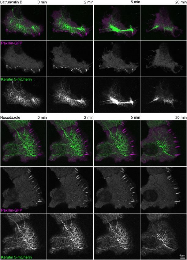
Int. J. Mol. Sci. 2021, 22, 2130                                                                                           2 of 24

                                   been shown to bind to the hemidesmosomal plakin family cytolinkers plectin 1a [15] and
                                   bullous pemphigoid antigen 1 (BPAG-1; also referred to as BPAG-1e or BP230) [16]. These
                                   linker molecules, in turn, facilitate the attachment to the hemidesmosome-specific α6β4-
                                   integrin heterodimers [6,17], which bind to laminin 332 in the basement membrane [18,19].
                                   Type II hemidesmosomes of simple epithelia consist only of α6β4-integrin heterodimers
                                   and plectin 1a-attached keratins, whereas type I hemidesmosomes, which occur in basal
                                   cells of pseudostratified and stratified epithelia, contain additional proteins, such as BPAG-
                                   1, the tetraspanin CD151, and the laminin 332-binding BPAG-2 (also referred to as BP180 or
                                   collagen type XVII) [6,20]. Recent studies suggest that type II hemidesmosomes mature
                                   into type I hemidesmosomes [21].
                                          The function and distribution of hemidesmosomes in physiological and pathological
                                   in vivo situations is poorly characterized [22]. But it is known that hemidesmosomes are
                                   abundant in various epithelial tissues throughout the animal kingdom [23–27]. Grazing
                                   sections revealed that hemidesmosomes are regularly arranged as linear arrays (e.g., Refer-
                                   ences [25,26,28]) reminiscent of the “beads-on-a-string” pattern described in recent in vitro
                                   studies [21,29], whereby keratin filaments (i.e., the “string”) interconnect hemidesmosomes
                                   (i.e., the “beads”) as is the case for desmosomal keratins [30]. The role of hemidesmosomes
                                   in wound-healing has also been cursorily investigated in vivo demonstrating that they are
                                   involved in the tongue-formation of keratinocytes invading the wound bed [31]. In the
                                   context of carcinogenesis, disassembly of hemidesmosomes and alternative association of
                                   the α6β4 integrin dimer with the actin cytoskeleton has been observed (cf. Reference [32]).
                                   Ultrastructural characterization of hemidesmosomes in invading tumor tissue, however, is
                                   scarce [33,34].
                                          The molecular composition of actin stress fiber bundle-associated focal adhesions,
                                   which also form prominent attachment sites to the ECM, is fundamentally different from
                                   that of hemidesmosomes (review in References [35,36]). The anchorage of focal adhesions
                                   to the ECM is mediated through integrin-dimers, such as α2β1, α3β1, and α9β1, that are ex-
                                   pressed in intact skin or α5β1, αVβ5 and αVβ6 that are upregulated during wound healing
                                   (cf. Reference [37]). Linker molecules between these integrins and the actin cytoskeleton
                                   include talin and vinculin [38,39]. Additional molecules with signaling functions, such as
                                   paxillin, focal adhesion kinase, and Src, are also recruited to focal adhesions [40–42].
                                          Focal adhesions perform central functions in force transmission during cell migra-
                                   tion [35]. The function of hemidesmosomes is less well defined. But an intricate interplay
                                   between focal adhesions and hemidesmosomes appears to take place during migration,
                                   which is of relevance during wound healing in human skin [29,31,32,37]. Even less is known
                                   about the coordination of the ECM-dependent organization of the different cytoskeletal
                                   systems although ECM-dependent coordinated regulation of cytoskeletal network dy-
                                   namics in epithelial cells has been reported [43,44]. Most recently, a force-dissipating
                                   function has been assigned to hemidesmosomes and their associated keratin filaments
                                   by demonstrating that prevention of α6β4 adhesion increases focal adhesion-mediated
                                   and actin-dependent traction force generation in human keratinocytes [45]. A major
                                   impediment to more detailed analyses of the cross talk between focal adhesions and
                                   hemidesmosomes in the context of cytoskeletal network organization has been the lack of
                                   bona fide hemidesmosomes in many epithelial cell lines grown on glass. It has been shown,
                                   however, that immortalized and primary keratinocytes form cell adhesions preferably on
                                   laminin 332-coated surfaces that contain α6β4-integrins, together with plectin 1a, BPAG-1,
                                   and BPAG-2, serving as attachment sites for keratin intermediate filaments (e.g., Refer-
                                   ences [21,29]). Keratin filaments and keratin filament bundles typically associate laterally
                                   with multiple hemidesmosomes [21,29]. These hemidesmosomal contacts are in close
                                   vicinity to focal adhesions indicative of structural, functional, and molecular links between
                                   both [6,29,45,46]. The precise nature of these links, however, and how they support the
                                   organization of the attached filament systems remains to be elucidated.
                                          Using the immortalized human keratinocyte cell line HaCaT, we show by high resolu-
                                   tion microscopy of fixed immunocytochemical samples and vital cells producing fluorescent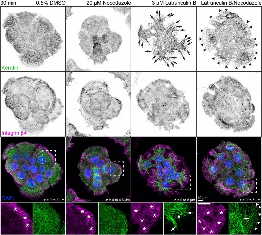
Int. J. Mol. Sci. 2021, 22, 2130                                                                                           3 of 24

                                   reporters that keratin filaments attach to hemidesmosomes, which are located in close
                                   proximity to focal adhesions. Treating these cells with inhibitors of actin and tubulin
                                   polymerization leads to loss of actin filaments, microtubules, and focal adhesions, while
                                   hemidesmosome-like entities with adhering keratin filaments remain. At the same time, the
                                   mesh size of the keratin cytoskeleton increases which is accompanied by bundling of ker-
                                   atin filaments. These findings demonstrate that established hemidesmosomes persist in the
                                   absence of focal adhesions and continue to serve as organizational hubs for the keratin sys-
                                   tem. Notably, keratin filament formation at hemidesmosomes is detected in this situation
                                   and also in migrating keratinocytes. The observed nucleation of growing keratin filaments
                                   that interconnect hemidesmosome assemblies is comparable to the recently described
                                   formation of keratins at desmosomal cell-cell adhesions [30]. Taken together, our findings
                                   confirm known and identify hitherto unknown features of the keratin-hemidesmosome
                                   system. We propose that these features confer special mechanophysical resilience and plas-
                                   ticity to epithelia against extreme stresses and strains that cannot be absorbed by the actin-
                                   and tubulin-based cytoskeletal components. Situations of extreme mechanical stress are
                                   particularly obvious in the epidermis. The epidermis is subjected to substantial pressure,
                                   especially the body weight-bearing foot sole epidermis, and is subjected to considerable
                                   viscous shear and tensile force as is the case for palmar epidermis of high bar gymnasts.
                                   These properties are reflected by epithelial superelasticity that has been recently linked to
                                   keratin intermediate filaments [47].

                                   2. Results
                                   2.1. Keratin Filaments Selectively Attach to and Interconnect Type I Hemidesmosomes That Are
                                   Juxtaposed to Focal Adhesions
                                        Immortalized human HaCaT keratinocytes were used to investigate interactions
                                   between the keratin cytoskeleton and ECM adhesions. HaCaT cells form a monolayer
                                   under standard cell culture conditions. Cells were grown for 3 days to ≈90% confluency on
                                   glass that had been pre-coated with a laminin 332-rich matrix. This favored the formation
                                   of hemidesmosomes and ensured that cells were extremely flat and, therefore, ideally
                                   suited for high-resolution microscopy. Peripheral keratin filaments were often but not
                                   always detected next to clustered integrin β5, which is a component of focal adhesions
                                   (Figure 1A). In contrast, hemidesmosomal integrin β4- and BPAG-1-containing complexes
                                   co-localized with keratin filaments in a “beads on a string” pattern (Figure 1B). This
                                   pattern was also detected with antibodies directed against integrin α6, BPAG-1 and keratin
                                   (Figure 1C). The integrin α6/β4- and BPAG-1-positive structures can, thus, be classified as
                                   type I hemidesmosomes. Conversely, the integrin β5-positive structures were also positive
                                   for paxillin, identifying them as focal adhesions (Figure 1E).
                                        To enable monitoring of the two types of ECM adhesions in living cells, fluorescent
                                   reporters were introduced into HaCaT keratinocytes by transfection of cDNA constructs.
                                   Hemidesmosomes were reliably labeled with integrin β4-green fluorescent protein (integrin
                                   β4-GFP) chimeras (Figure 1D), and either talin-GFP or paxillin-GFP reporters demarcated
                                   integrin β5-positive focal adhesions (Figure 1F,G).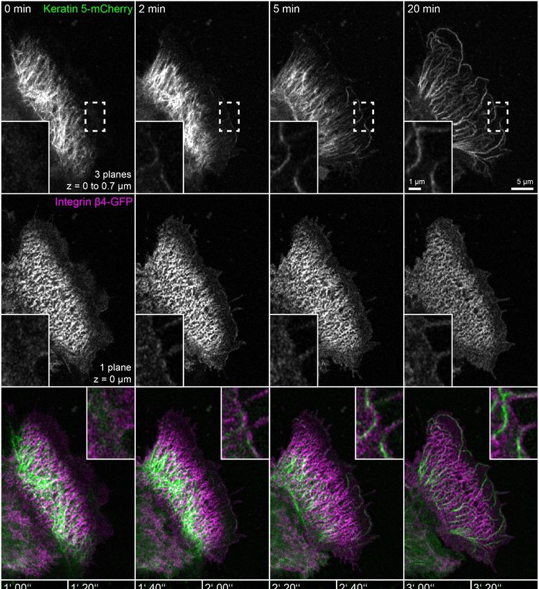
Int. J. Mol. Sci. 2021, 22, 2130 4 of 24
Int. J. Mol. Sci. 2020, 21, x FOR PEER REVIEW 4 of 26
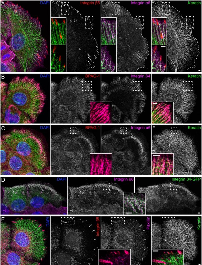
Figure 1. Keratin filaments are connected to hemidesmosomes, many of which are clustered next to focal adhesions. The
fluorescence micrographs show antibody staining, nuclear 40 ,6-diamidino-2-phenylindole (DAPI) staining (blue), and fluorescent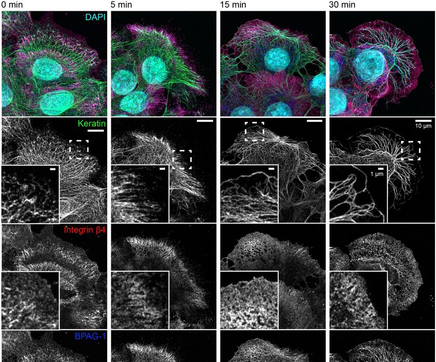
Int. J. Mol. Sci. 2021, 22, 2130                                                                                                  5 of 24

      reporters (at right in (D),(F),(G)) in methanol-fixed human keratinocytes (HaCaT). Enlargements of areas delineated by
      broken lines are shown as inserts. (A) The immunofluorescence images illustrate that the tips of keratin filament bundles
      localize next to but do not emanate from integrin β5-positive focal adhesions. Instead, peripheral keratin filaments co-
      localize with and connect integrin α6-positive hemidesmosomes (n = 8). (B,C) The pictures depict the co-distribution of
      bullous pemphigoid antigen 1 (BPAG-1) with integrin β4 and integrin α6 identifying the triple-positive structures as type I
      hemidesmosomes. Note, that they co-localize with peripheral keratin filament bundles, which are oriented along these
      extracellular matrix (ECM) adhesion sites (n = 7 and n = 11). (D) The images show two cells that were transfected with
      integrin β4-green fluorescent protein (integrin β4-GFP), which co-localizes with endogenous integrin α6 (n = 5). (E) The
      micrographs illustrate the co-distribution of integrin β5 with paxillin in focal adhesions and the lack of co-localization of
      focal adhesion markers with keratin filaments (n = 9). (F,G) The fluorescence recordings demonstrate that the fluorescent
      reporters talin-GFP and paxillin-GFP co-localize with integrin β5 in focal adhesions (n = 5 and n = 1). The image panels
      in (A–D) are maximum intensity projections of complete cells, and the image panels in (E–G) depict only the lower cell
      sections (also maximum intensity projections), where focal adhesions and hemidesmosomes are located.

                                   2.2. Hemidesmosomal Keratin Filament Anchorage Persists in the Absence of Actin Filaments
                                   and Microtubules
                                         To investigate the contribution of actin filaments and microtubules to the keratin-
                                   hemidesmosome scaffold, HaCaT keratinocytes were treated with latrunculin B and noco-
                                   dazole at different concentrations. Latrunculin B induced a dose-dependent inhibitory
                                   effect on mApple-actin polymerization as shown for 1 and 2 µM in time-lapse recordings
                                   (Video S1). For further experiments, a concentration of 3 µM was used to ensure reliable
                                   inhibition of actin polymerization. Twenty micromolar nocodazole treatment resulted
                                   in drastically reduced microtubule plus end-binding protein 3-green fluorescent protein
                                   (EB3-GFP) dynamics within seconds after inhibitor addition (Video S2; for efficient in-
                                   hibitory effects of 33 µM nocodazole in HaCaT keratinocytes, also see References [48,49]).
                                   In further experiments, both inhibitors were added simultaneously to cells and samples
                                   were immunostained at different time points. Minor alterations in the architecture of the
                                   microtubule system became evident 5 min after drug addition, as determined by β-tubulin
                                   immunostaining. After 15 min, several microtubules were still detected and only very few
                                   scattered microtubules remained after 30 min (upper panels in Figure A1). In comparison,
                                   phalloidin staining revealed alterations in the architecture of the actin filament system
                                   5 min after drug addition, near complete loss of actin filaments after 15 min, and the
                                   formation of differently sized granules after 30 min (middle panels in Figure A1). We,
                                   therefore, concluded that the chosen drug concentrations are highly efficient in disrupting
                                   both the actin and microtubule cytoskeleton within 15–30 min. We, furthermore, noted
                                   that a 30 min treatment with both drugs inhibited filopodia, lamellipodia, and ruffle for-
                                   mation and prevented cell migration. Cells instead retracted and rounded up, leading to
                                   increased cell height, which was reflected by expansion of the nucleus in the z-direction
                                   (Figure A2). We interpret this as a consequence of overall cell relaxation because of reduced
                                   intracellular tension.
                                         The next set of experiments concentrated on the response of the keratin/hemidesmosome
                                   scaffold to 3 µM latrunculin B and 20 µM nocodazole. Figure 2 depicts the distribution of
                                   integrin β4-positive hemidesmosomes and keratins in small cell clusters in the presence of
                                   the solvent, latrunculin B, nocodazole, or latrunculin B/nocodazole. Typical hemidesmo-
                                   somal distribution patterns were detectable and most prominent at the cell bottom of
                                   peripheral cells in each situation. On the other hand, treatment with latrunculin B induced
                                   retraction and straightening of keratin filaments which, however, still remained anchored
                                   in the cell periphery. Treatment with nocodazole affected keratin network organization only
                                   mildly inducing a slight increase in keratin filament straightness but no obvious keratin
                                   filament retraction. Notably, treatment with both drugs induced yet a different keratin
                                   network phenotype (also see Figure A3), while hemidesmosomal distribution was not
                                   visibly affected. In the latrunculin B/nocodazole-treated cells keratin networks retracted
                                   less than in cells treated with latrunculin B alone. In addition, keratin bundles appeared
                                   near the margin of peripheral cells that were not seen in the other conditions. Common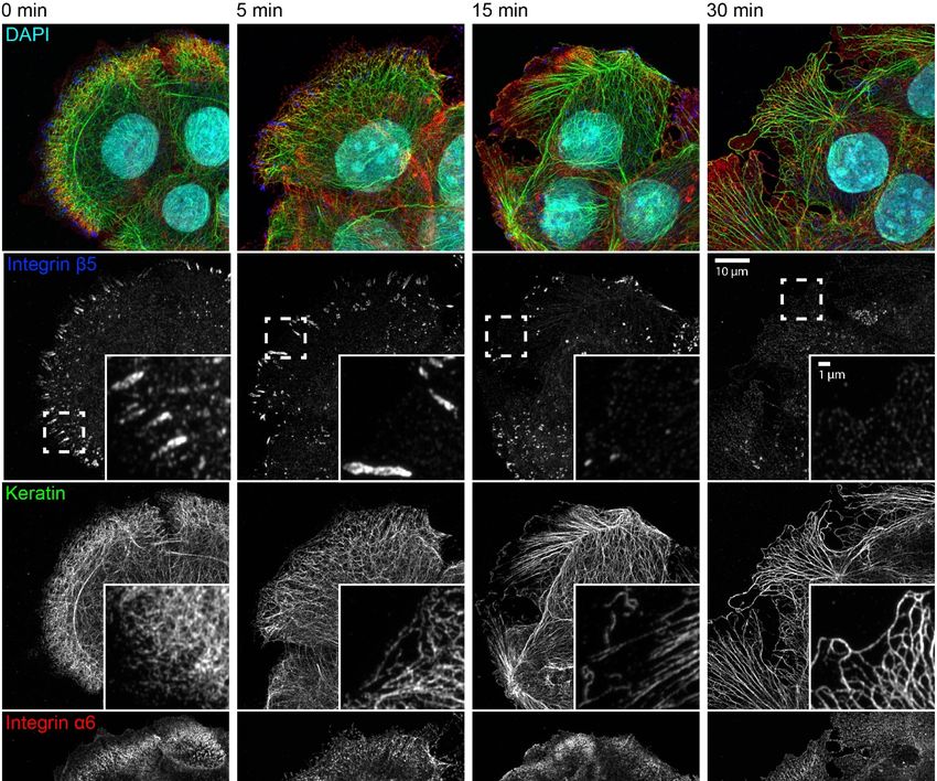
Int. J.
Int. J. Mol.
Mol. Sci.
Sci. 2020,
2021, 21,
22, x2130
FOR PEER REVIEW 66 of
of 24
26
bundles appeared near the margin of peripheral cells that were not seen in the other con-
ditions. Common to all drug-treated cells were perinuclear keratin network accumula-
to all which
tions, drug-treated cells werewith
were associated perinuclear keratin
radial keratin network
filaments. Theaccumulations,
differences in which were
straightness
associated that
suggested withthe
radial keratin
radial filaments.
filaments may beThe differences
under tension,inwhereas
straightness suggested that
the perinuclear fila-
the radial
ments are filaments
relaxed. Tomay be under
study tension,nature
the dynamic whereas the perinuclear
of keratin network filaments are relaxed.
reorganization, time-
To study
lapse the dynamic
fluorescence nature of was
microscopy keratin network in
performed reorganization,
HaCaT clonetime-lapse
B10, which fluorescence
stably ex-
microscopy was performed in HaCaT clone B10, which stably expresses
presses keratin 5-yellow fluorescent protein (keratin 5-YFP) [50]. The recording presentedkeratin 5-yellow
fluorescent
in Video S3protein (keratin
confirmed the 5-YFP) [50]. The recording
immunocytochemical presented in
observations. Video
The S3 confirmed
keratin network the re-
immunocytochemical
tracted observations.
shortly after addition of bothThe keratinbut
inhibitors network retracted
remained in partshortly
anchoredafterinaddition
the cell
of both inhibitors
periphery but remained
with straight in partfilaments.
and bundled anchored Coarsening
in the cell periphery withand
of mesh size straight and
filament
bundled filaments. Coarsening of mesh size and filament bundling occurred
bundling occurred throughout the entire network with local granule formation in some, throughout
the entire
but not all,network with
cells. The local granule
dynamic formation
restructuring wasinbest
some, butinnot
seen theall, cells.bottom
single The dynamic re-
plane, re-
structuring was best seen in the single bottom plane, revealing increasing
vealing increasing filament bundling and ongoing filament formation in the cell periph-filament bundling
and ongoing filament formation in the cell periphery.
ery.
Figure 2.
Figure 2. Disruption
Disruption ofof actin
actin filaments
filaments and microtubules alone
and microtubules alone or in combination
or in combination leads to different
leads to different changes
changes inin keratin
keratin
filament network architecture in each situation. The triple fluorescence micrographs (enlargements of boxed areas in the
filament network architecture in each situation. The triple fluorescence micrographs (enlargements of boxed areas in the
bottom panel) show the immunodetection of keratins (top panel) and integrin β4 (middle panel), together with nuclear
bottom panel) show the immunodetection of keratins (top panel) and integrin β4 (middle panel), together with nuclear
DAPI (merged images in lower panel), in methanol-fixed HaCaT keratinocytes. The actin cytoskeleton was disrupted by
DAPI (merged
treating images
cells with 3 µMin lower panel),
latrunculin B and inthe
methanol-fixed HaCaT keratinocytes.
microtubule cytoskeleton The by
was disrupted actin cytoskeleton
treating was
cells with 20 disrupted
µM noco-
by treating
dazole cells
for 30 min.with
The3 dimethyl
µM latrunculin B and
sulfoxide the microtubule
(DMSO) control on cytoskeleton
the left showswas disrupted
examples by treating
of typical cells
keratin with 20 and
networks µM
nocodazole
hemidesmosomalfor 30 min. The dimethyl
distribution sulfoxide
in untreated (DMSO)
HaCaT cells.control on the
Note that theleft showsβ4-demarcated
integrin examples of typical keratin networks
hemidesmosomal and
patterns
hemidesmosomal distribution
(asterisks) are not affected in untreated
by drug treatment,HaCaT cells.
for the most Note
part.that
Onthe
theintegrin
other hand, hemidesmosomal
major keratin
β4-demarcated filament network patterns
retrac-
tion is seen in latrunculin B treated cells (arrows), which is less pronounced in latrunculin B/nocodazole-treated cells (ar-
rows) and not apparent in cells treated with nocodazole alone. Note also that prominent keratin bundles are present at the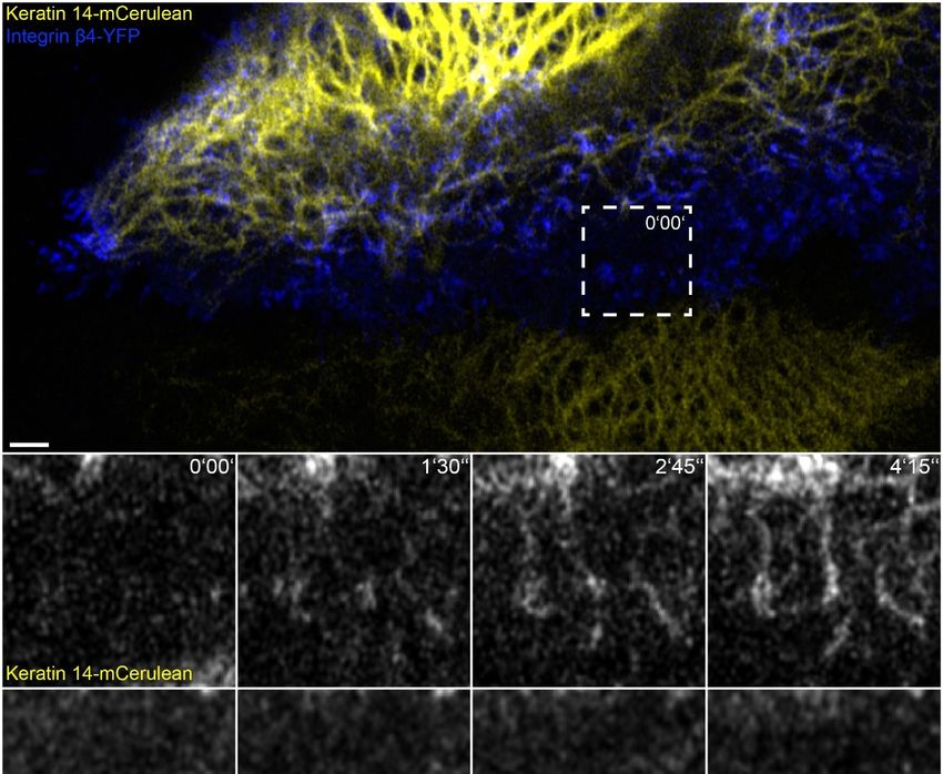
Int. J. Mol. Sci. 2021, 22, 2130                                                                                                    7 of 24

      (asterisks) are not affected by drug treatment, for the most part. On the other hand, major keratin filament network retraction
      is seen in latrunculin B treated cells (arrows), which is less pronounced in latrunculin B/nocodazole-treated cells (arrows)
      and not apparent in cells treated with nocodazole alone. Note also that prominent keratin bundles are present at the
      outermost margin of peripheral cells treated with both drugs (arrowheads) but not in untreated cells and cells treated with
      only one drug. All images are maximum intensity projections of entire HaCaT keratinocytes (n ≥ 10).

                                        The consequences of latrunculin B/nocodazole treatment for hemidesmosome-keratin
                                   distribution was assessed in more detail by immunocytochemistry in the next set of ex-
                                   periments (Figure 3). Five minutes after inhibitor addition, the keratin filament network
                                   retracted towards the cell center. It remained, however, attached to integrin β4-/BPAG-1-
                                   positive hemidesmosomes through radial, rather straight keratin filaments. After 15 and
                                   30 min, increasingly thicker keratin filament bundles were detected, and the mesh size
                                   of the keratin filament network increased. At the same time, additional keratin filaments
                                   were detected at the outermost cell periphery next to weakly fluorescent integrin β4-
                                   /BPAG-1-positive dotted structures. In some cells, these filaments formed a continuous
                                   rim adjacent to the plasma membrane interconnecting the integrin β4/BPAG-1 clusters.
                                   Antibodies against integrin α6 and keratin confirmed the findings obtained for anti-integrin
                                   β4/BPAG-1/keratin immunostainings (Figure 4).
                                        Taken together, we conclude that type I hemidesmosome-dependent keratin network
                                   organization persists after depletion of actin filaments and microtubules, resulting, however,
                                   in a novel keratin filament network phenotype.

                                   2.3. Focal Adhesions Are Not Essential for the Maintenance of Hemidesmosomal Keratin
                                   Filament Anchorage
                                        We next wanted to find out how the latrunculin B/nocodazole treatment affected
                                   focal adhesions in HaCaT cells. Immunocytochemistry showed that integrin β5-positive
                                   focal adhesions disassembled for the most part within 15 min and were virtually unde-
                                   tectable after 30 min (Figure 4). These findings were confirmed by time-lapse fluorescence
                                   microscopy of living cells, which also showed that latrunculin B affected focal adhesions
                                   but nocodazole alone did not (Figure A4 and corresponding Video S4).
                                        The triple immunofluorescence micrographs in Figure 4 demonstrate that areas devoid
                                   of integrin β5-containing focal adhesions still maintained clustered integrin α6, which
                                   co-localized with keratin filaments. Time-lapse fluorescence microscopy of HaCaT cells
                                   co-expressing talin-GFP and keratin 5-mCherry confirmed these observations (Figure 5
                                   and corresponding Video S5). They also supported the above-mentioned alterations in
                                   keratin filament organization and provided additional details. This included the retraction
                                   of the keratin network toward the nucleus, the bundling of keratin filaments coincident
                                   with network coarsening, and the appearance of filament bundles in the cell periphery,
                                   presumably at hemidesmosomal adhesions. In addition, the overall motility of the keratin
                                   cytoskeleton, including that of small keratin particles, ceased almost completely.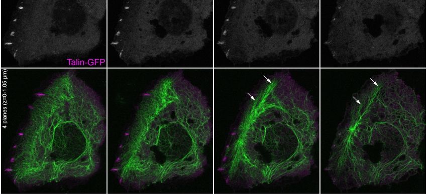
Int. J. Mol. Sci. 2021, 22, 2130                                                                                                                            8 of 24

        Int. J. Mol. Sci. 2020, 21, x FOR PEER REVIEW                                                                                             8 of 26

      FigureFigure
               3. The 3. The keratin filament network retracts but remains attached to hemidesmosomes through straightened and bun-
                          keratin filament network retracts but remains attached to hemidesmosomes through straightened and
             dled radial keratin filaments after actin filament and microtubule disruption. The quadruple fluorescence micrographs
      bundledshowradial   keratin filamentsofafter
                    the immunodetection                actinintegrin
                                                  keratin,    filamentβ4and
                                                                          and microtubule    disruption.
                                                                              BPAG-1, together            The quadruple
                                                                                                  with nuclear              fluorescence micrographs
                                                                                                                DAPI, in methanol-fixed     HaCaT
      show keratinocytes
             the immunodetection
                              before and of5, keratin,
                                              15, and 30  integrin       and BPAG-1,
                                                                     β4treatment
                                                             min after             with 3together   with nuclear
                                                                                           µM latrunculin  B and 20DAPI,   in methanol-fixed
                                                                                                                      µM nocodazole.     The insetsHaCaT
             depict the
      keratinocytes        areasand
                        before   marked
                                     5, 15,byand
                                              broken
                                                  30 minlines  at higher
                                                             after        magnification
                                                                   treatment  with 3 µM  and   as mergedBimages
                                                                                            latrunculin    and 20inµM
                                                                                                                    thenocodazole.
                                                                                                                         bottom panel. TheNote  thatdepict
                                                                                                                                             insets
             keratin
      the areas        filament
                  marked         bundles lines
                              by broken    are attached
                                                  at higher to integrin  β4-positive
                                                                magnification    andand   BPAG-1-positive
                                                                                      as merged             hemidesmosomes
                                                                                                    images in  the bottom panel.before   treatment.
                                                                                                                                      Note    that keratin
             The bulk of the keratin network retracts within 5 min of drug addition, while remaining attached to hemidesmosomes in
      filament   bundles     are attached    to integrin     β4-positive   and  BPAG-1-positive      hemidesmosomes       before   treatment.
             the cell periphery. After 15 min, the keratin network is composed of thicker keratin filament bundles and has a larger mesh
                                                                                                                                                 The bulk
      of the size.
              keratin    network retracts
                   Additionally,   new keratinwithin    5 minare
                                                   filaments     of detected
                                                                    drug addition,    while remainingdomain
                                                                             in the juxtamembraneous      attached   to hemidesmosomes
                                                                                                                  co-localizing with integrinin    the cell
                                                                                                                                                 β4-
             positive
      periphery.   Afterand15BPAG-1-positive
                               min, the keratin  clusters.
                                                     network A similar  and evenofmore
                                                                 is composed             pronounced
                                                                                    thicker           change inbundles
                                                                                              keratin filament   keratin network
                                                                                                                          and hasorganization
                                                                                                                                     a larger meshis size.
             seen at time
      Additionally,     newpoint  30 min
                              keratin      with a few,
                                       filaments          hemidesmosome-anchored
                                                    are detected                        thick keratin
                                                                     in the juxtamembraneous          bundles
                                                                                                    domain     and long keratin
                                                                                                             co-localizing   withfilaments
                                                                                                                                   integrinnext   to
                                                                                                                                               β4-positive
             the plasma membrane. All images are maximum intensity projections of entire HaCaT keratinocytes (n ≥ 7).
      and BPAG-1-positive clusters. A similar and even more pronounced change in keratin network organization is seen at time
      point 30 min with a few, hemidesmosome-anchored thick keratin bundles and long keratin filaments next to the plasma
      membrane. All images are maximum intensity projections of entire HaCaT keratinocytes (n ≥ 7).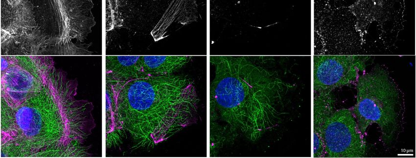
Int. J. Mol. Sci. 2021, 22, 2130                                                                                                                              9 of 24

        Int. J. Mol. Sci. 2020, 21, x FOR PEER REVIEW                                                                                              10 of 26

              Figure 4. Keratin network reorganization after actin filament and microtubule disruption is not dependent on focal adhe-
      Figure 4. Keratin network reorganization after actin filament and microtubule disruption is not dependent on focal
             sions. The microscopic fluorescence images show the immunodetection of integrin β5, keratin, and integrin α6, together
      adhesions.    The microscopic
             with nuclear  DAPI staining,   fluorescence    images
                                              in methanol-fixed       show
                                                                  HaCaT      the immunodetection
                                                                           keratinocytes                 of integrin
                                                                                          that were treated  with 3 µMβ5,      keratin,Band
                                                                                                                           latrunculin          integrin
                                                                                                                                           and 20  µM     α6,
      together  with nuclear
             nocodazole         DAPI staining,
                          for different             in methanol-fixed
                                          time periods.                    HaCaT
                                                         The inserts present    the keratinocytes
                                                                                    boxed regions that   were
                                                                                                    (broken      treated
                                                                                                              lines)       with magnification.
                                                                                                                      at higher   3 µM latrunculin The B and
      20 µMbottom     panel for
              nocodazole     depicts  merged
                                 different       images
                                              time       of the
                                                    periods.     boxed
                                                               The       regions.
                                                                    inserts        Before
                                                                             present   thetreatment,   keratin
                                                                                            boxed regions       filamentlines)
                                                                                                             (broken       bundles    are attached
                                                                                                                                 at higher          to
                                                                                                                                             magnification.
             integrin α6-positive hemidesmosomal structures that are next to integrin β5-positive focal adhesions. Focal adhesions
      The bottom panel depicts merged images of the boxed regions. Before treatment, keratin filament bundles are attached
             disassemble leading to granular integrin β5 clusters after 15 min and barely detectable clustered integrin β5 after 30 min
      to integrin  α6-positive
             of inhibitor         hemidesmosomal
                          treatment.   At the same time,  structures   that arebundle,
                                                             keratin filaments    next tostraighten,
                                                                                          integrin β5-positive       focal adhesions.
                                                                                                      and retract toward                   Focalwhile
                                                                                                                              the cell interior,  adhesions
      disassemble
             remaining attached to integrin α6-positive hemidesmosomes. Note that short keratin filaments elongate in the cell periph-30 min
                     leading   to granular      integrin β5   clusters  after 15  min   and  barely  detectable     clustered   integrin    β5 after
             ery, which
      of inhibitor      become more
                    treatment.    At the prominent    withkeratin
                                             same time,    time and   co-localizebundle,
                                                                    filaments     with integrin  α6 particles.
                                                                                           straighten,         All images
                                                                                                        and retract          are maximum
                                                                                                                        toward               intensity while
                                                                                                                                   the cell interior,
             projections  of entire cells  (n ≥ 7).
      remaining attached to integrin α6-positive hemidesmosomes. Note that short keratin filaments elongate in the cell periphery,
      which become more prominent with time and co-localize with integrin α6 particles. All images are maximum intensity
      projections of entire cells (n ≥ 7).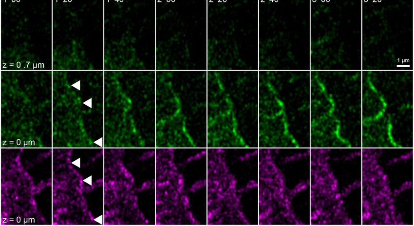
Int. J. Mol. Sci. 2021, 22, 2130 10 of 24
Int. J. Mol. Sci. 2020, 21, x FOR PEER REVIEW 11 of 26
Figure 5.
Figure 5. Keratin
Keratin filament
filament network
network reorganization
reorganization isis independent
independent of of focal
focal adhesions
adhesions inin the
the absence
absence of
of actin
actin filaments
filaments andand
microtubules. The images depict keratin 5-mCherry (top) and talin-GFP fluorescence (middle; merge
microtubules. The images depict keratin 5-mCherry (top) and talin-GFP fluorescence (middle; merge at bottom) in a vitalat bottom) in a vital
HaCaT keratinocyte before and after treatment with 3 µM latrunculin B and 20 µM nocodazole. The keratin network
HaCaT keratinocyte before and after treatment with 3 µM latrunculin B and 20 µM nocodazole. The keratin network retracts,
retracts, bundles, and remains partially attached in the cell periphery (arrows) independent of the continued, though di-
bundles, and remains partially attached in the cell periphery (arrows) independent of the continued, though diminishing or
minishing or even completely vanishing talin-GFP-fluorescence in some regions. All images are maximum intensity pro-
even completely
jections vanishing
of the lower talin-GFP-fluorescence
focal planes, as annotated in the in some
figureregions. All images
(n = 3). Note arecell
that the maximum
is next tointensity projections
non-transfected, i.e.,ofnon-
the
lower focal planes,
fluorescent cells. Theas complete
annotatedimage
in theseries
figureis(nprovided
= 3). Note inthat theS5.
Video cell is next to non-transfected, i.e., non-fluorescent cells.
The complete image series is provided in Video S5.
2.4. Keratin Filament Nucleation Occurs at Newly-Formed Hemidesmosomes and Does not
2.4. Keratin
Require ActinFilament Nucleation
Filaments Occurs at Newly-Formed Hemidesmosomes and Does Not
and Microtubules
Require Actin Filaments and Microtubules
To directly examine the dynamic behavior of hemidesmosomes and keratins in
To directly
latrunculin examine the dynamic
B/nocodazole-treated HaCaT behavior of specifically,
cells and, hemidesmosomes and keratins
to determine in la-
the origin of
trunculin B/nocodazole-treated HaCaT cells and, specifically, to determine
the peripheral keratin filaments, keratin 5-mCherry and integrin β4-GFP were imaged by the origin of
the peripheral
time-lapse keratin filaments,
fluorescence microscopykeratin 5-mCherry
(Figure and integrin β4-GFP
6 and corresponding were
Video S6). Twoimaged by
minutes
time-lapse fluorescence microscopy (Figure 6 and corresponding Video S6). Two minutes
after addition of the inhibitors, the keratin network started to retract, and keratin filaments
after addition
bundled, of the inhibitors,
extending the keratin
from the nucleus network
towards started
the cell to retract,
periphery. and
The keratin filaments
peripheral parts of
bundled, extending from the nucleus towards the cell periphery.
these filaments co-localized with integrin β4 and remained attached throughout The peripheral parts
the ob-
of these filaments co-localized with integrin β4 and remained attached throughout
servation period. It, furthermore, appeared as if the retracting keratin filaments transmit- the
observation period. It, furthermore, appeared as if the retracting keratin filaments trans-
ted force on the hemidesmosome-anchored keratin filaments, as evidenced by their
mitted force on the hemidesmosome-anchored keratin filaments, as evidenced by their
straightening and radial orientation, with respect to the nucleus-containing cell center.
straightening and radial orientation, with respect to the nucleus-containing cell center. No-
Noticeably, new integrin β4 clusters appeared at the plasma membrane. Unfortunately,
ticeably, new integrin β4 clusters appeared at the plasma membrane. Unfortunately, these
these initially rather weak fluorescence signals bleached considerably during the record-
ings. But it was evident that they served as nucleation sites for novel keratin filaments,Int. J. Mol. Sci. 2021, 22, 2130 11 of 24
initially rather weak fluorescence signals bleached considerably during the recordings.
But it was evident that they served as nucleation sites for novel keratin filaments, which
subsequently elongated locally (enlargements at the bottom of Figure 6). These keratin
filaments successively connected to establish the peripheral submembraneous scaffold
that had been detected by immunocytochemistry (Figures 3 and 4). A likely explanation
for this finding is that keratin filament nucleation proceeds in the absence of actin fila-
ments and microtubules, resulting in keratin filament self-assembly next to nucleation sites
without further intracellular distribution because of inactivation of actin filament- and
microtubule-dependent transport systems.
2.5. Keratin Filaments Nucleate at Nascent Hemidesmosomes in Migrating Cells
In this set of experiments, we wanted to find out whether the phenomena detected in
the absence of actin filaments, microtubules and focal adhesions are also of relevance for
cells containing these cytoskeletal components. To increase hemidesmosome formation,
cells were grown to complete confluence, and migration was induced by a scratch with
a 20 µL pipette tip. The wounded cell-layers typically started to close the wound within
minutes. During migration of the cells, new focal adhesions and hemidesmosomes were
formed at the leading edges and were disassembled at the trailing edges as previously
described for migrating primary keratinocytes [29]. At the same time, new keratin fila-
ments were formed preferentially at the leading edge that subsequently gained contact
to the main cytoskeletal keratin network (Figure 7 and corresponding Video S7; also see
References [44,51]). Shortly before the appearance of keratin particles, integrin β4-YFP clus-
ters could be detected at the same positions. These nucleating keratin particles remained
attached to the hemidesmosomal clusters. The nascent keratin particles subsequently elon-
gated in one direction and interconnected hemidesmosomal protein clusters. The newly
formed scaffold finally connected to pre-existing filaments of the more central keratin
filament network.Int. J. Mol. Sci. 2021, 22, 2130 12 of 24
Int. J. Mol. Sci. 2020, 21, x FOR PEER REVIEW 13 of 26
Figure
Figure 6. Growing
6. Growing keratin
keratin filaments
filaments appear
appear at at nascent
nascent hemidesmosomesafter
hemidesmosomes afteractin
actinand
andmicrotubule
microtubuledisruption
disruption(n(n= =6).
6).The
The fluorescence images show the distribution of keratin 5-mCherry and integrin β4-GFP in a living HaCaT cell before
fluorescence images show the distribution of keratin 5-mCherry and integrin β4-GFP in a living HaCaT cell before and after
and after treatment with 3 µM latrunculin B and 20 µM nocodazole. The images of keratin 5-mCherry fluorescence present
treatment
maximum with 3 µM latrunculin
intensity projectionsBof
and
the20 µM nocodazole.
lower focal planes, The
and images of keratin
the images of the 5-mCherry fluorescence
integrin β4-GFP present
fluorescence maximum
show only
intensity projections of the lower focal planes, and the images of the integrin β4-GFP fluorescence show only the bottom focalInt. J. Mol. Sci. 2021, 22, 2130 13 of 24
plane. The boxed regions are depicted at higher magnification in the insets and at higher temporal and spatial resolution in the
bottom panels to illustrate ongoing keratin filament nucleation and growth at integrin β4-positive clusters in the cell periphery. As a
result, new keratin filament bundles appear at the cell edge that co-localize and connect newly-formed integrin β4-GFP complexes.
Note that the weak integrin β4-GFP signal decreases significantly because of bleaching. The images also show that the drug
treatment induces keratin network retraction and bundling, while keratin filament bundles remain attached to hemidesmosomal
structures. The fluorescent cell is adjacent to non-transfected cells. The complete time-lapse recording is provided in Video S6.
Int. J. Mol. Sci. 2020, 21, x FOR PEER REVIEW 15 of 26
Figure 7. Keratin filaments are assembled at and interconnect hemidesmosomes in migrating cells. The fluorescence im-
Figure 7. Keratin filaments are assembled at and interconnect hemidesmosomes in migrating cells. The fluorescence images
ages depict the distribution of keratin 14-mCerulean and integrin β4-yellow fluorescent protein (integrin β4-YFP) in a
depictmigrating
the distribution of keratin
HaCaT cell 14-mCerulean
that is next andwhich
to another cell, integrin positive fluorescent
β4-yellow
is only protein (integrin
for keratin 14-mCerulean. that bothin
Noteβ4-YFP) a migrating
cells are
HaCaTsurrounded
cell that isby
next to another cell,
non-transfected which
cells. is only positive
The magnified for recorded
time series keratin 14-mCerulean. Note that
in the square delineated byboth cellsline
a broken are shows
surrounded
different stagescells.
by non-transfected of keratin filament assembly
The magnified at nascent
time series hemidesmosomal
recorded in the square integrin β4-YFP
delineated by aclusters.
brokenThe linegrowing filaments stages
shows different
interconnect hemidesmosomes and gain contact with the peripheral keratin cytoskeleton. All images were recorded at the
of keratin filament assembly at nascent hemidesmosomal integrin β4-YFP clusters. The growing filaments interconnect
bottom focal plane (n=20, all cells were migrating). The entire time-lapse series is shown in Video S7.
hemidesmosomes and gain contact with the peripheral keratin cytoskeleton. All images were recorded at the bottom focal
plane (n = 20, all cells were migrating). The entire time-lapse series is shown in Video S7.
3. Discussion
Keratin network organization in the combined absence of actin filaments and micro-
tubules has rarely been investigated. Wöll et al. [10] showed that simultaneous inhibition
of actin and microtubule polymerization results in a complete collapse of the keratin fila-Int. J. Mol. Sci. 2021, 22, 2130 14 of 24
3. Discussion
Keratin network organization in the combined absence of actin filaments and micro-
tubules has rarely been investigated. Wöll et al. [10] showed that simultaneous inhibition of
actin and microtubule polymerization results in a complete collapse of the keratin filament
network in non-epithelial, adrenal cortex-derived SW13 cells producing fluorescence-
tagged keratins 8 and 18. In contrast to those findings, we show that the keratin 5 and
14 positive filament network retracts towards the nucleus but remains partially extended
under comparable conditions in epithelial HaCaT keratinocytes. The obvious reason
for this difference is the presence of hemidesmosome-like structures in HaCaT cells and
their absence in SW13 cells. This finding emphasizes the importance of hemidesmosomal
anchorage for keratin network organization.
We further suggest that the obviously non-physiological situation created in our
cultured cells may be of relevance for certain in vivo situations, including, for example,
conditions of extreme stretch or at the tips of collectively migrating cells during wound
closure or invading tumor cells, when the hemidesmosome/desmosome-keratin system
kicks in because of insufficiency of the other less extensible and flexible cytoskeletal net-
works [47,52]. The tensegrity model proposes that the acto-myosin and microtubule-motor
protein systems are important determinants of cell shape [53,54]. Their depletion releases
these constraints and cells are expected to round up. This is exactly what we observed in the
latrunculin B/nocodazole-exposed HaCaT cells. The remaining hemidesmosome-anchored
keratin filaments that are connected to the perinuclear cage are thereby stretched. At the
same time, the finely grated keratin network lattice slides and compacts into thick filament
bundles. This idea is depicted in the scheme in Figure 8.
Our observations also show, for the first time, that keratin filaments nucleate at
hemidesmosomes. The proximity of hemidesmosomes to focal adhesions explains our
previous observations, which identified focal adhesions as “hotspots” of keratin filament
formation [55]. In contrast to vimentin, however, keratins do not bind to focal adhe-
sions [56,57]. The high-resolution images in the present study demonstrate that keratin
filaments nucleate and grow from hemidesmosomes where they remain anchored. The
nucleating and growing hemidesmosome-attached keratin particles are reminiscent of
the previously described motile keratin particles, which have been referred to as keratin
filament precursors [58,59] or keratin squiggles [11].
The current findings of keratin nucleation and growth at hemidesmosomes are strik-
ingly similar to those reported recently for desmosomes [30]. In both instances, nascent
spheroidal keratin particles are first detected next to clustered transmembrane adhesion re-
ceptors, i.e., β4 integrins and desmosomal cadherins, respectively. These particles elongate
into small rodlets that grow unidirectionally and interconnect adjacent adhesion clusters
forming submembraneous keratin lattices. Filaments bundle and connect to the remaining
network. The fact that keratin nucleation occurs in these molecularly distinct desmosomal
and hemidesmosomal domains suggest that common mechanisms apply. Thus, recruit-
ment of plakin-domain proteins, i.e., plectin 1a and BPAG-1 at hemidesmosomes and
desmoplakins at desmosomes, leads to an increase of keratin polypeptides, which have
been shown to bind to plakin domains [15,16,60]. The local increase in keratins may suffice
to exceed a threshold level needed for spontaneous keratin assembly. The same mechanism
may apply to the recently reported recruitment of keratins to apical microridges [61].
Taken together, our findings assign important mechanical functions on the keratin-
hemidesmosome scaffold, which is predominant in basal cells of homeostatic epidermis
reacting dynamically to wounding by localized reformation and providing protection
against extreme mechanical stress, resulting in deformation that cannot be absorbed by
actin filaments and microtubules.Int. J. Mol. Sci. 2021, 22, 2130 15 of 24
Int. J. Mol. Sci. 2020, 21, x FOR PEER REVIEW 17 of 26
Figure 8. The scheme summarizes our major findings and illustrates how the keratin network may provide mechanical
resilience to extreme cellular deformation, leading to network reorganization through filament bundling and filament
nucleation at
nucleation at nascent
nascent hemidesmosomes.
hemidesmosomes. Disruption
Disruption of of both
both the
the actin
actin filament
filament and
and microtubule systems induces
microtubule systems induces cell
cell shape
shape
changes because of loss of cortical actomyosin contractility and altered intracellular stiffness. Together, these
changes because of loss of cortical actomyosin contractility and altered intracellular stiffness. Together, these changes are changes are
predicted to lead to a rounding of the nucleus and the surrounding cell body, which is counterbalanced by the keratin
predicted to lead to a rounding of the nucleus and the surrounding cell body, which is counterbalanced by the keratin
intermediate filament cytoskeleton. Since the hemidesmosome-anchored keratin filament network persists, radial fila-
intermediate filament cytoskeleton. Since the hemidesmosome-anchored keratin filament network persists, radial filaments
ments become stretched, losing their waviness and forming thick bundles. Localized hemidesmosome formation and ker-
become stretched,
atin filament losingallows
nucleation their waviness
adjustment andofforming thick bundles.
the remaining Localized
cytoskeleton to cellhemidesmosome
shape. We propose formation and keratin
that deformations,
filament nucleation allows adjustment of the remaining cytoskeleton to cell shape. We propose that deformations,
which may, for example, occur in migrating cells after wounding or in epidermal cell layers subjected to tensile stress which
and
may, forshear
viscous example, occur
stress, in comparable
elicit migrating cells after wounding
responses or in epidermal
in the superelastic keratincell layers subjected
intermediate to tensile
filament stress and viscous
network.
shear stress, elicit comparable responses in the superelastic keratin intermediate filament network.
4. Materials and Methods
4. Materials
4.1. and Methods
Cell Culture
4.1. Cell Culture
Immortalized human HaCaT keratinocytes were kindly provided by Dr. Petra
Immortalized
Boukamp [62] andhuman
were HaCaT
grownkeratinocytes
at 37 °C in were
a 5%kindly provided byatmosphere
CO2 humidified Dr. Petra Boukamp [62]
and Dul-
and were grown at 37 ◦ C in a 5% CO humidified atmosphere and Dulbecco’s Modified
becco’s Modified Eagle’s Medium (DMEM) 2 containing l-alanyl-glutamine (Sigma-Al-
Eagle’sSt.Medium
drich, Louis, MO,(DMEM) containing
USA) and 10% (v/v) l-alanyl-glutamine
fetal bovine serum (Sigma-Aldrich,
(FBS) SeraPlus St.
(PANLouis, MO,
Biotech,
USA) and 10%
Aidenbach, (v/v) fetal For
Germany). bovine serum (FBS)
passaging, cellsSeraPlus (PAN Biotech,
were washed Aidenbach,
and incubated for Germany).
15 min in
For passaging, cells were
phosphate-buffered salinewashed and incubated
(PBS) without Ca2+/Mgfor 15 min in phosphate-buffered
2+ (Sigma-Aldrich). saline
They were thereafter
(PBS) without Ca 2+ /Mg2+ (Sigma-Aldrich). They were thereafter trypsinized for ≈5 min
trypsinized for ≈5 min in a solution of PBS without Ca /Mg (Biochrom, Schaffhausen,
2+ 2+
in a solution of PBS without 2+ /Mg2+ (Biochrom, Schaffhausen, Switzerland) containing
Ca(w/v)
Switzerland) containing 0.2% trypsin (Biochrom) supplemented with 0.02% (w/v)
0.2% (w/v)
EDTA trypsin (Biochrom)
(Sigma-Aldrich). supplemented
Cells were passaged once withper0.02%
week(w/v)
one EDTA (Sigma-Aldrich).
day after reaching con-
fluence and were seeded at a concentration of2 40,000–60,000 cells/cm2 in 6 mlwere
Cells were passaged once per week one day after reaching confluence and cell seeded
culture
2
medium in 25 cm cell culture flasks (Greiner Bio-One, Frickenhausen, Germany). Forcell
at a concentration 2 of 40,000–60,000 cells/cm in 6 ml cell culture medium in 25 cm ex-
culture flasks
periments, (Greiner
cells Bio-One,
were grown in Frickenhausen,
35-mm diameterGermany).dishes at aFor experiments,ofcells
concentration were
≈100,000
grown in 35-mm of ≈100,000 cells/cm 2 with 2 ml cell
cells/cm 2 with 2 mldiameter dishes
cell culture at a concentration
medium. For immunocytochemistry, glass cover slips were
culture medium. For immunocytochemistry, glass cover slips were precoated with laminin
precoated with laminin 332-rich matrix from 804G cells, as described in Reference [50,63].
332-rich matrix from 804G cells, as described in References [50,63]. For live cell microscopy,
For live cell microscopy, complete culture dishes were precoated with laminin 332-rich
complete culture dishes were precoated with laminin 332-rich matrix.
matrix.
Transfections were performed on day 2 after seeding by addition of 100 µl Xfect re-
action buffer, together with 5 µg plasmid DNA and 1.5 µl Xfect (Takara Bio, Kusatsu,Int. J. Mol. Sci. 2021, 22, 2130 16 of 24
Transfections were performed on day 2 after seeding by addition of 100 µL Xfect
reaction buffer, together with 5 µg plasmid DNA and 1.5 µL Xfect (Takara Bio, Kusatsu,
Shiga, Japan), according to the manufacturer’s protocol, but without medium removal. For
simultaneous transfection with two plasmids, 2.5 µg DNA was used for each.
Latrunculin B (AdipoGen, Liestal, Switzerland) was dissolved in pure dimethyl sul-
foxide (DMSO; Sigma-Aldrich) at a concentration of 1 mM and nocodazole (Sigma-Aldrich)
at a concentration of 10 mM. Both stock solutions were stored at −20 ◦ C for a maximum of
3 months, and thawed aliquots were used within one day. The final DMSO concentration
after drug addition to cells was 0.5% (v/v).
Wound closure assays were performed by inducing a vertical scratch through conflu-
ent cell monolayers with a 20 µL pipette tip (Starlab International, Hamburg, Germany)
and replacing the cell culture medium directly afterwards twice.
4.2. Immunocytochemistry
For immunocytochemistry, HaCaT cells were grown on 18-mm diameter high-precision
glass cover slips with a thickness of 170 µm (Paul Marienfeld, Lauda-Königshofen, Ger-
many) in six-well dishes (CytoOne®, Starlab International) for 3 days. Fixation was
performed by incubation in fresh 99.9% (v/v) methanol (Alfa Aesar, Heysham, United King-
dom) for 3 min at −20 ◦ C followed by washing in PBS (Biochrom) at room temperature for
5 min and optional storage over night at 4 ◦ C. Alternatively, cells for actin and β-tubulin
staining in Figure A1 were fixed in pre-warmed (37 ◦ C) 4% (v/v) paraformaldehyde (Merck,
Darmstadt, Germany) in PBS (pH 7.2–7.4; adjusted with NaOH at up to 60 ◦ C) for 15 min at
room temperature, and cell membrane permeabilization was accomplished by incubation
for 3 min in 0.2% (v/v) Triton-X100 (Sigma-Aldrich) in PBS. Blocking of cells was performed
with 5% (w/v) bovine serum albumin (BSA; SERVA, Heidelberg, Germany) in PBS for 1 h
(except for cells depicted in Figure A1). Primary and secondary antibodies were diluted
in 1% (w/v) BSA in PBS. The samples were incubated with primary antibodies for 1 h,
washed with PBS for 5–15 min, and incubated with secondary antibodies and 0.2 µg/ml
40 ,6-diamidino-2-phenylindole (DAPI; Hoffmann La Roche, Basel, Switzerland) for 40 min.
Finally, cells were washed with PBS for 20 min and deionized H2 O for 30 s before mounting
with Mowiol (Carl Roth, Karlsruhe, Germany) on glass slides (R. Langenbrinck, Emmendin-
gen, Germany). The prepared samples were dried over night at 4 ◦ C and stored at the same
temperature until recording, up to 2 weeks.
Guinea pig pan cytokeratin antibody cocktail (GP14) was from Progen Biotechnik
(Heidelberg, Germany). Monoclonal rat anti-integrin α6 antibody (clone GOH3) was
from R&D Systems (Minneapolis, MN, USA) and anti-integrin β4 (clone 439-9B) from BD
Pharmingen (San Diego, CA, USA). Monoclonal mouse anti paxillin (clone 349) was from
BD Transduction Laboratories (San Jose, CA, USA), anti-BPAG-1 (clone 279) from Cosmo
Bio (Carlsbad, CA, USA), and anti-α-tubulin (clone DM1A) from Thermo Fisher Scientific
(Waltham, MA, USA). Monoclonal rabbit anti integrin β5 (clone D24A5) was from Cell
Signaling Technology (Danvers, MA, USA). Alexa Fluor 546 Phalloidin was from Thermo
Fisher Scientific. Secondary antibodies coupled to the Alexa family of fluorophores were
from Thermo Fisher Scientific, and secondary antibodies coupled to cy3 or DyLight550 were
from Dianova (Hamburg, Germany). For combinations and concentrations of antibodies
used, see Table A1.
4.3. Microscopy
Microscopical recordings were performed with a laser scanning confocal microscope
(LSM 710) using Zen black 2.1 SP3 software (Carl Zeiss, Jena, Germany). The microscope
was equipped with an Airyscan detector, oil immersion objective (63×/1.40-N.A., DIC
M27), and a focus-shift correction system (DefiniteFocus; all from Carl Zeiss). For live-cell
imaging, the microscope was pre-warmed to 37 ◦ C in an incubation chamber. Living
HaCaT cells were imaged in glass-bottom dishes (12 mm glass-diameter, thickness 1.5#,
MatTek, Ashland, MA, USA) in 25 mM 4-(2-hydroxyethyl)-1-piperazineethanesulfonicInt. J. Mol. Sci. 2021, 22, 2130 17 of 24
acid-buffered DMEM without phenol red (Life Technologies, Carlsbad, CA, USA) sup-
plemented with 2% (v/v) FBS. Fluorescent reporter protein dynamics were recorded at
16 bit-depth with the Airyscan detector in “resolution vs. sensitivity” mode, and the signal
was processed with automatic 2D settings. The only exception was the recording in Video
S2, where the detector was used in conventional mode for best incident photon to current
efficiency. Immunostainings and reporter fluorescence in fixed cells were recorded in
“super resolution” mode, and the signal was processed with 3D automatic settings.
An argon-ion laser (module LGK 7872 ML8) was used at 458 nm for detection of
mCerulean, at 488 nm for GFP/Alexa488, and at 514 nm for YFP. For detection of mAp-
ple/mCherry/Alexa546/Alexa555/ DyLight550/cy3, a 543 nm HeNe-laser (module LGK
7786 P) was used. For detection of Alexa 647, a HeNe-laser (module LGK 7628-1F) was
used, and, for detection of DAPI, a 405 nm diode laser (laser cassette 405 cw) was used.
Live cell recordings of mCerulean, together with YFP, were performed at 465–505 nm for
mCerulean signal; otherwise, filters were not needed to prevent noticeable signal bleed
through resulting in faster acquisition speed. In immunostainings, DAPI was recorded
at 420–480 nm, Alexa 488 at 460–480 nm and 495–550 nm, Alexa 546/555 and cy3 and
DyLight550 at 570–620 nm and above 645 nm, Alexa 647 above 605 nm. In general, the
detector gain was set at 850–900 for living cells and 750–850 for fixed samples. The samples
were scanned at maximum speed at automatically calculated optimal resolution (except
for cells depicted in Figure 2, where a less precise resolution of 2048 × 2048 pixels for
133 × 133 µm was used). The z-resolution was set to 0.35 µm for living cells and to 0.25 µm
for fixed cells. The pinhole was set for all channels to a single value that was optimal (auto
setting) for the green emission range.
4.4. Plasmids
Human keratin 14-mCerulean was described before in Reference [30]. Murine talin-
GFP ((GFP)-talin) was a kind gift from Wolfgang Ziegler (then at University of Leipzig,
Germany [64]). EB3-GFP (end-binding protein 3-GFP) was a kind gift from Rainer Duden
(Universität zu Lübeck, Germany [65]). mApple-Actin was a kind gift from James Nelson
(Stanford University, Stanford, CA, USA).
Human integrin β4-GFP (GFP-hβ4) was a kind gift from Jonathan C.R. Jones (then at
Northwestern University, Evanston, IL, USA; [66]). We used this plasmid to generate a YFP
version. To this end, an in frame YFP-encoding sequence from pEYFP-N1 (Clontech Labora-
tories, Mountain View, CA, USA) was cloned with primers 50 -ATTCAGGATCCATCGCCAC
CATGGTGAGCAAGGG-30 and 50 -CAAATGTGGTATGGCTGATTATG-30 and subcloned
into pEYFP-N1 using BamHI/XbaI restriction sites, thereby substituting the original YFP-
encoding cassette. Next, the YFP-encoding fragment was excised with NheI/KpnI and was
used to replace the GFP-encoding part of GFP-hβ4, resulting in plasmid integrin β4-YFP.
Human keratin 5 (HK5) cDNA was kindly provided by Harald Hermann (German
Cancer Research Center, Heidelberg, Germany). The keratin 5 sequence was copied with
primers 50 -AAAAAGCTTATGTCTCGCCAGTCAAGTGTG-30 and 50 -AAAGGATCCGGGC
TCTTGAAGCTCTTCCGGGA-30 and integrated into pECFP-N1 (Clontech Laboratories)
using HindIII- and BamHI-restriction sites producing plasmid C-HK5-ECFP. In a next step,
the ECFP-encoding part was replaced by a cDNA fragment encoding mCherry from pRSET-
B mCherry (a kind gift from Roger Tsien, University of California, San Diego, CA, USA).
To this end, the mCherry fragment was subcloned into pEYFP-N1 using BamHI/NotI. The
fragment was then excised with XhoI/BamHI and integrated into C-HK5-ECFP after ECFP
removal generating plasmid keratin 5-mCherry.
Paxillin-dsRed2 was a kind gift from Alan Rick Horwitz (University of Virginia
School of Medicine, Charlottesville, VA, USA [67]) and was used to generate plasmid
paxillin-GFP. In short, the paxillin cDNA was first subcloned into NdeI/XbaI of pLVX-
Ires-puro (Clontech Laboratories) and then amplified from the plasmid DNA with primers
50 -TATGTCGACATGGACGACCTCGATG-30 and 50 -ATAGGATCCGAGTTTGAGAAAGC
AGTTCTG-30 . The amplified product was then further subcloned into the SalI/BamHI sitesYou can also read