COMPREHENSIVE HISTOPATHOLOGICAL DIAGNOSTICS OF AGGRESSIVE B-CELL LYMPHOMAS BASED ON THE UPDATED CRITERIA OF THE WORLD HEALTH ORGANISATION'S 2017 ...
←
→
Page content transcription
If your browser does not render page correctly, please read the page content below
DOI: https://doi.org/10.5114/pjp.2018.75332Pol J Pathol 2018; 69 (1): 1-19

R eview    paper

C omprehensive histopathological diagnostic s
of aggres sive B- cell lymphomas based on the updated
criteria of the W orld H ealth O rganisation ’ s 2017
clas sification

A nna S zumera -C iećkiewicz 1,2 , G rzegorz R ymkiewicz 2 , B eata G rygalewicz 3 ,
D orota J esionek -K upnicka 4 , A ndrzej G ruchała 5 , B ogna Z iarkiewicz -W róblewska 6 ,
K rystyna G ałązka 7 , J oanna R eszeć 8 , K atarzyna B org 1 , M onika P rochorec -S obieszek 1,2

1
  Department of Diagnostic Haematology, Institute of Haematology and Transfusion Medicine Warsaw, Poland
2
  Department of Pathology and Laboratory Diagnostics, Maria Sklodowska-Curie Institute-Oncology Centre, Warsaw,
  Poland
3
  Cytogenetics Laboratory, Maria Sklodowska-Curie Institute-Oncology Centre, Warsaw, Poland
4
  Department of Pathology, Chair of Oncology, Medical University of Lodz, Poland
5
  Department of Tumor Pathology, Centre of Oncology Maria Sklodowska-Curie Memorial Institute, Cracow Branch,
 Cracow, Poland
6
  Department of Pathology, Medical University, Warsaw, Poland
7
  Department of Pathomorphology, Faculty of Medicine, Jagiellonian University Medical College, Cracow, Poland
8
  Department of Medical Pathomorphology, Medical University of Bialystok, Bialystok, Poland

             Revision of the fourth edition of the World Health Organisation (WHO) Classifi-
             cation of Haematopoietic and Lymphatic Tissues, which was published in 2017, in-
             troduced important changes updating the biology, pathology, genetics, and clinical
             presentation of aggressive B-cell lymphomas. High grade B-cell lymphomas (HG-
             BLs) replaced B-cell lymphoma, unclassifiable, with features intermediate between
             diffuse large B-cell lymphoma and Burkitt lymphoma, the new provisional entity
             Burkitt-like lymphoma with 11q aberration was identified, and some categories
             were upgraded, e.g. EBV-positive diffuse large B-cell lymphoma, not otherwise
             specified. Still the histopathological diagnostics is based on morphology and im-
             munoprofile, but to define the HGBLs evaluation of MYC, BCL2, and BCL6 gene
             statuses is required. According to the presented WHO criteria, in the comprehen-
             sive histopathological diagnostics of aggressive B-cell lymphomas a highly special-
             ised diagnostic team including a pathologist, a molecular biologist, a geneticist,
             a haematologist, and immunophenotyping technicians is needed.

             Key words: aggressive B-cell lymphomas, high grade B-cell lymphomas, Burkitt-
             like lymphoma,11q aberration.

Introduction                                             ological, pathological, and clinical features. The
                                                         World Health Organisation (WHO) classification
   Aggressive lymphomas from mature B cells are          updated at the end of 2017 distinguishes 18 enti-
a heterogeneous group of diseases that differ in bi-     ties (Table I) with distinct clinical, pathological, and

                                                                                                               1

      Pobrano z https://publicum.umed.lodz.pl / Downloaded from Repository of Medical University of Lodz 2021-09-26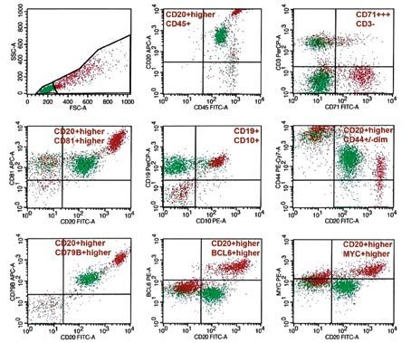
Anna Szumera-Ciećkiewicz, Grzegorz Rymkiewicz, Beata Grygalewicz, et al.

Table I. Aggressive, mature B-cell lymphomas according                 improvement of diagnostic criteria. The aim of this
the World Health Organisation’s 2017 classification                    study is to present practical diagnostic recommenda-
                                                                       tions necessary for the correct diagnosis of aggressive
 Aggressive, mature B-cell lymphomas
                                                                       B-cell lymphomas. The novelty concerning the three
 Diffuse large B-cell lymphoma, not otherwise specified                aggressive B-cell lymphomas has been discussed in
    Germinal centre B-cell type                                        detail: DLBCL, NOS, high-grade B-cell lymphomas
    Activated B-cell type                                              (HGBLs), and Burkitt lymphoma/Burkitt-like lym-
 T-cell/histiocyte-rich large B-cell lymphoma                          phoma with 11q aberration (BL/BLL,11q); for the
                                                                       remaining units, the most important features regard-
  iffuse large B-cell lymphoma, topographic site related
 D
                                                                       ing clinical and pathological presentation as well as
    Primary mediastinal (thymic) large B-cell lymphoma
   Primary diffuse large B-cell lymphoma of the central               genetic changes are highlighted [1, 2]. Moreover,
    nervous system                                                     general rules for preservation of biological material
   Primary cutaneous diffuse large B-cell lymphoma, leg               and tips on supporting diagnostic techniques (immu-
    type                                                               nohistochemistry, flow cytometry, genetics) are given.
    Intravascular large B-cell lymphoma
 Diffuse large B-cell lymphoma, EBV related                            Preservation of biological material
    EBV-positive diffuse large B-cell lymphoma, NOS
   Diffuse large B-cell lymphoma associated with                         Various materials are evaluated in the diagnosis
    chronic inflammation                                               of lymphoid tumours. Samples most often originate
    Lymphomatoid granulomatosis                                        from the main structures of the lymphatic system,
 Large B-cell lymphoma with terminal B-cell                            which include lymph nodes, spleen and thymus,
 differentiation                                                       Waldeyer’s ring (lymphatic tissue band surrounding
    Plasmablastic lymphoma                                             the oral throat with palatal tonsils, and lingual and
    ALK-positive large B-cell lymphoma                                 pharyngeal tonsils), appendix, and Peyer’s patches
    Primary effusion lymphoma                                          in the ileum. Other areas containing lymphatic tis-
    HHV8-positive diffuse large B-cell lymphoma, NOS#                  sue concern: bone marrow, mediastinum, liver, skin,
 Burkitt lymphoma                                                      pleura and gonads. Patients admitted with suspicion
    Burkitt-like lymphoma with 11q aberrations#                        of lymphoma for pathological diagnosis require bio-
 High grade B-cell lymphoma                                            logical material protection also for molecular, and/
   High-grade B-cell lymphoma, with MYC and BCL2                      or cytogenetic tests. Therefore, it is necessary to
    and/or BCL6 rearrangements                                         provide a standard operating procedure for material
    High-grade B-cell lymphoma, NOS                                    processing, including the following major steps:
 Mantle cell lymphoma                                                  ––tissue obtained for testing should be fresh and pro-
    Blastoid or pleomorphic variant                                      cessed according to type of material; entirely ex-
                                                                         cised lymph nodes should be cut into at least 5-mm
 B-cell lymphoma, unclassifiable, with features
 intermediated between diffuse large B-cell lymphoma                     thick slices along the long axis to ensure optimal
 and classic Hodgkin’s lymphoma                                          penetration of a fixative;
 # provisional entities
                                                                       ––description of fresh material should include the
                                                                         size, colour, texture, and the presence or absence
                                                                         of macroscopically visible haemorrhages, necrosis
                                                                         fields, or nodular architecture;
genetic features [1]. Some types of aggressive B-cell                  ––touch imprint cytological slides are made from
lymphomas are relatively frequent, i.e. diffuse large                    freshly cut material surface and are fixed in alcohol
B-cell lymphoma, not otherwise specified (DLBCL,                         or dried in the air;
NOS) which represents 25-35% non-Hodgkin’s lym-                        ––for cytogenetic tests (karyotype) a fresh tissue sam-
phomas depending on the population; others occur                         ple or viable cells from fine-needle aspiration biopsy
rarely and refer to specific groups of patients. Prog-                   (FNAB) should be collected into a sterile container
nosis and response to therapy are different in each                      with cell culture medium and antibiotics, always
clinical-pathological unit. Although currently used                      considering the requirements of the laboratory per-
treatment regimens are effective in the majority of                      forming the test;
patients, about 30% of patients develop progression                    ––immunophenotyping by flow cytometry (FCM)
to incurable disease [1, 2]. The results of genetic and                  requires protection of a fresh tissue sample or vi-
molecular research conducted in recent years allow                       able cells from FNAB in an appropriate transport
a better understanding of the mechanisms underly-                        medium (e.g. RPMI-1640), always considering the
ing the clinical and biological diversity of these tu-                   requirements of the laboratory performing the test;
mours and provide valuable information about new                       ––always indicate the type of fixative used and if pos-
therapeutic options, and at the same time allow                          sible determine the estimated time from tissue col-

2

    Pobrano z https://publicum.umed.lodz.pl / Downloaded from Repository of Medical University of Lodz 2021-09-26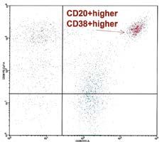
Histopathological diagnostics of aggressive B-cell lymphomas

  lection to fixation, because this is important for the     with architecture and tumour cytology. Moreover,
  recovery of RNA and phosphorylated proteins;               not all antibodies are available for immunohisto-
––for histopathological examination it is recommend-         chemical assessment, especially for fixed tissues,
  ed to use a fixative based on 10% buffered formal-         but the advantage of this method is the possibility
  dehyde (so-called formalin); most suitable for addi-       of using it in the archival materials embedded in
  tional studies such as immunohistochemistry and            paraffin. Both techniques can be used in the lym-
  fluorescence in situ hybridisation (FISH); to avoid        phomas diagnostics and are a source of clinically
  dilution and formalin buffering problems the use           relevant information (e.g. identification of mol-
  of commercially available ready-to-use (RTU) solu-         ecules necessary for the use of targeted therapies
  tions is strongly advised;                                 such as CD20);
––to ensure optimal antibody reactivity preservation       ––the importance of molecular research in lymphoid
  (for immunohistochemistry) extension of fixation           malignancies is constantly growing and allows the
  time (over 24 hours for small tissue sections fixated      determination of clonality and the origin of neo-
  in formalin) should be avoided;                            plastic cells. In specific entities, these tests are nec-
––if the size/volume of biological material and infra-       essary to make a definitive diagnosis i.e., Burkitt
  structure of the diagnostic centre allow, some sam-        lymphoma, Burkitt-like lymphoma, or high-grade
  ples should be biobanked through deep freezing             B-cell lymphoma with MYC and BCL2 and/or
  (short-term storage in –80°C freezers, long-term           BCL6 rearrangements;
  storage in liquid nitrogen at –170°C). The freez-        ––for an interpretation of immunohistochemical reac-
  ing technique should be complementary to further           tions a descriptive semi-quantitative scale is adopt-
  material processing methodology; the most popu-            ed: (+) positive reaction, (+/–) partially positive
  lar techniques are tumour snap frozen sections or          reaction, (–/+) positive reaction in a few, (–) nega-
  tumour viable cell freezing.                               tive reaction in the majority of cells; the percentage
                                                             of positive cells for each staining may be presented
General remarks on pathological                              in square brackets;
diagnostics of aggressive B-cell lymphoma:                 ––panB panel includes antibodies: CD19, CD20,
                                                             CD22, CD79a, PAX5;
––histopathological examination sustains a gold stan-      ––typical DLBCL immunophenotype includes:
  dard in diagnostics; however, in most cases precise        CD45(+), CD20(+), CD3(–) Ki-67 > 40% posi-
  determination of lymphoma type requires at least           tive cell nuclei;
  one additional method including immunopheno-             ––panel of IHC antibodies to establish DLBCL diag-
  typing, and molecular and/or cytogenetic tests [4,         nosis and GCB vs. non-GCB origin: CD20, CD3,
  5, 6, 7, 8, 9, 10]; in cases where material is inad-       CD5, CD10, BCL2, BCL6, Ki-67, IRF4/MUM1,
  equate or insufficient for diagnostic purposes, the        MYC with or without cell surface analysis by flow
  clinician should receive feedback and justify the          cytometry: k/l, CD45, CD3, CD5, CD19, CD10,
  above condition;                                           CD20, CD71;
––in certain circumstances, when a lymph node is not       ––additional IHC studies to establish lymphoma sub-
  easily accessible for surgical biopsy and in patients      type: Cyclin D1, PAX5, CD30, CD15, CD138,
  requiring immediate treatment, FNAB biopsy in              CD38, Epstein Barr virus (EBV) in situ hybridi-
  conjunction with FCM, karyotype, FISH for major            sation (ISH), EBV/LMP1, ALK, HHV8, SOX11,
  translocation, PCR for IGH, and TCR gene rear-             CD23, BOB1, OCT2, CD56, k/l, EMA;
  rangement may be sufficient for diagnosis;               ––the following cutoff points are recommended accord-
––haematopatholgy consultation should include re-            ing to WHO classification: BCL2 ≥ 50% strong pos-
  view of all slides with at least one paraffin block        itive cells, MYC ≥ 40% strong positive cell nuclei,
  representative of the tumour. Re-biopsy is recom-          for CD10, BCL6, IRF4/MUM1 ≥ 30% positive cells/
  mended if material is non-diagnostic;                      nuclei. The importance of choosing an appropriate
––immunophenotyping can be performed by im-                  antibody clone, especially among those that show re-
  munohistochemistry or flow cytometry [8]; each             action liability (i.e. BCL2, CD10, MYC), is highlight-
  method has its own advantages and disadvantag-             ed. For routine pathological diagnostics only certified
  es. Flow cytometry is fast (hours) and quantita-           antibodies (in vitro diagnostics, IVD) simultaneously
  tive method with evaluation of multiple antigens           with positive and negative controls are advised;
  simultaneously. Antigen detection, however, does         ––presence of EBV virus should be confirmed by in-
  not allow to correlate with tumour architecture and        tra-tissue hybridisation (EBER-ISH); it is a supe-
  its cytological features. Immunohistochemistry re-         rior technique to immunohistochemistry (LMP1)
  quires hours, sometimes days, and the quantitative         (Fig. 1);
  assessment is subjective, but its most important         ––karyotype and/or FISH for MYC, BCL2, BCL6 re-
  feature is the possibility of correlation of reaction      arrangements are recommended especially in cases

                                                                                                                             3

      Pobrano z https://publicum.umed.lodz.pl / Downloaded from Repository of Medical University of Lodz 2021-09-26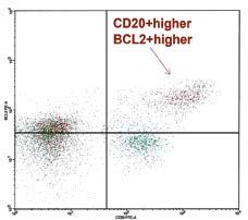
Anna Szumera-Ciećkiewicz, Grzegorz Rymkiewicz, Beata Grygalewicz, et al.

A                                                                          B

Fig. 1. EBV detection: A) immunohistochemical staining with LMP1 and B) EBER-ISH method (magnification 400×)

  with double expression of MYC and BCL2 and/or                        NF-kB pathway in the course of various mecha-
  having a GCB phenotype and high-grade morphol-                       nisms. Unique genetic changes, especially mutations
  ogy;                                                                 such as GNA13 and EZH2 in GCB and MYD88,
––despite advanced diagnostic methods, still there                     CARD11, and CD79B in ABC are found, respective-
  is a group of cases whose image does not meet all                    ly [6, 7]. Chromosomal translocations in DLBCL in-
  criteria and “escapes” from diagnostic guidelines;                   volving regions with BCL6 (3q27), BCL2 [t(14;18)
  the pathologist, taking into account the clinical                    (q32;q21.3)], and MYC (single hit) comprise, re-
  presentation and course, should maximally narrow                     spectively, approximately 30% (with predominance
  the differential diagnosis. It is worth remembering                  in ABC subtype), 20-30% (more commonly in GCB
  that the most powerful predictive factor determin-                   subtype), and 8-14% (similar distribution among
  ing the therapeutic approach remains the patho-                      ABC and GCB subtype) [8, 9, 10, 11, 12, 13, 14,
  logically confirmed type of lymphoma;                                15, 16, 17, 18]. It is estimated that approximate-
––diagnosis of lymphomas requires close cooperation                    ly 50% of DLBCL with MYC translocation demon-
  between haematopathologists and specialists in                       strate BCL2 and/or BCL6 rearrangement and that
  flow cytometry and genetics as well as clinicians                    cases should be transferred to the high-grade B-cell
  (oncologists, haematologists).                                       lymphoma category [1, 19, 20]. DLBCL with typ-
                                                                       ical morphology and isolated MYC translocation
Diffuse large B-cell lymphoma,                                         usually present higher mitotic index, but still such
not otherwise specified                                                cases meet the criteria of diagnosis of DLBCL, NOS
                                                                       [8, 21]. The pathologists are also discouraged from
   Diffuse large B-cell lymphoma, not otherwise                        preselecting aggressive B-cell lymphomas to FISH
specified (DLBCL, NOS) is morphologically, clini-                      testing upon Ki-67 result, while its range can be
cally and biologically heterogeneous. In developing                    variable [21]. Biological differences translate into
countries and in selected Eastern European countries,                  a distinct clinical course; so far in the majority of
such as Poland it represents 25-35% of non-Hod-                        studies, a worse prognosis was observed among pa-
gkin’s lymphomas and is a constant leader among                        tients with DLBCL, ABC in comparison with GCB
all aggressive mature B-cell lymphomas [3, 4]. The                     subtype. Moreover, the latest results of clinical tri-
identification of two molecular subtypes of this lym-                  als concerning combining R-CHOP regimen with
phoma based on gene expression profiling (GEP)                         bortezomib, lenalidomide, and ibrutinib in the ABC
was one of the most important achievements in un-                      subtype significantly stress the potential benefits of
derstanding its diversity. Subtyping DLBCL, NOS                        such treatment [22, 23]. The optimal method for
is based on different cell origins and includes lym-                   the molecular classification of DLBCL is the study
phomas derived from germinal centre cells (GCB) or                     of the gene expression profile, but it requires fresh
from activated B-cells (ABC) [5]. Additionally, these                  tissue and is not widely used in routine diagnostics.
DLBCL subgroups vary in the activated molecular                        Due to its clinical impact, pathologists are still ob-
pathways, chromosomal changes, and the occurrence                      ligated to precisely identify the molecular subtype.
of somatic mutations. In GCB type, the activation                      An up-to-date Hans algorithm based on a panel of
of the PI3K/AKT signalling pathway and over-ex-                        antibodies (CD10, BCL6, and IRF4/MUM1) deter-
pression of BCL6 protein are observed, whereas in                      mined by immunohistochemistry is available in most
ABC type there is a constitutive activation of the                     pathology departments and should be continuous-

4

    Pobrano z https://publicum.umed.lodz.pl / Downloaded from Repository of Medical University of Lodz 2021-09-26
Histopathological diagnostics of aggressive B-cell lymphomas
ly applied [24, 25, 26, 27]. The compliance of IHC in the pathological report referring to HGBL, R
with GEP reaches about 80-90%, but a small group and HGBL, NOS because the DLBCL morphology
of about 10% of DLBCL cases “slip out” of molec- may predict a better outcome compared to DLB-
ular classification and the ABC/GCB match is not CL/BL [35]. Neither morphology nor proliferation
possible [28]. One of the limitations of IHC is also index assessed by Ki-67 have sufficient sensitivi-
standardisation between haematopatholgy centres, ty and specificity to identify DH/THLs. MYC and
including quality of staining and evaluation repro- BCL2 protein expression, although independently
ducibility among pathologists [28, 29]. Recently valuable prognostic factors, are similarly not ideal
developed GEP tests based on RNA extracted from predictors of DH/THLs, and hence FISH (whenev-
formalin-fixed and paraffin-embedded tissues (e.g. er the karyotype assessment is not available) is the
the Lymph2Cx assay from NanoString Technologies, recommended modality to identify DH/THLs (Fig.
Seattle, WA, USA) can identify DLBCL molecular 3) [1, 2]. Despite the fact that a clear consensus has
subtypes [30]. Moreover, those methods are charac- not yet been reached to provide molecular testing
terised by high compliance level with GEP microar- guidelines, the 2017 WHO update strongly advises
rays and satisfactory reproducibility between labora- that all DLBCL cases should undergo genetic stud-
tories [30, 31, 32]. Possibly, tests based on GEP will ies for the detection of MYC, BCL2, and BCL6 re-
be a more precise diagnostic method than currently arrangements. Nevertheless, some pathologists and
approved by the WHO IHC methods in future [1, clinicians suggest that only cases with a GCB phe-
20]. The summary of DLBCL, NOS characteristic is notype and/or high-grade morphology or with >
presented in Table II. 40% MYC immunohistochemically positive cells
are worthy of deeper molecular testing [1] (Fig. 2).
High-grade B-cell lymphomas Nearly 100% of DHLs with MYC and BCL2 re-
arrangements harbour the t(14;18) translocation,
High-grade B-cell lymphoma (HGBL), with gene which results in BCL2 overexpression by IHC and
expression profile intermediate between molecular flow cytometry. BCL2 protein is expressed (in 30%
signatures of Burkitt lymphoma (BL) and non-BL of DHLs with MYC and BCL6 rearrangements) or
(mostly DLBCL), is a heterogeneous group of aggres- overexpressed (occurrence of BCL2 extra copy/am-
sive, mature B-cell lymphomas, which should not plification) in a higher proportion of DLBCL and
be classified separately due to biological and clini- HGBL, NOS. That phenomenon is often associated
cal reasons [1, 2]. High-grade B-cell lymphoma is with a concomitant expression of MYC, but still IHC
a newly introduced category in the updated 2017 evaluation cannot be used as a surrogate marker for
WHO classification, which primarily replaces B-cell DHL. Most DLBCL and HGBL do not carry both
lymphoma, unclassifiable, with features intermediate rearrangements (MYC and BCL2) and are referred to
between diffuse large B-cell lymphoma and Burkitt as “double-expressor lymphomas” (DELs) [1, 2, 33,
lymphoma (BCLU, DLBCL/BL) and at the moment 36]. DH/THL and DEL patients usually progress
comprises two entities: HGBL with MYC and BCL2 more rapidly, are resistant to R-CHOP chemo-im-
and/or BCL6 chromosomal rearrangements (HGBL, munotherapy [33, 35], and have very poor prognosis.
R) and high grade B-cell lymphomas, not otherwise Moreover, such cases may harbour TP53 mutations
specified (HGBL, NOS). HGBL, R is also called dou- or deletion, frequently observed in MYC/BCL2 DHLs
ble/triple-hit lymphoma (DH/THL). Most patients and blastoid morphology [37, 38, 39]. Worse prog-
develop de novo HGBL, R, while a minority have nosis in HGBL, R and HGBL, NOS compared with
a history of FL that progress to DH/THL secondari- the DLBCL groups were published lately [38, 40].
ly, presumably by acquisition of a MYC translocation DH/THLs show a common GCB immunophenotype
[1]. Double/triple-hit lymphomas are characterised (CD10+/CD81+higher/ BCL6+/CD44– or CD44+/–
by various morphologies including DLBCL, BL and dim
) by FCM, often presenting decreased expression of
intermediate features between DLBCL and BL (DL- CD20 or CD19, over expression of CD38 and BCL2
BCL/BL). HGBL, which lack co-occurring MYC and (Fig. 4) [41]. An aggressive biologic behaviour and
BCL2 and/or BCL6 rearrangements, falling in the poor clinical outcome of DH/THL may shortly influ-
category HGBL, NOS; such cases appear as blastoid ence to therapy selection including still controversial
or DLBCL/BL morphology (eventually resembling more intensive regimens initiation and CNS-directed
more closely BL than DLBCL), and in up to half of prophylaxis consideration. Under consideration is cy-
DLBCL/BL cases MYC rearrangements as one sep- togenetic testing by metaphase or cytogenetic anal-
arate hit are found [1, 2, 33, 34] (Fig. 2). Former ysis by karyotyping if biological material (fresh cells
criteria for BCLU, DLBCL/BL were vague, and the from FNAB or tissue surgical section) is available. As
diagnosis was not used uniformly, limiting its utility far as possible, a comprehensive histopathological di-
as a diagnostic category [1, 2]. The morphological agnostics of HGBL should include IHC, FISH, FCM
appearance should always be specified in a comment and cytogenetic studies (Table III) [42, 43, 44].
5
Pobrano z https://publicum.umed.lodz.pl / Downloaded from Repository of Medical University of Lodz 2021-09-266
Table II. Characteristics of diffuse large B-cell lymphoma, not otherwise specified
Diffuse large B-cell lymphoma, not otherwise specified
Presentation
Clinical 25-35% adult non-Hodgkin lymphomas;
Elderly patients;
Median age the 7th decade [rare cases in children and young adults];
M > F;
Most cases primary DLBCL, NOS [de novo];
Secondary DLBCL, NOS - transformation from less aggressive lymphoma [CLL/SLL, FL, MZL, NLPHL];
Risk factors: immunodeficiency, EBV-infection [3% in western to 10% in Asian / Latin American populations; cases with EBV positivity in most
lymphoma cells should be diagnosed as EBV-positive DLBCL, NOS/another specific type of EBV-positive lymphoma];
Rapidly enlarging tumour mass at single/multiple nodal/extranodal sites;
~50% of patients are in stage I or II disease – inclusion PET/CT raised up the initial stage
Pathological Morphology
Three most common variants:
Centroblastic [medium-sized to large, oval to round vesicular nuclei with fine chromatin, 2-4 nucleoli close to nuclear membrane, cytoplasm scant and
amphophilic/basophilic];
Immunoblastic [a single centrally located nucleolus, more abundant basophilic cytoplasm; immunoblasts with plasmacytoid differentiation may be
observed];
Anna Szumera-Ciećkiewicz, Grzegorz Rymkiewicz, Beata Grygalewicz, et al.
Anaplastic [large to very large cells with bizarre nuclei, some cells may resemble: HRS cells/anaplastic large cell lymphoma/undifferentiated carcinoma;
may show a sinusoidal and/or cohesive growth pattern];
Rare morphological variants: myxoid stroma, fibrillary matrix, pseudorosette formation, spindle-shaped/signet ring lymphoma cells
Architecture
Diffuse or partial [interfollicular and/or less commonly sinusoidal] nodal involvement;
Infiltration of perinodal tissue [often];
Sclerosis [broad/fine bands];
Background may contain high number of T cells and/or histiocytes
IHC PanB(+), surface and cytoplasmic immunoglobulins [50-75%, IgM(+)>IgG(+)>IgA(+], CD5(–/+) [5-10%, positive cases usually DLBCL de novo
not CLL/SLL transformation], CD30(–/+) [10-20% anaplastic variant], cyclin D1/SOX11(–) [rare cases with cyclin D1/SOX11 weakly positive without
CCND1 translocation], CD10(+/–) [30-50%], BCL6(+/–) [60-90%], IRF4/MUM1(+/-) [35-65%], BCL6 and IRF4/MUM1 co-expression in 50% of
cases, FOXP1(–/+) [20%; more often in ABC subtype with IRF4/MUM1(+) and BCL2(+) without t(14;18)(q32;q21.3)], GCET1(+/–) [40-50%, in
GCB subtype], LMO2(+/–) [45%, more often in GCB subtype with CD10(+), BCL6(+), HGAL(+) but IRF4/MUM1(–) or BCL2(–)], BCL2(+/–) [47-
84%], p53(+/–) [20-60%, expression more frequent than mutation], Ki-67 high [≥ 40% up to > 90% in some cases]
Hans algorithm
(+) GCB
(+) ABC
CD 10 (+) MUM1
(–) BLC6 (–) GCB
(–) ABC
Pobrano z https://publicum.umed.lodz.pl / Downloaded from Repository of Medical University of Lodz 2021-09-26Histopathological diagnostics of aggressive B-cell lymphomas
Burkitt Lymphoma and Burkitt-like
lymphoma with 11q aberration
20-25%
EZH2
↓
Burkitt lymphoma (BL) is defined by the WHO
classification as a highly aggressive lymphoid neo-
plasm, often presenting with extra nodal site in-
MYC, single hit
volvement or as an acute leukaemia composed of
GNA13
5-8%
5-8%
25%
monomorphic, medium-sized B-cells with basophil-
↓
ic cytoplasm and a high mitotic index. Translocation
involving the MYC oncogene (8q24) and immuno-
globulin IG genes is the constant features in 90% of
cases [1]. Case analysis without MYC rearrangement
have shown at least several mechanisms for the alter-
native activation of MYC i.e. microRNA, amplifica-
CREBBP
tion, transcriptional increase of MYC activity. Results
10%
30%
of next-generation sequencing revealed the BL pro-
file of somatic mutations; in about 70% of classic BL
mutations of transcription factor TCF3 (E2A) and
its negative regulator ID3 were found and its role in
PI3K pathway signalling was observed. Furthermore
Rearrangements
NFκB activation
CCND3, RHOA, TP53, ARID1A and SMARCA4
PI3K/AKT
Mutations
Pathways
KMT2D
25-30%
mutations were identified in 30% of patients with BL.
BCL6
15%
35%
40%
Recently, a subset of MYC translocation-negative
aggressive B-cell lymphomas resembling BL, char-
acterized by proximal gains and distal losses of the
long arm of chromosome 11 was described [45, 46].
In the 2017 WHO classification, these MYC-nega-
tive lymphomas were recognized as a new provision-
CARD11
10-15%
10-15%
al entity, “Burkitt-like lymphoma with 11q aberra-
tion” (BLL,11q) [1] (Fig. 5). MYC-negative BLL,11q
shows a number of clinico-pathological similarities to
MYC-positive BL, but also harbour some significant
differences in immunoprofile [1, 45, 46, 47, 48, 49,
50]. BLL,11q usually express CD43/LMO2/CD56
in IHC and CD16/CD56/CD38/CD45/CD8/CD43
in FCM. That characteristics may contribute to the
20-25%
CD79B
< 5%
BCL2
40%
differential diagnosis of BLL, 11q and BL [49]. The
↓
11q aberrations in BLL, 11q (11q-gain/loss) were
described as an inverted duplication of a part of the
long arm of chromosome 11 with mono- or biallel-
MYD88
ic telomeric deletion of 11q (Figure 6) [45, 46, 47,
35%
49]. Coincidence of duplication and deletion of 11q
↓
(11q23 and 11q24-qter, respectively) suggests a pos-
sibility of simultaneous up-regulation of oncogenes
and down-regulation of tumour suppressor genes.
The candidate oncogene is commonly up-regulated
GCB subtype
GCB subtype
GCB subtype
ABC subtype
ABC subtype
ABC subtype
PAFAH1B2. FLI1 and ETS1, located in the region of
deletion, are often down-regulated and/or mutated,
and are postulated to be candidate tumour suppres-
sor genes affected by this aberration [46, 47]. The di-
agnostic algorithm for diagnosing BL and BLL, 11q,
besides standard histopathological and immunohis-
Table II. Cont.
tochemical examination, incorporates flow cytometry
*↓ Rare/Uncommon
with a broad panel of monoclonal antibodies as well
Genetic
as genetic analysis (conventional cytogenetic analysis
with fluorescence in situ hybridisation and molecular
techniques) [1, 48]. Some cases with 11q aberration
7
Pobrano z https://publicum.umed.lodz.pl / Downloaded from Repository of Medical University of Lodz 2021-09-26Anna Szumera-Ciećkiewicz, Grzegorz Rymkiewicz, Beata Grygalewicz, et al.
A B C
D E F
G H I
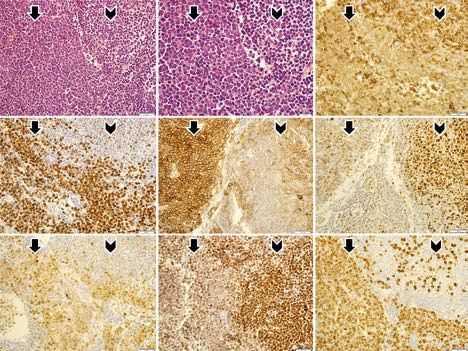
Fig. 2. Histopathology and immunohistochemistry of HGBL, R (A 400×; B, 600×; arrow); a case of a secondary
triple-hit lymphoma (transformation from follicular lymphoma – arrowhead; HGBL, R – arrow) with DLBCL/BL mor-
phology. Immunohistochemical staining on HGBL, R cells: C – BCL2(+) weaker, D – MYC(+), E – CD38(+) higher,
F – BCL6(+) weaker, G – MUM1(+), H – FOXP1(+) weaker, I – Ki-67 index (>90%) (for all IHC 400×)
also have MYC rearrangement and are diagnosed as younger patients (mainly in the third decade of life
BL or high-grade B-cell lymphoma, not otherwise and almost three times more often in men) [1, 20].
specified (HGBL, NOS). From the other point of DLBCL EBV(+), NOS is characterised by a different
view, the 11q-gain/loss are not exclusively specific for clinical picture and pathological features comparing
BLL, 11q [47]. Therefore, the conventional cytoge- with DLBCL, NOS. Mainly it occurs in patients with
netic analysis by metaphase karyotyping is needed to immunological disorders that impair the control of
provide correct BLL, 11q diagnosis (Table IV) [49]. viral infections. Histopathology reveals large atypical
B cells that may resemble Hodgkin’s and Reed-Ster-
Other aggressive B-cell lymphomas nberg cells, and variable inflammatory infiltrates in-
volving cytotoxic T lymphocytes [CD8(+)], plasmo-
Changes in the WHO 2017 classification and di- cytes, and histiocytes. In addition, tumour necrosis
agnostic criteria of other aggressive lymphomas from is present in most cases. Histopathological features
mature B cells are less significant. may suggest a diagnosis, but it should be confirmed
In the group of large B-cell lymphomas associated by testing for the presence of EBV virus by in situ
with infection with EBV the most common lympho- hybridisation, which is a superior method to IHC
ma is EBV-positive diffuse large B-cell lymphoma, staining. EBV is found in most lymphoma cells and
not otherwise specified [DLBCL EBV(+), NOS]. is usually in the second and third phase of latency
In the previous WHO 2008 classification, this unit [52]. Pictures of EBER-ISH and EBV-LMP1(IHC)
was called EBV-positive DLBCL of the elderly, due are shown in Fig. 1.
to significantly more frequent occurrence in patients In the group of large B-cell lymphomas classified on
over the age of 50 years with a peak of incidence in the basis of topography, the changes concern mainly
the eighth decade of life [51]. Recent studies have primary mediastinal (thymic) large B-cell lymphoma
shown that these lymphomas may also occur in (PMBL) and primary DLBCL of central nervous sys-
8
Pobrano z https://publicum.umed.lodz.pl / Downloaded from Repository of Medical University of Lodz 2021-09-26Histopathological diagnostics of aggressive B-cell lymphomas
A B
C D
Fig. 3. A) Conventional cytogenetic diagnosis of double-hit lymphoma (DHL). Complex karyotype presenting DHL with
diffuse large B-cell lymphoma morphology: 52,XX,+5,+7,t(8;14)(q24;q32),+der(12),t(6;12)(p11.2;p11.2) x2,t(14;18)
(q32;q21),+21,+mar, arrows indicate MYC, BCL2 and both IGH copies rearrangements. B) FISH with MYC break apart
probe, split signal shows rearrangement of one copy of MYC gene in interphase nucleus. C) FISH with BCL2 break apart
probe, split signal indicates rearrangement of one copy of BCL2 gene in interphase nucleus. D) FISH with IGH break
apart probe, two split signals display rearrangement of two copies of IGH gene in interphase nucleus
tem (DLBCL, CNS). PMBL cells frequently express logical features related to the immunologically priv-
PDL1/PDL2 markers (rearrangement in about 20% ileged location in which it develops and the lack of
of cases), CD30 and CD23, and usually are negative expression of HLA class I and II proteins, which allow
for IG and HLA class I and II antigens [53]. Primary tumour cells to avoid immune control. It should be
mediastinal lymphoma has a specific gene expression differentiated with other large cell lymphomas occur-
profile that can be useful in differential diagnosis of ring in the CNS, particularly with those associated
PMBL and DLBCL, NOS involving mediastinum or with immunosuppression [56]; they are characterised
other outside thorax locations. Differences between with deletions and loss of gene expression within the
PMBL and DLBCL at the molecular level relate to HLA system and MYD88 L265P [> 50%], CD79B
CIITA rearrangements (38% vs. rarely respective- [20%], and CARD11 [16%] mutations are often ob-
ly), which leads to the activation of NFκB and JAK/ served, which may have potential therapeutic signif-
STAT signalling pathways and the reduction of the icance [57, 58].
antigen expression of the major histocompatibility B-cell lymphomas with terminal B-cell differ-
complex of class II [5, 54, 55]. entiation include a heterogeneous group of aggres-
The primary DLBCL, CNS constitutes less than sive lymphomas characterised by immunoblastic or
1% of non-Hodgkin’s lymphomas and approximate- plasmablastic cell morphology, a plasma cell pheno-
ly 2.4-3% of all brain tumours. It has separate bio- type with no or reduced expression of B-cell markers
9
Pobrano z https://publicum.umed.lodz.pl / Downloaded from Repository of Medical University of Lodz 2021-09-26Anna Szumera-Ciećkiewicz, Grzegorz Rymkiewicz, Beata Grygalewicz, et al.
A
B
Fig. 4. Flow cytometry [FCM] of high grade B-cell lymphoma, triple hit [HGBL, TH]. A) FNAB/FCM analysis case with
MYC/BCL2/BCL6 rearrangements. Forward scatter/side scatter picture showing the larger HGBL lymphoma cells (red
cells) coexisting with of small normal T/B lymphocytes (green cells). HGBL express: CD45/ CD20+higher/ CD19+(C-
D20>CD19) / CD71+++/ CD81+higher / CD10+/ CD44+/−dim/ CD79β+higher / BCL6+ higher / MYC+ higher
/ BCL2+ higher, and CD38+ higher [“higher” or “dim” means higher or more heterogeneous (from very weak to high)
expression compared to expression on normal (green cells) B lymphocytes, respectively]. B) Higher level of BCL2 and
CD38 expression (marked by an arrow) than in normal T/B lymphocytes correlates with BCL2 and MYC rearrangements,
respectively. Dot-plots
10
Pobrano z https://publicum.umed.lodz.pl / Downloaded from Repository of Medical University of Lodz 2021-09-26Table III. Characteristics of high-grade B-cell lymphomas
High grade B-cell lymphomas
Presentation HGBL, R HGBL, NOS
Clinical Elderly patients, the 6th - 7th decade; Elderly patients
M:F (with slight male predominance); Still too few epidemiological data
Advanced disease (in 70-100% patients Ann Arbor IV stage)
More than one extranodal localisation including bone marrow and CNS
High IPI
Elevated LDH
HGBL DH more often in patients with diagnosed DLBCL who do not respond to
R-CHOP regimen or who have early relapses after complete remission
Pathological Morphology: Morphology:
~50% DLBCL, NOS (5-10% of all primary DLBCLs are double-hit lymphomas DLBCL/BL (most cases, mimics more closely BL);
[DHL], while 30–40% are MYC/BCL2 protein co-expressing lymphomas); Blastoid;
~50% DLBCL/BL; Usually not “classical” DLBCL;
Rare cases with blastoid appearance (closely mimic true lymphoblasts). Architecture:
Architecture: Completely diffuse;
Completely diffuse; No follicular component, stromal reaction nor fibrosis
Sometimes with follicular component/fibrosis
IHC GCB in 100% of BCL2/MYC DHL and in 50%-80% of BCL6/MYC DHL; GCB (70%) or ABC (30%) (simultaneously with BCL2R and BCL6R
or BCL6R or extra copies of BCL2 and BCL6);
CD19(+), CD20(+), CD79a(+), PAX5(+), TdT(–), sIg(–/+), CD10(+/–) CD20(+) [100%], BCL6(+/–), CD10(–/+)*[variable], IRF4/
i BCL6(+/–) [75-90%], IFR4/MUM1(–/+) [20%], BCL2(+) [strong cytoplasmic MUM1(–), Ki-67 [variable], MYC [variable, depending on MYCR or
in contrast to absent/weaker in BL], Ki-67 variable [BL morphology: high 80-95%; extra copies of MYC]
morphology DLBCL may be low: even below 30%]
FCM/IHC CD20(+) [often weaker], CD19(+) [often weaker], CD79a(+), CD79β(+), CD20(+),CD19(+),CD22(+),CD79a(+),CD79β(+),
CD10(+) [75-90%], BCL6(+) [75-90%], MYC(+) [but MYC negative cases CD10(+/–)*,CD43(+/–), CD81(+)higher or CD81(+)*, CD44(–) or
exist], CD38(+)higher, BCL2(+)higher [if BCL2R], or BCL2 (+)weak or (–) [if BCL6R], (+/–)dim or (+)*, CD5(–), CD71(+++) [100%]
CD44(–) or CD44(+/–)dim, MUM1(–/+) [20% if BCL6R], CD5(–), CD43(+/–),
IgM(+), or IgM/IGD(+), TdT(–) [if blastoid morphology], CD71(+++) [100%]
Genetic By definition: By definition:
MYC (8q24) and BCL2 (18q21) and/or BCL6 (3q27) rearrangements; MYC (8q24) and BCL2 (18q21) and/or BCL6 (3q27) rearrangements
should be excluded;
Complex karyotype [cases with DLBCL/BL morphology can have a low complex 20-35% MYC, R with or without increased copy numbers or, rarely,
or simple karyotype closer to BL; D/THLs show a significantly higher genomic amplification of 18q21 [BCL2];
complexity than BL]; Still too few coherent data
60-65% IG/MYC (juxtaposition of MYC to one of the immunoglobulin loci: IGH at
14q32, IGK at 2p12, or IGL at 22q11);
35%-40% non–IG/MYC [partners such as at 9p13 (gene unknown), 3q27 (BCL6) or
other loci];
Combination of a chromosomal rearrangement of one gene and copy-number increase
11
Histopathological diagnostics of aggressive B-cell lymphomas
or amplification of other genes [e.g. MYC, R(8q24) with gain or amplification
BCL2(18q21)] are not sufficient to classify a case as DHL
Pobrano z https://publicum.umed.lodz.pl / Downloaded from Repository of Medical University of Lodz 2021-09-26
*depending on GCB vs. ABC subtypeAnna Szumera-Ciećkiewicz, Grzegorz Rymkiewicz, Beata Grygalewicz, et al.
(CD20 and PAX5), and strong expression of plasma
cell antigens (CD38, CD138, IRF4/MUM1, and
PRDM1/BLIMP1) [1]. Highly aggressive plasmab-
lastic lymphoma (PBL) develops in immunocompro-
mised patients, mainly caused by HIV infection, as
well as during iatrogenic immunosuppression (after
transplantations, autoimmune diseases). It is usual-
ly located extranodally within the head and neck,
including the oral cavity, also in the gastrointesti-
nal tract. In majority of cases the generalised disease
stage is determined at the time of diagnosis (over
75% of patients with HIV infection) [59, 60, 61].
In some cases, PBL involves bones and with its mor-
phological and immunophenotype features overlaps
with plasmablastic plasma cell myeloma. The differ-
Fig. 5. Histopathological features of Burkitt-like lym- ential diagnosis should take into account the clini-
phoma with 11q aberration (BLL,11q). Diffuse growth is
cal picture of PBL and immunodeficiency and EBV
composed of medium-sized lymphoid cells showing jigsaw
puzzle effect of cytoplasmic borders with a starry pattern
infection. The EBV virus in the first type of latency
due to admixed macrophages (but in this case, you see a re- is present in about 70% of cases. MYC translocation
duced phagocytosis and apoptotic bodies). The nuclei are occurs in about 50% of patients, usually with the IG
similar in size and shape (HE, magnification 600×) partner [62].
A B
C D
Fig. 6. Conventional cytogenetic diagnosis of Burkitt-like lymphoma with 11q aberration (BLL,11q). A) A low complexi-
ty karyotype of BLL,11q: 45,X,-Y,del(6)(q21), dup(11)(q23q21), arrow indicates 11q gain/loss. B) FISH with centromere
11 (Aqua) and KMT2A (break apart) probes, four red-green signals show multiplication of this region on duplicated chro-
mosome 11 (arrow). C) FISH with centromere 11 (Aqua) and ATM (locus specific, red) probes, two red signals indicate
a duplication in 11q aberration region (arrow). D) FISH with centromere 11 (Aqua) and telomeric 11q (red) probes, one
red 11q telomeric signal indicates normal chromosome 11, lack of red signal in 11q aberration confirms terminal deletion
(arrow)
12
Pobrano z https://publicum.umed.lodz.pl / Downloaded from Repository of Medical University of Lodz 2021-09-26Table IV. Characteristics of Burkitt lymphoma and Burkitt-like lymphoma with 11q aberration
Burkitt lymphoma and Burkitt-like lymphoma with 11q aberration
Presentation BL BLL,11q
Clinical Three clinical variants are: endemic, sporadic, and immunodeficiency-associated BL; Still not very well described entity;
The incidence is low, accounting for only 1-2% of all lymphomas in Europe and Two clinical settings of BLL,11q are classical one and post-transplantation;
in the USA; The incidence is rare, probably less than 10% of all BL cases;
HIV infection positive in some cases; HIV/EBV infection negative;
EBV-EBER positive [15%-30%] in some cases; Young males;
Mainly children, young males but also rare cases in elderly patients are reported; Median age at time of diagnosis: 25 years,
Median age at time of diagnosis among adults: 30 years; M : F > 10 : 1;
M : F = 2-3 : 1; Nodal and Tonsilar>>Extranodal, often a bulky tumor;
Extranodal >>Nodal involvement, often a bulky tumor; No bone marrow nor cerebrospinal fluid involvement
Bone marrow and cerebrospinal fluid involvement more frequently in children
and HIV-positive patients
Pathological Medium-sized cells, with diffuse, monotonous growth type; Diverse morphology – mainly BL, frequently slightly differed from
Squared-off borders [cytoplasm retraction as a result of formalin fixation]; classical BL features by the reduced number of macrophages and apoptotic
Round nuclei with numerous basophilic, medium-sized nucleoli; bodies (loss of the “starry-sky” appearance), sometimes of DLBCL/BL, and
Basophilic cytoplasm with lipid vacuoles [visible in touch imprint slides or fine- sporadically DLBCL
needle aspiration cytology];
“Starry-sky” image, numerous macrophages and granulomatous background;
Less often nuclear pleomorphism or features of plasma differentiation [eccentric
basophilic cytoplasm, single central nucleus];
Rarely DLBCL/BL morphology
IHC Pan B (+), CD20(+), CD10(+), MYC(+), LMO2(–)^, BCL6(+),BCL2(–) Pan B (+),CD20(+), CD10(+), MYC(+), LMO2(+)^, BCL6(+),
[positive suggest HGBL], MUM1(–), CD43(+)^, CD44(–), IgM(+)[moderate BCL2(–), MUM1(–), CD43(+/–)#, CD44(–), IgM(+), CD56(+/–)#,
to strong membrane reaction, light chain restriction],CD56(–), EBV(+/– EBV(–),Ki-67 [high, ~100%]
)#,Ki-67 [high, ~100%]
FCM/IHC CD45(+)weaker/CD38(+)higher/CD16/CD56(–)/ CD8(–) /CD43(+) or* CD45(+)bright/CD38(+)/CD16/CD56(+) or* CD16/CD56(–) /CD8(+)
CD43(+/–) or* CD8(–)/ CD43(+/–) or* CD43(+) or* CD43(–)
Genetic The simple karyotype [40% of cases: MYC translocation as the sole abnormality]. The more- complex karyotype than BL and lack MYC rearrangement.
Molecular hallmark is MYC translocation 8q24 to one of three immunoglobulin Typical 11q aberrations: inverted duplication dup(11)(q23q13) with
loci: in 85% the IGH locus on 14q32, in 5%, the IGK on 2p11 and 10% the IGL mono- or biallelic telomeric loss of 11q as a recurrent 11q abnormality.
on 22q11; The most frequent additional changes comprised deletions of 6q, and
10% cases without MYC rearrangement alternative mechanisms of MYC trisomy 12
activation i.e. microRNA, amplification, transcriptional increase of MYC
activity.
Additional recurrent abnormalities: gains in chromosomes 1q, 7 and 12, and
losses of 6q, 13q32–34 and 17p
^usually negative or positive, #sometimes positive, *alternative expressions are ordered from more to less frequent
13
Histopathological diagnostics of aggressive B-cell lymphomas
Pobrano z https://publicum.umed.lodz.pl / Downloaded from Repository of Medical University of Lodz 2021-09-26Table V. Characteristics of other aggressive B cell lymphomas
14
T-cell/histiocyte-rich large B-cell lymphoma
Pathology Scattered, single, large B cells [do not form aggregates/sheets] in background of T-cells/ histiocytes [bland = looking non-epithelioid];
Large B cells may mimic: LP cells of NLPHL, centroblasts, HRS cells;
Absent meshwork of follicular dendritic cells;
No eosinophils / plasma cells;
Spectrum of B cell size, morphology, and distribution (clusters/sheets of medium-sized to large B cells) - consider DLBCL, NOS
IHC PanB(+), BCL6(+), BCL2(–/+) [variable], EMA(–/+) [variable], CD15(–), CD30(–), CD138(–); the background: histiocytes with CD68(+) and
CD163(+), T cells CD3(+) and CD5(+); no residual IgD-positive mantle cells; if EBV(+) cells, consider the spectrum EBV-positive DLBCL
Diffuse large B-cell lymphoma, topographic site related
Primary mediastinal (thymic) large B-cell lymphoma
Pathology Variable morphology, usually diffuse pattern with fibrosis;
Medium-sized to large cells, pale cytoplasm, round to ovoid nuclei;
Some cases: more pleomorphic – mimic HRS cells
IHC PanB+, slg(-/+) [commonly lacks of expression despite IgG functional rearrangement], PAX5(+), BOB1(+), OCT2(+), PU1(+), CD30(+) [>
80%, weak and heterogeneous], CD15(-/+) [rare cases], CD23(+) [70%], MAL(+) [70%], PDL1/PDL2(+) [70%], EBV(-), IFR4/MUM1(+) [75%],
BCL2(+) [55-80%], BCL6(+) [45-100%], CD10(-/+) [8-32%], CD11c(–/+) [40%], MYC(–/+) [30%, independent of MYC alterations], CD54(+),
FAS/CD95(+), TRAF1(+), REL(+) [nuclear expression], HLA I/II(–) [often]
Anna Szumera-Ciećkiewicz, Grzegorz Rymkiewicz, Beata Grygalewicz, et al.
Primary diffuse large B-cell lymphoma of the central nervous system
Pathology Highly cellular, medium-sized to large round, oval, irregular or pleomorphic nuclei and distinct nucleoli [centroblastic/immunoblastic morphology];
Rarely monomorphic cell population mixed with macrophages [mimics Burkitt lymphoma];
Central areas of geographical necrosis, perivascular infiltration, infiltration of cerebral blood vessels with fragmentation of argyrophilic fibre network
IHC PAX5(+), CD20(+), CD22(+), CD79a(+), sIgM(+), sIgD(+), sIgG(–) [either kappa or lambda light chain restriction], BCL6(+/–) [60-80%], IRF4/
MUM1(+) [90%], CD138/CD38(-), CD10(–/+) [< 10%, if positive check because it may be DLBCL with CNS dissemination], HLA-A/HLA-B/
HLA-C/HLA-DR [variable], HLA I(–/+) / HLA II(–/+) [lost in 50%], BCL2(+) [82% high expression not related to t(14;18)(q32;q21) and MYC-high
phenotype], Ki-67 high [> 70%, even up to > 90%], EBV(–) [positive if immunodeficiency in background]
Primary cutaneous diffuse large B-cell lymphoma, leg type
Pathology Monotonous, diffuse, non-epidermotropic infiltrate of confluent sheets of centroblasts/immunoblasts
IHC sIG(+), CD20(+), CD79a(+), BCL2(+) [together with IRF4/MUM1 absent in 10%], IRF4/MUM1(+), FOXP1(+), MYC(+), cIgM(+) with
coexpression of IgD(+) [50%], BCL6(+) [weak], CD10(–) [usually]
Intravascular large B-cell lymphoma
Pathology Large cells with prominent nucleoli in small or intermediate-sized vessels;
Sinusoidal involvement in spleen, liver, and bone marrow
IHC PanB(+), CD5(–/+) [38%], CD10(–/+) [13%], sIG(+),IRF4/MUM1(+) [almost all negative cases with CD10], CD29/integrin beta-1(–), CD54/
ICAM1(–)
Pobrano z https://publicum.umed.lodz.pl / Downloaded from Repository of Medical University of Lodz 2021-09-26Table V. Cont.
Diffuse large B-cell lymphomas, EBV related
EBV-positive diffuse large B-cell lymphoma, not otherwise specified
Pathology Large transformed cells/immunoblasts and HRS-like cells;
Reactive background: small lymphocytes, plasma cells, histiocytes and epithelioid cells;
Geographical necrosis and angioinvasion
IHC PanB(+), IRF4/MUM1(+), CD10(–), BCL6(–), CD30(+) [frequently], CD15(–/+) [sometimes with CD30 co-expression but lacks other phenotypic
features of HRS cells], EBNA2(–/+) [7-36%, type II > type III EBV latency], LMP1(+) [> 90%], PDL1/PDL2(+/–), EBER(+) [>80%, mandatory
technique for diagnosis]
Diffuse large B-cell lymphoma associated with chronic inflammation
Pathology DLBCL, NOS phenotype;
Massive necrosis and angiocentric growth
IHC CD20(+/–), CD79a(+/–), CD138(–/+), IRF4/MUM1(–/+), CD30(–/+) [occasional], T-cell markers(–/+) [occasional, one or more; CD2, CD3, CD4
and/or CD7], LMP1(+/–), EBER(+/–)
Lymphomatoid granulomatosis
Pathology Angiocentric and angiodestructive polymorphous lymphoid infiltrate [small number of EBV-positive large B cells and prominent inflammatory
background]
IHC CD20(+), CD30(+/–) [variably], CD15(–), LMP1(+/–), EBER(+), the background T-cells CD3(+), CD4(+)>CD8(+)
Large B-cell lymphomas with terminal B-cell differentiation
Plasmablastic lymphoma
Pathology Morphological spectrum;
Diffuse and cohesive cells with immunoblastic to plasmablastic morphology;
IHC CD38(+), CD138(+), Vs38c(+), IRF4/MUM1(+), PRDM1/BLIMP1(+), XBP1(+), CD79a(+/–) [40%], CD45(–/+) and CD20(–/+) and PAX5(–
/+) [negative or weakly positive; if strongly positive the diffuse LBCL, NOS should be diagnosed], cIgG(+), light chains lambda(+) or kappa(+),
CD56(–/+) [25%], EMA(+/–) and CD30(+/–) [frequently], Ki-67 high [> 90%], BCL2(–), BCL6(–), CD10(–/+) [20%], Cyclin D1(–), CD43 or
CD45RO(–/+) [rare cases], EBER(+/–) [60-75%; more frequently in HIV-positive and post-transplant cases], LMP1(–/+), HHV8(–)
ALK-positive large B-cell lymphoma
Pathology Diffuse and a sinusoidal growth pattern;
Monomorphic large immunoblast-like cells with round pale nuclei and large central nucleolus and abundant amphophilic cytoplasm
IHC ALK(+) [granular cytoplasmic staining – expression CLTC-ALK fusion protein, cytoplasmic, nuclear and nucleolar staining – expression NPM1-ALK
fusion protein, cytoplasmic staining other ALK partners], EMA(+), CD138(+), VS38(+), PRDM1/BLIMP1(+), XBP1(+), PanB(–) [positive only
occasional cases], CD45(–/+), IRF4/MUM1(+), CD30(–), cIgA>cIgG(+) [with light chain restriction], cytokeratins(–/+) [single cases; be aware of
carcinoma diagnosis], EBER(–), HHV8(–), CD4(–/+), CD57(–/+), CD43(–/+), perforin(–/+)
Primary effusion lymphoma
Pathology Large immunoblastic/plasmablastic cells to more anaplastic morphology; Variable morphology
15
Histopathological diagnostics of aggressive B-cell lymphomas
Pobrano z https://publicum.umed.lodz.pl / Downloaded from Repository of Medical University of Lodz 2021-09-26Anna Szumera-Ciećkiewicz, Grzegorz Rymkiewicz, Beata Grygalewicz, et al.
In Table V pathological and immunohistochemi-
sIgM/IgD(+) [more frequently with lambda than kappa restriction], BCL2(+), CD5(+), CD43(+), IRF4/MUM1(+/–) [sometimes positive], CD10(–),
cal presentation of other aggressive B cell lymphomas
were presented.
BCL6(–), CD23(–) [or weakly positive], cyclin D1(+) [> 95%], SOX11(+) [> 90%], LEF1(+) [blastoid/pleomorphic variant], CD200(+) [more
CD45+, PanB(–), surface/cytoplasmic Ig(–), BCL6(–), HLA-DR(+/–), CD30(+/–), CD38(+/–), VS38c(+/–), CD138(+/–), EMA(+/–), HHV8/
Summary
In the diagnosis of aggressive B-cell lymphomas,
CD20(+/–), CD79a(+/–), CD30(+), CD15(+/–), PAX-5(+/–), OCT-2(+/–), BOB.1(+/–), BCL6 (+/–) CD10(–), ALK(–), EBER(–)
parallel genetic tests should be done to determine the
B-cell lymphoma, unclassifiable, with features intermediated between diffuse large B-cell lymphoma and classic Hodgkin lymphoma
status of the MYC gene; these changes are complex
and, although mostly involving classic translocations
with one of the IG partners, also translocations with
HHV8/LANA1(+), cIgM(+) [light chain restriction], CD20(+/–), CD79a(–), CD38(–/+), CD138(–), CD27(–), EBER(–)
Morphological spectrum – areas of classical Hodgkin lymphoma and primary mediastinal (thymic) large B-cell lymphoma
Pleomorphic: pleomorphic cells, large with oval to irregular nuclei, pale cytoplasm, prominent nucleoli [some of the cells]
non-IG partners, mutations, transcriptional up-reg-
ulations, amplifications, and microRNA-dependent
mechanisms are possible. This has a direct impact
on the amount of protein product and its expression.
Large plasmablastic cells with vesicular often eccentric nuclei with one/two nucleoli and amphophilic cytoplasm
BCL2 and BCL6 rearrangement and translocation
Fields of pleomorphic cells mimic HRS / lacunar cells with fibrosis, inflammatory infiltrates, necrotic areas mechanisms are well established. Two basic methods
include the fluorescence in situ technique in break-
apart probe or dual-fusion; these are convenient and
stable methods and can be used on formalin-fixed
paraffin-embedded material. It makes these meth-
HHV8-positive diffuse large B-cell lymphoma, NOS
ods more available and allows the FFPE to be sent to
a standardised FISH laboratory. FISH analysis can
detect critical gene rearrangements or abnormalities
in aggressive B-cell lymphomas. If fresh biopsy or
The phenotype with intermediate features between cHL and PMBL: CD45 (+),
tissue material is available karyotype analysis can
Blastoid: lymphoblasts-like with dispersed chromatin and a high mitotic rate;
Mantle cell lymphoma
reveal additional chromosome aberrations giving a
more accurate picture of genetic changes clarifying
the diagnosis.
In the comprehensive histopathological report, the
following components should be determined:
––cell morphology [blastoid, DLBCL, BL, DLBCL/
BL];
––immunophenotype with reference to origin accord-
ing to Hans’s algorithm [GCB vs. non-GCB sub-
type];
––MYC and BCL2 immunohistochemistry [double
LANA1/ORF73(+), EBER(+), LMP1(–)
expressor vs. non-double expressor];
frequent in leukemic non-nodal variant]
––rearrangement of MYC, BCL2, BCL6 genes [pres-
ence or absence of MYC and BCL2 and/or BCL6
rearrangements].
A summary of all diagnostic procedures in aggres-
sive B-cell lymphomas is shown in Fig. 7.
This work has been implemented using the Project infra-
structure POIG.02.03.00-14-111/13 funded by Opera-
tional Programme Innovative Economy 2007-2013, Pri-
ority II. R&D Infrastructure, Measure 2.3. Investments
connected with development of IT infrastructure of Science.
The authors declare no conflict of interest.
Table V. Cont.
Pathology
Pathology
Pathology
IHC
IHC
IHC
IHC
16
Pobrano z https://publicum.umed.lodz.pl / Downloaded from Repository of Medical University of Lodz 2021-09-26You can also read