SUPPLEMENTARY MATERIALS FOR - PLITIDEPSIN HAS POTENT PRECLINICAL EFFICACY AGAINST SARS-COV-2 BY TARGETING THE HOST PROTEIN EEF1A
←
→
Page content transcription
If your browser does not render page correctly, please read the page content below
science.sciencemag.org/cgi/content/full/science.abf4058/DC1

                              Supplementary Materials for
  Plitidepsin has potent preclinical efficacy against SARS-CoV-2 by targeting
                                      the host protein eEF1A

  Kris M. White*, Romel Rosales, Soner Yildiz, Thomas Kehrer, Lisa Miorin, Elena Moreno,
         Sonia Jangra, Melissa B. Uccellini, Raveen Rathnasinghe, Lynda Coughlan,
  Carles Martinez-Romero, Jyoti Batra, Ajda Rojc, Mehdi Bouhaddou, Jacqueline M. Fabius,
   Kirsten Obernier, Marion Dejosez, María José Guillén, Alejandro Losada, Pablo Avilés,
  Michael Schotsaert, Thomas Zwaka, Marco Vignuzzi, Kevan M. Shokat, Nevan J. Krogan*,
                                   Adolfo García-Sastre*

*Corresponding author. Email: kris.white@mssm.edu (K.M.W.); nevan.krogan@ucsf.edu (N.J.K.); adolfo.garcia-
sastre@mssm.edu (A.G.-S.)

                              Published 25 January 2021 on Science First Release
                                        DOI: 10.1126/science.abf4058

This PDF file includes:
Materials and Methods
Figs. S1 to S3
References

Other supplementary material for this manuscript includes:
MDAR Reproducibility Checklist (PDF)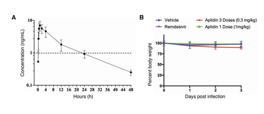
Supplemental Material

MATERIAL AND METHODS

Cells and Viruses
Vero E6 (ATCC, CRL-1586) and HEK293T (ATCC, CRL-3216; kind gift of Dr. Viviana
Simon), were maintained in DMEM (Corning) supplemented with 10% FB (Peak Serum)
and Penicillin/Streptomycin (Corning) at 37°C and 5% CO2. hACE2-293T cells were
generated for this study. Briefly, HEK293T cells were transduced with a lentiviral vector
expressing human ACE2. Puromycin resistant cells with hACE2 surface expression were
sorted after staining with AlexaFluor 647-conjugated goat anti-hACE2 antibodies. Cells
were then single-cell-cloned and screened for their ability to support SARS-CoV-2
replication.. All cell lines used in this study were regularly screened for mycoplasma
contamination using the Universal Detection Kit (ATCC, 30-1012K). Cells were infected
with SARS-CoV-2, isolate USA-WA1/2020 (BEI Resources NR-52281) under biosafety
level 3 (BSL3) containment in accordance to the biosafety protocols developed by the
Icahn School of Medicine at Mount Sinai. Viral stocks were grown in Vero E6 cells as
previously described (22) and were validated by genome sequencing.

Viral growth and cytotoxicity assays in the presence of inhibitors.

2,000 Vero E6 or hACE2-293T cells were seeded into 96-well plates in DMEM (10% FBS)
and incubated for 24 h at 37C, 5% CO2. Two hours before infection, the medium was
replaced with 100 µl of DMEM (2% FBS) containing the compound of interest at
concentrations 50% greater than those indicated, including a DMSO control. Plates were
then transferred into the BSL3 facility and 100 PFU (MOI = 0.025) was added in 50 µl of
DMEM (2% FBS), bringing the final compound concentration to those indicated. Plates
were then incubated for 48 h at 37C. After infection, supernatants were removed and
cells were fixed with 4% formaldehyde for 24 hours prior to being removed from the
BSL3 facility. The cells were then immunostained for the viral NP protein (an in-house
mAb 1C7, provided by Dr. Thomas Moran, Thomas.Moran@mssm.edu) with a DAPI
counterstain. Infected cells (488 nM) and total cells (DAPI) were quantified using the
Celigo (Nexcelcom) imaging cytometer. Infectivity was measured by the accumulation of
viral NP protein in the nucleus of the Vero E6 cells (fluorescence accumulation). Percent
infection was quantified as ((Infected cells/Total cells) - Background) *100 and the DMSO
control was then set to 100% infection for analysis. The IC50 and IC90 for each experiment
were determined using the Prism (GraphPad Software) software. Cytotoxicity was also
performed using the MTT assay (Roche), according to the manufacturer’s instructions.
Cytotoxicity was performed in uninfected VeroE6 cells with same compound dilutions
and concurrent with viral replication assay. All assays were performed in biologically
independent triplicates. Remdesivir was purchased from Medkoo Bioscience inc., while
plitidepsin was provided by PharmaMar.

Human pneumocyte-like cell generation

Human embryonic stem cells (H9, WiCell, WA09) were cultured with mTeSR (STEMCELL
Technologies, 85850) on Vitronectin XF (STEMCELL Technologies, 07180)-coated tissue
culture plates and split in a ratio of 1:6 to 1:12 every four to six days with Gentle Cell
Dissociation Reagent (STEMCELL Technologies, 07174). Alveolar differentiation was
induced as previously described (61). Briefly, cells were collected at 70-80% confluency,
and 2 million cells per 10 cm2 were plated on Vitronectin-coated tissue culture plates in
mTeSR. The next day, definitive endoderm differentiation was induced following the
protocol described by Jacob et al. (23) for four days. Cells were split and further
differentiated following an adapted alveolar differentiation protocol (24) in IMDM medium
(Life Technologies, 31980030) supplemented with 10% FBS (Sigma, F4135), 2 mM
L-glutamine (Life Technologies 25030081), 0.5 µM all-trans-retinoic acid (Sigma, R2626),
10 ng/ml FGF-10 (R&D Systems, 345-FG-025), 10 ng/ml EGF (R&D Systems,
236-EG-01M), 100 ng/ml Wnt3a (R&D Systems, 5036-WN-010), 10 ng/ml KGF (R&D
Systems, 251-KG-050) and 5 ng/ml BMP-4 (R&D Systems, 314-BP-010). Viral infections
were performed on day nine after induction of differentiation, and the cells were analyzed
two days post-infection.

Pneumocyte-like cell treatment and infection

Human stem cell derived pneumocyte-like cells were treated in 96-well format with
appropriate drug concentration for 1 hour at 37⁰C followed by SARS-CoV2 infection
(4000 PFU, MOI 0.1) in BSL3 facility for 48 hours at 37⁰C. After infection, supernatants
were removed and cells were fixed with 4% formaldehyde for 24 hours prior to being
removed from the BSL3 facility. Viral infection and cytotoxicity of compounds was
quantified using the same procedure outlined in the viral growth and cytotoxicity assays
in the presence of inhibitors section.

CRISPR/Cas9 meditated gene editing of EEF1A1 in HEK293T cells

HEK293T (ATCC® CRL-3216™), referred to as 293T here on, were a kind gift from Dr.
Lisa Miorin. Cells were maintained in Dulbecco’s Modified Eagle’s Medium (DMEM,
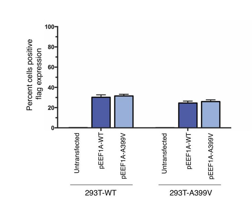
Corning) supplemented with Fetal Bovine Serum (FBS, 10% final concentration, PEAK).
For generation of 293T cells encoding endogenous EEF1A1 protein with previously
reported Alanine to Valine modification at position 399 (293T-EFF1A1-A399V),
CRISPR-Cas9 technology with Homology directed repair (HDR) approach was used.
Briefly, EEF1A1 gene was targeted at the genomic location: GRCh38.p11-Human 38.p11
chromosome 6, 73518097-100 with the intended change of sequence from GCT to GTT.
For this purpose, CRISPR-Cas9 cRNA, 5’ GCC AGG AAC CAU AUC AAC AAG UUU UAG
AGC UAU GCU 3’, and two HDR Donor Oligos for positive and negative strands, 5’ AAG
CTC TCA ACA CAC ATG GGC TTG CCA GGA ACC ATA TCA ACA ATG GCA ACA TCA
CCA GAC TTC AAG AAT TTA GGG CCA TCT TCC AGC TTT 3’ and 5’ AAA GCT GGA
AGA TGG CCC TAA ATT CTT GAA GTC TGG TGA TGT TGC CAT TGT TGA TAT GGT
TCC TGG CAA GCC CAT GTG TGT TGA GAG CTT 3’ respectively, were designed and
purchased through the use of Alt-R HDR Design Tool (IDT). CRISPR-RiboNucleoProtein
(CRISPR-RNPs) complexes with Cas9, tracrRNA and selected crRNA were prepared
according to manufacturer’s protocol (IDT). CRISPR-RNPs together with HDR donor
oligos were electroporated into 293T cells (2x10⁵) in suspension using SF Cell Line
4D-Nuclofector X Kit (LONZA) on a 4D-Nucleofector X unit (LONZA) based on the
guidelines provided. Following 3-day incubation in DMEM supplemented with 10% FBS
for recovery, cells were pre-selected by supplementing media with plitidepsin. Genomic
sequence of EEF1A1 gene covering the intended mutation (Exon 5-6) of genetically
modified cells were amplified using a manually designed primer pair, Forward 5’ GGC
TTC ACT GCT CAG GTA ACA ATT 3’ and Reverse 5’ CAC GAA CAG CAA AGC GAC CTA
TT 3’ (SnapGene, NCBI Blast). Purified PCR product was analyzed by SANGER
sequencing for confirmation of the mutation (Eton Bioscience) in comparison to wild-type
equivalents. A single cell clone was selected and expanded and used for further
experimentation, which showed approximately a 50% distribution of intended sequence
change.

Plasmid transfection experiments

A Plasmid encoding hEEF1A1 on a pCMV6 backbone with Myc and DDK tags at
C-terminus were purchased from Origene, EEF1A1 (NM_001402) Human Tagged ORF
Clone CAT#: RC209697. pCMV6-EEF1A1 with A399V mutation was created through
site-directed mutagenesis with PCR overlap extension. For drug efficacy tests, wild type
293T      or   293T-EEF1A1-A399V       cells    (4000/well)   were    transfected    with
pCAGGS-hACE2-10His(C-ter), created in this study, together with pCMV6-EEF1A1,
EEF1A1-A399V      using    LT1    reagent      (Mirus)   according   to   manufacturer’s
reverse-transfection protocol on a 96-well format. Following 48 hours of incubation, cells
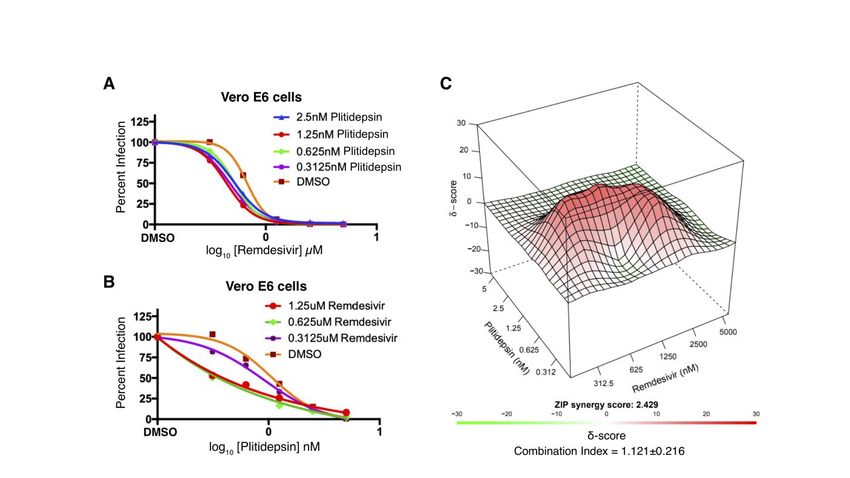
were used in drug efficacy tests with virus infection as detailed previously.

Antiviral combination assay

Like previous antiviral assay, 2,000 Vero E6 cells were seeded into 96-well plates in
DMEM (10% FBS) and incubated for 24 h at 37°C, 5% CO2. Two hours before infection,
the medium was replaced with 100µl of DMEM (2% FBS) containing the combination of
plitidepsin and remdesivir following a dilution combination matrix. A 6 by 6 matrix of drug
combinations was prepared in triplicate by making serial two-fold dilutions of the two
drugs on each axis, including a DMSO control column and row. The resulting matrix had
no drug in the right upper well, a single drug in rising 2-fold concentrations in the vertical
and horizontal axes starting from that well, and the remaining wells with rising
concentrations of drug mixtures reaching maximum concentrations of both drugs at the
lower left well. Plates were then transferred into the BSL3 facility and SARS-CoV-2 (MOI
0.025) was added in 50µl of DMEM (2% FBS), bringing the final compound concentration
to those indicated. Plates were then incubated for 48 h at 37ºC. After infection, cells were
fixed with final concentration of 5% formaldehyde for 24 hours prior to being removed
from the BSL3 facility. The cells were then immunostained for the viral NP protein (an
in-house mAb 1C7, provided by Dr. Thomas Moran, Thomas.Moran@mssm.edu) with a
DAPI counterstain. Infected cells (AlexaFluor 488) and total cells (DAPI) were quantified
using the Celigo (Nexcelcom) imaging cytometer. Infectivity was measured by the
accumulation of viral NP protein in the nucleus of the Vero E6 cells (fluorescence
accumulation). Percent infection was quantified as ((Infected cells/Total cells) -
Background) *100 and the DMSO control was then set to 100% infection for analysis.
The combination antiviral assay was performed in biologically independent triplicates.

The apparent IC90 for each combination in the matrix was determined using the Prism
(GraphPad Software) software. The IC90 for plitidepsin and remdesivir were calculated for
each drug treatment alone and in combination. This combination data was analyzed
using SynergyFinder (27) and by the isobologram method, combination indices were
calculated as previously described (60).

We assessed synergy using 2 different methodologies: the Chou-Talalay method based
on Loewe additivity (60) and Synergyfinder, scoring synergism using 3 main models,
Loewe, Bliss and zero interaction potency (ZIP) (27). Vero E6 cells were infected with
SARS-CoV-2 in the presence of the two compounds which were titrated against each
other in 2-fold serial dilutions and viral replication was determined using our
immunofluorescence-based assay. We did not observe a clear synergistic or
antagonistic impact on the inhibition curves of either compound in the presence of the
other (Supp Figure 2A,B). Using Synergyfinder, a positive ZIP synergy score is indicative
of a synergistic interaction, but a score between -10 and +10 is generally considered to
be additive in nature. Red regions with synergy scores > 1 indicate synergism, whereas
the green regions indicate antagonism. We found that while there are some
concentrations of plitidepsin and remdesivir that do show some synergism (Supp Figure
2C), the Loewe synergy score of -3.5, the Bliss synergy score of 2.42, and the ZIP
synergy score of 2.429 suggest an overall additive effect. Using the Chuo-Talalay
method, we calculated the combination index for the plitidepsin–remdesivir combined
treatment to be 1.121±0.216, also indicating an additive interaction.

Antiviral effect of Plitidepsin in a time course viral infection

2.5x10⁵ Vero E6 cells were seeded in 24-well plates in DMEM (10% FBS) and incubated
for 24 h at 37°C, 5% CO2. Two hours before infection, the medium was replaced with 300
µl of DMEM (2% FBS) containing plitidepsin 3 nM, remdesivir 5 µM or DMSO as control.
Plates were then transferred into the BSL3 facility and SARS-CoV-2 (MOI 1) was added
in 150 µl of DMEM (2% FBS), bringing the final compound concentration to those
indicated. Samples were collected immediately after infection, at time 0 and 4, 8, 12 and
24 hours post infection. Before sample collection, cells were washed twice in PBS. To
determine viral protein expression, protein lysates were collected in RIPA Lysis and
Extraction Buffer (Thermo Fisher Scientific, USA) supplemented with 1% SDS. To
determine viral RNA levels, samples were harvested using TRIzol reagent (Thermo Fisher
Scientific, USA) according to manufacturer instructions.

Viral protein expression analysis

Protein extract concentration was quantified using the Pierce BCA Protein Assay,
according to manufacturer instructions. 10 µg of protein extracts were resolved in 10%
Mini-PROTEAN®      TGX    Stain-Free™       Protein   Gels   (Biorad)   then   transferred    to
nitrocellulose membranes (0.45 μm; Bio-Rad). Blots were incubated with an in-house
mAb 1C7 anti-viral NP protein diluted at 1:2000 (Provided by Dr. Thomas Moran,
Thomas.Moran@mssm.edu).           Protein       expression       was       normalized         to
glyceraldehyde-3-phosphate dehydrogenase (GAPDH) (GTX100118; Genetex). The
HRP-conjugated      anti-mouse    IgG    antibody      (GTX26820,       Genetex)    and      the
HRP-conjugated anti-rabbit IgG antibody (GTX26802, Genetex) were used to detect the
primary antibodies at 1:10000. HRP was detected using Clarity™ Western ECL Substrate
(Biorad). Digital images were acquired and analyzed with a The ChemiDoc XRS+ System
(Biorad). Each protein band was quantified by ImageJ and normalized to GAPDH levels.

Viral RNA isolation and quantitative PCR

Genomic RNA was extracted for each time point using Trizol (ThermoScientific) and
Direct-zolTM RNA Miniprep Plus (Zymo Research) according to manufacturer instructions.
1 µg of total RNA was used for RT-PCR using the High-Capacity cDNA Reverse
Transcription Kits (4368814, Applied Biosystems™). The qPCR amplification was
performed with a dilution 1:10 of cDNA using the LightCycler® 480 SYBR Green I Master,
2x conc (04887352001, Roche).

We amplified two regions of the viral genome of SARS-COV-2. The first set of primers
LeaderF          (5’-CCAACCAACTTTCGATCTCTTGTA-3’)                       and         Nsub-R
(5’-TCTGGTTACTGCCAGTTGAATCTG-3’)             amplified     a   region    specific   to   the
sub-genomic      N      RNA.      The     second         set    of      primers     Nsp3-F
(5’-GGAATTTGGTGCCACTTC-3’) and Nsp3-R (5’-CTATTCACTTCAATAGTCTGAACA-3’)
amplified a fragment of the NSP3 as a measure of genomic RNA. The fold change in viral
RNA was normalized with the amplification of a fragment of 18S rRNA using these
primers    18S-rRNA-F      (5’-GTAACCCGTTGAACCCCATT-3’)                 and    18S-rRNA-R
(5’-CCATCCAATCGGTAGTAGCG-3’). Reaction volumes were 20 µL and all pairs of
primers were tested at 0.5 µM (final each) with PCR parameters recommended for a PCR
run with the LightCycler® 480 SYBR Green I Master using a LightCycler® 480 Multiwell
Plate 384 described by the manufacturer (Roche, USA). The results were analyzed using
software 1.5.1.62 SP3. Each assay was performed in triplicate with two technical
replicates, and each assay included no-template negative controls.

siRNA knockdown of EEF1A1.

293T-hACE2 cells were transfected with 50 nM of siRNA against Human EEF1A1
(M-017199-02-0005, DharmaconTM). A negative control siGENOME Non-Targeting siRNA
(D-001206-13-05, DharmaconTM) was used at the same concentrations of the siRNA
EE1FA1 described above. Gene knockdown was performed using 1 × 10⁵ cells per well
using Lipofectamine RNAiMAX Transfection Reagent following manufacture protocol for
HEK293 cells (Invitrogen). After 48h post transfection, plates were then transferred into
the BSL3 facility, transfection media was removed, and cells were infected with
SARS-COV2 at MOI of 1 and cells were harvest at 8 h post infection using RIPA Lysis
and Extraction Buffer (Thermo) with protease inhibitor cocktail (P8340, Sigma). These
lysates were used in western blotting to determine decrease in EEF1A1 and viral protein
N expression. Briefly, we incubated membranes with eEF1A1 Antibody (NBP1-32122,
Novus Biologicals) diluted at 1:1000. Viral N protein was inmunostained with an in-house
mAb 1C7 (provided by Dr. Thomas Moran, Thomas.Moran@mssm.edu) diluted at 1:2000.
The HRP-conjugated anti-mouse IgG antibody (GTX26820, Genetex) and the
HRP-conjugated anti-rabbit IgG antibody (GTX26802, Genetex) were used to detect the
primaries antibodies at 1:10000. HRP was detected using Clarity™ Western ECL
Substrate (Biorad). Digital images were acquired and analyzed with a The ChemiDoc
XRS+ System (Biorad). Each protein band was quantified by ImageJ and normalized to
GAPDH levels.

Plitidepsin pharmacokinetic study in C57BL/6J mice

Female C57BL/6J mice between 4 to 8 weeks of age (16-21 g) were purchased from
Charles River Laboratories (France). Animals were housed in individually ventilated cages
(Sealsafe® Plus, Techniplast S.p.A.), 10 mice per cage, on a 12-h light–dark cycle at
21–23°C and 40–60% humidity. Mice were allowed free access to irradiated standard
rodent diet (Tecklad 2914C) and sterilized water. Animal protocols were reviewed and
approved according to regional Institutional Animal Care and Use Committees.

After a single subcutaneous administration (dose: 0.30 mg/kg), 4 animals per sampling
time point (5 min, 15 min, 30 min, 1, 2, 4, 12, 24 and 48 hours post-dosing) blood
samples were collected from CO2 euthanized mice, in suitable polypropylene blood
collection tubes containing k3 EDTA, were centrifuged at 3,000 rpm for 15 min at ca. 4°C
and the resulting plasma was frozen at -20°C until bioanalysis. Briefly, plasma samples
were subjected to supported liquid-liquid extraction with tert-butyl methyl ether (TBME),
followed by a gradient phase chromatography. Plitidepsin was separated on an Atlantis
T3, 3.0 μm, 2.1 x 20 mm column (Waters, Milford, mA, USA) heated to 50 ºC. Liquid
chromatography was carried out at a flow rate of 0.6 mL/min using a gradient of water (A)
in acetonitrile (B), both with 0.1% formic acid: 90 to 10 % A from 0.0 to 1.5 min; isocratic
for 1.0 min and then, initial conditions recovered over 0.1 min, which were kept for 2.4
min up to the total time of 5.0 min. The mass spectrometry instrument (API 4000 AB
Sciex, Framingham, MA, USA) was operated in the positive ESI mode using multiple
reaction monitoring (MRM). Plitidepsin was monitored at mass transitions of m/z 1110.7
→ 100.3. A non-compartmental pharmacokinetic analysis of plitidepsin was performed
using WinNonlin® 8.2 Phoenix® 64 software (Certara USA, Inc., NJ, USA) according to a
composite sampling method. Pharmacokinetic parameters were calculated by the
linear-log trapezoidal rule; Cmax values were obtained from observed data.

After a s.c. administration of plitidepsin (0.3 mg/kg) to C57BL/6J female mice, Cmax (ca.
7 ng/mL) were observed at 1 h post-dosing, followed by a gradual decrease as observed
from the plasma concentration-time profiles (Supp Figure 3). Results obtained
demonstrated low systemic clearance (ca. 4.5 L/h/kg), large apparent volume of
distribution (ca. 85 L/kg) and long terminal half-life (13 h) in plasma, with a calculated
AUC0-∞ of 72 ng·h/mL.

Animal models of SARS-CoV-2 infection experiments

All the antiviral studies were performed in animal biosafety level 3 (BSL3) facility at the
Icahn school of Medicine in Mount Sinai Hospital, New York City. All work was
conducted under protocols approved by the Institutional Animal Care and Use
Committee (IACUC). We performed two studies in mice to evaluate the in vivo efficacy of
plitidepsin as antiviral in SARS-CoV-2 infection. First, we use a model of BALB/c mice
transduced intranasally with an adenovirus expressing human ACE2 (hACE2); and
second, we used a model of mice hemizygous for the expression of hACE2 gene
(K18-ACE2).

We utilized 16 female 12-week-old specific pathogen–free BALB/c mice (the Jackson
laboratory strain 000651). Five days prior to infection with SARS-CoV-2, BALB/c mice
were infected intranasally with 2.5x10⁸ PFU of an adenovirus carrying the gene for
hACE2. Viral seed stocks for non-replicating E1/E3 deleted viral vectors based on human
adenovirus type-5 expressing the human angiotensin-converting enzyme 2 (Ad-ACE2)
receptor under the control of a CMV promoter, were obtained from the Iowa Viral Vector
Core Facility. Viral stocks were amplified to high titers following infection of T-RexTM-293
cells and purification using two sequential rounds of cesium chloride (CsCl)
ultracentrifugation, as described previously (62, 63). The infectious titer was determined
using a tissue culture infectious dose-50 (TCID50) end-point dilution assay, and physical
particle titer quantified by micro-bicinchoninic acid (microBCA) protein assay, both
described previously (62). This experiment was repeated independently twice, a total of
10 mice were used in the vehicle and control groups and 8 mice were used in the
plitidepsin   treated   groups.   Remdesivir     and    plitidepsin   were   administrated
subcutaneously (s.c.), each once per day for 3 days . We administered vehicle (2%
cremophor, 2% ethanol, 0.75% NaCl), remdesivir or plitidepsin, 2 hours before intranasal
infection with 1 × 10⁴ PFU of SARS-CoV-2 in 50 μl of PBS. Mice were anesthetized with a
mixture of ketamine/xylazine before each intranasal infection. Each day mouse body
weight was measured. 3 days post infection (dpi) animals were humanely euthanized.
Whole left lungs were harvested and homogenized in PBS with silica glass beads then
frozen at −80°C for viral titration via TCID50. Briefly, infectious supernatants were
collected at 48h post infection and frozen at −80°C until later use. Infectious titers were
quantified by limiting dilution titration using Vero E6 cells. Briefly, Vero E6 cells were
seeded in 96-well plates at 20,000 cells/well. The next day, SARS-CoV-2-containing
supernatant was applied at serial 10-fold dilutions ranging from 10−1 to 10−6 and, after 5
days, viral cytopathic effect (CPE) was detected by staining cell monolayers with crystal
violet. TCID50/ml were calculated using the method of Reed and Muench. The Prism
software (GraphPad) was used to determine differences in lung titers using an unpaired T
test on log-transformed data.

For the second animal study, we utilized 50 female 12-week-old specific pathogen–free
Hemizygous     for   Tg(K18-ACE2)2Prlmn     (Strain   B6.Cg-Tg(K18-ACE2)2Prlmn/J,      the
Jackson laboratory strain 034860) mice. We performed an efficacy study in mice where
we initiated treatment 2 hours before infection with 1 × 10⁴ PFU of SARS-CoV-2 in 50 μl
of PBS. 5 mice were used for the plitidepsin and vehicle groups, while 3 mice were used
in the remdesivir group. All dosing was performed through the subcutaneous route. On
day 3 post SARS-CoV-2 infection, animals were humanely euthanized and lung tissues
were harvested. Lungs were homogenized in PBS with silica glass beads and frozen at
−80°C for viral titration by TCID50.

Mouse lung histological analysis

Paraffin-embedded lung tissue blocks for mouse lungs were cut into 5μm sections.
Sections were stained with hematoxylin and eosin (H&E) and analyzed by Histowiz
(Brooklyn, NY). Digital light microscopic scans of whole lung processed in toto were
examined by an experienced veterinary pathologist. Hematoxylin Eosin stained sections
of lung from K18 hACE2 mice were examined by implementing a semi quantitative, 5
point grading scheme (0 - within normal limits, 1 - mild, 2 - moderate, 3 - marked, 4 -
severe). that took into account four different histopathological parameters: 1)
perivascular inflammation 2) bronchial or bronchiolar epithelial degeneration or necrosis
3) bronchial or bronchiolar inflammation and 4) alveolar inflammation. These changes
were absent (grade 0) in lungs from vehicle and plitidepsin treated uninfected mice from
groups that were utilized for this assessment. All mice from plitidepsin, remdesivir, and
vehicle treated infected groups exhibited multifocal pulmonary lesions. Individual total
pathology scores varied from 5/16 to 6/16 for the vehicle group, from 2/16 to 3/16 for the
remdesivir group and were equal to 1/16 for the plitidepsin group.
Supplemental Figures

Supplemental Figure 1. Flag tag expression from eEF1A expression plasmids. Wild
type 293T or 293T-EEF1A1-A399V cells were transfected with pCAGGS-hACE2-10His,
together with pCMV6-EEF1A1-Flag or EEF1A1-A399V-Flag. Following 48 hours of
incubation, cells were immunostained with anti-Flag antibody and quantified with the
Celigo imaging cytometer to confirm transfection efficiency.

Supplemental Figure 2. Combination of plitidepsin and remdesivir exhibits additive
antiviral activity against SARV-CoV-2 infection. Antiviral activity curves for (A)
remdesivir (0.3 – 5 μM) in the presence of fixed amounts of plitidepsin or (B) plitidepsin
(0.3 – 5 nM) in the presence of fixed amounts of remdesivir. (C) Plitidepsin and remdesivir
act in additive manner in the inhibition of SARS-CoV-2 replication. Vero E6 were infected
with SARS-CoV-2 and treated for 48 h with the indicated concentrations of plitidepsin
and remdesivir and viral replication was assessed by immunofluorescence. Synergy
scores were calculated using the Synergyfinder software. ZIP Synergy scores > 0 indicate
synergism (red regions) and scores < 0 indicate antagonism (green regions). The
combination index was calculated using the Chou-Talalay method and demonstrates an
additive antiviral effect between the drugs against SARS-CoV-2.

Supplemental Figure 3. Pharmacokinetic and toxicity analysis of plitidepsin in mice.
(A) Concentration-time curves (mean±SD) of plitidepsin in plasma of female C57BL/6J
mice after a subcutaneous administration dose at 0.3 mg/kg. Dotted line represents the
IC90 for plitidepsin (0.88 nM; 0.98 ng/mL; Molecular Weight, 1110.4). Results obtained
demonstrated low systemic clearance (ca. 4.5 L/h/kg), large apparent volume of
distribution (ca. 85 L/kg) and long terminal half-life (13 h) in plasma, with a calculated
AUC0-∞ of 72 ng·h/mL. (B) Body weight measurements from infected mice prior to lung
harvest in the Ad-ACE2 model from Figure 4.
Supplementary Figure 1
Supplementary Figure 2
Supplementary Figure 3
References

1. World Health Organization, “Middle East respiratory syndrome coronavirus (MERS-CoV)”;
       www.who.int/emergencies/mers-cov/en/.

2. E. de Wit, N. van Doremalen, D. Falzarano, V. J. Munster, SARS and MERS: Recent insights
       into emerging coronaviruses. Nat. Rev. Microbiol. 14, 523–534 (2016).
       doi:10.1038/nrmicro.2016.81 Medline

3. D. Wang, B. Hu, C. Hu, F. Zhu, X. Liu, J. Zhang, B. Wang, H. Xiang, Z. Cheng, Y. Xiong, Y.
       Zhao, Y. Li, X. Wang, Z. Peng, Clinical Characteristics of 138 Hospitalized Patients With
       2019 Novel Coronavirus-Infected Pneumonia in Wuhan, China. JAMA 323, 1061–1069
       (2020). doi:10.1001/jama.2020.1585 Medline

4. Y. Z. Zhang, E. C. Holmes, A Genomic Perspective on the Origin and Emergence of
       SARS-CoV-2. Cell 181, 223–227 (2020). doi:10.1016/j.cell.2020.03.035 Medline

5. K. G. Andersen, A. Rambaut, W. I. Lipkin, E. C. Holmes, R. F. Garry, The proximal origin of
       SARS-CoV-2. Nat. Med. 26, 450–452 (2020). doi:10.1038/s41591-020-0820-9 Medline

6. B. Hu, X. Ge, L. F. Wang, Z. Shi, Bat origin of human coronaviruses. Virol. J. 12, 221 (2015).
       doi:10.1186/s12985-015-0422-1 Medline

7. S. J. Anthony, K. Gilardi, V. D. Menachery, T. Goldstein, B. Ssebide, R. Mbabazi, I.
       Navarrete-Macias, E. Liang, H. Wells, A. Hicks, A. Petrosov, D. K. Byarugaba, K.
       Debbink, K. H. Dinnon, T. Scobey, S. H. Randell, B. L. Yount, M. Cranfield, C. K.
       Johnson, R. S. Baric, W. I. Lipkin, J. A. K. Mazet, Further Evidence for Bats as the
       Evolutionary Source of Middle East Respiratory Syndrome Coronavirus. mBio 8,
       e00373-17 (2017). doi:10.1128/mBio.00373-17 Medline

8. V. D. Menachery, B. L. Yount Jr., A. C. Sims, K. Debbink, S. S. Agnihothram, L. E. Gralinski,
       R. L. Graham, T. Scobey, J. A. Plante, S. R. Royal, J. Swanstrom, T. P. Sheahan, R. J.
       Pickles, D. Corti, S. H. Randell, A. Lanzavecchia, W. A. Marasco, R. S. Baric, SARS-like
       WIV1-CoV poised for human emergence. Proc. Natl. Acad. Sci. U.S.A. 113, 3048–3053
       (2016). doi:10.1073/pnas.1517719113 Medline

9. V. D. Menachery, B. L. Yount Jr., K. Debbink, S. Agnihothram, L. E. Gralinski, J. A. Plante, R.
       L. Graham, T. Scobey, X.-Y. Ge, E. F. Donaldson, S. H. Randell, A. Lanzavecchia, W. A.
       Marasco, Z.-L. Shi, R. S. Baric, A SARS-like cluster of circulating bat coronaviruses
shows potential for human emergence. Nat. Med. 21, 1508–1513 (2015).
doi:10.1038/nm.3985 Medline
10. P. C. Woo, S. K. P. Lau, K. S. M. Li, R. W. S. Poon, B. H. L. Wong, H. W. Tsoi, B. C. K. Yip,
Y. Huang, K. H. Chan, K. Y. Yuen, Molecular diversity of coronaviruses in bats. Virology
351, 180–187 (2006). doi:10.1016/j.virol.2006.02.041 Medline
11. Y. Liang, M.-L. Wang, C.-S. Chien, A. A. Yarmishyn, Y.-P. Yang, W.-Y. Lai, Y.-H. Luo,
Y.-T. Lin, Y.-J. Chen, P.-C. Chang, S.-H. Chiou, Highlight of Immune Pathogenic
Response and Hematopathologic Effect in SARS-CoV, MERS-CoV, and SARS-Cov-2
Infection. Front. Immunol. 11, 1022 (2020). doi:10.3389/fimmu.2020.01022 Medline
12. C. D. Spinner, R. L. Gottlieb, G. J. Criner, J. R. Arribas López, A. M. Cattelan, A. Soriano
Viladomiu, O. Ogbuagu, P. Malhotra, K. M. Mullane, A. Castagna, L. Y. A. Chai, M.
Roestenberg, O. T. Y. Tsang, E. Bernasconi, P. Le Turnier, S.-C. Chang, D. SenGupta, R.
H. Hyland, A. O. Osinusi, H. Cao, C. Blair, H. Wang, A. Gaggar, D. M. Brainard, M. J.
McPhail, S. Bhagani, M. Y. Ahn, A. J. Sanyal, G. Huhn, F. M. Marty, Effect of Remdesivir
vs Standard Care on Clinical Status at 11 Days in Patients With Moderate COVID-19: A
Randomized Clinical Trial. JAMA 324, 1048–1057 (2020). doi:10.1001/jama.2020.16349
Medline
13. Y. Wang, D. Zhang, G. Du, R. Du, J. Zhao, Y. Jin, S. Fu, L. Gao, Z. Cheng, Q. Lu, Y. Hu, G.
Luo, K. Wang, Y. Lu, H. Li, S. Wang, S. Ruan, C. Yang, C. Mei, Y. Wang, D. Ding, F. Wu,
X. Tang, X. Ye, Y. Ye, B. Liu, J. Yang, W. Yin, A. Wang, G. Fan, F. Zhou, Z. Liu, X. Gu,
J. Xu, L. Shang, Y. Zhang, L. Cao, T. Guo, Y. Wan, H. Qin, Y. Jiang, T. Jaki, F. G.
Hayden, P. W. Horby, B. Cao, C. Wang, Remdesivir in adults with severe COVID-19: A
randomised, double-blind, placebo-controlled, multicentre trial. Lancet 395, 1569–1578
(2020). doi:10.1016/S0140-6736(20)31022-9 Medline
14. RECOVERY Collaborative Group, Dexamethasone in Hospitalized Patients with
Covid-19—Preliminary Report. N. Engl. J. Med. NEJMoa2021436 (2020).
doi:10.1056/NEJMoa2021436
15. WHO Solidarity Trial Consortium, Repurposed Antiviral Drugs for COVID-19—Interim
WHO SOLIDARITY Trial Results. N. Engl. J. Med. NEJMoa2023184 (2020).
doi:10.1056/NEJMoa2023184
16. D. E. Gordon, G. M. Jang, M. Bouhaddou, J. Xu, K. Obernier, K. M. White, M. J. O’Meara, V.
V. Rezelj, J. Z. Guo, D. L. Swaney, T. A. Tummino, R. Hüttenhain, R. M. Kaake, A. L.Richards, B. Tutuncuoglu, H. Foussard, J. Batra, K. Haas, M. Modak, M. Kim, P. Haas, B.
J. Polacco, H. Braberg, J. M. Fabius, M. Eckhardt, M. Soucheray, M. J. Bennett, M. Cakir,
M. J. McGregor, Q. Li, B. Meyer, F. Roesch, T. Vallet, A. Mac Kain, L. Miorin, E.
Moreno, Z. Z. C. Naing, Y. Zhou, S. Peng, Y. Shi, Z. Zhang, W. Shen, I. T. Kirby, J. E.
Melnyk, J. S. Chorba, K. Lou, S. A. Dai, I. Barrio-Hernandez, D. Memon, C.
Hernandez-Armenta, J. Lyu, C. J. P. Mathy, T. Perica, K. B. Pilla, S. J. Ganesan, D. J.
Saltzberg, R. Rakesh, X. Liu, S. B. Rosenthal, L. Calviello, S. Venkataramanan, J.
Liboy-Lugo, Y. Lin, X.-P. Huang, Y. Liu, S. A. Wankowicz, M. Bohn, M. Safari, F. S.
Ugur, C. Koh, N. S. Savar, Q. D. Tran, D. Shengjuler, S. J. Fletcher, M. C. O’Neal, Y. Cai,
J. C. J. Chang, D. J. Broadhurst, S. Klippsten, P. P. Sharp, N. A. Wenzell, D.
Kuzuoglu-Ozturk, H.-Y. Wang, R. Trenker, J. M. Young, D. A. Cavero, J. Hiatt, T. L.
Roth, U. Rathore, A. Subramanian, J. Noack, M. Hubert, R. M. Stroud, A. D. Frankel, O. S.
Rosenberg, K. A. Verba, D. A. Agard, M. Ott, M. Emerman, N. Jura, M. von Zastrow, E.
Verdin, A. Ashworth, O. Schwartz, C. d’Enfert, S. Mukherjee, M. Jacobson, H. S. Malik,
D. G. Fujimori, T. Ideker, C. S. Craik, S. N. Floor, J. S. Fraser, J. D. Gross, A. Sali, B. L.
Roth, D. Ruggero, J. Taunton, T. Kortemme, P. Beltrao, M. Vignuzzi, A. García-Sastre, K.
M. Shokat, B. K. Shoichet, N. J. Krogan, A SARS-CoV-2 protein interaction map reveals
targets for drug repurposing. Nature 583, 459–468 (2020).
doi:10.1038/s41586-020-2286-9 Medline
17. D. E. Gordon, J. Hiatt, M. Bouhaddou, V. V. Rezelj, S. Ulferts, H. Braberg, A. S. Jureka, K.
Obernier, J. Z. Guo, J. Batra, R. M. Kaake, A. R. Weckstein, T. W. Owens, M. Gupta, S.
Pourmal, E. W. Titus, M. Cakir, M. Soucheray, M. McGregor, Z. Cakir, G. Jang, M. J.
O’Meara, T. A. Tummino, Z. Zhang, H. Foussard, A. Rojc, Y. Zhou, D. Kuchenov, R.
Hüttenhain, J. Xu, M. Eckhardt, D. L. Swaney, J. M. Fabius, M. Ummadi, B. Tutuncuoglu,
U. Rathore, M. Modak, P. Haas, K. M. Haas, Z. Z. C. Naing, E. H. Pulido, Y. Shi, I.
Barrio-Hernandez, D. Memon, E. Petsalaki, A. Dunham, M. C. Marrero, D. Burke, C. Koh,
T. Vallet, J. A. Silvas, C. M. Azumaya, C. Billesbølle, A. F. Brilot, M. G. Campbell, A.
Diallo, M. S. Dickinson, D. Diwanji, N. Herrera, N. Hoppe, H. T. Kratochvil, Y. Liu, G. E.
Merz, M. Moritz, H. C. Nguyen, C. Nowotny, C. Puchades, A. N. Rizo, U.
Schulze-Gahmen, A. M. Smith, M. Sun, I. D. Young, J. Zhao, D. Asarnow, J. Biel, A.
Bowen, J. R. Braxton, J. Chen, C. M. Chio, U. S. Chio, I. Deshpande, L. Doan, B. Faust, S.
Flores, M. Jin, K. Kim, V. L. Lam, F. Li, J. Li, Y.-L. Li, Y. Li, X. Liu, M. Lo, K. E. Lopez,
A. A. Melo, F. R. Moss 3rd, P. Nguyen, J. Paulino, K. I. Pawar, J. K. Peters, T. H. Pospiech
Jr., M. Safari, S. Sangwan, K. Schaefer, P. V. Thomas, A. C. Thwin, R. Trenker, E. Tse, T.K. M. Tsui, F. Wang, N. Whitis, Z. Yu, K. Zhang, Y. Zhang, F. Zhou, D. Saltzberg, A. J.
Hodder, A. S. Shun-Shion, D. M. Williams, K. M. White, R. Rosales, T. Kehrer, L. Miorin,
E. Moreno, A. H. Patel, S. Rihn, M. M. Khalid, A. Vallejo-Gracia, P. Fozouni, C. R.
Simoneau, T. L. Roth, D. Wu, M. A. Karim, M. Ghoussaini, I. Dunham, F. Berardi, S.
Weigang, M. Chazal, J. Park, J. Logue, M. McGrath, S. Weston, R. Haupt, C. J. Hastie, M.
Elliott, F. Brown, K. A. Burness, E. Reid, M. Dorward, C. Johnson, S. G. Wilkinson, A.
Geyer, D. M. Giesel, C. Baillie, S. Raggett, H. Leech, R. Toth, N. Goodman, K. C.
Keough, A. L. Lind, R. J. Klesh, K. R. Hemphill, J. Carlson-Stevermer, J. Oki, K. Holden,
T. Maures, K. S. Pollard, A. Sali, D. A. Agard, Y. Cheng, J. S. Fraser, A. Frost, N. Jura, T.
Kortemme, A. Manglik, D. R. Southworth, R. M. Stroud, D. R. Alessi, P. Davies, M. B.
Frieman, T. Ideker, C. Abate, N. Jouvenet, G. Kochs, B. Shoichet, M. Ott, M. Palmarini, K.
M. Shokat, A. García-Sastre, J. A. Rassen, R. Grosse, O. S. Rosenberg, K. A. Verba, C. F.
Basler, M. Vignuzzi, A. A. Peden, P. Beltrao, N. J. Krogan, QCRG Structural Biology
Consortium, Zoonomia Consortium, Comparative host-coronavirus protein interaction
networks reveal pan-viral disease mechanisms. Science 370, eabe9403 (2020).
doi:10.1126/science.abe9403 Medline
18. J. T. Ernst, P. A. Thompson, C. Nilewski, P. A. Sprengeler, S. Sperry, G. Packard, T. Michels,
A. Xiang, C. Tran, C. J. Wegerski, B. Eam, N. P. Young, S. Fish, J. Chen, H. Howard, J.
Staunton, J. Molter, J. Clarine, A. Nevarez, G. G. Chiang, J. R. Appleman, K. R. Webster,
S. H. Reich, Design of Development Candidate eFT226, a First in Class Inhibitor of
Eukaryotic Initiation Factor 4A RNA Helicase. J. Med. Chem. 63, 5879–5955 (2020).
doi:10.1021/acs.jmedchem.0c00182 Medline
19. M. Ito, J. Ito, H. Kitazawa, K. Shimamura, T. Fukami, S. Tokita, K. Shimokawa, K. Yamada,
A. Kanatani, D. Uemura, (–)-Ternatin inhibits adipogenesis and lipid metabolism in
3T3-L1 cells. Peptides 30, 1074–1081 (2009). doi:10.1016/j.peptides.2009.02.008
Medline
20. Spanish Clinical Trials Registry, 2020-001993-31:
reec.aemps.es/reec/estudio/2020-001993-31.
21. ClinicalTrials.gov, NCT04382066:
https://clinicaltrials.gov/ct2/show/NCT04382066?term=plitidepsin&draw=2&rank=8.
22. F. Amanat, K. M. White, L. Miorin, S. Strohmeier, M. McMahon, P. Meade, W.-C. Liu, R. A.
Albrecht, V. Simon, L. Martinez-Sobrido, T. Moran, A. García-Sastre, F. Krammer, An InVitro Microneutralization Assay for SARS-CoV-2 Serology and Drug Screening. Curr.
Protoc. Microbiol. 58, e108 (2020). doi:10.1002/cpmc.108 Medline
23. A. Jacob, M. Vedaie, D. A. Roberts, D. C. Thomas, C. Villacorta-Martin, K.-D. Alysandratos,
F. Hawkins, D. N. Kotton, Derivation of self-renewing lung alveolar epithelial type II cells
from human pluripotent stem cells. Nat. Protoc. 14, 3303–3332 (2019).
doi:10.1038/s41596-019-0220-0 Medline
24. M. Ghaedi, E. A. Calle, J. J. Mendez, A. L. Gard, J. Balestrini, A. Booth, P. F. Bove, L. Gui, E.
S. White, L. E. Niklason, Human iPS cell-derived alveolar epithelium repopulates lung
extracellular matrix. J. Clin. Invest. 123, 4950–4962 (2013). doi:10.1172/JCI68793
Medline
25. R. A. Siemieniuk, J. J. Bartoszko, L. Ge, D. Zeraatkar, A. Izcovich, E. Kum, H.
Pardo-Hernandez, B. Rochwerg, F. Lamontagne, M. A. Han, Q. Liu, A. Agarwal, T.
Agoritsas, D. K. Chu, R. Couban, A. Darzi, T. Devji, B. Fang, C. Fang, S. A. Flottorp, F.
Foroutan, M. Ghadimi, D. Heels-Ansdell, K. Honarmand, L. Hou, X. Hou, Q. Ibrahim, A.
Khamis, B. Lam, M. Loeb, M. Marcucci, S. L. McLeod, S. Motaghi, S. Murthy, R. A.
Mustafa, J. D. Neary, A. Qasim, G. Rada, I. B. Riaz, B. Sadeghirad, N. Sekercioglu, L.
Sheng, A. Sreekanta, C. Switzer, B. Tendal, L. Thabane, G. Tomlinson, T. Turner, P. O.
Vandvik, R. W. M. Vernooij, A. Viteri-García, Y. Wang, L. Yao, Z. Ye, G. H. Guyatt, R.
Brignardello-Petersen, Drug treatments for covid-19: Living systematic review and
network meta-analysis. BMJ 370, m2980 (2020). doi:10.1136/bmj.m2980 Medline
26. Y. Yokoyama, A. Briasoulis, H. Takagi, T. Kuno, Effect of remdesivir on patients with
COVID-19: A network meta-analysis of randomized control trials. Virus Res. 288, 198137
(2020). doi:10.1016/j.virusres.2020.198137 Medline
27. A. Ianevski, A. K. Giri, T. Aittokallio, SynergyFinder 2.0: Visual analytics of multi-drug
combination synergies. Nucleic Acids Res. 48, W488–W493 (2020).
doi:10.1093/nar/gkaa216 Medline
28. P. Krastel, S. Roggo, M. Schirle, N. T. Ross, F. Perruccio, P. Aspesi Jr., T. Aust, K. Buntin, D.
Estoppey, B. Liechty, F. Mapa, K. Memmert, H. Miller, X. Pan, R. Riedl, C. Thibaut, J.
Thomas, T. Wagner, E. Weber, X. Xie, E. K. Schmitt, D. Hoepfner, Nannocystin A: An
Elongation Factor 1 Inhibitor from Myxobacteria with Differential Anti-Cancer Properties.
Angew. Chem. Int. Ed. 54, 10149–10154 (2015). doi:10.1002/anie.201505069 Medline29. J. D. Carelli, S. G. Sethofer, G. A. Smith, H. R. Miller, J. L. Simard, W. C. Merrick, R. K. Jain,
N. T. Ross, J. Taunton, Ternatin and improved synthetic variants kill cancer cells by
targeting the elongation factor-1A ternary complex. eLife 4, e10222 (2015).
doi:10.7554/eLife.10222 Medline
30. S. H. van den Worm, K. Knoops, J. C. Zevenhoven-Dobbe, C. Beugeling, Y. van der Meer, A.
M. Mommaas, E. J. Snijder, Development and RNA-synthesizing activity of coronavirus
replication structures in the absence of protein synthesis. J. Virol. 85, 5669–5673 (2011).
doi:10.1128/JVI.00403-11 Medline
31. D. Bojkova, K. Klann, B. Koch, M. Widera, D. Krause, S. Ciesek, J. Cinatl, C. Münch,
Proteomics of SARS-CoV-2-infected host cells reveals therapy targets. Nature 583, 469–
472 (2020). doi:10.1038/s41586-020-2332-7 Medline
32. S. G. Sawicki, D. L. Sawicki, Coronavirus minus-strand RNA synthesis and effect of
cycloheximide on coronavirus RNA synthesis. J. Virol. 57, 328–334 (1986).
doi:10.1128/JVI.57.1.328-334.1986 Medline
33. S. G. Sawicki, D. L. Sawicki, S. G. Siddell, A contemporary view of coronavirus transcription.
J. Virol. 81, 20–29 (2007). doi:10.1128/JVI.01358-06 Medline
34. S. G. Sawicki, D. L. Sawicki, Coronaviruses use discontinuous extension for synthesis of
subgenome-length negative strands. Adv. Exp. Med. Biol. 380, 499–506 (1995).
doi:10.1007/978-1-4615-1899-0_79 Medline
35. J. Delgado-Calle, N. Kurihara, E. G. Atkinson, J. Nelson, K. Miyagawa, C. M. Galmarini, G.
D. Roodman, T. Bellido, Aplidin (plitidepsin) is a novel anti-myeloma agent with potent
anti-resorptive activity mediated by direct effects on osteoclasts. Oncotarget 10, 2709–
2721 (2019). doi:10.18632/oncotarget.26831 Medline
36. L. Yao, Aplidin PharmaMar. IDrugs 6, 246–250 (2003). Medline
37. P. E. Morande, S. R. Zanetti, M. Borge, P. Nannini, C. Jancic, R. F. Bezares, A. Bitsmans, M.
González, A. L. Rodríguez, C. M. Galmarini, R. Gamberale, M. Giordano, The cytotoxic
activity of Aplidin in chronic lymphocytic leukemia (CLL) is mediated by a direct effect
on leukemic cells and an indirect effect on monocyte-derived cells. Invest. New Drugs 30,
1830–1840 (2012). doi:10.1007/s10637-011-9740-3 Medline
38. C. S. Mitsiades, E. M. Ocio, A. Pandiella, P. Maiso, C. Gajate, M. Garayoa, D. Vilanova, J. C.
Montero, N. Mitsiades, C. J. McMullan, N. C. Munshi, T. Hideshima, D. Chauhan, P.Aviles, G. Otero, G. Faircloth, M. V. Mateos, P. G. Richardson, F. Mollinedo, J. F.
San-Miguel, K. C. Anderson, Aplidin, a marine organism-derived compound with potent
antimyeloma activity in vitro and in vivo. Cancer Res. 68, 5216–5225 (2008).
doi:10.1158/0008-5472.CAN-07-5725 Medline
39. R. Nalda-Molina, B. Valenzuela, A. Ramon-Lopez, B. Miguel-Lillo, A. Soto-Matos, J. J.
Perez-Ruixo, Population pharmacokinetics meta-analysis of plitidepsin (Aplidin) in cancer
subjects. Cancer Chemother. Pharmacol. 64, 97–108 (2009).
doi:10.1007/s00280-008-0841-4 Medline
40. A. Soto-Matos, S. Szyldergemajn, S. Extremera, B. Miguel-Lillo, V. Alfaro, C. Coronado, P.
Lardelli, E. Roy, C. S. Corrado, C. Kahatt, Plitidepsin has a safe cardiac profile: A
comprehensive analysis. Mar. Drugs 9, 1007–1023 (2011). doi:10.3390/md9061007
Medline
41. R. Salazar, R. Plummer, A. Oaknin, A. Robinson, B. Pardo, A. Soto-Matos, A. Yovine, S.
Szyldergemajn, A. H. Calvert, Phase I study of weekly plitidepsin as 1-hour infusion
combined with carboplatin in patients with advanced solid tumors or lymphomas. Invest.
New Drugs 29, 1406–1413 (2011). doi:10.1007/s10637-010-9488-1 Medline
42. R. L. Piekarz, A. R. Frye, J. J. Wright, S. M. Steinberg, D. J. Liewehr, D. R. Rosing, V.
Sachdev, T. Fojo, S. E. Bates, Cardiac studies in patients treated with depsipeptide, FK228,
in a phase II trial for T-cell lymphoma. Clin. Cancer Res. 12, 3762–3773 (2006).
doi:10.1158/1078-0432.CCR-05-2095 Medline
43. M. H. Shah, P. Binkley, K. Chan, J. Xiao, D. Arbogast, M. Collamore, Y. Farra, D. Young, M.
Grever, Cardiotoxicity of histone deacetylase inhibitor depsipeptide in patients with
metastatic neuroendocrine tumors. Clin. Cancer Res. 12, 3997–4003 (2006).
doi:10.1158/1078-0432.CCR-05-2689 Medline
44. J. A. Maroun, K. Belanger, L. Seymour, S. Matthews, J. Roach, J. Dionne, D. Soulieres, D.
Stewart, R. Goel, D. Charpentier, G. Goss, E. Tomiak, J. Yau, J. Jimeno, G. Chiritescu,
Phase I study of Aplidine in a daily×5 one-hour infusion every 3 weeks in patients with
solid tumors refractory to standard therapy. A National Cancer Institute of Canada Clinical
Trials Group study: NCIC CTG IND 115. Ann. Oncol. 17, 1371–1378 (2006).
doi:10.1093/annonc/mdl165 Medline
45. R. Rathnasinghe, S. Strohmeier, F. Amanat, V. L. Gillespie, F. Krammer, A.
García-Sastre, L. Coughlan, M. Schotsaert, M. Uccellini, Comparison of Transgenic andAdenovirus hACE2 Mouse Models for SARS-CoV-2 Infection. Emerg. Microbes Infect. 9,
2433–2445 (2020). doi:10.1080/22221751.2020.1838955
46. T. P. Sheahan, A. C. Sims, R. L. Graham, V. D. Menachery, L. E. Gralinski, J. B. Case, S. R.
Leist, K. Pyrc, J. Y. Feng, I. Trantcheva, R. Bannister, Y. Park, D. Babusis, M. O. Clarke,
R. L. Mackman, J. E. Spahn, C. A. Palmiotti, D. Siegel, A. S. Ray, T. Cihlar, R. Jordan, M.
R. Denison, R. S. Baric, Broad-spectrum antiviral GS-5734 inhibits both epidemic and
zoonotic coronaviruses. Sci. Transl. Med. 9, eaal3653 (2017).
doi:10.1126/scitranslmed.aal3653 Medline
47. E. S. Winkler, A. L. Bailey, N. M. Kafai, S. Nair, B. T. McCune, J. Yu, J. M. Fox, R. E. Chen,
J. T. Earnest, S. P. Keeler, J. H. Ritter, L. I. Kang, S. Dort, A. Robichaud, R. Head, M. J.
Holtzman, M. S. Diamond, SARS-CoV-2 infection of human ACE2-transgenic mice
causes severe lung inflammation and impaired function. Nat. Immunol. 21, 1327–1335
(2020). doi:10.1038/s41590-020-0778-2 Medline
48. D. Li, T. Wei, C. M. Abbott, D. Harrich, The unexpected roles of eukaryotic translation
elongation factors in RNA virus replication and pathogenesis. Microbiol. Mol. Biol. Rev.
77, 253–266 (2013). doi:10.1128/MMBR.00059-12 Medline
49. W. Abbas, A. Kumar, G. Herbein, The eEF1A Proteins: At the Crossroads of Oncogenesis,
Apoptosis, and Viral Infections. Front. Oncol. 5, 75 (2015). doi:10.3389/fonc.2015.00075
Medline
50. N. S. Heaton, N. Moshkina, R. Fenouil, T. J. Gardner, S. Aguirre, P. S. Shah, N. Zhao, L.
Manganaro, J. F. Hultquist, J. Noel, D. Sachs, J. Hamilton, P. E. Leon, A. Chawdury, S.
Tripathi, C. Melegari, L. Campisi, R. Hai, G. Metreveli, A. V. Gamarnik, A. García-Sastre,
B. Greenbaum, V. Simon, A. Fernandez-Sesma, N. J. Krogan, L. C. F. Mulder, H. van
Bakel, D. Tortorella, J. Taunton, P. Palese, I. Marazzi, Targeting Viral Proteostasis Limits
Influenza Virus, HIV, and Dengue Virus Infection. Immunity 44, 46–58 (2016).
doi:10.1016/j.immuni.2015.12.017 Medline
51. S. Sammaibashi, S. Yamayoshi, Y. Kawaoka, Strain-Specific Contribution of Eukaryotic
Elongation Factor 1 Gamma to the Translation of Influenza A Virus Proteins. Front.
Microbiol. 9, 1446 (2018). doi:10.3389/fmicb.2018.01446 Medline
52. T. Wei, D. Li, D. Marcial, M. Khan, M.-H. Lin, N. Snape, R. Ghildyal, D. Harrich, K. Spann,
The eukaryotic elongation factor 1A is critical for genome replication of theparamyxovirus respiratory syncytial virus. PLOS ONE 9, e114447 (2014).
doi:10.1371/journal.pone.0114447 Medline
53. X. Zhang, H. Shi, J. Chen, D. Shi, C. Li, L. Feng, EF1A interacting with nucleocapsid protein
of transmissible gastroenteritis coronavirus and plays a role in virus replication. Vet.
Microbiol. 172, 443–448 (2014). doi:10.1016/j.vetmic.2014.05.034 Medline
54. B. W. Neuman, J. S. Joseph, K. S. Saikatendu, P. Serrano, A. Chatterjee, M. A. Johnson, L.
Liao, J. P. Klaus, J. R. Yates 3rd, K. Wüthrich, R. C. Stevens, M. J. Buchmeier, P. Kuhn,
Proteomics analysis unravels the functional repertoire of coronavirus nonstructural protein
3. J. Virol. 82, 5279–5294 (2008). doi:10.1128/JVI.02631-07 Medline
55. A.-K. Reuschl et al., Host-directed therapies against early-lineage SARS-CoV-2 retain
efficacy against B.1.1.7 variant. bioRxiv 427991 [preprint]. 24 January 2021.
56. S. V. Rajkumar, Multiple myeloma: 2020 update on diagnosis, risk-stratification and
management. Am. J. Hematol. 95, 548–567 (2020). doi:10.1002/ajh.25791 Medline
57. New and Emerging Respiratory Virus Threats Advisory Group, NERVTAG meeting on
SARS-CoV-2 variant under investigation VUI-202012/01 (18 December 2020);
https://khub.net/documents/135939561/338928724/SARS-CoV-2+variant+under+investi
gation%2C+meeting+minutes.pdf/962e866b-161f-2fd5-1030-32b6ab467896?t=1608491
166921.
58. A. Rambaut et al., Preliminary genomic characterisation of an emergent SARS-CoV-2 lineage
in the UK defined by a novel set of spike mutations (18 December 2020);
https://virological.org/t/preliminary-genomic-characterisation-of-an-emergent-sars-cov-2-
lineage-in-the-uk-defined-by-a-novel-set-of-spike-mutations/563.
59. I. Spicka, E. M. Ocio, H. E. Oakervee, R. Greil, R. H. Banh, S.-Y. Huang, J. M. D’Rozario, M.
A. Dimopoulos, S. Martínez, S. Extremera, C. Kahatt, V. Alfaro, A. M. Carella, N.
Meuleman, R. Hájek, A. Symeonidis, C.-K. Min, P. Cannell, H. Ludwig, P. Sonneveld, M.
V. Mateos, Randomized phase III study (ADMYRE) of plitidepsin in combination with
dexamethasone vs. dexamethasone alone in patients with relapsed/refractory multiple
myeloma. Ann. Hematol. 98, 2139–2150 (2019). doi:10.1007/s00277-019-03739-2
Medline
60. M. Leisch, A. Egle, R. Greil, Plitidepsin: A potential new treatment for relapsed/refractory
multiple myeloma. Future Oncol. 15, 109–120 (2019). doi:10.2217/fon-2018-0492
Medline61. T. C. Chou, Theoretical basis, experimental design, and computerized simulation of synergism
and antagonism in drug combination studies. Pharmacol. Rev. 58, 621–681 (2006).
doi:10.1124/pr.58.3.10 Medline
62. L. Riva, S. Yuan, X. Yin, L. Martin-Sancho, N. Matsunaga, L. Pache, S.
Burgstaller-Muehlbacher, P. D. De Jesus, P. Teriete, M. V. Hull, M. W. Chang, J. F.-W.
Chan, J. Cao, V. K.-M. Poon, K. M. Herbert, K. Cheng, T. H. Nguyen, A. Rubanov, Y. Pu,
C. Nguyen, A. Choi, R. Rathnasinghe, M. Schotsaert, L. Miorin, M. Dejosez, T. P. Zwaka,
K.-Y. Sit, L. Martinez-Sobrido, W.-C. Liu, K. M. White, M. E. Chapman, E. K. Lendy, R.
J. Glynne, R. Albrecht, E. Ruppin, A. D. Mesecar, J. R. Johnson, C. Benner, R. Sun, P. G.
Schultz, A. I. Su, A. García-Sastre, A. K. Chatterjee, K.-Y. Yuen, S. K. Chanda, Discovery
of SARS-CoV-2 antiviral drugs through large-scale compound repurposing. Nature 586,
113–119 (2020). doi:10.1038/s41586-020-2577-1 Medline
63. L. Coughlan, A. C. Bradshaw, A. L. Parker, H. Robinson, K. White, J. Custers, J. Goudsmit, N.
Van Roijen, D. H. Barouch, S. A. Nicklin, A. H. Baker, Ad5:Ad48 hexon hypervariable
region substitutions lead to toxicity and increased inflammatory responses following
intravenous delivery. Mol. Ther. 20, 2268–2281 (2012). doi:10.1038/mt.2012.162 Medline
64. L. Coughlan, S. Vallath, A. Saha, M. Flak, I. A. McNeish, G. Vassaux, J. F. Marshall, I. R.
Hart, G. J. Thomas, In vivo retargeting of adenovirus type 5 to alphavbeta6 integrin results
in reduced hepatotoxicity and improved tumor uptake following systemic delivery. J.
Virol. 83, 6416–6428 (2009). doi:10.1128/JVI.00445-09 MedlineYou can also read