ZIF 8 as a promising drug delivery system for benznidazole: development, characterization, in vitro dialysis release and cytotoxicity - Nature
←
→
Page content transcription
If your browser does not render page correctly, please read the page content below
www.nature.com/scientificreports
OPEN ZIF‑8 as a promising drug
delivery system for benznidazole:
development, characterization,
in vitro dialysis release
and cytotoxicity
Leslie Raphael de Moura Ferraz1*, Alinne Élida Gonçalves Alves Tabosa1,
Débora Dolores Souza da Silva Nascimento1, Aline Silva Ferreira1,
Victor de Albuquerque Wanderley Sales1, José Yago Rodrigues Silva2, Severino Alves Júnior2,
Larissa Araújo Rolim3, Jorge José de Souza Pereira4 & Pedro José Rolim‑Neto1
Chagas disease (CD), caused by the flagellate protozoan Trypanosoma cruzi, is one of the major
public health problems in developing countries. Benznidazole (BNZ) is the only drug available for CD
treatment in most countries, however, it presents high toxicity and low bioavailability. To address
these problems this study used Zeolitic Imidazolate Framework-8 (ZIF-8), which has garnered
considerable attention due to its potential applications, enabling the controlled delivery of drugs. The
present work developed and characterized a BNZ@ZIF-8 system, and the modulation of BNZ release
from the ZIF-8 framework was evaluated through the in vitro dialysis release method under sink
conditions at different pH values. Moreover, the in vitro evaluation of cell viability and cytotoxicity
by MTT assay were also performed. The dissolution studies corroborated that a pH sensitive Drug
Delivery System capable of vectorizing the release of BNZ was developed, may leading to the
improvement in the bioavailability of BNZ. The MTT assay showed that no statistically significant
toxic effects occurred in the developed system, nor significant effects on cell viability.
Chagas disease or American trypanosomiasis is a neglected tropical disease caused by the flagellated protozoan
Trypanosoma cruzi. Although it is endemic in about 21 developing countries in Latin America, it is a public
health problem on other continents due to the intense immigration movement of the infected population. It is
estimated that, globally, 6 to 7 million people are infected with T. cruzi whose contamination occurs through
contact with the waste of infected blood-sucking triatomine bugs or by blood transfusion, work accidents and
maternal transmission1–3.
T. cruzi infection is potentially fatal. It induces distinct clinical aspects, presenting an acute phase that might
be asymptomatic and last an average of 2 months, being succeeded by a chronic phase which is more severe and
typically extends throughout the life of the host. Its clinical manifestations are characterized by the Romaña
sign or the skin lesion called “chagoma”, followed by fever, myocarditis, electrocardiographic changes, lym-
phadenopathy and hepatosplenomegaly; in the most severe cases. Currently, chemotherapy treatment uses
Benznidazole (BNZ) as the first choice. BNZ, chemically N-benzyl-2-(2-nitro-1H-imidazol-1-yl)acetamide
1
Laboratório de Tecnologia Dos Medicamentos, Department of Pharmaceutical Sciences, Federal University
of Pernambuco, Av. Prof. Arthur de Sá, s/n, Cidade Universitária, Recife, PE 50740‑521, Brazil. 2Laboratório
de Terras Raras, Departamento de Química Fundamental, Federal University of Pernambuco, Av. Jornalista
Aníbal Fernandes, s/n ‑ Cidade Universitária, Recife, PE 50740‑560, Brazil. 3Central de Análise de Fármacos,
Medicamentos e Alimentos, Federal University of Vale Do São Francisco, Av. José de Sá Maniçoba, s/n, Centro,
Petrolina, PE 56304‑917, Brazil. 4Laboratory of Immunopathology Keizo Asami (LIKA), Federal University of
Pernambuco, Recife, Brazil. *email: prof.raphaelferraz@gmail.com
Scientific Reports | (2020) 10:16815 | https://doi.org/10.1038/s41598-020-73848-w 1
Vol.:(0123456789)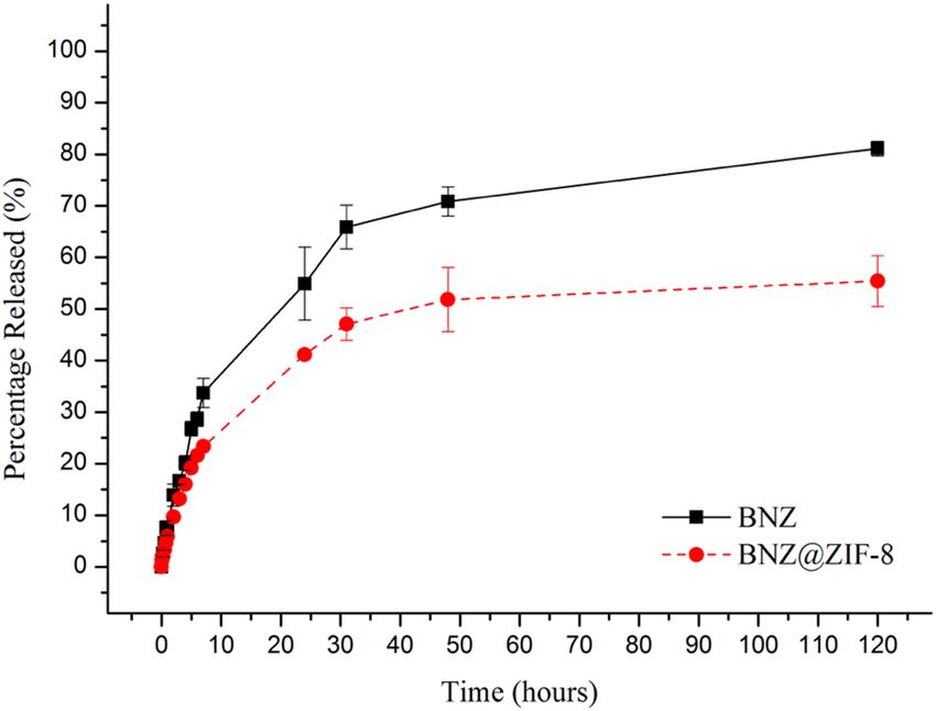
www.nature.com/scientificreports/
(C12H12N4O3—260.25 g mol−1), is the only drug available for treatment of Chagas disease in countries such as
Brazil, Argentina, Chile and Uruguay and the only one marketed for this purpose in Latin A merica2–5.
−1
The recommended treatment is 5 to 7 and 5 to 10 mg kg orally, divided into two or three daily doses for
60 days for children and adults, respectively. First, BNZ was formulated only as tablets for oral administra-
tion (100 mg), culminating in treatment limitations mainly in child patients, where the drug was grossly frag-
mented, increasing the risk of incorrect doses and the incidence of toxic effects or therapeutic failure. Then, in
2011, a pediatric dose of BNZ (12.5 mg) tablets began to be marketed, the result of an initiative by the Drugs
for Neglected Diseases Initiative and Brazilian LAFEPE, the only producer of the drug. Nowadays, efforts are
being made to develop more effective dosage forms, capable of increasing patient compliance and reduced toxic
effects4–6.
Although BNZ has been used since the 1970s, its mechanism of action is not well understood. Several obser-
vations in the literature converge to formation of toxic metabolites to the parasite’s DNA and RNA, proteins
and lipid molecules by reducing the nitro group of the drug mediated by T. cruzi type I nitroreductase. Effective
against epimastigotes, trypomastigotes and amastigotes, these studies suggest that BNZ increases the concentra-
tion of superoxide anions and nucleophilic metabolites at the mitochondrial level, meanwhile forms covalent
bond of its nitro group with T. cruzi macromolecules reducing the parasite’s metabolism. Other findings also
suggest that BNZ increases the phagocytosis and lysis of T. cruzi through an interferon-gamma-dependent
mechanism (INF-g) and inhibits NADH-fumarate reductase enzyme and therefore the growth of the p arasite7–9.
Furthermore, according to the biopharmaceutical classification system, BNZ is classified as class II, thus, it
has low solubility in aqueous fluids (about 0.2 mg mL−1). Consequently, it has limited absorption due to the low
dissolution rate. In addition, its low bioavailability requires a considerable dose of drug, which may be related
with its high toxicity, leading to some serious adverse reactions, such as skin allergies, bone marrow suppres-
sion and peripheral neuropathy. Nevertheless, it is still used in the acute phase, with chances of cure, and in the
chronic phase, preventing the disease progression. Therefore, as it is a worldwide public health problem, there
is a certain urgency for the development of therapeutic a lternatives6,10–12.
Due to the low solubility in aqueous medium, BNZ has a reduced absorption in the gastrointestinal tract, and
therefore, has low bioavailability. Thus, to address these problems, increasing solubility and modulating drug
release through smart excipients the Drug Delivery System (DDS) has been developed (Gomes et al.13; Lima
et al.14; Soares-Sobrinho et al.15). Among the most recent DDS the Metal Organic Frameworks (MOFs) have
received growing attention. MOFs are organic–inorganic hybrid materials that have promising properties to be
used as carriers of drugs, such as: improving bioavailability and biocompatibility of drugs, large surface area,
high thermal stability, insertion capacity of ionic compounds and small molecules, and possibility of promoting
controlled release of substances13–20.
Zeolitic Imidazolate Framework (ZIF) is a subclass of MOF composed of tetrahedral coordination metals,
usually zinc or cobalt, interconnected through imidazoles or imidazolates linkers. Although they are similar to
zeolites, ZIFs are getting more attention due to a larger surface area, permanent porosity, different topologies and
high thermal stability. Among the ZIFs, ZIF-8 has recently been used for several purposes, such as: membrane
constituents, gas exchange and storage, biosensors, catalysis agents and drug carriers for the development of
DDS20–24.
The ZIF-8 has tetrahedral zinc as a coordination metal and is stabilized by the bridges formed by 2-meth-
ylimidazole at an angle of 145°, just like sodalites. It presents itself as a lamellar structure maintained through
compensation between π–π stacking interaction and hydrogen bonds, resulting in a one-dimensional polymeric
chain of densely interconnected lamellae. This conformation gives the coordination framework a central octahe-
dral pattern shared by tetragonal and hexagonal faces, with cavities of about 11.6 Å and 0.4 nm3 that contribute
to a wide surface area and that can be used for the delivery of drugs and the use of the ZIF-8 as a c arrier25–30.
Because it is the second most abundant metal in the human body and the imidazole group is found in the
amino acid histidine, ZIF-8 has good biocompatibility and safety26. Thus, ZIF-8 has potential as a pharmaceutical
adjuvant due to its adsorption capacity followed by the release of drugs, whether hydrophilic or hydrophobic
given its organic–inorganic c omposition28–30. In addition, as it has a flexible structure, the ZIF-8 presents a
phenomenon called breathing, which ends up promoting a modulated release of drugs. The molecule has a
simple and fast synthesis and is still highly chemically and thermally stable, withstanding temperatures of up to
400 °C before decomposition, which allows a series of industrial operations31,32. The development of DDS using
ZIF-8 as smart excipient might be an important tool for the transport and control of drug release kinetics from
a pharmaceutical dosage form with the main objective of establishing constant therapeutic plasma levels, within
the therapeutic index, with high bioavailability and less toxic e ffects33–36.
Therefore, this study aimed to analyze the release kinetics and cell viability of a new drug delivery system
based on BNZ carried by the ZIF-8 framework, using in vitro dialysis and cytotoxicity tests in order to provide
the necessary subsidies for the development of new pharmaceutical products for Chagas disease.
Results and discussion
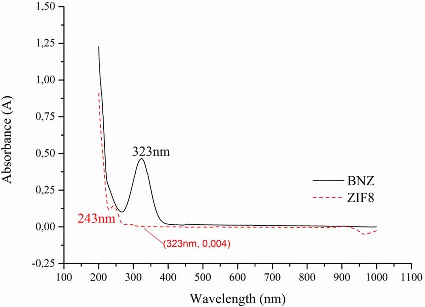
Evaluation of drug incorporation in ZIF‑8 system. Through absorption spectroscopy in the UV–Vis
region it was possible to determine the reading wavelength to be used for the quantification of BNZ incorpora-
tion. Based on the scan (200–1000 nm) (Fig. 1), the BNZ solution (15 μg mL−1) had maximum absorption at
323 nm, while the ZIF-8 at 243 nm. In order to prove that ZIF-8 would not be a contaminant capable of interfer-
ing with the selectivity of the BNZ quantification method, a quantity of 10 mg of ZIF-8 was suspended in 100 mL
of ultra-pure water, followed by filtration (pore filter: 0.22 μm) and quantification using UV–Vis.
Scientific Reports | (2020) 10:16815 | https://doi.org/10.1038/s41598-020-73848-w 2
Vol:.(1234567890)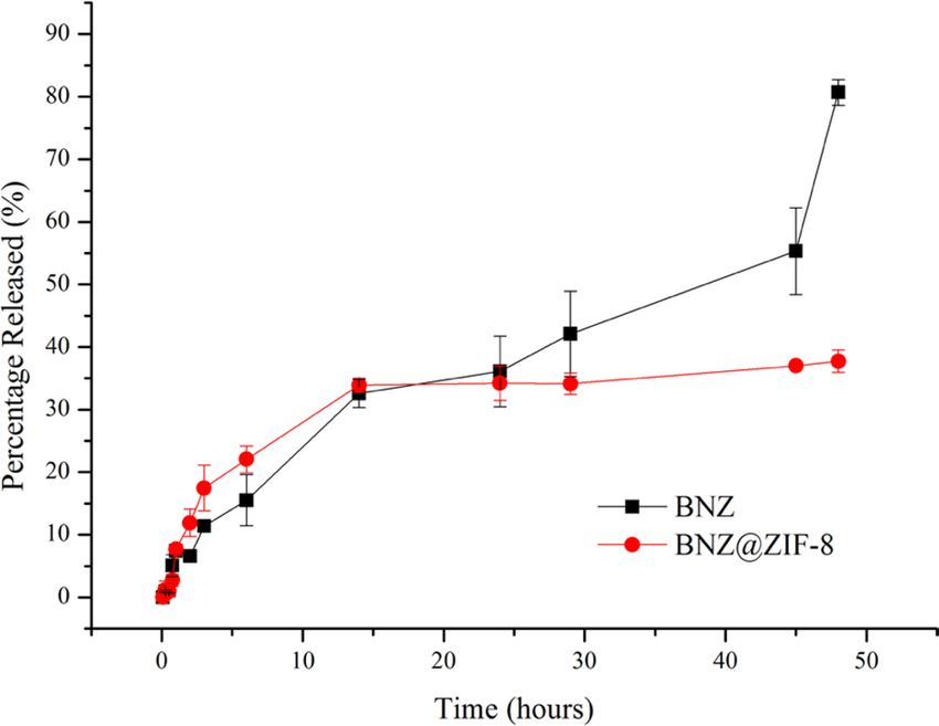
www.nature.com/scientificreports/
Figure 1. UV–Vis spectra of BNZ and ZIF-8.
Figure 2. TG curves of BNZ, ZIF-8, physical mixture (PM) and BNZ@ZIF-8 (β = 10 °C min−1).
Although ZIF-8 also shows peak absorption in the UV–Vis region, the method did not present selectivity
problems because the material does not absorb at the same wavelength. In addition, ZIF-8 is insoluble in water
and in nonpolar solvents, being excluded from the analyte the moment the sample is fi ltered26.
Thus, it was possible to build daily calibration curves, based on the method developed by Soares-Sobrinho
et al.15 making it possible to calculate the apparent incorporation of BNZ into the ZIF-8 network. The experi-
ment aimed to analyze the drop in drug concentration, justified by its incorporation into the ZIF-8 network. This
decrease was evaluated daily and the percentage of incorporated BNZ was measured by subtracting the value
of the actual concentration used in the experiment by the dissolved concentration found, represented in terms
of percentage, as described by Eq. 1 in experimental section. From the incorporation curves it was possible to
observe that, after 4 days of intermittent agitation, the value of IE% was 38%.
Thermal analysis (DSC, TG/DTG). BNZ TG curve (Fig. 2) showed a mass loss of 1.62%, between 30 and
105 °C relative to the water content in the sample. Thermal degradation of the isolated drug was observed in only
one single event (257.15–297.13) (DTG peak = 287.43 °C). A mass loss of 45.66% was found.
In the TG curve of ZIF-8 (Fig. 2), the high thermal stability of the molecule was observed31,32. First, it was
possible to show the mass loss of up to 2.16% in the temperature range between 30 and 125 °C. This value may be
related to the loss of water molecules in cavities or on the surface of ZIF-831. At 460.40 °C, there were loss of only
7% of the initial mass, which evidences the high thermal stability of the ZIF-8. This value can also indicate the exit
of water molecules connected to the ZIF-8 network. In the range of 535.69–578.16 °C it was possible to observe
a significant mass loss—better evidenced by the DTG curve—of 24.32%, related to the molecule organic portion
degradation, the imidazolate. Since then, the sample presents slow decay, with low resolution, and no apparent
peaks, being difficult to identify them even by the DTG curve, probably caused by thermal decomposition of
Scientific Reports | (2020) 10:16815 | https://doi.org/10.1038/s41598-020-73848-w 3
Vol.:(0123456789)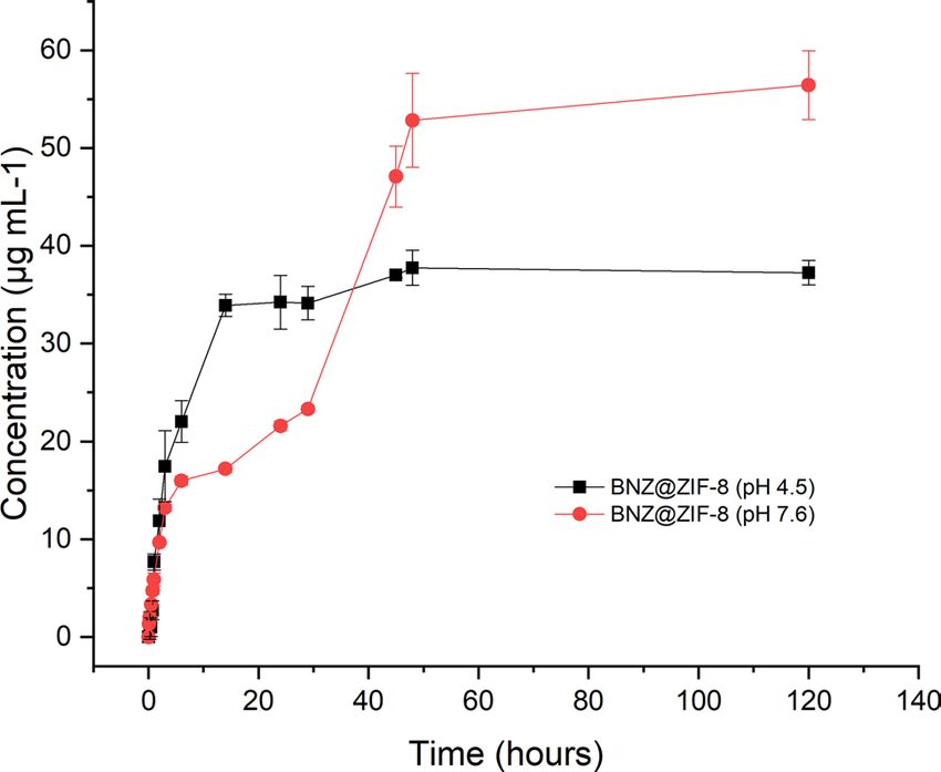
www.nature.com/scientificreports/
Figure 3. DSC curves of BNZ, ZIF-8, physical mixture (PM) and BNZ@ZIF-8 (β = 10 °C min−1).
the inorganic portion of ZIF-8, until the formation of zinc oxide (620.35–720.11 °C—41.91% mass loss). These
results are in accordance with previous s tudies37–39.
In the TG curve of physical mixture (PM) (Fig. 2) it was possible to see the anticipation of the thermal degra-
dation of the isolated drug, now occurring in the range of 229.89–247.23 °C (DTGpeak = 238.04 °C), subsequent
to drug melt. However, a significant reduction of mass loss (24.49%) was observed, suggesting a certain thermal
protection in the reduction of mass loss. In relation to ZIF-8 degradation, the first event of thermal degrada-
tion—related to degradation of imidazolate—was observed in a range quite different from that found for ZIF-8
alone (between 410.89 and 448.54 °C), also presenting reduction of mass loss (11.55%). This fact suggests that
the physical interaction between drug and ZIF-8 may destabilize the organic portion of the latter. Then, the TG
curve decayed to approximately 975.07 °C corresponding to residual zinc oxide (36.78% of mass loss).
In the analysis of the BNZ@ZIF-8 (Fig. 2), a new mass loss event was observed, between 50.13 and 87.94 °C,
with a mass loss of 1.97%. Probably, this event is related to the volatilization of acetone that still remains inside the
ZIF-8 network, since the decay can be observed from room temperature, with acetone having low vapor pressure
and boiling point. This value, smaller than that found for the isolated ZIF-8, still shows that there is a smaller
amount of water (moisture) present in the cavity of the ZIF-8, and therefore, gives space for the connection with
the molecules of the drug. The rest of the TG curve showed some consonance with the previous results, from
the aqueous medium obtained system. An anticipation of the thermal degradation of the drug over the isolated
BNZ (223.39–245.34 °C) was also o bserved23,26,40.
However, there was an even more significant reduction in mass loss (26.49%). The degradation of the inor-
ganic portion of ZIF-8 occurred in the range of 321.02–434.88 °C, with a mass loss of 16.15%; followed by
thermal decomposition to zinc oxide (568.51–613.31 °C and 32.78% mass loss). Although it has been shown
that ZIF-8 degradation values were anticipated, both showed a significant reduction of the decomposed content.
The difference between this thermal profile and that evidenced by PM may be a great indication of the actual
formation of systems, corroborating the fact that the drug is actually present in the ZIF-8 cavities23,40. However,
new investigations about this fact should be made in order to analyze what, in fact, promoted this behavior.
BNZ DSC curve (Fig. 3) showed an intense and defined endothermic peak in the temperature range between
190.04 and 194.36 °C (Tpeak = 191.44 °C) (ΔH = 195, 52 mJ). Then, the exothermic peak related to drug degra-
dation was observed in the range of 272.57–292.79 °C (Tpeak = 286.90 °C), which showed large energy release
(ΔH = 1.8 J). Similar results were described by Santos et al.41
The DSC curve of ZIF-8 (Fig. 3) demonstrates the absence of peaks in the temperature range used, a feature
inherent in the nature of the molecule. Since it is an organic–inorganic hybrid molecule, the ZIF-8 degradation
events were analyzed through the DTA curve (Fig. 4), which enabled the sample to heat up to 900 °C. In that
point, it was possible to observe two endothermic events regarding phase transition: the first one between 535.69
and 578.16 °C; and the second, between 620.35 and 720.11 °C. These values and their mass losses are described
in the discussion of the TG curve of ZIF-8. Such behavior can be observed in zinc-containing materials. Similar
results were discussed by Blachnik and Siethoff42. Such solid–solid phase transitions suggest conformational
changes of the alkyl chains, where the number and size of the transformations occur as a function of the length
and number of the alkyl chains42.
From the DSC curve of the PM (Fig. 3) it was possible to evidence the anticipation of the drug fusion event
(187.82–194.28 °C). Although this fact can identify interactions between the components of a formulation, the
curve also showed the reduction of enthalpy energy related to the event (ΔH = 195.52 mJ). This phenomenon is
characteristic of polymeric materials, such as: PEG, PVP, HPMC; which makes it possible to increase the solubil-
ity of the material, since there is need for a smaller amount of energy to solubilize the drug. Such behavior has
already been widely discussed by previous studies of the g roup43,44.
Scientific Reports | (2020) 10:16815 | https://doi.org/10.1038/s41598-020-73848-w 4
Vol:.(1234567890)www.nature.com/scientificreports/
Figure 4. DTA curve of ZIF-8 (β = 10 °C min−1).
Thus, it is clear that simple physical mixing promotes the formation of a drug–excipient interaction, even
without causing the formation of the system itself. On the other hand, there was an intense anticipation of the
event related to the degradation of the drug (Tpeak = 231.08 °C), almost occurring subsequent to the fusion of
the same. However, there was lower energy release (ΔH = 1.09 J). These values are in line with those evidenced
by the DTA curve.
BNZ@ZIF-8 DSC curve (Fig. 3) shows an endothermic event between 54.17 and 62.85 °C, regarding the
volatilization of acetone. This behavior, different from that presented in PM, can characterize the formation of
the BNZ@ZIF-8. A much more discrete anticipation (184.44–192.91 °C) (Tpeak = 189.97 °C) was observed in
the drug fusion. This variation—approximately 2%—is described by many authors as an acceptable range of
compatibility between components of the same formulation43,45. Therefore, this behavior can identify that the
system obtained in acetone is, in fact, more efficient for drug incorporation. However, the value for the enthalpy
change was higher in comparison to the isolated drug (ΔH = 261.25 mJ), which may indicate that the system
was formed, since a new thermal profile was evidenced. Regarding drug degradation, the exothermic event was
anticipated (216.72–244.86 °C) (Tpeak = 234.14 °C), releasing an energy of 1.8 J, identical to that presented by
the isolated drug.
All the above-mentioned information shows the importance of thermal analysis as a technique for the char-
acterization of DDS. Through the TG/DTG, DTA and DSC curves it was possible to corroborate the fact that the
acetone system made possible a more efficient incorporation of the drug, either by changing the thermal profile
or by the compatibility between the used components.
X‑ray diffraction spectroscopy. It was possible to identify the characteristic peaks of both BNZ crystals
(7.36°, 16.28° and 21.86°) and ZIF-8 (7.44°, 10.46° and 12.78°, corresponding to reflections 011, 002 and 112,
relating to a body-centered hub). These data were used to calculate the basal spacing in order to establish a
reference value to characterize the insertion of the BNZ molecule in ZIF-8. These results are in line with recent
works12,31,46.
The BNZ@ZIF-8 system also presented crystalline behavior, resulting in the sum of the XRD profiles of the
isolated substances. However, different from PM, the system showed the most characteristic peaks of the BNZ
(7.36°, 10.88°, 16.82° and 21.88°), which suggests the formation of the coupled system, even though it is only
characterized by the physical adsorption of the BNZ on the surface of ZIF-8.
In this material, a reduction of the peak intensity characteristic of ZIF-8 (7.44°) was observed indicating the
strong attraction between the BNZ and the ZIF-8 network. Similar results were evidenced by Liédana et al.20 It is
important to notice that the obtained XRDs demonstrate that the structural integrity of ZIF-8 remains unchanged
after the adsorption of the BNZ. This is of great importance because it shows that even though the formation of
the systems promotes some reduction of the peaks of the molecule, and the crystalline structural integrity of the
ZIF-8 is m aintained34–36,40. The XRD results of all materials are summarized in Fig. 5.
Basal spacing values, defined by the Bragg equation, were used as a parameter to confirm the insertion
of the drug into the ZIF-8 molecule. Values greater than baseline suggest that the drug was inserted into the
molecule and, therefore, dilates the space between the lamellae. However, the discrete increase in basal spacing
was observed when the isolated ZIF-8 was compared to the PM and to the obtained system (Table 1), mainly
at the peak of reflection 110, the most characteristic of ZIF-8 crystallinity and of the rhombic dodecahedral of
its crystals (He et al.41). This fact suggests the formation of the system but suggests that it is the product of an
Scientific Reports | (2020) 10:16815 | https://doi.org/10.1038/s41598-020-73848-w 5
Vol.:(0123456789)www.nature.com/scientificreports/
Figure 5. XRD of the BNZ, ZIF-8, PM and the BNZ@ZIF-8 with their respective main peaks.
Sample Peak (° 2θ) Reflection Basal spacing (Å)
7.44 110 11.87
ZIF-8 10.46 002 8.71
12.78 112 6.94
7.32 110 12.04
PM 10.4 002 8.51
12.70 112 6.97
7.3 110 12.11
BNZ@ZIF-8 10.32 002 8.10
12.7 112 6.97
Table 1. Calculation of the basal spacing of the reflection peaks 110, 002 and 112 of ZIF-8, Physical Mixture
and Systems obtained in water and acetone.
adsorption of the drug to the surface of the ZIF-8, characterizing a physical interaction, since the increase of
the basal spacing is very small.
Fourier transform infrared absorption spectroscopy. The BNZ spectrum (Fig. 6) shows characteris-
tic peaks, especially when considering typical bands of amides (N–H stretching vibration), carbonyl stretching
(amide band I) and N–H deformation (amide band II), in addition to vibrations resulting from the benzyl and
imidazole groups, and the nitro group. The band from the N–H stretching vibrations is located at 3266 cm−1, the
carbonyl stretching band at 1664 cm−1 and the NH (amide II) deformation at 1552 cm−1, characterizing the sec-
ondary amide. In addition, the band at 1292 cm−1 is attributed to the C–N stretch. The set of bands at 3033, 3068,
3112 and 3269 cm−1 arise from the symmetrical and asymmetric stretching vibrations of the benzene group and
the stretching of the aromatic C–H, as shown in Fig. 6.
The region of the harmonics and combination bands (2000 to 1667 cm−1), often useful in determining the
number and position of substituents on aromatic rings, appears to be less informative in the spectrum, not being
observed the four small bands that would characterize mono-substituted benzene present in the molecule. The
band at 1355 cm−1 refers to vibration of the symmetrical stretching of the nitro g roup44.
Looking at Fig. 6 referring to ZIF-8, at 3132, 2962 and 2936 cm−1, the presence of C–H aromatic com-
pounds, asymmetric axial deformation and aliphatic C–H stretching relative to the imidazole ring, respectively.
In 1670 cm−1 a stretch band of C=C was observed, while the absorption C–N bands appeared at 1100 to 1400 cm−1
Scientific Reports | (2020) 10:16815 | https://doi.org/10.1038/s41598-020-73848-w 6
Vol:.(1234567890)www.nature.com/scientificreports/
Figure 6. Infrared spectra of BNZ, ZIF-8, physical mixture (PM) and BNZ@ZIF-8.
region (Park et al.31). It is not possible to observe the behavior of the Zn–N stretch at 450 and 400 cm−1, consider-
ing that the analyzes were done in equipment that operated up to 600 cm−1.
The PM spectrum (Fig. 6) corresponds to the overlapping of the same bands of BNZ and ZIF-8 when these
are analyzed in isolation, it is possible to note the presence of their characteristic peaks. This result suggests a
physical interaction only between the drug and ZIF-8 due to the sum of the profiles of the isolated materials.
Analyzing the BNZ@ZIF-8 (Fig. 6), the peaks of the isolated BNZ and ZIF-8 are not well evidenced due to
their overlapping. However, it was possible to suggest the presence of the drug in question, due to the presence,
in 3266 cm−1 and 1292 cm−1, of the axial deformation of the N–H and C–N bond, respectively. In 1664 cm−1 there
is a decrease in the intensity in the carbonyl band, at 1355 cm−1 for the nitro group and 1552 cm−1 corresponding
to the secondary amide. In addition, there was a decrease in the intensity of the absorption band related to the
C–N group of ZIF-8 (1400 and 1302 cm−1), confirming that it interacts effectively with B NZ26.
Thus, when comparing the infrared spectra of BNZ@ZIF-8, PM and the isolated substances, it was observed
that the system presented the main peaks in a lower intensity, indicating, therefore, the formation of the system
due to the signs of interaction between BNZ and ZIF-840.
In vitro dialysis release method and the breathing phenomena. The in vitro release assay through
a dialysis membrane was performed in order to estimate the BNZ permeation from the ZIF-8 network. The dif-
fusion of the drug through the dialysis membrane pores (1000 Da) is possible due to its molecular size (about
260.25 Da). Thus, the BNZ molecules are able to be released through the dialysis membrane while the ZIF-8
network is dissociated or eroded. In contrast, ZIF-8 cannot permeate through the dialysis membrane due to its
large structural size and rigid polymeric matrix.
At pH 4.5 (Fig. 7), the BNZ alone achieved 80% of release after 48 h with an almost linear release kinetics.
On the other hand, the BNZ@ZIF-8 system, in the same time interval, presented a slower release of BNZ (34%),
without oscillations. After 25 h, the drug and the BNZ@ZIF-8 system exhibit a similar release, approximately
39 and 35%, respectively. However, after this point the system presents the formation of a plateau, stabilizing
the release of the drug in this same range until the end of the experiment, while the drug alone continues to be
released in a linear kinetics, reaching the percentage expected for the end of the test.
In order to compare the dissolution profiles between the BNZ and the system, the model-independent method
that calculates the value of ƒ2 was used. The value of 190.87 shows that the release profile among them is differ-
ent, once ƒ2 > 100.
At pH 7.6 (Fig. 8), the isolated BNZ achieved 80% of release after 120 h of assay, due to its low aqueous
solubility. In contrast, the BNZ@ZIF-8 system presented a slower release, excluding the burst effect presented
by the BNZ alone. After 7 h, the BNZ alone achieved 40% of release, while the BNZ@ZIF-8 system released only
23% of the drug. This difference of percentage—approximately 20%—remains constant up to 120 h, where the
system reaches only 57% of drug release.
It was evident that, in fact, the drug release modulation occurred, since the release profiles were not similar
due to the obtained ƒ2 value: 213.06 (> 100).
Figure 9 provides a comparison between the results obtained from dialysis in sink conditions at different pH
values. It was possible to observe a fast release at pH 4.5, with a pronounced and almost linear burst effect, releas-
ing about 34% in 14 h. Afterwards, a plateau concentration maintenance is observed, reaching approximately
Scientific Reports | (2020) 10:16815 | https://doi.org/10.1038/s41598-020-73848-w 7
Vol.:(0123456789)www.nature.com/scientificreports/
Figure 7. In vitro cumulative drug release (%) of BNZ and BNZ@ZIF-8 system through dialysis membrane
(10,000 Da) under sink conditions at pH 4.5.
Figure 8. In vitro cumulative drug release (%) of BNZ and BNZ@ZIF-8 system through dialysis membrane
(10,000 Da) under sink conditions at pH 7.6.
38% of release until the end of the experiment. MOFs usually show the structural transition phenomena upon the
input of external stimuli, known as the breathing phenomenon. This phenomenon is common among MOFs. Due
to the flexibility of these molecules in an acidic environment, they open exit gates for the adsorbed substances
that were previously trapped by the coordination p olymer33,46.
Also called “easing”, the breathing phenomenon of ZIF-8 is extremely important in drug delivery applications,
allowing the adsorption and diffusion of large molecules. The breathing flexing of the ZIF-8 framework is the
process in which the movement/twisting of the imidazolate linkers in the network, providing a greater opening
of the crystal pore windows, causing a dilation or contraction of the pores, directly interfering with the adsorbed
load and in the diffusion of molecules. ZIF-8 presents the beginning of the dissociation of the coordination net-
work in the pH range between 5.0 and 6.0, and intense erosion in an even more acidic pH, which can promote
the network’s “breathing”, opening of spaces between the polymeric mesh and, consequently, intensifying the
drug release which, in this case, can be modulated by changing the p H37,38,47.
At pH 7.6, an attenuation of the burst effect is observed, since only 17% of BNZ is released in the same time
interval of 14 h. The release follows slowly, modulated, with about 53% released in 48 h, where a balance is estab-
lished, since the release is about 56% until 120 h. Then, the dissolution studies corroborated that a pH sensitive
DDS capable of vectorizing the release of BNZ was developed.
It is evident that a lower percentage of drug is released from the BNZ@ZIF-8 system in a time interval, com-
paring to the BNZ alone. This behavior should be explained by the interactions between the drug that is closely
linked to the ZIF-8 coordination network, although only physically adsorbed. It should be noted that, while the
drug dissociates and diffuses, a new sorption and desorption equilibrium of the drug can be achieved, which
further prolongs even more, the release time interval.
Scientific Reports | (2020) 10:16815 | https://doi.org/10.1038/s41598-020-73848-w 8
Vol:.(1234567890)www.nature.com/scientificreports/
Figure 9. Comparative profile of the BNZ@ZIF-8 system through dialysis membrane (10,000 Da) under sink
conditions at pH 4.5 and 7.6.
An important factor to be considered during dialysis is that the membrane acts as a barrier between ZIF-8
suspension after drug release and the external environment. However, to be quantified in the external environ-
ment, the drug must diffuse not only through the ZIF-8 network, but also through the dialysis membrane.
The pH values used in this study are extremely interesting for the pharmaceutical field in the development
of pH-dependent DDS. For example, by modulating the release of the drug to occur at an acidic pH only, it is
possible to avoid an intense drug release in bloodstream and thus, reducing the incidence of adverse effects and
increasing the selectivity of the treatment. This fact is particularly interesting for the treatment of Chagas disease.
In the life cycle of T. cruzi, the parasite provides mechanisms of action capable of favoring its entry into the
host-cell cytosol. For example, the rupture of the parasites phagocytic vacuole marks the transformation of the
trypomastigote phase into amastigote. At this stage, about 70% of the parasites will be free in the cytosol 2 h after
infection. Some studies suggest that this is due to the secretion of acidic substances, such as hemolysin, which has
maximum activity at pH 5.5. Some of this studies showed that the rupture of the vacuole occurs around pH 6.0
and, with the addition of slightly more basic substances (pH 6.2), the escape of parasites is inhibited. Therefore,
it is inferred that the release of the parasites and the continuity of the life cycle of the parasite are related to a
change in pH to slightly more acidic l evels39,48,49.
In vitro evaluation of cell viability and cytotoxicity using the MTT assay. The low cytotoxic activ-
ity of the materials was verified. Even with the highest concentrations tested (100 μg mL−1), all samples induced
low levels of cell death (viability > 80%) over a 24-h period (Fig. 10A). After 24 h, ZIF-8 (150 μg mL−1) showed
low cytotoxic potential (viability of 98.2% vs control 99.6%). The BNZ showed viability varying between 83.9
and 87.6%. For BNZ@ZIF-8 systems, cell viability values were 88.6%, 87.7% and 84%, for the doses of 1, 10 and
100 μg mL−1, respectively. There were significant differences between the tested and control groups (**p < 0.05,
***p < 0.001), and a significant difference between the cytotoxic activity of ZIF-8 alone and when combined in
the systems (#p < 0.05). Furthermore, there was a difference between the system at 10 μg mL−1 when compared
to the drug alone at the same dosage (δp < 0.05).
After 48 h of exposure (Fig. 10B), the viability of the ZIF-8 exposed cells alone remained high (97.3%)
versus a range of 80 to 87% of the BNZ groups. In the BNZ@ZIF-8 system, the values were 88.5%, 87.7% and
82.4%, for 1, 10 and 100 μg mL−1, respectively. There were significant differences between the tested and control
groups (**p < 0.05, ***p < 0.001), and a significant difference between the ZIF-8 alone activity when compared
to the BNZ@ZIF-8 systems (p < 0.05). Moreover, it was possible to observe a difference between the BNZ@ZIF-8
system at 10 μg mL−1 and the BNZ alone at the same concentration, as well as the system BNZ@ZIF-8 activity
(100 μg mL−1) when compared to the BNZ alone at this same concentration (δδp < 0.05). The slow release of
the drug by the BNZ@ZIF-8 system may have positively affected the toxicity reduction, in comparison with the
drug alone.
Thus, the low cytotoxic activity of the materials was evident. Even after 48 h of treatment, no statistically
significant toxic effects occurred in the concentrations tested on cell viability. The slow release of the drug by
the BNZ@ZIF-8 system may have positively affected the toxicity reduction, in comparison with the drug alone.
ZIF-8 and BNZ@ZIF-8 system did not show significant cytotoxicity in the cells tested by the MTT method.
ZIF-8 has been shown to be chemically inert, evidencing the fact that it may be a carrier as a pharmaceutical
adjuvant. These results are in accordance with the data presented by Alves and Ren et al.50,51
Finally, the present work suggests that the BNZ@ZIF-8 system can be used as an antitrypanosomal API
in pharmaceutical forms, since it has been shown to be safe due to its low cytotoxic potential at the times and
Scientific Reports | (2020) 10:16815 | https://doi.org/10.1038/s41598-020-73848-w 9
Vol.:(0123456789)www.nature.com/scientificreports/
Figure 10. Effects of BNZ, ZIF-8 and BNZ@ZIF-8 at different concentrations on human cell viability (PBMC)
according to the MTT assay at exposure time: (A) 24 h and (B) 48 h (**p < 0.05, ***p < 0.001 are related to
significant differences between the tested and control groups; #p < 0.05 to the comparison between activity of
ZIF-8 alone and when combined in the systems and δp < 0.05 the difference between the system and the drug
alone, both at 10 μg/mL).
concentrations tested. However, further studies are required in order to evaluate the in vitro and in vivo efficacy
and cytotoxicity of these materials.
Conclusions
The dialysis assay provided an estimative of the pH-dependent release from its own carrier (ZIF-8) and through
the dialysis membrane. A faster release of BNZ in the system was observed at pH 4.5, when compared with pH
7.6. Furthermore, the BNZ concentration in both situations was lower than in the BNZ alone, keeping a pro-
longed release without oscillations and the burst effect observed in the drug alone. The drug release modulation
could be proven by the model-independent method. By adjusting the release profiles, it was possible to suggest
the mechanism of drug release from the BNZ@ZIF-8 system, which occurs depending on the pH tested.
Experimental
Materials. BNZ was acquired from the Pharmaceutical Laboratory of the State of Pernambuco (LAFEPE),
lot 301045 (99.5%), while the ZIF-8, lot S45328-308, was donated by the Laboratório de Terras Raras of UFPE.
Acetone (Modern Chemistry, lot 03196) and ultra-pure water (Mili-Q) were also used. Potassium phosphate
(Vetec, lot 1007140) and sodium hydroxide (Sigma-Aldrich, batch SLBM7637) were used for the preparation of
the dialysis test medium. The dialysis bags (10,000–12,000 Da) were obtained from Sartorius, Germany.
Development of BNZ@ZIF‑8 systems. The obtaining method used in the present study was based on
the work of Horcajada et al.36 and optimized by our research group, which promoted the drug adsorption using
a ZIF-8 suspension though an ex situ method36. The procedure used the molar ratio of BNZ@ZIF-8 1:1, based on
the molecular weights of BNZ and ZIF, which are 260.25 and 229.61 g mol−1, respectively.
Initially the drug was solubilized with acetone in Erlenmeyer flasks with capacity of 250 mL, yielding an initial
concentration of 20 μg mL−1. The BNZ solution was sonicated for 10 min in a Limp Sonic sonicator to ensure
complete solubilization. At the end of the shaking, the ZIF-8 was suspended in acetone and added at the BNZ
solution. Subsequently, the volume was filled with the respective solvent, submitting the mixture to intermittent
stirring on Magnetic Stirrer MA089 Marconi with the aid of a magnetic bar for 4 days.
The supernatant was collected daily (0.33 mL, followed by solvent replenishment) and filtered through a
hydrophobic filter with 0.22 μm pore aperture for further quantification by absorption spectroscopy in the
Ultraviolet–Visible region (UV–Vis) with the aid of a previously performed calibration curve (Line equation:
y = 0.0316x − 0.0003; R2 = 0.9994) for the construction of a BNZ incorporation curve into the ZIF-8 network. This
process was used to determine the maximum incorporation efficiency (IE%), defined by Eq. 1.
[BNZ theoretical concentration] × [BNZ real concentration]
IE% = × 100 (1)
[BNZ theoretical concetration]
At the end of the process, the material was centrifuged at 2000 RPM for 20 min in order to remove residual
BNZ. The supernatant was discarded and the precipitate was washed with acetone. The same procedure was
repeated twice. Then, the material was dry in a drying oven with 35 °C airflow (Shel Lab) until completely dry
(about 4 h). Once dry, the material was used to quantify the EI% of the ZIF-8 network. Finally, the system pre-
sented an IE% = 38% (± 1.28%) and then it was called BNZ@ZIF-8. The BNZ@ZIF-8 system, composed by the
Scientific Reports | (2020) 10:16815 | https://doi.org/10.1038/s41598-020-73848-w 10
Vol:.(1234567890)www.nature.com/scientificreports/
association BNZ:ZIF-8 in 1:1 mol/mol ratio, presented itself as the best system among the tests carried out with
other ratios in three levels (SM Fig. 1).
This entire procedure was done in triplicate and was performed in the absence of light due to the inherent
photosensitivity of BNZ, as observed in previous studies. Due to the presence of acetone, the Erlenmeyer flasks
were sealed in order to reduce the volatilization of the solvent. However, the volatilized content was constantly
restored after verification of mass loss, based on the value of 0.79 g mL−1 for the acetone’s density44.
Systems characterization. The materials were characterized by several analytical techniques to confirm
the formation of the BNZ@ZIF-8 systems: Scanning Electron Microscopy (SEM), Polarized Light Microscopy
(MLP), X-ray Diffraction (XRD), Thermal Analysis: Thermogravimetry (TG), Calorimetry Differential Scan-
ning (DSC), Fourier transform infrared absorption spectroscopy (FTIR), Laser particle size and surface area
analysis, and pore size and volume.
TG curves were performed using a Shimadzu Thermocouple, model TGA Q60, under a flowing nitrogen
atmosphere of 50 mL min−1, sample mass (3.00 mg ± 0.05) for isolate BNZ and ZIF-8 and (6.00 mg ± 0.05) for
platinum crucible Physical Mixtures (PM) and system in the temperature range of 25 to 1000 °C at the heating
rate (β) of 10 °C min−1. Prior to the tests, thermobalance was checked with calcium oxalate. The BNZ and PM
DSC curves were obtained using Shimadzu DSC-60 Calorimeter, interconnected to the Shimadzu TA-60WS soft-
ware, with 50 mL min−1 nitrogen atmosphere and 10 °C min−1 heating rate in the range temperature of 25–500 °C.
The samples were placed in a hermetically sealed aluminum sample holder with a mass of 3.0 ± 0.2 mg for BNZ
and ZIF-8 and 6.0 ± 0.2 mg for PM and BNZ@ZIF-8 system. The determinations were performed in triplicate
and indium and zinc were used to calibrate the temperature scale and the enthalpy response.
UV–Vis Spectra was obtained by scanning from 190 to 1000 nm. The ultraviolet spectrophotometer Shimadzu
UV-2401 PC and quartz cuvettes with a cross section of 1 cm were used.
The FTIR spectra of BNZ, PM and BNZ@ZIF-8 were obtained using the PerkinElmer equipment (Spectrum
400) with an attenuated total reflectance (ATR) device of selenium crystal. The samples to be analyzed were
transferred directly into the ATR device compartment. The results were obtained by scans from 4500 to 600 cm−1.
XRD analysis was performed on a Shimadzu XRD-7000 diffractometer, equipped with a copper anode at a
scanning speed of 1.2° min−1, in the 2θ angle range from 5° to 45°. Basal spacing was calculated using the Bragg
equation.
In vitro dialysis release method under sink conditions. The in vitro release assay of BNZ was per-
formed by dialysis method in order to define the modulated release profile by the BNZ@ZIF-8 system. Samples
of 18.33 mg of BNZ were used in the dialysis studies, in which, if completely dissolved in a 250 mL of dissolution
medium phosphate buffer release medium at pH 4.5 and 7.6, and 37 ± 0.5 °C, it would present a concentration
of 73.32 μg mL−1, establishing, thus, the sink condition. The dissolution medium was chosen based on recent
studies showing the modulation of the pH-dependent release of the drug from the ZIF-8 n etwork20,26,41. A lower
pH value was not used due to the rapid structural dissociation of ZIF-8, which would result in an extremely
accelerated drug release33.
Sink condition was determined based on a quantitative solubility assay of BNZ performed by our research
group where the maximum solubility in the dissolution media used (about 240 μg mL−1) was measured after
7 days of intermittent stirring. The experiments were performed under the suitable conditions described by
Rohrs52 and the determination of Sink index, which employs dissolution medium volumes of 3 to 10 fold of the
volume present in the saturated BNZ solution for sink condition.
During the experiment, 2 mL of the BNZ@ZIF-8 system formulation and the same volume of drug alone were
both placed in dialysis bags (10.000–12.000 Da) (Sartorius, Germany). The dialysis membranes were immersed
in 250 mL of dissolution medium, under continuous agitation at 75 RPM (Revolutions per minute). Aliquots
(2 mL) from the external medium were collected at time intervals of 0.083, 0.25, 0.5, 0.75, 1, 2 h, followed by an
exploratory collection until the drug release reaches at least 80%. The release medium was replenished at each
collection to maintain the sink conditions. The amount of BNZ released was assessed by UV–Vis (BNZ absorb-
ance wavelength = 323 nm) (Fig. 1).
In order to compare the dissolution profiles between BNZ and the system, the nonlinear method that calcu-
lates the value of ƒ2, known as similarity factor, was used. The ƒ2 metric is a function of the reciprocal of mean
square root transform of the sum of square distances. The ƒ2 can be obtained through Eq. 253.
�
� n
�−0.5
�
� � ��
1
2
f 2 = 50 × log 1+ (Rt − Tt) × 100 (2)
�
n
t=1
where n is the number of collection times considered for calculation purposes; Rt is the percentage value dis-
solved at time t obtained with the reference or comparator curve; and Tt is the percent dissolved value of the test
curve, or the altered formulation at time t.
In vitro evaluation of cell viability and cytotoxicity by MTT assay. For in vitro cellular cytotoxicity
assay, whole human blood (5 mL) from healthy and non-smoking volunteers was obtained by venipuncture (with
informed consent and approved by the National Ethics Committee. Reference number: 30667014.5.0000.5208).
Peripheral blood mononuclear cells (PBMC) were obtained from whole blood by Ficoll-quantum method
(Ficoll-Paque Plus)54,55. Human PBMC were cultured at 5.1 × 10³ cells/well in a 96-wells plate containing Dul-
becco MEM culture medium (DMEM) supplemented with 10% phosphate buffered saline.
Scientific Reports | (2020) 10:16815 | https://doi.org/10.1038/s41598-020-73848-w 11
Vol.:(0123456789)www.nature.com/scientificreports/
The cells were incubated with BNZ and ZIF-8 on 3 different conditions: (1) ZIF-8 at 150 μg mL−1 concentra-
tion, (2) BNZ at 1, 10 and 100 µg mL−1 concentrations and (3) BNZ@ZIF-8 system under the same conditions as
the drug alone. Cells were incubated for 24 and 48 h at 37 °C in 5% C O2. Then the culture medium was replaced
by 100 µL of fresh DMEM with 10 µL of tetrazolium dye 3-(4,5-dimethylthiazol-2-yl)-2,5-diphenyltetrazolium
bromide (MTT) (5 mg mL−1 in phosphate buffer) per well, proceeding with the incubation for 4 h at 37 °C in
the dark in 5% C O2. Untreated cells, MTT and buffer-phosphate were used as negative control and the medium
alone (without cells), MTT and buffer-phosphate were used as the positive control. Dead cells (0.1–1% Triton-
X), MTT and buffer-phosphate were used as the blank. Finally, absorbance (at 570 nm) was recorded by ELISA
microplate reader.
Regarding the statistical analysis, each trial was performed in biological duplicates and technical triplicates,
and the results were expressed as mean ± standard deviation. Statistical analysis was performed using GraphPad
Prism software version 5.0. p values less than 0.05 were considered statistically significant. The results were
compared by analysis of variance (ANOVA), followed by Dunn’s multiple comparison test and Student’s t test.
The concentration required for the half maximal inhibitory concentration (IC 50) was graphically estimated by
linear regression analysis, and correlation indices were calculated using the Pearson (r) coefficient.
Ethical statement. The author declares that all the experiment protocol involving humans is in accord-
ance to guidelines of the National Research Ethics Committee (CONEP) of the National Health Council—Brazil
Ministry of Health (CNS / MS). The blood used in the MTT assay was collected from volunteers after signing the
informed consent form, registered under reference number: 30667014.5.0000.5208.
Received: 27 May 2020; Accepted: 24 August 2020
References
1. Antinori, S. et al. Chagas disease in Europe: a review for the internist in the globalized world. Eur. J. Intern. Med. 43, 6–15. https
://doi.org/10.1016/j.ejim.2017.05.001 (2017).
2. Coura, J. R. & Viñas, P. A. Chagas disease: a new worldwide challenge. Nature 465, 6–7. https://doi.org/10.1038/nature09221
(2010).
3. WHO—World Health Organization. Chagas disease (American trypanosomiasis). https: //www.who.int/health -topics /chagas -disea
se#tab=tab_1 Accessed 30 June 2020.
4. Castro, J. A., De Mecca, M. M. & Bartel, L. C. Toxic side effects of drugs used to treat Chagas’ disease (American trypanosomiasis).
Hum. Exp. Toxicol. 25, 471–479. https://doi.org/10.1191/0960327106het653oa (2006).
5. Kratz, J. M., Bournissen, F. G., Forsyth, C. J. & Sosa-Estani, S. Clinical and pharmacological profile of benznidazole for treatment
of Chagas disease. Expert Rev. Clin. Pharmacol. 11(10), 943–957. https://doi.org/10.1080/17512433.2018.1509704 (2018).
6. Correia, J. P. R. et al. Pharmacotherapeutic follow-up of patients with Chagas disease using benznidazole: drug-related problems
and pharmaceutical interventions. Rev. Soc. Bras. Med. Trop. 50, 334–340. https://doi.org/10.1590/0037-8682-0474-2016 (2017).
7. Campos, M. C. O. et al. Benznidazole-resistance in Trypanosoma cruzi: evidence that distinct mechanisms can act in concert. Mol.
Biochem. Parasitol. 193(1), 17–19 (2014).
8. Hall, B. S. & Wilkinson, S. R. Activation of benznidazole by trypanosomal type I nitroreductases results in glyoxal formation.
Antimicrob. Agents Chemother. 56(1), 115–123. https://doi.org/10.1128/AAC.05135-11 (2014).
9. Rajão, M. A. et al. Unveiling benznidazole’s mechanism of action through overexpression of DNA repair proteins in Trypanosoma
cruzi. Environ. Mol. Mutagen 55(4), 309–321. https://doi.org/10.1002/em.21839 (2014).
10. Ferraz, L. R. M. et al. Technological innovation strategies for the specific treatment of Chagas disease based on Benznidazole. Acta
Trop. 185, 127–132. https://doi.org/10.1016/j.actatropica.2018.02.008 (2018).
11. Field, M. et al. Anti-trypanosomatid drug discovery: an ongoing challenge and a continuing need. Nat. Rev. Microbiol. 15, 217–231.
https://doi.org/10.1038/nrmicro.2016.193 (2017).
12. Santos, F. L. A. et al. Pesquisa, desenvolvimento e inovação para o controle das doenças negligenciadas. Rev. Cienc. Farm. Bas. Apl.
33, 37–47 (2012).
13. Gomes, T. D. A. et al. Estratégias utilizadas para o incremento da solubilidade do fármaco antiretroviral classe II: Efavirenz. Rev.
Ciênc. Farm Básica Apl. 36, 239–249 (2015).
14. Lima, ÁA. N. et al. The use of solid dispersion systems in hydrophilic carriers to increase benznidazole solubility. J. Pharm. Sci.
100, 2443–2451. https://doi.org/10.1002/jps.22436 (2011).
15. Soares-Sobrinho, J. L. et al. Benznidazole drug delivery by binary and multicomponent inclusion complexes using cyclodextrins
and polymers. Carbohydr. Polym. 89, 323–330. https://doi.org/10.1016/j.carbpol.2012.02.042 (2012).
16. Anwekar, H., Patel, S. & Singhai, A. K. Liposome as drug carriers. Int. J. Pharm. Life Sci. 2, 945–951 (2011).
17. Aguilera-Sigalat, J. & Bradshaw, D. Synthesis and applications of metal-organic framework–quantum dot (QD@MOF) composites.
Coord. Chem. Rev. 307, 267–291. https://doi.org/10.1016/j.ccr.2015.08.004 (2016).
18. Hamman, J. & Steenekamp, J. Excipients with specialized functions for effective drug delivery. Expert Opin. Drug Deliv. 9, 219–230.
https://doi.org/10.1517/17425247.2012.647907 (2012).
19. Huxford, R. C., Della Rocca, J. & Lin, W. Metal-organic frameworks as potential drug carriers. Curr. Opin. Chem. Biol. 14, 262–268.
https://doi.org/10.1016/j.cbpa.2009.12.012 (2010).
20. Liédana, N., Galve, A., Rubio, C., Teĺlez, C. & Coronas, J. CAF@ZIF-8: one-step encapsulation of caffeine in MOF. ACS Appl. Mater.
Interfaces 4, 5016–5021. https://doi.org/10.1021/am301365h (2012).
21. Zornoza, B., Téllez, C. & Coronas, J. Metal organic framework based mixed matrix membranes: an increasingly important
field of research with a large application potential. Microporous Mesoporous Mater. 166, 67–78. https://doi.org/10.1016/j.micro
meso.2012.03.012 (2013).
22. Della-Rocca, J. & Lin, W. Nanoscale metal-organic frameworks: magnetic resonance imaging contrast agents and beyond. Eur. J.
Inorg. Chem. 24, 3725–3734. https://doi.org/10.1002/ejic.201000496 (2010).
23. Fang, M. et al. ZIF-8/PDMS mixed matrix membranes for propane/nitrogen mixture separation: experimental result and permea-
tion model validation. J. Membr. Sci. 474, 103–113. https://doi.org/10.1016/j.memsci.2014.09.040 (2015).
24. Hara, N. et al. Diffusive separation of propylene/propane with ZIF-8 membranes. J. Membr. Sci. 450, 215–223. https://doi.
org/10.1016/j.memsci.2013.09.012 (2014).
Scientific Reports | (2020) 10:16815 | https://doi.org/10.1038/s41598-020-73848-w 12
Vol:.(1234567890)www.nature.com/scientificreports/
25. Karagiaridi, O. et al. Opening ZIF-8: a catalytically active zeolitic imidazolate framework of sodalite topology with unsubstituted
linkers. J. Am. Chem. Soc. 134, 18790–18796. https://doi.org/10.1021/ja308786r (2012).
26. Vasconcelos, I. B. et al. Cytotoxicity and slow release of the anti-cancer drug doxorubicin from ZIF-8. RSC Adv. 2, 9437–9442.
https://doi.org/10.1039/C2RA21087H (2012).
27. Phan, A. et al. Synthesis, structure, and carbon dioxide capture properties of zeolitic imidazolate frameworks. Acc. Chem. Res.
43(1), 58–67 (2010).
28. Zhang, Y., Jia, Y., Li, M. & Hou, L. Influence of the 2-methylimidazole/zinc nitrate hexahydrate molar ratio on the synthesis of
zeolitic imidazolate framework-8 crystals at room temperature. Sci. Rep. 8, 9597. https://doi.org/10.1038/s41598-018-28015-7
(2018).
29. Tiwari, A. et al. Curcumin encapsulated zeolitic imidazolate frameworks as stimuli responsive drug delivery system and their
interaction with biomimetic environment. Sci. Rep. 7, 12598. https://doi.org/10.1038/s41598-017-12786-6 (2017).
30. Proenza, Y. G. & Longo, R. L. Simulation of the adsorption and release of large drugs by ZIF-8. J. Chem. Inf. Model. 60(2), 644–652.
https://doi.org/10.1021/acs.jcim.9b00893 (2020).
31. Park, K. S. et al. Exceptional chemical and thermal stability of zeolitic imidazolate frameworks. Proc. Natl. Acad. Sci. USA 103,
10186–10191. https://doi.org/10.1073/pnas.0602439103 (2006).
32. Xing, T., Lou, Y., Bao, Q. & Chen, J. Surfactant-assisted synthesis of ZIF-8 nanocrystals in aqueous solution via microwave irradia-
tion. Cryst. Eng. Commun. 16, 8994–9000. https://doi.org/10.1039/C4CE00947A (2014).
33. Zhuang, J. et al. Optimized metal-organic-framework nanospheres for drug delivery: evaluation of small-molecule encapsulation.
ACS Nano 8(3), 2812–2819 (2014).
34. Hu, X., Yan, X., Zhou, M. & Komarneni, S. One-step synthesis of nanostructured mesoporous ZIF-8/silica composites. Microporous
Mesoporous Mater. 219, 311–316. https://doi.org/10.1016/j.micromeso.2015.06.046 (2016).
35. Ho, P. H., Salles, F., Di Renzo, F. & Trens, P. One-pot synthesis of 5-FU@ZIF-8 and ibuprofen@ZIF-8 nanoparticles. Inorg. Chim.
Acta 500, 119229. https://doi.org/10.1016/j.ica.2019.119229 (2020).
36. Horcajada, P. et al. Porous metal-organic-framework nanoscale carriers as a potential platform for drug delivery and imaging. Nat
mater. 9, 172–178. https://doi.org/10.1038/nmat2608 (2010).
37. Coudert, F. X. Molecular mechanism of swing effect in zeolitic imidazolate framework ZIF-8: continuous deformation upon
adsorption. ChemPhysChem 18(19), 2732–2738. https://doi.org/10.1002/cphc.201700463 (2017).
38. Sun, C.-Y. et al. Zeolitic imidazolate framework-8 as efficient pH-sensitive drug delivery vehicle. Dalton Trans. 41(23), 6906. https
://doi.org/10.1039/c2dt30357d (2012).
39. Cestari, I., Ansa-Addo, E., Deolindo, P., Inal, J. M. & Ramirez, M. I. Trypanosoma cruzi immune evasion mediated by host cell-
derived microvesicles. J. Immunol. 188, 1942–1952. https://doi.org/10.4049/jimmunol.1102053 (2012).
40. Rodrigues, M. O. et al. Metal organic frameworks for drug delivery and environmental remediation: a molecular docking approach.
Int. J. Quantum Chem. 112, 3346–3355. https://doi.org/10.1002/qua.24211 (2012).
41. He, L. et al. Carbon nanodots@zeolitic imidazolate framework-8 nanoparticles for simultaneous pH-responsive drug delivery and
fluorescence imaging. Cryst. Eng. Commun. 16, 3259–3263. https://doi.org/10.1039/c3ce42506a (2014).
42. Blachnik, R. & Siethoff, C. Thermoanalytical and X-ray study of some alkylammonium tetrachlorozincates. Thermochim. Acta
278, 39–47. https://doi.org/10.1016/0040-6031(95)02768-8 (1996).
43. Costa, S. P. M. et al. Thermal behavior and compatibility analysis of the new chemical entity LPSF/FZ4. Thermochim. Acta 562,
29–34. https://doi.org/10.1016/j.tca.2013.03.003 (2013).
44. Santos, F. L. A. et al. A study of photostability and compatibility of the anti-chagas drug Benznidazole with pharmaceutics excipi-
ents. Drug Dev. Ind. Pharm. 41, 63–69. https://doi.org/10.3109/03639045.2013.845842 (2015).
45. Melo, C. M. et al. A compatibility study of the prototype epiisopiloturine and pharmaceutical excipients aiming at the attainment
of solid pharmaceutical forms. J. Therm. Anal. Calorim. 120, 689–697 (2014).
46. Papporello, R. L., Mir, E. E. & Zamaro, J. M. Secondary growth of ZIF-8 films on to copper-based foils. Insight into surface interac-
tions. Microporous Mesoporous Mater. 211, 64–72. https://doi.org/10.1016/j.micromeso.2015.02.049 (2015).
47. Xia, Y. et al. Amine-functionalized ZIF-8 as a fluorescent probe for breath volatile organic compound biomarker detection of lung
cancer patients. ACS Omega 5(7), 3478–3486. https://doi.org/10.1021/acsomega.9b03793 (2020).
48. Dias, J. C. P., Coura, J. R. & Yasuda, M. A. S. The present situation, challenges, and perspectives regarding the production and
utilization of effective drugs against human Chagas disease. Rev. Soc. Bras. Med. Trop. 47, 123–125. https://doi.org/10.1590/0037-
8682-0248-2013 (2014).
49. Souza, W. & Carvalho, U. M. T. Active penetration of Trypanosoma cruzi into host cells: historical considerations and current
concepts. Front. Immunol. 4, 2–4. https://doi.org/10.3389/fimmu.2013.00002 (2013).
50. Alves, I. B. V. Metal-Organic Frameworks (MOFs): alternativa inteligente para carreamento de fármacos anti-inflamatório e
antineoplásico. Ph.D. Thesis, Federal University of Pernambuco, 2013.
51. Ren, H. et al. Polyacrylic acid@zeolitic imidazolate framework-8 nanoparticles with ultrahigh drug loading capability for pH-
sensitive drug release. Chem. Comm. 50, 1000–1002. https://doi.org/10.1039/c3cc47666a (2014).
52. Rohrs, B. R. Dissolution method development for poorly soluble compounds. Dissolut. Technol. 8(3), 1–5. https: //doi.org/10.14227
/DT080301P6 (2001).
53. Dash, S., Murthy, P. N., Nath, L. & Chowdhury, P. Kinetic modeling on drug release from controlled drug delivery systems. Acta
Pol. Pharm. 67, 217–223 (2010).
54. Pohla, H. PBMC Isolation, cryopreservation and thawing. Clin. Coop. Gr. Immune Monit. 1640, 1–6 (2007).
55. Immune Tolerance Network. Isolation and Cryopreservation of PBMC—CPT without Plasma Collection. https://www.immun
etolerance.org/sites/default/files/ITN_Protocol_PBMC-CPT.pdf. (2019). Accessed 25 September 2020.
Acknowledgements
We gratefully acknowledge the National Council for Scientific and Technological Development (CNPq) for grant-
ing the scholarship and the Laboratory of Immunopathology Keizo Asami (LIKA) from the Federal University
of Pernambuco, Brazil.
Author contributions
The following authors L.R.F., A.T., D.N. and A.F. worked on the conception of the work and on the collection,
analysis and interpretation of data. V.S. helped with the data processing, obtaining the figures and writing the
final draft. J.S. and Professor S.J. were responsible for donating the ZIF-8, as well as assisting in the treatment of
data related to X-ray diffraction and scanning electron microscopy. J.P. was responsible for the cytotoxicity tests
performed at the Laboratory of Immunopathology K.A. (LIKA). Professors L.R. and P.R. were responsible for
the critical review and final approval of the paper.
Scientific Reports | (2020) 10:16815 | https://doi.org/10.1038/s41598-020-73848-w 13
Vol.:(0123456789)You can also read