Transdermal delivery of gentamicin using dissolving microneedle arrays for potential treatment of neonatal sepsis
←
→
Page content transcription
If your browser does not render page correctly, please read the page content below
Transdermal delivery of gentamicin using dissolving microneedle
arrays for potential treatment of neonatal sepsis
González-Vázquez, P., Larrañeta, E., McCrudden, M. T. C., Jarrahian, C., Rein-Weston, A., Quintanar-Solares,
M., Zehrung, D., McCarthy, H., Courtenay, A. J., & Donnelly, R. F. (2017). Transdermal delivery of gentamicin
using dissolving microneedle arrays for potential treatment of neonatal sepsis. Journal of Controlled Release,
265(Nov 2017), 30. https://doi.org/10.1016/j.jconrel.2017.07.032
Published in:
Journal of Controlled Release

Document Version:
Publisher's PDF, also known as Version of record

Queen's University Belfast - Research Portal:
Link to publication record in Queen's University Belfast Research Portal

Publisher rights
Copyright 2017 Elsevier.
This manuscript is distributed under a Creative Commons Attribution-NonCommercial-NoDerivs License
(https://creativecommons.org/licenses/by-nc-nd/4.0/), which permits distribution and reproduction for non-commercial purposes, provided the
author and source are cited.

General rights
Copyright for the publications made accessible via the Queen's University Belfast Research Portal is retained by the author(s) and / or other
copyright owners and it is a condition of accessing these publications that users recognise and abide by the legal requirements associated
with these rights.

Take down policy
The Research Portal is Queen's institutional repository that provides access to Queen's research output. Every effort has been made to
ensure that content in the Research Portal does not infringe any person's rights, or applicable UK laws. If you discover content in the
Research Portal that you believe breaches copyright or violates any law, please contact openaccess@qub.ac.uk.

Download date:15. Sep. 2021
Journal of Controlled Release xxx (xxxx) xxx–xxx

                                                                        Contents lists available at ScienceDirect

                                                                 Journal of Controlled Release
                                                         journal homepage: www.elsevier.com/locate/jconrel

Transdermal delivery of gentamicin using dissolving microneedle arrays for
potential treatment of neonatal sepsis
Patricia González-Vázqueza, Eneko Larrañetaa, Maelíosa T.C. McCruddena, Courtney Jarrahianb,
Annie Rein-Westonb, Manjari Quintanar-Solaresb, Darin Zehrungb, Helen McCarthya,
Aaron J. Courtenaya, Ryan F. Donnellya,⁎
a
    Queen's University Belfast, School of Pharmacy, Medical Biology Centre, 97 Lisburn Road, Belfast BT9 7BL, Northern Ireland, United Kingdom
b
    PATH, PO Box 900922, Seattle, WA 98109, United States

A R T I C L E I N F O                                           A B S T R A C T

Keywords:                                                       Neonatal infections are a leading cause of childhood mortality in low-resource settings. World Health
Neonatal sepsis                                                 Organization guidelines for outpatient treatment of possible serious bacterial infection (PSBI) in neonates and
Gentamicin                                                      young infants when referral for hospital treatment is not feasible include intramuscular gentamicin (GEN) and
Microneedle                                                     oral amoxicillin. GEN is supplied as an aqueous solution of gentamicin sulphate in vials or ampoules and requires
                                                                health care workers to be trained in dose calculation or selection of an appropriate dose based on the patient's
                                                                weight band and to have access to safe injection supplies and appropriate sharps disposal. A simplified for-
                                                                mulation, packaging, and delivery method to treat PSBI in low-resource settings could decrease user error and
                                                                expand access to lifesaving outpatient antibiotic treatment for infants with severe infection during the neonatal
                                                                period. We developed dissolving polymeric microneedles (MN) arrays to deliver GEN transdermally. MN arrays
                                                                were produced from aqueous blends containing 30% (w/w) of GEN and two polymers approved by the US Food
                                                                and Drug Administration: sodium hyaluronate and poly(vinylpyrrolidone). The arrays (19 × 19 needles and
                                                                500 μm height) were mechanically strong and were able to penetrate a skin simulant to a depth of 378 μm. The
                                                                MN arrays were tested in vitro using a Franz Cell setup delivering approximately 4.45 mg of GEN over 6 h.
                                                                Finally, three different doses (low, medium, and high) of GEN delivered by MN arrays were tested in an animal
                                                                model. Maximum plasma levels of GEN were dose-dependent and ranged between 2 and 5 μg/mL. The time
                                                                required to reach these levels post-MN array application ranged between 1 and 6 h. This work demonstrated the
                                                                potential of dissolving MN arrays to deliver GEN transdermally at therapeutic levels in vivo.

1. Introduction                                                                                   potentially ototoxic and nephrotoxic. GEN is excreted almost entirely
                                                                                                  unchanged by the kidney, mostly by glomerular filtration and has a
    Neonatal infections, including sepsis, are a significant cause of                              short plasma elimination half-life (2 − 3 h) in adults with normal renal
childhood mortality in low-resource settings; they cause an estimated                             function [5]. The elimination half-life of GEN in neonates and children
26% to 36% of the neonatal deaths that occur globally in low- to                                  depends on the patient age [6–8]. Consequently, when this drug is
middle-income countries [1,2]. Approximately 421,000 newborns die                                 administered to neonates, careful dose calculation and close monitoring
each year from sepsis, primarily in low-resource settings [3]. Recently                           are required to prevent GEN-induced toxicity. Ideally, close monitoring
updated WHO guidelines for outpatient treatment of severe bacterial                               of GEN concentration in serum should be conducted, but this is not
infections (sepsis) in neonates (0–28 days old) and young infants                                 commonly available in low-resource settings and is currently not part of
(0–59 days old) when referral for hospital treatment is not feasible in-                          the WHO recommendations specifically for the outpatient setting.
clude intramuscular (IM) gentamicin and oral amoxicillin [4].                                         An easy-to-use, less-invasive, affordable delivery method for GEN
    GEN is a polar, water-soluble compound, which presents very poor                              paired with oral amoxicillin has the potential to expand access to life-
dermal and intestinal permeability. GEN for injection is presented as an                          saving outpatient antibiotic treatment for infants with severe infection
aqueous solution of gentamicin sulphate in vials or ampoules. It has a                            during the neonatal period. Dissolving MNs are drug delivery systems
narrow therapeutic index and, like other aminoglycosides, it is                                   with potential to administer GEN while fulfilling these requirements

    ⁎
        Corresponding author.
        E-mail address: r.donnelly@qub.ac.uk (R.F. Donnelly).

http://dx.doi.org/10.1016/j.jconrel.2017.07.032
Received 19 May 2017; Received in revised form 18 July 2017; Accepted 25 July 2017
0168-3659/ © 2017 The Authors. Published by Elsevier B.V. This is an open access article under the CC BY license (http://creativecommons.org/licenses/BY/4.0/).

    Please cite this article as: González-Vázquez, P., Journal of Controlled Release (2017), http://dx.doi.org/10.1016/j.jconrel.2017.07.032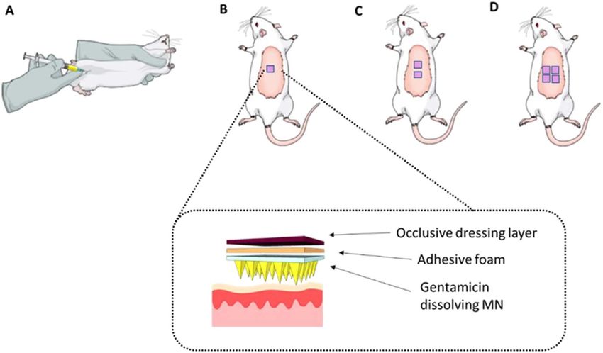
P. González-Vázquez et al. Journal of Controlled Release xxx (xxxx) xxx–xxx
[9–11]. They are made of safe water-soluble biodegradable or bio- 2.2.2. Preparation of films as a backing layer for dissolving MNs
compatible polymers that act as a matrix containing the drug to be Thirty grams of formulation containing 15% w/w PVP (MW
delivered. Following insertion into skin and subsequent contact with 360 kDa) were poured into 10 × 10 cm moulds consisting of a release
skin interstitial fluid, MNs dissolve and release the encapsulated drug. liner with the siliconised surface facing upwards secured to a Perspex®
This MN system has successfully delivered numerous substances, in- base with wing nuts. Previously, formulation was centrifuged at
cluding low molecular weight (MW) drugs [12,13], macromolecules 3500 rpm (rpm) for 15 min to ensure removal of entrapped air. Films
[14–17], DNA [18], and vaccines [19–21]. Recently, the work of our were placed on a levelled surface and required at least 48 h to dry at
research group has focused on delivering drugs at clinically-relevant room temperature. Once dried, the film was cut into small portions of
doses using MN arrays [22,23]. McCrudden et al. have formulated and approximately 1 cm2.
tested dissolving MNs for delivery of therapeutically-relevant con-
centrations of ibuprofen sodium [24]. In addition, in vitro delivery of a 2.2.3. Microneedle mechanical characterisation
combination of cardiovascular drugs from a single-dissolving MN array The mechanical strength of dissolving MN arrays was investigated
has recently been investigated by Quinn et al. [25]. by using a TA.XT2 Texture Analyser (Stable Micro Systems, Haslemere,
GEN delivery by a dissolving MN array could enable administration UK) in compression mode, as previously described [26]. Briefly, MN
in both inpatient and outpatient settings. Compared to parenteral de- arrays were attached to the moveable cylindrical probe (length 5 cm,
livery, this novel delivery system could reduce training requirements cross-sectional area 1.5 cm2) of the Texture Analyser and the probe was
for calculation of GEN dose and administration, thereby expanding programmed to move vertically downward at a rate of 1.19 mm/s. The
access to antibiotics to treat neonates with possible serious bacterial test station compressed MN arrays against a flat block of aluminium of
infections, particularly in resource-limited settings. The present work dimensions 9.2 × 5.2 cm, at defined rates for 30 s using a force of
evaluates the possibility of using dissolving MN arrays for the delivery 32 N/array [27]. The Leica EZ4D digital microscope was utilised for
of GEN. To the best of our knowledge, this is the first study that uses visualisation of MN arrays before and after application of the com-
dissolving MN arrays for the delivery of therapeutically relevant doses pression load. The heights of individual MNs were measured before and
of an antibiotic. after testing, using the ruler function of ImageJ® software (US National
Institutes of Health, Bethesda, Maryland, USA) so that the percentage
change in MN height could be calculated using Eq. (1), where, HBC is
2. Material and methods
the Height before compression and HAC is the Height after compression.
2.1. Materials HBC − HAC
%Compression = x 100
HBC (1)
N-acetylcysteine (NAC), methanol (HPLC grade), poly(vinyl al-
In order to evaluate MN insertion, the Parafilm M® (Bemis Company
cohol) (PVA) (80% hydrolysed, MW = 9000–10,000 Da), poly(vi-
Inc., Soignies, Belgium) insertion model developed by Larrañeta et al.
nylpyrrolidone) (PVP), (MW 360 kDa) and poly(ethylene glycol) (PEG)
was used [27]. MN arrays were inserted using the Texture Analyser, at
(MW 400 Da) were obtained from Sigma-Aldrich (Dorset, UK). Gantrez®
32 N/array for 30 s, into eight-layer folded Parafilm M® sheets. After
S-97 (PMVE/MA) (a copolymer of methyl vinyl ether and maleic acid,
the insertion, the MN arrays were removed from the polymeric layers.
MW 1,500,000 Da) and plasdone® K-29/32 (PVP) (MW 58 kDa) were
Parafilm M® layers were unfolded and the number of holes in each layer
kindly donated by Ashland (Kidderminster, UK). Glycerol bidistilled
was evaluated using the digital microscope and two polarizer filters.
(AnalaR NORMAPUR®, 99.5%) and sodium chloride (AnalaR
NORMAPUR® ACS) were obtained from VWR International
2.2.4. Dissolution studies and recovery of GEN content from dissolving MNs
(Leicestershire, UK). Gentamicin sulphate and o-phthaldialdehyde
Dissolution kinetics and drug content evaluations were carried out
(OPA) were obtained from Tokyo Chemical Industry UK Ltd. (Oxford,
on candidate GEN-loaded dissolving MN arrays to ensure that there
UK). Heparin Sodium (200 units in 2 mL) was provided by Wockhardt®
were no compatibility issues between GEN and the polymers selected as
UK Ltd. (Wrexham, UK). Hyabest®(S) LF-P (sodium hyaluronate 99.9%
a matrix for MN formation. Individual MNs were placed in a 20 mL
purity, MW 250–400 kDa range) was obtained from Kewpie
volume of phosphate buffered saline (PBS), pH 7.4 in a glass vial. The
Corporation Fine Chemical Division (Tokyo, Japan).
time required for complete dissolution of the sample using continuous
stirring at 600 rpm at 37 °C was recorded. Following appropriate dilu-
2.2. Methods tions, the GEN content obtained from the dissolving MNs was de-
termined by reversed-phase high-performance liquid chromatography
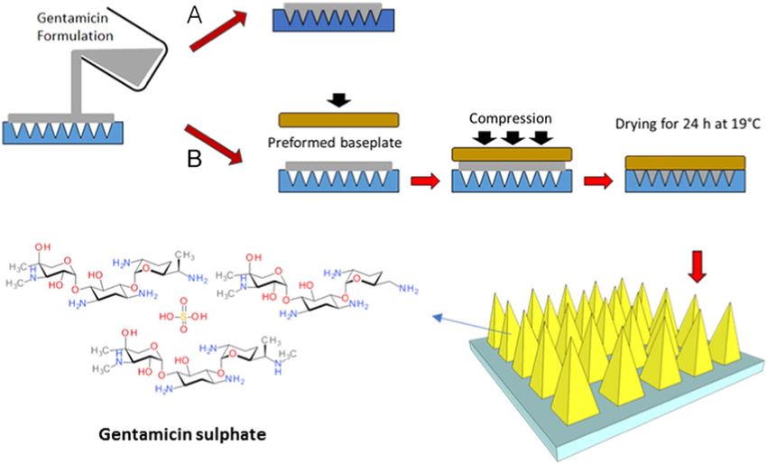
2.2.1. Fabrication of dissolving MNs (RP-HPLC), as detailed in Section 2.2.8.
Selected polymers were mixed with GEN and dissolved in deionized
water as potential matrices in the formation of MN. The blend was then 2.2.5. In-skin dissolution kinetics of dissolving MNs
mixed and sonicated at 37 °C for 1–2 h depending on the composition. Optical coherence tomography (OCT) (EX1301 OCT microscope,
The resulting formulations (100 mg) were poured into MN moulds and Michelson Diagnostics Ltd., Kent, UK) was employed to investigate the
positive pressure (3–4 bar) was applied for 15 min to fill the moulds. dissolution rate of dissolving MNs in skin, as previously described [28].
MN arrays were allowed to dry for 24 h in a controlled temperature Neonatal porcine skin was used as a skin model, due to its similarities to
room at 19 °C. The MN array design used to cast the formulations was human skin in terms of general structure, thickness, hair density, pig-
an array consisting of 361 pyramidal needles (19 rows of 19 needles) mentation, collagen and lipid composition [29]. Neonatal porcine skin
each with heights of 500 μm and array area of 0.45 cm2. In addition, a samples were obtained from stillborn piglets and immediately (< 24 h
two-step method was investigated with different dissolving MN for- after birth) excised and trimmed to a thickness of 500 μm using an
mulations. For this purpose, aqueous blends (100 mg) containing GEN electric dermatome instrument (Integra® Life-Sciences Corporation,
were poured into MN moulds. Post-casting, a preformed baseplate, Ratingen, Germany). Skin samples were stored in sealed Petri dishes at
prepared as detailed in Section 2.2.2, was immediately added on top of − 20 °C until use. Skin samples were shaved and equilibrated in PBS,
the GEN formulation. Positive pressure (3–4 bar) was applied for pH 7.4 for 15 min prior to use. One section of 500 μm thick neonatal
15 min to fill the moulds and then MNs were dried for 24 h at 19 °C. porcine skin was placed, dermal side facing downwards, onto a 500 μm
Once dried, dissolving MNs were visually inspected using a Leica EZ4D piece of Suprasorb® G wound dressing (Lohmann & Rauscher GmbH,
digital light microscope (Leica Microsystems, Milton Keynes, UK). Fig. 1 Rengsdorf, Germany). The Suprasorb® G wound dressing was equili-
shows a diagrammatic representation of the fabrication method. brated for 48 h in a copious volume of PBS, pH 7.4 at 37 °C prior to use.
2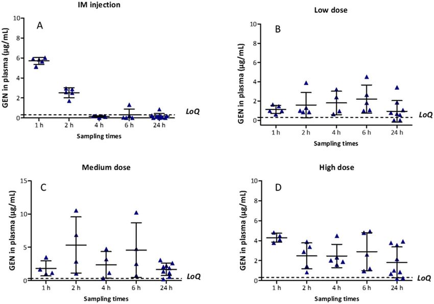
P. González-Vázquez et al.                                                                                                  Journal of Controlled Release xxx (xxxx) xxx–xxx

                                   Fig. 1. Diagrammatic representation of MN fabrication process in (A) one-step and (B) two-step.

The piece of Suprasorb® G wound dressing was pinned, using drawing                   of sample: 100 μL of derivatising reagent. Samples were centrifuged for
pins, into a Styrofoam™ platform which was wrapped in aluminium foil                 10 min and then filtered through 0.2 μL syringe filters. Finally, the
and Parafilm M®. The skin was pinned on top of these wound dressing                   samples were analysed by RP-HPLC.
sections. Dissolving MN arrays were adhered to a piece of Sellotape®
and manually applied into the skin. This experimental design main-
tained a hydrated membrane. The swept-source Fourier domain OCT                      2.2.7. In vivo delivery of GEN
system has a laser centre wavelength of 1305.0 ± 15.0 nm, facilitating                   An in vivo experiment using dissolving MNs was carried out to
real-time high-resolution imaging of the upper skin layers (7.5 μm lat-              evaluate profiles of GEN in plasma after the application of MN arrays.
eral and 10.0 μm vertical resolution). The skin was scanned at a frame               Female Sprague-Dawley rats weighing 208.65 ± 21.48 g and aged
rate of up to 15 B-scans (2D cross-sectional scans) per second (scan                 10 weeks were acclimatised to laboratory conditions for a 7-day period.
width = 2.0 mm). The 2D images were examined using the imaging                       Animals were separated in four groups (n = 10 per group) (Fig. 2). In
software ImageJ®. To allow differentiation between MN and skin layer,                 the control group (A), animals received an IM injection of GEN based on
false colours were applied to images using Microsoft® PowerPoint 2016                individual rat weight, 7.5 mg/kg of gentamicin sulphate to mimic the
(Microsoft Corporation, Redmond, Washington, USA). OCT images                        WHO higher neonatal dose [4]. Thus, a solution of 15 mg/mL of GEN
were captured at designated time points over the course of a 1 h ex-                 was freshly prepared in sterile water for injection. The IM injection was
periment.                                                                            given into the muscles of the thigh and the volume injected was
                                                                                     ≤ 100 μL. The transdermal treatments groups were denominated: low
                                                                                     dose (B), medium dose (C) and high dose (D), in which rats were treated
2.2.6. In vitro permeation study of GEN from dissolving MNs                          with one, two or four dissolving MNs, respectively. Dissolving MN ar-
    Transdermal permeation of GEN released from dissolving MNs was                   rays were prepared to contain an average of 30 mg of GEN per array.
investigated in vitro using Franz diffusion cells, as described previously                Animals were anaesthetized using gas anaesthesia (2–4% isoflurane
[14,24]. A section of the neonatal porcine skin (350 μm thick), obtained             in oxygen) and the backs of the rats were shaved using an animal hair
as described above, was secured to the donor compartment of the dif-                 clipper and hair removal cream, prior to application of MNs. Dissolving
fusion cell using cyanoacrylate glue with the stratum corneum facing                 MNs were secured onto an open “frame” of adhesive foam and were
upwards in the donor compartment. MNs were inserted into the centre                  applied using firm finger pressure onto a pinched section of skin on the
of the skin section by applying manual pressure for 30 s to a syringe                back of the animals. To keep the MN array in place, an occlusive
plunger, with the flat end pressing on the baseplate. A stainless-steel               dressing layer, Tegaderm™ (3 M, St Paul, Minnesota, USA), was placed
cylinder (diameter 11.0 mm, 5.0 g mass) was then placed on top of the                on top of the dissolving MNs, and Micropore™ tape (3 M UK Plc,
MNs to hold them in place. The donor compartments were set onto the                  Bracknell, Berkshire, UK) was used to wrap the back of the animals.
receptor compartments of the Franz cells, which were sealed using                    Blood samples (≤ 200 μL) were collected into heparinised tubes at de-
Parafilm M® to reduce evaporation. In the receiver solution, PBS                      signated time points: 1, 2, 4, 6 and 24 h via tail vein bleeds. The MN
(pH 7.4) was thermostatically maintained at 37 ± 1 °C and stirred at                 arrays were kept in place for 24 h. It was not possible to track plasma
600 rpm. Samples (≤ 200 μL) were removed from the sampling arms of                   level of GEN at each time point from the same animal due to restrictions
the Franz cells at predetermined time intervals (15 min, 30 min, 1 h,                in blood sampling volumes by our Institutional Project Licence. To
2 h, 3 h, 4 h, 5 h, 6 h and 24 h) using 1 mL syringes with 8 cm needles              overcome this, blood samples were taken from the first five rats at 1 h
and 200 μL of PBS was subsequently added to replace the volume taken.                and 4 h. The other five animals were bled at 2 h and 6 h and all animals
Derivatisation of the samples was carried out using volumes of 100 μL                (n = 10) were sampled at 24 h. After collection of rat blood, plasma

                                                                                 3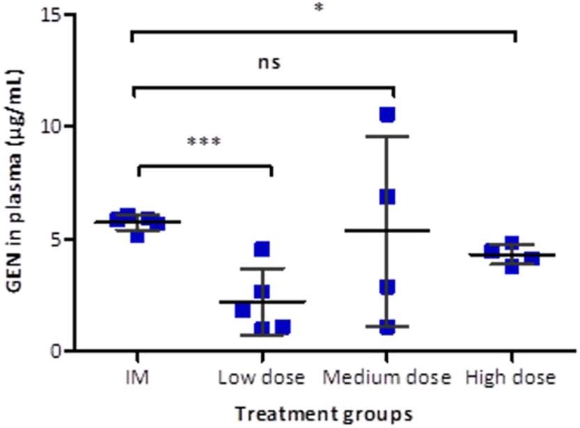
P. González-Vázquez et al.                                                                                                            Journal of Controlled Release xxx (xxxx) xxx–xxx

Fig. 2. Schematic representation showing the four treatment groups investigated in the in vivo experiment. (A) IM administration of GEN, 7.5 mg/kg. Transdermal application of (B) one,
(C) two and (D) four MN arrays to the hairless back of the animals and the setup used to ensure the MNs were kept in place.

separation was performed by centrifuging the blood at 3000 relative                           detection with excitation wavelength set at 328 nm and emission at
centrifugal force (RCF) for 10 min at 4 °C in a refrigerated centrifuge (Z                    423 nm. The gain was optimal at a reading of 12. Phase separation was
216 MK, HERMLE Labortechnik GmbH, Wehingen, Germany). Plasma                                  performed on a Phenomenex® SphereClone™ 5 μm ODS (1) column
samples were collected in 0.5 mL microtubes and stored in a freezer at                        (150 mm × 4.60 mm with 5 μm packing; Phenomenex, Cheshire, UK)
− 80 °C prior to analysis. To extract GEN, plasma samples were vor-                           at ambient temperature. All analytical runs were preceded by a security
texed for 10 s and transferred to Amicon® Ultra-0.5 centrifugal filter                         guard cartridge of matching chemistry. Data acquisition and analysis
devices (10 kDa) (EMD Millipore Corporation, Billerica, Massachusetts,                        were performed using Agilent Rapid Res® software (Agilent Technolo-
USA). Samples were centrifuged at 15,000 RCF for 30 min at 4 °C to                            gies UK Ltd., Stockport, UK). All samples were centrifuged at
remove proteins prior to HPLC analysis. A pre-column derivatisation                           14,000 rpm using an Eppendorf MiniSpin® centrifuge (Eppendorf UK
was carried out with the filtered samples obtained after centrifugation.                       Limited, Stevenage, UK) for 10 min prior to HPLC analysis. Unknown
In all cases, the ratio of derivatising reagent mixture and samples was                       concentrations of test samples were calculated using external standards.
1:1, depending on the volume of sample recovered. Approval for animal                             The HPLC method for quantification of GEN substances in PBS and
experiments was obtained from the Committee of the Biological                                 rat plasma were developed and validated in accordance with the
Research Unit, Queen's University Belfast. The work was carried out                           International Committee of Harmonisation Q2(R1) guidelines for vali-
under Project Licence PPL 2794 and Personal Licence PIL 1466. All in                          dation of analytical procedures [31]. Table 1 shows the slope, y-inter-
vivo experiments were conducted according to the policy of the fed-                           cept and coefficient of determination (R2 value) obtained from least
eration of European Laboratory Animal Science Associations and the                            squares linear regression analysis, followed by correlation analysis of
European Convention for the protection of vertebrate animals used for                         the calibration plots for derivatised GEN in PBS and plasma.
experimental and other scientific purposes, with implementation of the
principles of the 3Rs (replacement, reduction, and refinement). Values
for area under the curve (AUC) were calculated by the trapezoidal                             2.2.9. Statistical analysis
method using GraphPad Prism®.                                                                      All data were expressed as means ± standard deviation. The sta-
                                                                                              tistical analyses were performed using GraphPad Prism® version 5.3
2.2.8. GEN analytical method                                                                  (GraphPad Software, San Diego, California, USA). Where appropriate,
    Chemical derivatisation of GEN components was accomplished to                             the Mann-Whitney U test was performed for comparison of two groups,
increase sensitivity and allow low level quantification by RP-HPLC. The                        when n < 5. The unpaired t-test was used for comparison of two
derivatisation of GEN was carried out using o-phthaldialdehyde (OPA)                          groups, when n > 5 and data were normally distributed. The Kruskal-
and N-acetylcysteine (NAC) as reagents by slight modification of the                           Wallis test with post-hoc Dunn's test was used for comparison of mul-
procedure developed by Kowalczuk et al. [30]. Briefly, the derivatisa-                         tiple groups. In all cases, p < 0.05 was used to denote statistical sig-
tion reagents were prepared by dissolving 20 mg of OPA in 1 mL of                             nificance.
methanol and 100 mg of NAC was dissolved in 1 mL of deionized water.
To the mixture of OPA and NAC, 8 mL of 0.05 M borate buffer (pH 9.3)
were added. OPA and NAC were freshly prepared for each derivatisa-
                                                                                              Table 1
tion. For GEN quantification, 500 μL of sample was reacted with 500 μL
                                                                                              Calibration curve properties for derivatised GEN quantification in PBS, pH 7.4 and rat
of derivatising reagent mixture in microtubes in a water-bath at 50 °C                        plasma and limits of detection (LoD) and quantification (LoQ) for GEN.
for 20 min. After cooling, samples were analysed by RP-HPLC. The
mobile phase was a mixture of methanol and an aqueous solution of                                               Slope       y-Intercept    R2         LoD (μg/mL)      LoQ (μg/mL)
0.02 M of sodium hexanesulphonate and glacial acetic acid (65.5:33.5,
                                                                                                PBS, pH 7.4     291.46      −2.366         0.9987     0.049            0.149
v/v), with a flow rate of 1 mL/min, an injection volume of 40 μL and a                           Rat plasma      670.98      −92.982        0.9955     0.099            0.301
run time of 25 min per sample. GEN was detected using fluorescence

                                                                                          4
P. González-Vázquez et al.                                                                                         Journal of Controlled Release xxx (xxxx) xxx–xxx

Table 2
Formulations selected for the production of GEN loaded dissolving MNs.

  Formulation                     Composition (% w/w)                                                                     MN morphology

                                  GEN                      HA            PVP                     Glycerol

  F1                              30                       3.4           1                       –

  F2                              30                       3             1                       0.5

  F3                              30                       3             2                       0.5

  F4                              30                       5             5                       –

  F5                              30                       5             2.5                     –

  F6                              30                       3.4           1                       0.5

3. Results                                                                         hyaluronate (HA), MW 250–400 kDa range, in combination with low
                                                                                   MW PVP (58 kDa) formed the most promising formulations. Low con-
3.1. Formulations investigated in the production of GEN-loaded dissolving          centrations of glycerol were used in some formulations as plasticisers.
MNs                                                                                Dissolving MNs that appeared to be fully formed upon visual ex-
                                                                                   amination under a light microscope (Table 2) were chosen for further
   Several polymers were investigated to evaluate their potential as               dissolution and drug content evaluations.
matrices in the formation of dissolving MNs loaded with a high con-
centration of GEN. Some of the selected polymers, such as PVP high
                                                                                   3.2. Mechanical characterisation of dissolving MN arrays
MW (360 kDa) or PVA were not able to form a homogeneous aqueous
blend with a high concentration of GEN. In contrast, sodium
                                                                                      Dissolving MNs prepared with the selected formulations were used

                                                                               5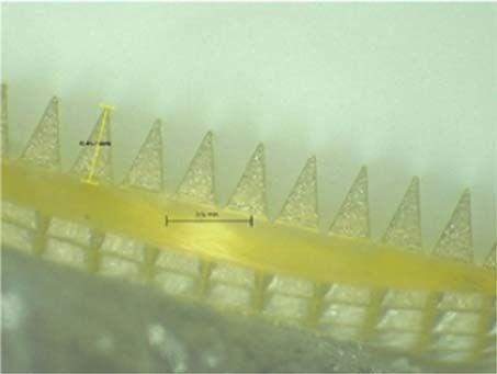
P. González-Vázquez et al. Journal of Controlled Release xxx (xxxx) xxx–xxx
Fig. 3. (A) Comparison of percentage of MN height reduc-
tion of dissolving MN formulations tested, observed fol-
lowing the application of a force of 32 N/array (means
+ S.D., n = 4). (B) Percentage of holes created in each
Parafilm M® layer following insertion of dissolving MN
formulations investigated. MN arrays used were 19 × 19
with a needle height of 500 μm, base width of 300 μm and a
base interspacing of 50 μm (means + S.D., n = 4).
to investigate the effects of compression tests on the heights of in- 3.4. In vitro permeation study of GEN from dissolving MNs
dividual needles on the MN arrays. Fig. 3A shows the reduction in MN
height for all the selected formulations. The application force of 32 N/ Two-layered MNs were fabricated from formulation F1 containing
array reduced the height of all dissolving MNs. The percent reduction in approximately 30 mg of GEN per array. The amount of GEN loaded in
MN height for formulation F2 and F5 was < 9% and < 5%, respec- the needles was approximately 4.8 mg, calculated considering the
tively. The percent reduction in the original MN height was < 2% for density of the formulation and the volume of the 361 square pyramidal
formulations F3 and F6 and < 3% for formulations F1 and F4. The needles. These arrays were used to evaluate the in vitro permeation of
percent reduction of F2 was significantly different compared to all other GEN across neonatal porcine skin (Fig. 5). Dissolving MNs made of F1
formulations (p = 0.0084). delivered 1.53 ± 0.28 mg of GEN in 1 h, 3.08 ± 0.5 mg of GEN in 3 h
The percentage of holes created in each Parafilm M® layer for dif- (10.25%) and a total of 22.56 ± 5.36 mg of GEN in 24 h (75%), de-
ferent dissolving MN formulations is shown in Fig. 3B. The in vitro in- livered across the neonatal porcine skin. All GEN loaded in the needles
sertion depths obtained for formulations tested showed a similar trend was delivered within 6 h and most of the GEN content forming the
and all were capable of piercing three layers of Parafilm M®. The baseplate was delivered within 24 h.
average thickness of a Parafilm M® layer is 126 ± 7 μm; this suggests
that MNs were inserted up to 378 μm of the total 500 μm height, ap-
proximately 75.6% of MN tip height. These results indicate that total 3.5. In vivo delivery of GEN
needle length inserted correlated with previously reported studies,
where the insertion was equivalent to 60% of the total needle length The in vivo plasma profile of GEN post-IM injection is shown in
[27,32]. However, it is important to note that formulations F2, F3, F4 Fig. 6A. Therapeutic levels of GEN in plasma (5.72 ± 0.35 μg/mL)
and F5 were not selected for further investigation because these for- were achieved within an hour following IM injection. The mean plasma
mulations showed some individual needles remained in the Parafilm level of GEN progressively decreased to 2.52 ± 0.49 μg/mL at 2 h,
M® after the insertion test. after which most concentrations were found to be below the LoQ.
The mean concentration of GEN in plasma for the low dose treat-
ment group was 1.13 ± 0.42 μg/mL at 1 h, 1.58 ± 1.31 μg/mL at 2 h
3.3. Fabrication of MN in two-steps and 1.80 ± 1.22 μg/mL at 4 h. A maximal concentration of GEN
(2.21 ± 1.46 μg/mL) was achieved at 6 h and then was slightly re-
After optimising the formulation of the MN, a two-step manu- duced (0.93 ± 1.11 μg/mL) at 24 h (Fig. 6B).
facturing process was tested to provide a degree of flexibility to the The mean concentration of GEN in plasma for the medium dose
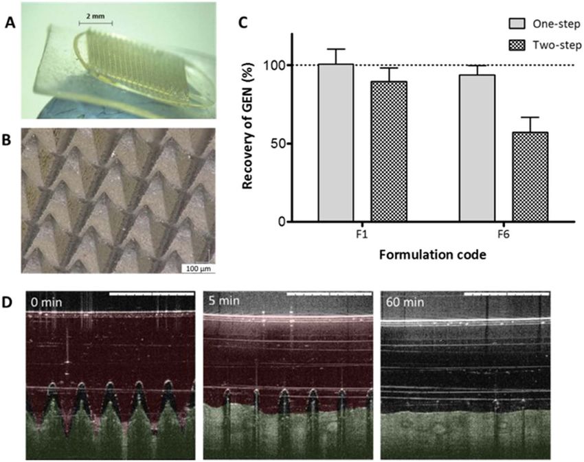
baseplate of the MN array. Fig. 4A and B shows a two-layered MN array. treatment group increased from 1.83 ± 1.13 μg/mL at 1 h to
Formulation F6, which contained 0.5% glycerol to reduce fragility, 5.34 ± 4.23 μg/mL at 2 h. The mean GEN concentration in plasma
and prepared with a baseplate made of 15% w/w of PVP, MW 360 kDa: dropped to 2.37 ± 1.81 μg/mL at 4 h, increased again to
2.5% glycerol, showed a remarkable reduction of recovery of drug 4.58 ± 4.11 μg/mL at 6 h and dropped to 1.68 ± 0.94 μg/mL at 24 h
compared to F1 fabricated in two-step which contained no glycerol in (Fig. 6C).
the needles or baseplate (Fig. 4C). The percentage of GEN recovery was The plasma profile for the high dose treatment group is presented in
found to be 89.56 ± 8.82% and 57.27 ± 9.38% for the two-layered Fig. 6D. A therapeutic concentration of GEN in plasma was achieved
MNs prepared using F1 and F6, respectively. However, the percentage within an hour (4.30 ± 1.47 μg/mL). After 1 h, the mean drug con-
of GEN recovery was 100.7 ± 9.69% and 89.56 ± 8.82% for for- centration in plasma dropped and remained consistently < 3 μg/mL for
mulations F1 and F6 prepared as a single layer. Besides, MNs containing subsequent sampling points.
glycerol in the preformed baseplate became flexible, losing mechanical The IM control treatment group presented the highest plasma con-
strength, after 10 days of storage at room temperature. This was un- centration of GEN at 1 h (Cmax = 5.72 ± 0.35 μg/mL) (Fig. 7). The
surprising given the highly hygroscopic nature of glycerol. Conse- Cmax for the IM control group was statistically higher than those for the
quently, F6 was discarded. MN arrays prepared in two-step, made of F1 low dose Cmax (p = 0.0008) and the high dose treatment group Cmax
in the needles and 15% w/w of PVP high MW (360 kDa) as a preformed (p = 0.0159). However, the maximum plasma concentration of GEN for
baseplate showed adequate recovery of GEN and desirable mechanical the medium dose transdermal treatment group was not different com-
strength. Fig. 4D displays the in vitro dissolution kinetics of F1 in neo- pared to the IM control Cmax (p = 1.000). Pharmacokinetic parameters
natal porcine skin using OCT. MN arrays prepared from F1 effectively are presented in Table 3.
pierced the skin and the in-skin MNs dissolved in the first five min. As expected, the lowest GEN Cmax was observed following ad-
After 1 h, skin was completely recovered. Consequently, this formula- ministration of an individual dissolving MN patch and the time of
tion was taken forward for in vitro permeation studies to investigate the maximum plasma concentration (Tmax) was 6 h following application.
efficacy of drug delivery. Formulation F1 retained its mechanical The comparison between Cmax of the transdermal groups was not
strength properties after storage in a plastic container covered with statistically significant (p = 0.1629). However, increasing the number
aluminium foil at 19 °C for > 4 weeks. of GEN MN arrays resulted in an earlier Tmax.
6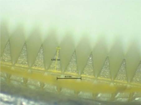
P. González-Vázquez et al.                                                                                                          Journal of Controlled Release xxx (xxxx) xxx–xxx

Fig. 4. Digital pictures showing two-layered MN array taken with (A) the Leica EZ4D digital microscope and (B) the Keyence VHX-700F digital microscope (Keyence, Milton Keynes, UK).
(C) Comparison of percentage recovery of GEN from dissolving MNs prepared from F1 and F6 in one-step and two-step (means ± S.D., n = 3). (D) Representative OCT images showing in
vitro dissolution kinetics of a 19 × 19 MN array prepared using F1 and inserted manually in neonatal porcine skin. False colours were applied to the skin and MNs. The original OCT
images can be found on the Supplementary content (Fig. S1). The white scale bar at top right represents a length of 1 mm.

                                                                                             strength [10].
                                                                                                 We formulated numerous dissolving MNs using water-soluble bio-
                                                                                             compatible or biodegradable polymers to investigate their compat-
                                                                                             ibility with a high dose of GEN (30% w/w). Some polymers, such as
                                                                                             PVP [25], PVA [33,34], sodium hyaluronate [35,36] and Gantrez®
                                                                                             copolymers [12,24,25,37,38] have frequently been used as matrices in
                                                                                             the formation of dissolving MNs. PVP of high MW (360 kDa) and PVA
                                                                                             (MW 9–10 kDa), showed incompatibilities with high doses of GEN. In
                                                                                             contrast, MNs formulated using low concentrations of sodium hyalur-
                                                                                             onate (MW 300 kDa) and PVP (MW 58 kDa) formed MNs that, upon
                                                                                             visual inspection, were fully formed. Plasticisers, such as glycerol and
                                                                                             PEG 400, were used to decrease MN brittleness, a property that can
                                                                                             prevent successful insertion in skin and thus inhibit drug delivery.
                                                                                             Glycerol is commonly incorporated into films to overcome brittleness
Fig. 5. In vitro permeation profile of GEN across 350 μm neonatal porcine skin when           [39,40]. Moga et al. have reported the use of plasticisers such as castor
delivered from dissolving MNs made of formulation F1 over a 24 h period.                     oil, Tween® 80, glycerol, PEG 400 and trimethyl citrate to impart
Means ± S.D., n = 4.                                                                         flexibility to the blends of a PVP/poly(vinylacetate) copolymer used as
                                                                                             a substrate for dissolving MNs [41]. Among all the prepared formula-
4. Discussion                                                                                tions, a few were selected based on the formation of intact and re-
                                                                                             producible MNs possessing rapid dissolution rates (Table 2). The main
    Despite the limitation of drug loading in dissolving MNs, several                        compounds used to formulate these arrays were PVP, HA and glycerol.
studies have highlighted the ability of this platform to enhance trans-                          This study was conceived as a proof of concept and the main ob-
dermal delivery of an extensive range of drug molecules [9,10]. The                          jective was to explore the possibility of administering therapeutically
initial objective of this work was to formulate well-formed MNs using                        relevant doses of GEN using dissolving MNs. To the best of our
biocompatible materials approved by the US Food and Drug Adminis-                            knowledge there are no previous articles describing similar drug de-
tration (FDA) with high drug loading of GEN. Ideal GEN MN formula-                           livery systems. Accordingly, we maximise the amounts of GEN loaded
tions should have sufficient mechanical strength for a successful ap-                          in the MN arrays. The arrays were manufactured containing GEN in the
plication into the skin by the patient or a healthcare provider, rapidly                     needle tips and in a small baseplate. In this way GEN can be adminis-
dissolve, and yield high drug recovery. The geometry of the MN and                           tered from the needle tips and subsequently from the baseplate. The
physicochemical properties of the drug and polymers affect mechanical                         drug contained in the baseplate can permeate through the created pores

                                                                                         7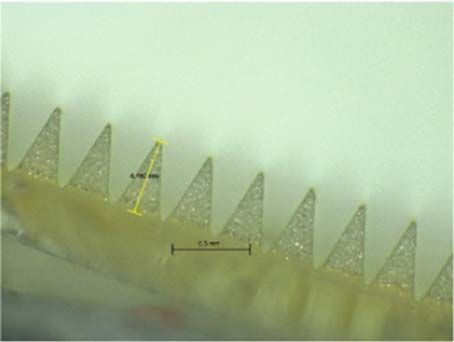
P. González-Vázquez et al.                                                                                                              Journal of Controlled Release xxx (xxxx) xxx–xxx

Fig. 6. (A) In vivo plasma profile of GEN following IM injection of gentamicin sulphate dissolved in sterilised water for injections (dose was 7.5 mg/kg). Means ± S.D., n = 5 at 1 h, 2 h,
4 h and 6 h; n = 10 at 24 h. (B) Transdermal delivery by using one dissolving MN (low dose). Means ± S.D., n = 5 at 1 h, 2 h and 6 h; n = 4 at 4 h; n = 10 at 24 h. (C) Transdermal
delivery by using two dissolving MNs (medium dose). Means ± S.D., n = 4 at 1 h, 2 h, 4 h and 6 h; n = 9 at 24 h. (D) Transdermal delivery by using four dissolving MNs (high dose).
Means ± S.D., n = 4 at 1 h; n = 5 at 2 h, 4 h and 6 h; n = 10 at 24 h. Dashed lines represent limit of quantification for GEN (LoQ = 0.30 μg/mL).

                                                                                                characterisation tests has been developed to test their mechanical
                                                                                                strength and insertion characteristics. Such tests include MN insertion
                                                                                                into skin [42], MN compression test [26] and OCT [28], among others.
                                                                                                Lutton and co-workers have recently highlighted an urgent need for
                                                                                                consistency to characterise MN products in order to demonstrate MN
                                                                                                strength and insertion [43]. The in vitro insertion test developed by
                                                                                                Larrañeta et al. was used to assess the ability of the dissolving MNs to
                                                                                                pierce a skin simulant, Parafilm M®, thus avoiding dissolution of the
                                                                                                MNs in biological tissue. The mechanical testing of dissolving MN
                                                                                                provided information about which concentrations of HA and PVP
                                                                                                yielded the best strength and insertion capabilities. In summary, for-
                                                                                                mulations investigated showed to form robust needles that were not
                                                                                                fractured upon compression. However, some formulations were deemed
                                                                                                unsuitable for further investigation because needles tips were not en-
                                                                                                tirely formed and also some individual needles retained in the Parafilm
Fig. 7. Comparison of mean peak plasma concentrations of GEN (Cmax) following IM
                                                                                                M® post-insertion test. Furthermore, dissolving MNs were prepared in
injection and transdermal delivery of GEN at increasing doses. Means ± S.D., n = 5 for
the control group and the low dose and n = 4 for medium and high dose. (ns) indicates p         two-step to provide certain flexibility to the baseplate of the MN arrays.
values > 0.05, (*) indicates p values ≤ 0.05 and (***) indicates p values ≤ 0.001.              Ideally, the baseplate should have some inherent flexibility to allow
                                                                                                conformation to the skin surface without cracking; a variety of tech-
                                                                                                niques have been used to fabricate dissolving MNs in a two-step process
Table 3
Pharmacokinetic parameter of GEN applied to rats for the IM injection and the three
                                                                                                to localise the active compounds to the needles, thereby reducing drug
transdermal treatments groups: low dose, medium dose and high dose. Means ± S.D.,               waste [15,17,18,44,45].
n = 5 for the IM group and low dose and n = 4 for medium and high doses.                            The rate of dissolution of dissolving MNs in-skin depends on the
                                                                                                solubility in water of the polymers and drug studied and also the
  Parameter           IM injection     Low dose         Medium dose       High dose
                                                                                                composition [10]. The non-invasive technique based on light reflection,
  AUC (μg. h/mL)      12.03            37.05            74.58             56.13                 OCT, was used to investigate the in-skin dissolution kinetics of dissol-
  Tmax (h)            1                6                2                 1                     ving MNs. This technique has been widely used in clinical care, pre-
  Cmax (μg/mL)        5.72 ± 0.35      2.21 ± 1.46      5.34 ± 4.23       4.30 ± 1.47           dominantly for ophthalmic applications, and in research, providing
                                                                                                high-resolution cross-sectional images of the object of study [46]. A
                                                                                                number of studies have highlighted the potential of OCT as a valuable
as suggested by McCrudden et al. [24].
                                                                                                tool to assess MN insertion in skin, allowing measurement of the MN
    The delivery of the drug via dissolving MNs depends on their ability
                                                                                                depth and visualisation of microchannels created in skin [26,28,47,48].
to pierce the stratum corneum; dissolving MNs must therefore possess
                                                                                                Likewise, OCT has been used for in vivo visualisation of dissolution of
sufficient mechanical strength. Accordingly, a range of mechanical

                                                                                            8
P. González-Vázquez et al.                                                                                               Journal of Controlled Release xxx (xxxx) xxx–xxx

MNs into murine skin [16,17] and in situ dissolution of MNs into neo-            Consequently, a patch size of approximately 15 cm2 could potentially
natal porcine skin [28]. The in vivo swelling of hydrogel-forming MNs            deliver therapeutic doses of GEN and should be removed after 3 h to
has also been examined using OCT in the skin of human volunteers                 avoid possible GEN side effects. For the high dose treatment, the GEN
[49–51] and in vitro in neonatal porcine skin [52]. The GEN-loaded               Cmax was observed at 1 h post-application, reaching therapeutic levels.
dissolving MNs were robust enough to penetrate skin in vitro and the             Thus, the high dose treatment group yielded a more rapid delivery of
complete dissolution of the in-skin MNs was achieved in the first five             GEN, and the data showed less variability than the data obtained from
min.                                                                             the medium dose treatment group when the GEN Cmax was observed.
    The in vitro permeation studies carried out with two-layered dis-            Therefore, a patch size not larger than 30 cm2 could potentially deliver
solving MNs (F1) across neonatal porcine skin via the Franz diffusion             therapeutic doses of GEN in newborns and should be removed after
cell technique deduced that 14.85% (4.45 mg) of GEN loadings were                1–2 h in order to avoid oto- and nephrotoxicity. Nevertheless, Tanira
delivered over a 6 h experimental period and approximately 75% of the            et al. have reported shorter half-lives and larger clearances of GEN in
GEN contained in the MN array was successfully delivered in 24 h. As             young rats (2–3 months old) compared to older rats (22–24 months old)
expected, GEN contained in the baseplate was delivered through the               [56]. This is unsurprising due to small mammals such as rats tend to
created pores after the dissolution of the needle tips. On the other hand,       metabolise some drugs more rapidly than larger mammals [57,58]. In
the results obtained in the in vivo experiment confirmed that the use of          addition, GEN pharmacokinetics is affected by renal function and
dissolving MN arrays enabled transdermal delivery of GEN. It is im-              neonates present reduced glomerular filtration rate. Therefore, it is
portant to highlight that once the GEN Cmax was reached, regardless of           expected that neonates have lower renal clearance of GEN compared to
the dose, GEN levels in plasma were shown to be sustained over time.             older infants and adults [59–62]. Considering this, the estimated patch
This may be indicative of drug still being released from the MN patch            sizes could probably be reduced significantly. A transdermal patch of
while concurrently being cleared from the body of the animals. It is             such size could potentially be applied to the thigh or to the upper back
important to notice that GEN is contained in the needle tips and in the          of the neonate, as both sites provide a relatively flat surface. The
baseplate of the MN array. And after the dissolution of the needle tips,         manual application of larger than 1 cm2 MN patches is feasible as
GEN continued to be administered from the baseplate as the patches               shown recently by Ripolin et al. [53]. The acceptability and feasibility
were covered by an occlusive dressing. Consequently, MNs provide a               of delivery of GEN MN arrays at these sites should be assessed. A range
sustained release and that explains why the AUC obtained for MN ad-              of patch sizes would be required according to different weight cate-
ministration are higher than the AUC obtained for the IM injection.              gories. MN arrays allow the delivery of GEN without the need of in-
Besides, this explains why the Tmax is higher for the lower GEN dose             jections and could enable expanded access to treatment outside of in-
administered using MNs. In this case, GEN delivered from the baseplate           patient facilities. Additionally, this approach reduces dangerous sharps
is required to reach the higher maximum drug levels. The patches with            waste as dissolving MN arrays are self-disabling and cannot be re-
higher GEN doses contained enough drug in the needle tips to achieve             inserted into another patient after being used [11]. Future work will
the maximum drug levels within shorter periods of time.                          focus on optimisation of the two-step method to cast only GEN for-
    As described before, this study was planned as a proof of concept            mulation in the tips of the MN arrays. A transdermal MN patch design
and after evaluating the in vivo results it is obvious that the system           with targeted GEN loading may reduce the risk of prolonged adminis-
should be optimized for further applications. In order to achieve a rapid        tration of GEN and toxicity if the patch is inadvertently worn too long.
delivery and prevent the sustained delivery the MN arrays can be
manufactured containing the drug only in the needle tips. In this way,           5. Conclusion
the drug can be administered in a quicker way preventing sustained
release of GEN from the baseplate. Thus, drug clearance in animals will              The work presented here reports the successful formulation and
require further investigation. Regardless of the number of MN arrays             mechanical characterisation of dissolving MN arrays containing the
used, it was observed that MNs were completely dissolved when arrays             antibiotic, GEN. Furthermore, in in vivo experimentation, ther-
were removed from the back of the animal. Transient mild erythema                apeutically relevant doses of the antibiotic were delivered to rats,
was observed after removing the patches and, in line with previous               highlighting the potential for exploitation of this delivery route for GEN
publications, the erythema disappears quickly after MN removal                   administration. This promising technology could simplify administra-
[53,54]. It is worth noting that concentrations of the HA, a FDA-ap-             tion of GEN in developing countries, thus expanding access to lifesaving
proved biodegradable and biocompatible polymer, and low MW PVP, a                outpatient antibiotic treatment for infants with severe infection during
biocompatible FDA-approved pharmaceutical excipient, were low in                 the neonatal period. This is an exploratory study and before this tech-
the formulation investigated. However, elimination of both polymers              nology can be applied to patients several studies need to be conducted
from the body should be investigated.                                            such as a formulation stability evaluation, a comprehensive pharma-
    The therapeutic plasma levels of GEN in humans ranges between 4              cokinetic study and patient usability/acceptability study. Finally,
and 10 μg/mL [8,55]. GEN exhibits a concentration-dependent bacter-              evaluating the potential pathway to commercialisation of this tech-
icidal effect, in which a linear relationship exists among higher peaks,          nology, and the cost of GEN-loaded MN arrays when manufactured at
the minimum inhibitory concentration ratio and improved clinical re-             scale, will also be important factors in the potential health impact of
sponse [6]. With once-daily administration, higher peak concentrations           this delivery approach in resource-limited settings.
may be observed. Peak and trough GEN serum concentrations should be                  Supplementary data to this article can be found online at http://dx.
therapeutically effective, while being nontoxic. Prolonged peak serum             doi.org/10.1016/j.jconrel.2017.07.032.
concentrations above 10–12 μg/mL and trough serum concentrations
above 2 μg/mL should be avoided, as these levels may be potentially              References
toxic [8]. Predicting pharmacokinetics from animals to humans require
further investigation. However, one can prudently extrapolate the in-             [1] J.E. Lawn, S. Cousens, J. Zupan, 4 million neonatal deaths: when? Where? Why?
formation obtained from the animals and approximate the patch size                    Lancet 365 (2005) 891–900, http://dx.doi.org/10.1016/S0140-6736(05)71048-5.
                                                                                  [2] S. Vergnano, M. Sharland, P. Kazembe, C. Mwansambo, P. Heath, Neonatal sepsis:
necessary to use in neonates. A neonate has an average body weight of                 an international perspective, Arch. Dis. Child. Fetal Neonatal Ed. 90 (2005)
3 kg, which is approximately 15 times greater than the average body                   F220–F224, http://dx.doi.org/10.1136/adc.2002.022863.
weight of rats used in this experiment (0.209 kg). For the medium dose            [3] L. Liu, S. Oza, D. Hogan, J. Perin, I. Rudan, J.E. Lawn, S. Cousens, C. Mathers,
                                                                                      R.E. Black, Global, regional, and national causes of child mortality in 2000–13, with
treatment group, which applied two MN arrays of size 0.45 cm2,                        projections to inform post-2015 priorities: an updated systematic analysis, Lancet
the < 1 cm2 MN patch was capable of delivering GEN achieving a mean                   385 (2015) 430–440, http://dx.doi.org/10.1016/S0140-6736(14)61698-6.
plasma concentration of 5.34 μg/mL at 2 h upon application.                       [4] World Health Organization, GUIDELINE Managing Possible Serious Bacterial

                                                                             9
P. González-Vázquez et al. Journal of Controlled Release xxx (xxxx) xxx–xxx
Infection in Young Infants When Referral Is Not Feasible, (2015). [26] R.F. Donnelly, R. Majithiya, T.R.R. Singh, D.I.J. Morrow, M.J. Garland, Y.K. Demir,
[5] L. Šoltés, Aminoglycoside antibiotics — two decades of their HPLC bioanalysis, K. Migalska, E. Ryan, D. Gillen, C.J. Scott, A.D. Woolfson, Design, optimization and
Biomed. Chromatogr. 13 (1999) 3–10, http://dx.doi.org/10.1002/(SICI)1099- characterisation of polymeric microneedle arrays prepared by a novel laser-based
0801(199902)13:13.0.CO;2-T. micromoulding technique, Pharm. Res. 28 (2011) 41–57, http://dx.doi.org/10.
[6] G.L. Darmstadt, M. Batra, A.K.M. Zaidi, Parenteral antibiotics for the treatment of 1007/s11095-010-0169-8.
serious neonatal bacterial infections in developing country settings, Pediatr. Infect. [27] E. Larrañeta, J. Moore, E.M. Vicente-Pérez, P. González-Vázquez, R. Lutton,
Dis. J. 28 (2009) S37–S42, http://dx.doi.org/10.1097/INF.0b013e31819588c3. A.D. Woolfson, R.F. Donnelly, A proposed model membrane and test method for
[7] K.W. Hindmarsh, R.L. Nation, G.L. Williams, E. John, J.N. French, microneedle insertion studies, Int. J. Pharm. 472 (2014) 65–73, http://dx.doi.org/
Pharmacokinetics of gentamicin in very low birth weight preterm infants, Eur. J. 10.1016/j.ijpharm.2014.05.042.
Clin. Pharmacol. 24 (1983) 649–653, http://dx.doi.org/10.1007/BF00542216. [28] M.J. Garland Donnelly, D.I.J. Morrow, K. Migalska, T.R.R. Singh, R. Majithiya,
[8] B. Chattopadhyay, Newborns and gentamicin-how much and how often? J. A.D. Woolfson, Optical coherence tomography is a valuable tool in the study of the
Antimicrob. Chemother. 49 (2002) 13–16, http://dx.doi.org/10.1093/jac/49.1.13. effects of microneedle geometry on skin penetration characteristics and in-skin
[9] T.-M. Tuan-Mahmood, M.T.C. McCrudden, B.M. Torrisi, E. McAlister, M.J. Garland, dissolution, J. Control. Release 147 (2010) 333–341, http://dx.doi.org/10.1016/j.
T.R.R. Singh, R.F. Donnelly, Microneedles for intradermal and transdermal drug jconrel.2010.08.008.
delivery, Eur. J. Pharm. Sci. 50 (2013) 623–637, http://dx.doi.org/10.1016/j.ejps. [29] A. Summerfield, F. Meurens, M.E. Ricklin, The immunology of the porcine skin and
2013.05.005. its value as a model for human skin, Mol. Immunol. 66 (2015) 14–21, http://dx.doi.
[10] J.W. Lee, J.-H. Park, M.R. Prausnitz, Dissolving microneedles for transdermal drug org/10.1016/j.molimm.2014.10.023.
delivery, Biomaterials 29 (2008) 2113–2124, http://dx.doi.org/10.1016/j. [30] D. Kowalczuk, R. Pietraś, B. Paw, A. Czerkies, Applying liquid chromatography with
biomaterials.2007.12.048. fluorescence detection to determine gentamicin, Pol. J. Environ. Stud. 19 (2010)
[11] E. Larrañeta, R.E.M. Lutton, A.D. Woolfson, R.F. Donnelly, Microneedle arrays as 587–591.
transdermal and intradermal drug delivery systems: materials science, manufacture [31] I.C.H. ICH, Topic Q2 (R1) Validation of Analytical Procedures: Text and
and commercial development, Mater. Sci. Eng. R. Rep. 104 (2016) 1–32, http://dx. Methodology, (2005).
doi.org/10.1016/j.mser.2016.03.001. [32] R.E.M. Lutton, E. Larrañeta, M.-C. Kearney, P. Boyd, A.D. Woolfson, R.F. Donnelly,
[12] Y.A. Gomaa, M.J. Garland, F. McInnes, L.K. El-Khordagui, C. Wilson, R.F. Donnelly, A novel scalable manufacturing process for the production of hydrogel-forming
Laser-engineered dissolving microneedles for active transdermal delivery of na- microneedle arrays, Int. J. Pharm. 494 (2015) 417–429, http://dx.doi.org/10.
droparin calcium, Eur. J. Pharm. Biopharm. 82 (2012) 299–307, http://dx.doi.org/ 1016/j.ijpharm.2015.08.049.
10.1016/j.ejpb.2012.07.008. [33] L.Y. Chu, S.-O. Choi, M.R. Prausnitz, Fabrication of dissolving polymer micro-
[13] M.J. Garland, K. Migalska, T.-M. Tuan-Mahmood, T. Raghu Raj Singh, R. Majithija, needles for controlled drug encapsulation and delivery: bubble and pedestal mi-
E. Caffarel-Salvador, C.M. McCrudden, H.O. McCarthy, A. David Woolfson, croneedle designs, J. Pharm. Sci. 99 (2010) 4228–4238, http://dx.doi.org/10.
R.F. Donnelly, Influence of skin model on in vitro performance of drug-loaded so- 1002/jps.22140.
luble microneedle arrays, Int. J. Pharm. 434 (2012) 80–89, http://dx.doi.org/10. [34] L.Y. Chu, M.R. Prausnitz, Separable arrowhead microneedles, J. Control. Release
1016/j.ijpharm.2012.05.069. 149 (2011) 242–249, http://dx.doi.org/10.1016/j.jconrel.2010.10.033.
[14] K. Migalska, D.I.J. Morrow, M.J. Garland, R. Thakur, A.D. Woolfson, R.F. Donnelly, [35] K. Matsuo, Y. Yokota, Y. Zhai, Y.-S. Quan, F. Kamiyama, Y. Mukai, N. Okada,
Laser-engineered dissolving microneedle arrays for transdermal macromolecular S. Nakagawa, A low-invasive and effective transcutaneous immunization system
drug delivery, Pharm. Res. 28 (2011) 1919–1930, http://dx.doi.org/10.1007/ using a novel dissolving microneedle array for soluble and particulate antigens, J.
s11095-011-0419-4. Control. Release 161 (2012) 10–17, http://dx.doi.org/10.1016/j.jconrel.2012.01.
[15] M.-H. Ling, M.-C. Chen, Dissolving polymer microneedle patches for rapid and ef- 033.
ficient transdermal delivery of insulin to diabetic rats, Acta Biomater. 9 (2013) [36] S. Liu, M. Jin, Y. Quan, F. Kamiyama, K. Kusamori, H. Katsumi, T. Sakane,
8952–8961, http://dx.doi.org/10.1016/j.actbio.2013.06.029. A. Yamamoto, Transdermal delivery of relatively high molecular weight drugs using
[16] M.T.C. McCrudden, B.M. Torrisi, S. Al-Zahrani, C.M. McCrudden, M. Zaric, novel self-dissolving microneedle arrays fabricated from hyaluronic acid and their
C.J. Scott, A. Kissenpfennig, H.O. McCarthy, R.F. Donnelly, Laser-engineered dis- characteristics and safety after application to the skin, Eur. J. Pharm. Biopharm. 86
solving microneedle arrays for protein delivery: potential for enhanced intradermal (2014) 267–276, http://dx.doi.org/10.1016/j.ejpb.2013.10.001.
vaccination, J. Pharm. Pharmacol. 67 (2015) 409–425, http://dx.doi.org/10.1111/ [37] M.J. Garland, E. Caffarel–Salvador, K. Migalska, A.D. Woolfson, R.F. Donnelly,
jphp.12248. Dissolving polymeric microneedle arrays for electrically assisted transdermal drug
[17] M. Zaric, O. Lyubomska, O. Touzelet, C. Poux, S. Al-Zahrani, F. Fay, L. Wallace, delivery, J. Control. Release 159 (2012) 52–59, http://dx.doi.org/10.1016/j.
D. Terhorst, B. Malissen, S. Henri, U.F. Power, C.J. Scott, R.F. Donnelly, jconrel.2012.01.003.
A. Kissenpfennig, Skin dendritic cell targeting via microneedle arrays laden with [38] R.F. Donnelly, D.I.J. Morrow, F. Fay, C.J. Scott, S. Abdelghany, R.R.T. Singh,
antigen-encapsulated poly-D,L-lactide-co-glycolide nanoparticles induces efficient M.J. Garland, A. David Woolfson, Microneedle-mediated intradermal nanoparticle
antitumor and antiviral immune responses, ACS Nano 7 (2013) 2042–2055, http:// delivery: potential for enhanced local administration of hydrophobic pre-formed
dx.doi.org/10.1021/nn304235j. photosensitisers, Photodiagn. Photodyn. Ther. 7 (2010) 222–231, http://dx.doi.
[18] J. McCaffrey, C.M. McCrudden, A.A. Ali, A.S. Massey, J.W. McBride, org/10.1016/j.pdpdt.2010.09.001.
M.T.C. McCrudden, E.M. Vicente-Perez, J.A. Coulter, T. Robson, R.F. Donnelly, [39] M.G.A. Vieira, M.A. Da Silva, L.O. Dos Santos, M.M. Beppu, Natural-based plasti-
H.O. McCarthy, Transcending epithelial and intracellular biological barriers; a cizers and biopolymer films: a review, Eur. Polym. J. 47 (2011) 254–263, http://dx.
prototype DNA delivery device, J. Control. Release 226 (2016) 238–247, http://dx. doi.org/10.1016/j.eurpolymj.2010.12.011.
doi.org/10.1016/j.jconrel.2016.02.023. [40] M.L. Sanyang, S.M. Sapuan, M. Jawaid, M.R. Ishak, J. Sahari, Effect of glycerol and
[19] S.P. Sullivan, D.G. Koutsonanos, M. del Pilar Martin, J.W. Lee, V. Zarnitsyn, S.- sorbitol plasticizers on physical and thermal properties of sugar palm starch based
O. Choi, N. Murthy, R.W. Compans, I. Skountzou, M.R. Prausnitz, Dissolving films, Recent Adv. Environ. Ecosyst. Dev. Eff. (2015) 157–162.
polymer microneedle patches for influenza vaccination, Nat. Med. 16 (2010) [41] K.A. Moga, L.R. Bickford, R.D. Geil, S.S. Dunn, A.A. Pandya, Y. Wang, J.H. Fain,
915–920, http://dx.doi.org/10.1038/nm.2182. C.F. Archuleta, A.T. O'Neill, J.M. DeSimone, Rapidly-dissolvable microneedle pat-
[20] K. Matsuo, S. Hirobe, Y. Yokota, Y. Ayabe, M. Seto, Y.-S. Quan, F. Kamiyama, ches via a highly scalable and reproducible soft lithography approach, Adv. Mater.
T. Tougan, T. Horii, Y. Mukai, N. Okada, S. Nakagawa, Transcutaneous im- 25 (2013) 5060–5066, http://dx.doi.org/10.1002/adma.201300526.
munization using a dissolving microneedle array protects against tetanus, diph- [42] J.S. Kochhar, T.C. Quek, W.J. Soon, J. Choi, S. Zou, L. Kang, Effect of microneedle
theria, malaria, and influenza, J. Control. Release 160 (2012) 495–501, http://dx. geometry and supporting substrate on microneedle array penetration into skin, J.
doi.org/10.1016/j.jconrel.2012.04.001. Pharm. Sci. 102 (2013) 4100–4108, http://dx.doi.org/10.1002/jps.23724.
[21] C. Edens, M.L. Collins, J.L. Goodson, P.A. Rota, M.R. Prausnitz, A microneedle patch [43] Lutton, J. Moore, E. Larrañeta, S. Ligett, A.D. Woolfson, R.F. Donnelly, Microneedle
containing measles vaccine is immunogenic in non-human primates, Vaccine 33 characterisation: the need for universal acceptance criteria and GMP specifications
(2015) 4712–4718, http://dx.doi.org/10.1016/j.vaccine.2015.02.074. when moving towards commercialisation, Drug Deliv. Transl. Res. 5 (2015)
[22] M.-C. Kearney, E. Caffarel-Salvador, S.J. Fallows, H.O. McCarthy, R.F. Donnelly, 313–331, http://dx.doi.org/10.1007/s13346-015-0237-z.
Microneedle-mediated delivery of donepezil: potential for improved treatment op- [44] Q. Wang, G. Yao, P. Dong, Z. Gong, G. Li, K. Zhang, C. Wu, Investigation on fab-
tions in Alzheimer's disease, Eur. J. Pharm. Biopharm. 103 (2016) 43–50, http://dx. rication process of dissolving microneedle arrays to improve effective needle drug
doi.org/10.1016/j.ejpb.2016.03.026. distribution, Eur. J. Pharm. Sci. 66 (2015) 148–156, http://dx.doi.org/10.1016/j.
[23] R.F. Donnelly, M.T.C. McCrudden, A. Zaid Alkilani, E. Larrañeta, E. McAlister, ejps.2014.09.011.
A.J. Courtenay, M.-C. Kearney, T.R.R. Singh, H.O. McCarthy, V.L. Kett, E. Caffarel- [45] K. Fukushima, A. Ise, H. Morita, R. Hasegawa, Y. Ito, N. Sugioka, K. Takada, Two-
Salvador, S. Al-Zahrani, A.D. Woolfson, Hydrogel-forming microneedles prepared layered dissolving microneedles for percutaneous delivery of peptide/protein drugs
from ‘super swelling’ polymers combined with lyophilised wafers for transdermal in rats, Pharm. Res. 28 (2011) 7–21, http://dx.doi.org/10.1007/s11095-010-
drug delivery, PLoS One 9 (2014) e111547, , http://dx.doi.org/10.1371/journal. 0097-7.
pone.0111547. [46] M. Adhi, J.S. Duker, Optical coherence tomography – current and future applica-
[24] M.T.C. McCrudden, A.Z. Alkilani, C.M. McCrudden, E. McAlister, H.O. McCarthy, tions, Curr. Opin. Ophthalmol. 24 (2013) 213–221, http://dx.doi.org/10.1097/ICU.
A.D. Woolfson, R.F. Donnelly, Design and physicochemical characterisation of 0b013e32835f8bf8.
novel dissolving polymeric microneedle arrays for transdermal delivery of high [47] R.F. Donnelly, K. Moffatt, A.Z. Alkilani, E.M. Vicente-Pérez, J. Barry,
dose, low molecular weight drugs, J. Control. Release 180 (2014) 71–80, http://dx. M.T.C. McCrudden, A.D. Woolfson, Hydrogel-forming microneedle arrays can be
doi.org/10.1016/j.jconrel.2014.02.007. effectively inserted in skin by self-application: a pilot study centred on pharmacist
[25] H.L. Quinn, L. Bonham, C.M. Hughes, R.F. Donnelly, Design of a dissolving mi- intervention and a patient information leaflet, Pharm. Res. 31 (2014) 1989–1999,
croneedle platform for transdermal delivery of a fixed-dose combination of cardi- http://dx.doi.org/10.1007/s11095-014-1301-y.
ovascular drugs, J. Pharm. Sci. 104 (2015) 3490–3500, http://dx.doi.org/10.1002/ [48] S.A. Coulman, J.C. Birchall, A. Alex, M. Pearton, B. Hofer, C. O'Mahony, W. Drexler,
jps.24563. B. Považay, In vivo, in situ imaging of microneedle insertion into the skin of human
10You can also read