The chromatin remodeler Snf2h is essential for oocyte meiotic cell cycle progression
←
→
Page content transcription
If your browser does not render page correctly, please read the page content below
Downloaded from genesdev.cshlp.org on October 1, 2020 - Published by Cold Spring Harbor Laboratory Press

The chromatin remodeler Snf2h
is essential for oocyte meiotic cell
cycle progression
Chunxia Zhang,1,2,3,8 Zhiyuan Chen,1,2,3,8 Qiangzong Yin,1,2,3 Xudong Fu,1,2,3 Yisi Li,1,2,3,4
Tomas Stopka,5 Arthur I. Skoultchi,5 and Yi Zhang1,2,3,6,7
1
 Howard Hughes Medical Institute, Boston Children’s Hospital, Boston, Massachusetts 02115, USA; 2Program in Cellular
and Molecular Medicine, Boston Children’s Hospital, Boston, Massachusetts 02115, USA; 3Division of Hematology/Oncology,
Department of Pediatrics, Boston Children’s Hospital, Boston, Massachusetts 02115, USA; 4Department of Automation, Tsinghua
University, Beijing 100084, China; 5Department of Cell Biology, Albert Einstein College of Medicine, Bronx, New York 10461,
USA; 6Department of Genetics, Harvard Medical School, Boston, Massachusetts 02115, USA; 7Harvard Stem Cell Institute, Boston,
Massachusetts 02115, USA

Oocytes are indispensable for mammalian life. Thus, it is important to understand how mature oocytes are gener-
ated. As a critical stage of oocytes development, meiosis has been extensively studied, yet how chromatin remod-
eling contributes to this process is largely unknown. Here, we demonstrate that the ATP-dependent chromatin
remodeling factor Snf2h (also known as Smarca5) plays a critical role in regulating meiotic cell cycle progression.
Females with oocyte-specific depletion of Snf2h are infertile and oocytes lacking Snf2h fail to undergo meiotic re-
sumption. Mechanistically, depletion of Snf2h results in dysregulation of meiosis-related genes, which causes failure
of maturation-promoting factor (MPF) activation. ATAC-seq analysis in oocytes revealed that Snf2h regulates
transcription of key meiotic genes, such as Prkar2b, by increasing its promoter chromatin accessibility. Thus, our
studies not only demonstrate the importance of Snf2h in oocyte meiotic resumption, but also reveal the mechanism
underlying how a chromatin remodeling factor can regulate oocyte meiosis.
[Keywords: chromatin remodeling; Snf2h; transcriptional regulation; germ cell development; meiotic resumption;
MPF activity]
Supplemental material is available for this article.
Received July 25, 2019; revised version accepted December 16, 2019.

Germ cell meiotic defect is one of the major causes of fe-                     Meiotic progression is controlled by the maturation-
male reproductive disorders including aneuploidy, mis-                      promoting factor (MPF), a protein complex composed of
carriage, and infertility. Oocytes initiate meiosis during                  cyclin-dependent kinase 1 (Cdk1) and its coactivator cy-
fetal development and then remain arrested at the pro-                      clin B1 (Cyclin B1). MPF is inactive when GV oocytes
phase I stage until puberty (Holt et al. 2013). After puber-                are arrested at prophase I and its activation is a perquisite
ty, the luteinizing hormone (LH) surge triggers oocytes to                  for meiotic resumption (Holt et al. 2013). In GV oocytes,
re-enter the meiotic cell cycle, which is termed as meiotic                 Cdk1 is inactivated by Wee1b-mediated phosphorylation
resumption. The first visible sign of meiotic resumption                    at Thr14 and Tyr15 (Han et al. 2005) and Cyclin B1 is
is breakdown of the nuclear membranes, which is referred                    maintained at minimal level by anaphase-promoting
to as germinal vesicle (GV) breakdown (GVBD). Follow-                       complex/cyclosome (APC/C)-mediated degradation (Mar-
ing GVBD, oocytes undergo metaphase I (MI) spindle                          angos et al. 2007). To trigger meiotic resumption, LH
formation, microtubule-to-kinetochore attachments, ana-                     surge decreases oocyte cAMP level and its downstream
phase-to-telophase transition, and first polar body extru-                  protein kinase A (PKA) activity, which activates Cdk1
sion. After completion of meiosis I, oocytes enter                          by reducing the phosphorylation at Thr14/Tyr15. Active
meiosis II and stay arrested at the metaphase II (MII) until                Cdk1 then represses APC/C activity to allow accumula-
fertilization. Upon fertilization, oocytes complete meio-                   tion of Cyclin B1 and leads to MPF activation (Holt
sis II with the extrusion of second polar body (Clift and                   et al. 2013). Besides the cAMP-PKA signaling cascade,
Schuh 2013).
                                                                            © 2020 Zhang et al. This article is distributed exclusively by Cold Spring
                                                                            Harbor Laboratory Press for the first six months after the full-issue publi-
8
 These authors contributed equally to this work.                            cation date (see http://genesdev.cshlp.org/site/misc/terms.xhtml). After
Corresponding author: yzhang@genetics.med.harvard.edu                       six months, it is available under a Creative Commons License (Attribu-
Article published online ahead of print. Article and publication date are   tion-NonCommercial 4.0 International), as described at http://creative-
online at http://www.genesdev.org/cgi/doi/10.1101/gad.331157.119.           commons.org/licenses/by-nc/4.0/.

                  GENES & DEVELOPMENT 34:1–13 Published by Cold Spring Harbor Laboratory Press; ISSN 0890-9369/20; www.genesdev.org                   1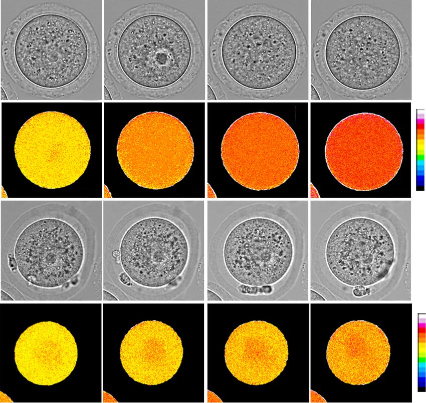
Downloaded from genesdev.cshlp.org on October 1, 2020 - Published by Cold Spring Harbor Laboratory Press

Zhang et al.

additional factors such as phosphatases (e.g., Dusp7 and           oocytes manifest to cause gene expression changes in ful-
Ppp2cb) (Su et al. 2012; Pfender et al. 2015; Tischer and          ly grown GV oocytes underlying the meiotic resumption
Schuh 2016) and kinases (e.g., Fyn) (Zheng et al. 2007;            defects in CKO Snf2h female mice.
Levi et al. 2010) have been reported to control meiotic re-
sumption through regulating the MPF activity. Thus, the
role of posttranslational regulation of the cAMP–PKA–              Results
MPF pathway in meiosis has been well established.
                                                                   Snf2h is required for MII oocyte generation
   In addition to regulation of MPF activity, meiotic pro-
gression is also accompanied with dramatic chromatin               A previous study in bovine indicated that Snf2h protein is
remodeling (Sasaki and Matsui 2008) and epigenetic                 transiently detectable in GV but not GVBD oocytes, im-
changes. For example, depletion of the CxxC zinc finger            plying that Snf2h may have a specific function for oocyte
protein Cfp1, a major mediator of H3K4me3, delays mei-             maturation (Wee et al. 2010). In this study, we used mouse
otic resumption and causes defective spindle assembly              oocyte as a model system to test this hypothesis. We first
(Sha et al. 2018). In addition, histone lysine methyltrans-        assessed Snf2h level by immunostaining during mouse
ferase Setdb1 and demethylase Lsd1 have been shown to              oocyte maturation and found Snf2h signal is readily
be required for chromatin condensation and meiotic re-             detectable in the nucleus of GV oocytes but appears dis-
sumption in mouse oocytes (Kim et al. 2015, 2016; Eym-             persed in GVBD and MII oocytes (Fig. 1A). Western blot
ery et al. 2016). However, depletion of these epigenetic           analysis confirmed that Snf2h protein is present with a
regulators only delays meiotic resumption, but not                 similar level at all three stages (Fig. 1B).
completely blocks GVBD, suggesting that either function-              To assess the Snf2h function in oogenesis, an oocyte-
al redundancy between these epigenetic factors or the key          specific knockout of Snf2h (CKO) was generated by cross-
regulator(s) of meiotic resumption functioning at chroma-          ing the Zp3 Cre mice (Lewandoski et al. 1997) with the
tin level is yet to be identified.                                 Snf2hfloxed/floxed mice (Snf2hfl/fl) (Alvarez-Saavedra et al.
   ATP-dependent chromatin remodeling complexes play               2014). The Zp3-Cre is an oocyte-specific Cre line, in
important roles in regulating gene transcription and con-          which Cre is expressed in early growing oocytes of mice
sist of four subfamilies: the switching/sucrose nonfer-            at postnatal day five (P5) (Lan et al. 2004). In this study,
menting (SWI–SNF) family, the imitation switch (ISWI)              Snf2hfl/fl and Zp3cre-Snf2hfl/fl female mice served as wild
family, the chromodomain helicase DNA-binding protein              type (WT) and CKO, respectively. In CKO oocytes, exon
(CHD) family, and the inositol requiring 80 (INO80) fam-           5 of Snf2h is depleted and a truncated protein without
ily (Clapier and Cairns 2009). All chromatin remodeling            the ATPase domain is expected to be produced (Supple-
complexes use ATP hydrolysis to alter histone–DNA con-             mental Fig. S1A). Immunostaining with an antibody
tacts, and the catalytic subunits share a similar ATPase-          against the full length Snf2h protein confirmed that
containing domain. Since ATP-dependent chromatin                   Snf2h was undetectable in the CKO GV oocytes (Fig. 1C).
remodeling complexes are mainly characterized in bio-                 The Snf2h CKO female mice are infertile as no pups were
chemical assays or cell lines (Clapier et al. 2017), their         obtained when they were crossed with WT fertile male
physiological roles remain less understood. With regard            mice for 6 mo (Fig. 1D). Remarkably, almost no MII oocytes
to meiosis, ISWI has been shown to be required for germ-           can be collected from CKO female mice (Fig. 1E), indicating
line stem cell (GSC) self-renewal and early oogenesis in           a severe defect in meiotic maturation. The sizes of WT and
Drosophila (Deuring et al. 2000; Xi and Xie 2005). Consis-         CKO ovaries are similar (Supplemental Fig. S1B) and com-
tently, depletion of Acf1, a subunit of an ISWI complex,           parable number of GV oocytes can be retrieved from both
CHRAC, leads to defects in oogenesis in Drosophila                 groups (Supplemental Fig. S1C). The ratio of GV oocytes
(Börner et al. 2016). In addition, Snf2h, the catalytic sub-       with nonsurrounded nucleolus (NSN) and surrounded nu-
unit of ISWI family complexes, has been postulated to              cleolus (SN) chromatin configuration showed no differ-
play a role in meiotic resumption given its transient pres-        ence between WT and CKO (Supplemental Fig. S1D).
ence in bovine GV oocytes (Wee et al. 2010). However,              Interestingly, the average diameter of CKO GV oocytes
whether ISWI complex is functionally important for                 (66 µm) is significantly smaller than that of the WT GV oo-
mammalian meiotic progression remains unknown.                     cytes (75 µm) (Fig. 1F). Since the meiotic and developmen-
   Through oocyte-specific depletion, we demonstrate               tal competences are acquired gradually during oocyte
here that Snf2h (also known as Smarca5) plays an essential         growth and the competences correlate with oocyte size,
role in oocyte meiotic resumption in mouse. Loss of func-          chromatin conformation, and cytoplasmic maturation
tion of Snf2h results in dysregulation of a number of genes        (Wickramasinghe et al. 1991; Eppig et al. 1994), the above
involved in MPF activation and oocyte maturation, in-              data suggests that the CKO GV oocytes may have im-
cluding Ccnb2 (Li et al. 2018), Prkaca (Bornslaeger et al.         paired meiotic and/or developmental competence.
1986), Prkar2b (Yoon et al. 2018), and Ndc80 (Gui and
Homer 2013). In addition, ATAC-seq analysis revealed a
                                                                   Oocyte-specific depletion of Snf2h blocks meiotic
global alteration in chromatin accessibility in Snf2h-
                                                                   resumption
deficient oocytes and a significant decrease of chromatin
accessibility at the Prkar2b promoter. Our study supports          To determine when Snf2h CKO oocytes arrest during mei-
the notion that Snf2h deficiency-mediated chromatin                otic maturation, we monitored the meiotic progression of
remodeling defects of the key meiotic genes in growing             GV oocytes collected from 6- to 8-wk female mice by in

2      GENES & DEVELOPMENT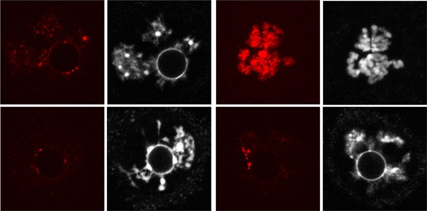
Downloaded from genesdev.cshlp.org on October 1, 2020 - Published by Cold Spring Harbor Laboratory Press
Chromatin remodeling and oocyte meiotic resumption
A B C Figure 1. Snf2h is essential for female fer-
GV GVBD MII tility in mouse. (A) Immunostaining of
Snf2h WT Snf2h CKO
Snf2h in oocytes at germinal vesicle (GV),
GVBD
germinal vesicle breakdown (GVBD), and
GV
MII
Snf2h Snf2h
meiosis metaphase II (MII) stages. Oocytes
Snf2h
25/25 20/20
were collected from adult ovaries (6–8 wk).
a-Tubulin Scale bar, 20 µm. (B) Western blotting of
DAPI DAPI Snf2h at GV, GVBD, and MII stages. a-Tubu-
20 μm 20 μm
lin was used as a loading control. (C ) Immu-
nostaining of Snf2h in WT and CKO GV
oocytes. Oocytes were collected from adult
D E F WT and CKO ovaries (6–8 wk). The numbers
p < 0.0001 p < 0.0001
Superovulated MII oocytes
40 90
0
represent the number of oocytes showing
Diameter of GV oocytes
50 Snf2h WT (n=4)
Snf2h CKO (n=4) the staining pattern and the total number
Pups/Female
40 30 80
0
of oocytes analyzed, respectively. Scale bar,
/Female
30
(um)
20 70
0 20 µm. (D) Fertility test of WT and CKO fe-
20
10 60
0
male mice. The WT (n = 6) and CKO (n = 6)
10
female mice were co-caged with WT fertile
0 0 50
0 male mice for 6 mo and the total numbers
1 2 3 4 5 6 Snf2h WT CKO Snf2h WT CKO
Months n=10 n=10 n=40 n=68 of pups per female are shown. (E) The num-
bers of MII oocytes per adult female mice
(6–8 wk) after superovulation. The WT (n = 10 from three independent biological replicates) and CKO (n = 10 from three independent bi-
ological replicates) female mice were used for the analysis. Data are presented as mean ± SEM. Each dot represents a single female mouse
analyzed. (∗∗∗∗ ) P < 0.0001 by two-tailed Student’s t-tests. (F) Quantification of oocyte size by measuring the diameters of GV oocytes (n =
40 from WT and n = 68 from CKO adult female mice, N = 3 independent biological replicates). Each dot represents a single oocyte analyzed.
Data are presented as mean ± SEM. (∗∗∗∗ ) P < 0.0001 by two-tailed Student’s t-tests.
vitro maturation (IVM). After 2 h, most (92.2%) WT GV Snf2h is required for cytoplasmic MPF activation
oocytes underwent GVBD, but almost all (98.6%) CKO in oocytes
oocytes were still arrested at prophase I (Fig. 2A). At 16
h, while 69% of the WT oocytes developed to MII stage, At the initial stages of meiotic resumption, oocytes under-
as evident by the extrusion of second polar body, only a go chromatin condensation and nuclear membrane break-
few CKO oocytes entered into MI stage and most (90%) down. As a chromatin remodeling factor, Snf2h can
CKO oocytes still maintain the intact nuclear envelope participate in this process by directly shaping the chroma-
(Fig. 2B). Similar meiotic resumption defects were also ob- tin or indirectly through regulating the expression of
served in oocytes collected from CKO mice without prior genes important for meiotic resumption. To differentiate
hormone (pregnant mare serum gonadotrophin [PMSG]) between these two possibilities, we first analyzed the
administration (Supplemental Fig. S2A), ruling out the chromatin organizations by immunofluorescence in WT
possibility that such defects are artifacts caused by and CKO GV oocytes. During the NSN-to-SN transition
hormone stimulation. Interestingly, CKO oocytes from in GV oocytes, chromatin becomes condensed and the
3-wk-old female mice showed a weaker phenotype, in number of chromocenters (aggregation of heterochroma-
which ∼35% of oocytes can develop to MII stage after tin regions, indicated by the immunostaining of HP1β) is
16 h of in vitro culture (Supplemental Fig. S2B,C). This ob- decreased from approximately seven to approximately
servation suggests that the longer oocyte growth phase in three per oocyte (Supplemental Fig. S3A,B; Bonnet-
adult female mice may lead to accumulation of more Garnier et al. 2012). However, in CKO GV oocytes, the
severe defects caused by Snf2h deficiency, which further number of chromocenters was only slightly decreased
impair the meiotic competence. To better characterize during NSN-to-SN transition (from approximately seven
the Snf2h deficiency caused oogenesis defects, we used to approximately six per oocyte). Thus, more chromocen-
the GV oocytes from 6- to 8-wk-old female mice in the ters in CKO SN oocytes were observed than that in WT
subsequent experiments. SN oocytes (Supplemental Fig. S3A,B). Furthermore, as in-
We next performed tubulin and DNA immunostaining dicated by HP1β staining, while chromocenters in WT SN
at different time points of oocyte maturation to monitor GV oocytes were mostly scattered away from the nucleo-
the chromatin configuration changes. Contrary to WT lus-like body (NLB), they were still in proximity to the
oocytes, which underwent chromatin condensation, spin- perinucleolar chromatin in CKO SN GV oocytes (Supple-
dle formation and first polar body extrusion during IVM, mental Fig. S3A,C,D). Therefore, Snf2h depletion affects
most CKO oocytes were arrested at GV stage without both the number of chromocenters and their localization
any apparent chromatin configuration changes (Fig. 2C). during NSN-to-SN transition in GV oocytes.
Furthermore, CKO oocytes did not exhibit increased To determine whether the chromatin organization de-
γH2AX signals at GVBD as observed in WT oocytes (Fig. fects are the cause of the meiotic arrest of CKO GV oo-
2D), indicating a defect in chromatin remodeling at meiot- cytes, we exchanged the nucleus between WT and CKO
ic resumption. oocytes and monitored the meiotic progression by IVM
GENES & DEVELOPMENT 3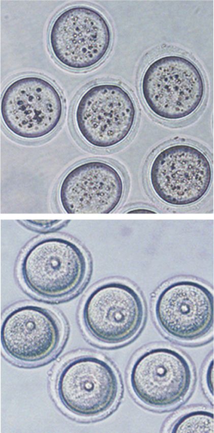
Downloaded from genesdev.cshlp.org on October 1, 2020 - Published by Cold Spring Harbor Laboratory Press

Zhang et al.

A                                                    B                                                             Figure 2. Oocyte-specific depletion of
            IVM-2h                                               IVM-16h                                           Snf2h blocks meiotic resumption. (A)
                                     IVM-2h                                                   IVM-16h              GVBD ratio of WT and Snf2h CKO oocytes
                            120           4       5                                    120                         after in vitro maturation (IVM) for 2 h. The
                                       14      15              WT                                 66    61
   WT
                            100     n=      n=                                         100     n=    n=            left panel shows the representative bright
                                                    No GVBD                                                MII
                                                                                                                   field images. Arrowheads point to germinal

                                                                                Percentage (%)
                          Percentage (%)
                             80                     GVBD                                80                 MI
        50 μm                                                      50 μm                                           vesicles. Scale bar, 50 µm. The right panel
                             60                                                         60                 GV
                                                                                                                   shows the quantification of GVBD ratio for
                             40                                                         40                         WT and CKO oocytes. One-hundred-forty-
  CKO                        20                               CKO                       20                         four WT and 155 CKO GV oocytes from
                               0                                                          0
                                                                                                                   adult ovaries (6–8 wk) were used for the anal-
                                   WT CKO                                                     WT CKO               ysis. N = 6 independent biological replicates.
                                                                                                                   (B) Oocyte maturation ratio of WT and CKO
C                                                           D              IVM-0h                IVM-2.5h          oocytes after IVM for 16 h. Left panel shows
          IVM-0h     IVM-2h     IVM-5h         IVM-16h               rH2AX        DAPI      rH2AX          DAPI    the representative bright field images. Ar-
     GV          GVBD        MI             MII
                                                                 N                      N
                                                                                                                   rows point to first polar bodies and arrow-
  WT                                                         WT                                                    heads point to germinal vesicles. Scale bar,
      27/27      24/25       26/30          23/30     20 μm     35/35                  30/30                 20 μm 50 µm. Right panel shows the quantification
                                                                 N
                                                                                                                   of maturation ratio for GV oocytes. Sixty-six
                                                                                        N
 CKO
                                                                                                                   WT and 61 CKO GV oocytes from adult ova-
                                                            CKO
                                                                                                                   ries (6–8 wk) were used for the analysis. N = 3
      31/31      32/32       25/32          33/38               27/27                  27/27                       independent biological replicates. (C )
                                                                                                                   Immunostaining showing the maturation
process of WT and CKO oocytes. Stages are determined based on DAPI and α-Tubulin staining. The numbers represent the combined num-
ber of oocytes exhibiting the staining pattern and the total number of oocytes analyzed in three independent biological replicates. Scale
bar, 20 µm. (D) Immunostaining showing the level of γH2AX in WT and CKO oocytes during IVM. The numbers represent the combined
number of oocytes exhibiting the staining pattern and the total number of oocytes analyzed in three independent biological replicates.
Scale bar, 20 µm.

(Fig. 3A). Although most GV oocytes reconstructed with                                           derstand how Snf2h deficiency impairs MPF activation,
WT cytoplasm and CKO nucleus successfully went                                                   we performed RNA sequencing (RNA-seq) analysis of
through meiotic resumption after 2.5-h of IVM, none of                                           growing oocytes from early secondary follicles (growing
the GV oocytes reconstructed with CKO cytoplasm and                                              oocytes 1 [GO1], 40–45 µm) and secondary follicles (grow-
WT nucleus underwent GVBD (Fig. 3B). This result indi-                                           ing oocytes 2 [GO2], 50–55 µm), and fully grown oocytes
cates that cytoplasm defects, but not the chromatin de-                                          from preovulatory follicles (FGO, >70 µm) (Fig. 4A). The
fects, in the CKO oocytes are the major causes of the                                            transcriptomes of two replicates from each group exhibit-
meiotic arrest. These data also suggest that Snf2h contrib-                                      ed high correlations (R > 0.975) (Supplemental Fig. S4A,B)
utes to oocyte maturation, likely through regulating the                                         and Snf2h was completely depleted at all three stages
expression of meiosis-related genes.                                                             (Supplemental Fig. S4C). Notably, our transcriptome
   It is well established that meiotic arrest of GV oocytes is                                   data sets are highly correlated to a previously published
maintained by high levels of cAMP. cAMP activates PKA                                            RNA-seq data set (Supplemental Fig. S4D; Zhang et al.
to phosphorylate downstream effector proteins, which in                                          2016), validating our oocyte staging, collection, and
turn suppresses the MPF (Cdk1/Cyclin B1) activity (Holt                                          RNA-seq analysis procedures.
et al. 2013). To determine whether MPF fails to activate                                            Comparative analysis revealed that the most significant
in CKO oocytes and causes the meiotic arrest, we used a                                          transcriptome differences between WT and CKO were ob-
fluorescence resonance energy transfer (FRET) assay to                                           served in fully grown oocytes with 1104 genes up-regulat-
monitor the MPF activity during IVM (Levasseur et al.                                            ed and 1221 genes down-regulated (FC > 1.5, P < 0.05, and
2019). As expected, the MPF activity of WT GV oocytes                                            RPKM > 1 as a cutoff) (Fig. 4B; Supplemental Fig. S4E; Sup-
increased during the 2-h IVM period and underwent                                                plemental Table S1). This result is consistent with the
GVBD. In contrast, the MPF activity of CKO oocytes                                               principle component analysis (PCA) (Supplemental Fig.
only slightly increased and reached a plateau by 50 min                                          S4B). One likely explanation for the milder gene expres-
and GVBD did not take place (Fig. 3C,D). These data indi-                                        sion changes in growing oocytes compared with those in
cate that Snf2h CKO GV oocytes fail to activate MPF to                                           fully grown oocytes is that Snf2h depletion by Zp3-Cre oc-
initiate meiotic resumption.                                                                     curs at P5 and the transcriptional changes are accumulat-
                                                                                                 ed during oocyte growth. Gene Ontology (GO) analysis
                                                                                                 revealed that the up-regulated genes in fully grown GV oo-
Depletion of Snf2h leads to dysregulation of genes
                                                                                                 cytes is enriched for the GO term of chromatin assembly
involved in MPF activation
                                                                                                 (Fig. 4C), consistent with the chromatin remodeling func-
Since no active transcription takes place after GV NSN-                                          tion of Snf2h. Additionally, both up- and down-regulated
to-SN transition, the inability of cytoplasmic products                                          genes in GV oocytes are enriched for the GO terms of cel-
to activate MPF should reflect the cumulative defects                                            lular and metabolic processes (Fig. 4C) known to be impor-
caused by Snf2h deficiency during oocyte growth. To un-                                          tant for oocyte maturation (Gu et al. 2015).

4       GENES & DEVELOPMENT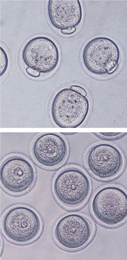
Downloaded from genesdev.cshlp.org on October 1, 2020 - Published by Cold Spring Harbor Laboratory Press
Chromatin remodeling and oocyte meiotic resumption
A B Figure 3. Cytoplasmic defects cause CKO
WT cytoplasm 120 oocytes unable to activate maturation-pro-
CKO nucleus WT (n=47)
WT
100 moting factor (MPF) for meiotic resumption.
GVBD ratio (%)
CKO (n=32)
IVM 80 WT-N/CKO-C (n=43) (A) Schematic diagram of the experimental
for 3h 60 CKO-N/WT-C (n=31)
strategy for nuclei exchange experiments.
40
IVM 20
The GV nuclei from WT or CKO mice were
for 3h 0 removed and reconstructed with enucleated
CKO cytoplasm 0 0.5 1 1.5 2 2.5
CKO
Time (hours after IVM initiation) CKO or WT oocytes, respectively. (B) GVBD
WT nucleus
ratio of WT, CKO, and reconstructed oo-
C D cytes at different time points during IVM.
00:00:00 00:40:00 01:20:00 02:00:00 N = 3 independent biological replicates and
n > 10 oocytes were used for each replicate.
WT
Bright field 125 Data are presented as mean ± SEM. (C ) Rep-
20 μm
resentative images (one image per 40 min)
(% of initial value)
120 WT (n=14)
1.7
Emission ratio
WT 0.9 115 CKO (n=14) showing the dynamic FRET signal during
Emission ratio
0.0 110 IVM of WT and CKO oocytes. (D) Quantifi-
105 cation of the emission ratio over time in
CKO
Bright field
100 WT (n = 14) and CKO (n = 14) oocytes from
95
0 N = 3 independent biological replicates.
12 0
10 0
13 0
0
80
0
20
40
10
50
60
70
30
11
9
1.5
CKO Time (minutes after IVM initiation) Data are presented as mean ± SEM.
0.7
Emission ratio
0.0
Although unlikely, these transcriptional changes might The meiotic resumption defect can be rescued by
be caused by hormone administration. To rule out this restoring MPF activity
possibility, we performed RNA-seq using non-hormone-
stimulated fully grown GV oocytes (nFGO). Comparative The cAMP-PKA-MPF signaling pathway plays a critical
analysis revealed a good correlation not only between the role in regulating meiotic resumption (Fig. 5A). Prkar2b
two replicates of nFGO, but also between the FGO sam- and Prkaca, respectively, encode the regulatory and cata-
ples collected with or without hormone stimulation (Sup- lytic subunit of PKA (Kirschner et al. 2009). After binding
plemental Fig. S5A). Similar to the FGO collected under to cAMP, the PKA catalytic subunit is released from the
hormone stimulation, a significant transcriptome change regulatory subunit and becomes capable of phosphorylat-
was observed between WT and CKO nFGO with 880 ing downstream substrates (e.g., Wee1b) to inhibit MPF
genes up-regulated and 1065 genes down-regulated (Sup- activity (Fig. 5A). Previous studies have shown that the ra-
plemental Fig. S5B), and these differentially expressed tio of catalytic to regulatory subunit (C/R ratio) is impor-
genes were highly correlated with those identified under tant for PKA activity (Nishimura et al. 2012) and that
hormone stimulated conditions (Supplemental Fig. S5C). overexpression of Prkaca could inhibit meiotic resump-
These data strongly support the notion that the gene ex- tion (Bornslaeger et al. 1986). Thus, simultaneous up-
pression change is due to loss of Snf2h function rather regulation of Prkaca and down-regulation of Prkar2b in
than caused by hormone stimulation. CKO GV oocytes (Fig. 4E) may elevate the C/R ratio, lead-
To establish a link between the transcriptome changes ing to the increase in PKA activity and ultimate inhibition
and the MPF activation defect, we analyzed expression of MPF and meiotic resumption. We attempted to rescue
levels of genes involved in cAMP signaling and female the meiotic arrest phenotype by Prkar2b mRNA overex-
meiotic cell cycle and found a number of meiosis-related pression or Prkaca siRNA-mediated knockdown (Fig.
genes were indeed dysregulated in both CKO FGO and 5A). Importantly, both manipulations partially rescued
nFGO (Fig. 4D; Supplemental Fig. S5D). We validated the meiotic resumption defects (Fig. 5B,C). Similarly,
the differential expression of Ccnb2, Ndc80, Prkar2b, overexpression of a constitutively active form of Cdk1
and Prkaca in CKO oocytes using quantitative PCR and (Cdk1AF), which cannot be phosphorylated at Thr14/
confirmed that the protein abundance of Prkaca, Prkar2b, Tyr15 residues (Adhikari et al. 2016), also partially res-
and Cyclin B2 were also obviously altered in Snf2h-deplet- cued the phenotype (Fig. 5D).
ed oocytes (Supplemental Fig. S6A,B). Based on previous Cyclin B2 (encoded by Ccnb2), similar to Cyclin B1 (en-
studies (Yin et al. 2007; Gui and Homer 2013; Li et al. coded by Ccnb1), can associate with Cdk1 (Satyanarayana
2018), a combined up-regulation of Adcy9, Prkaca, and and Kaldis 2009) and compensate for Cyclin B1 deficiency
Cdc20, and down-regulation of Ccnb2, Ccnb3, Prkar2b, in Ccnb1 CKO oocytes (Li et al. 2018). In addition,
and Ndc80 is likely to be responsible for the failure of Ndc80 (also known as Hecl) has been shown to protect
MPF activation in CKO oocytes. Since most genes in- Cyclin B2 from destruction by Cdh1-activated anaphase-
volved in cAMP signaling and female meiosis was not as promoting complex (APCCdh1) and depletion of Ndc80 or
severely altered in growing oocytes (i.e., GO1 and GO2) Ccnb2 by antisense morpholinos resulted in severe de-
as compared with the fully grown oocytes (Fig. 4E; Supple- fects in GVBD (Gui and Homer 2013). In Snf2h CKO oo-
mental Fig. S6A), it is likely that the overall defects accu- cytes, we found that expression levels of both Ccnb2
mulated during oocyte growth are responsible for the MPF and Ndc80 were decreased (Fig. 4E), which resulted in a
activation defect. decrease of Cyclin B2 protein abundance (Supplemental
GENES & DEVELOPMENT 5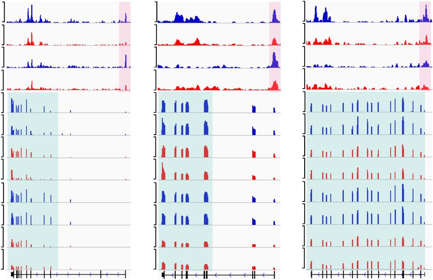
Downloaded from genesdev.cshlp.org on October 1, 2020 - Published by Cold Spring Harbor Laboratory Press
Zhang et al.
A B Figure 4. Depletion of Snf2h leads to dysre-
Oocyte collection information FGO [Log2(normalized counts)]
gulation of genes related to MPF activation.
20 .5
Up-regulated: =1 (A) Schematic illustration of oocytes collec-
Sample ID Type Age of mice Oocyte size FC 1.
5
1104 (8.13%) =- tion for RNA-seq. Oocytes were collected
15 FC
Oocytes from based on the age of the mice and the size of
GO1 P7-12 40-45 μm
early secondary follicles
CKO
10 the oocytes. Twenty oocytes were used for
GO2
Oocytes from
P9-14 50-55 μm
Snf2h each RNA-seq library and two RNA-seq rep-
secondary follicles 5 licates were performed for WT and CKO
Down-regulated:
FGO
Oocytes from
6-8 weeks >70 μm 1221 (8.99%) groups at each stage. (B) Scatter plots com-
preovulatory follicles 0
paring the gene expression profiles of WT
0 5 10 15 20
WT and CKO fully grown GV oocytes. Two repli-
C cates for WT and CKO FGO oocytes were
Up-regulated genes Down-regulated genes used for differential gene expression analyses
[GO:0019222] Regulation of metabolic process [GO:0044238] Primary metabolic process (FC > 1.5, P-value 1). (C ) Gene
[GO:0031497] Chromatin assembly [GO:0009116] Nucleoside metabolic process ontology analysis of the differentially ex-
[GO:0007242] Intracellular signaling cascade [GO:0044237] Cellular metabolic process
pressed genes in CKO FGO oocytes. (D)
Functional classification of differentially ex-
[GO:0006333] Chromatin assembly or disassembly [GO:0008152] Metabolic process
pressed genes associated with cAMP signal-
[GO:0009987] Cellular process [GO:0009987] Cellular process
ing, oocyte meiosis, meiotic M phase,
0 2 4 6 0 2 4 6 abnormal female meiosis, and other reported
-Log10(p-value) -Log10(p-value)
factors involved in oogenesis. Up-regulated
D E Log2FC: CKO vs. WT and down-regulated genes are colored with
List of DEGs that are involved in meiosis
1 2 2 1 0 -1 -2 red and blue, respectively. (E) Heat map
O
GO GO FG 1 2 O showing the log2 (fold change) of genes listed
Acox1, Adcy9, Akt1, Arap3, Cnga2, Gli3 GO GO FG
cAMP signaling
Creb3, Fshr, Gria4, Nfatc1, Pik3r2, Tex15 Cnga2 in D for GO1, GO2, and FGO oocytes.
Prkaca, Rapgef3, Slc9a1, Vav3 Ppp2r5b Pabpc1
[PATH: mmu04024] Tet1 Adcy9
Atp1a3, Atp1b1, Gli3, Plce1, Atp1a3 Slc9a1
Rapgef4, Rras2 Oog3 Ppp2r5e
Cul1 Tex12
Adcy9, Ccne1, Cdc20, Ppp2r5a, Ndc80 Rapgef3
Ppp3cc Acox1
Oocyte meiosis Ppp2r5e, Prkaca, Rps6ka1 Ccnb2 Cdc20
[PATH: mmu04114] Ccnb2, Cul1, Ppp2r5b, Ppp3cc, Plce1 Ppp2r5a
Tex11 Creb3
Rps6ka6, Smc1b Zp2 Ccne1
Prkar2b Prkaca
Meiotic M phase Ccnb2, Ccnb3, Mnd1, Msh4, Cdc14b Nfatc1
[GO: mmu04114] Piwil1, Smc1b, Tex11, Tex15 Ccnb3 Akt1
Rapgef4 Vav3
Cdc20, Hormad2, Pabpc1, Patl2, Rras2 Rps6ka1
Abnormal female Wee1 Gria4
meiosis Sycp3, Tex12, Trp73 Rps6ka6 Fshr
[MP: 0005168] Ccnb3, Msh4, Smc1b, Tet1 Atp1b1 Arap3
Smc1b Pik3r2
Dppa3 Sycp3
Other factors related Cdc14b, Ndc80, Prkar2b, Oog3, Mnd1 Hormad2
to oogenesis Dppa3, Wee1, Zp2 Msh4 Patl2
Piwil1 Trp73
Fig. S6B). Consistently, overexpression of Ccnb2 fully res- accessibility by performing ATAC-seq analysis of growing
cued the defects of meiotic resumption in CKO oocytes, oocytes (GO1) and fully grown GV oocytes (FGO) from
even when they were cultured in the MEM medium con- WT and CKO female mice with two replicates (Supple-
taining cAMP phosphodiesterase inhibitor 3-Isobutyl-1- mental Table S2). After confirming the data reproducibil-
methylxanthine (IBMX) (Fig. 5E). Since Cyclin B1 and ity (Supplemental Fig. S7A), we combined the data for
Cyclin B2 have redundant functions in meiotic cell cycle subsequent analysis. Consistent with previous reported
regulation, we also overexpressed Ccnb1 mRNA and chromatin “open-to-close” transition at the transcription
achieved almost a complete rescue of the meiotic pheno- start sites (TSS) during oocyte growth (Gu et al. 2019),
type (Fig. 5F). Collectively, the above results support the lower level of ATAC-seq signal at TSS was observed in ful-
notion that Snf2h regulates meiotic resumption by regu- ly grown GV oocytes when compared with that in GO1
lating transcription of genes involved in MPF activation. oocytes from WT mice (Fig. 6A). Compared with WT
oocytes, an overall reduction of ATAC-seq signal was ob-
served in both CKO GO1 and FGO (Fig. 6A,B), indicating a
Snf2h regulates transcription by altering chromatin
global decrease of chromatin accessibility due to Snf2h
accessibility
depletion.
We next set out to understand how Snf2h may regulate To determine whether the mRNA abundance differenc-
transcription of meiosis-related genes during oogenesis. es in CKO fully grown GV oocytes correlate with chroma-
Since chromatin remodeling factors are known to regulate tin accessibility changes during oocyte growth, we
gene expression by altering nucleosome positioning and integrated the ATAC-seq data sets with the transcriptome
promoter accessibility (Clapier and Cairns 2009), we hy- of WT and CKO oocytes. We found that the down-regulat-
pothesize that dysregulation of meiotic genes may be ed genes in CKO fully grown GV oocytes, as a whole,
due to the alternation of promoter chromatin accessibility exhibited a more dramatic decrease of promoter ATAC
in the CKO oocytes. To this end, we assessed chromatin signal in CKO GO1 compared with the CKO FGO (Fig.
6 GENES & DEVELOPMENT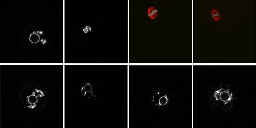
Downloaded from genesdev.cshlp.org on October 1, 2020 - Published by Cold Spring Harbor Laboratory Press
Chromatin remodeling and oocyte meiotic resumption
A B Figure 5. Meiotic resumption defects caused
Prophase I arrest by Snf2h deficiency can be rescued by restor-
Adcy3 Gs-Gpr3 LH 120 WT (n=46) ing MPF activity. (A) Schematic illustration
GVBD ratio (%)
100
cAMP Pde3 CKO (n=49) of the cAMP-PKA-MPF pathway. Red words
80
60
with yellow background positively regulate
PKA Prkar2b mRNA CKO+
Prkaca siRNA 40 Prkar2b cAMP-PKA activity, which leads to meiotic
mRNA (n=59)
Cdc25b Wee1b
20 arrest at prophase I stage. Black words with
CDK1AF mRNA
Cdk1 0 gray background negatively regulate cAMP-
MPF 0 0.5 1 1.5 2 2.5
CyclinB1 CyclinB1/2 mRNA Time (hours after IVM initiation) PKA activity, which activates MPF and pro-
motes meiotic resumption. Red words with
Meiotic Resumption
no background are the strategies used for the
C D rescue experiments. (B–F ) GVBD ratio at
120 120 WT (n=43)
WT (n=39) different time points during IVM for oocytes
GVBD ratio (%)
GVBD ratio (%)
100 100
CKO (n=35) CKO (n=39) injected with Prkar2b mRNA (B), Prkaca
80 80
CKO+ siRNA (C), Cdk1AF mRNA (D), Ccnb2
60 CKO+ 60 Cdk1AF
siPrkaca mRNA (n=36) mRNA (E), or Ccnb1 mRNA (F). N = 3 inde-
40 (n=51) 40
20 20
pendent biological replicates for each rescue
0 0 experiment and n > 10 oocytes were used for
0 0.5 1 1.5 2 2.5 0 0.5 1 1.5 2 2.5 each replicate. Data are presented as mean ±
Time (hours after IVM initiation) Time (hours after IVM initiation)
SEM.
E F
120 120
WT (n=42) WT (n=40)
GVBD ratio (%)
GVBD ratio (%)
100 100
80 CKO (n=48) 80 CKO (n=37)
60 60
CKO+ CKO+
40 CyclinB2 40 CyclinB1
mRNA (n=34) mRNA (n=37)
20 20
0 0
0 0.5 1 1.5 2 2.5 0 0.5 1 1.5 2 2.5
Time (hours after IVM initiation) Time (hours after IVM initiation)
6C). To analyze this correlation in more detail, we gener- gress towards understanding the molecular mechanism
ated a heat map followed by clustering analysis, which of how chromatin remodeling contributes to meiotic
revealed that 70% of down-regulated genes were associat- resumption.
ed with decreased ATAC-seq signals in GO1 and/or FGO
(Supplemental Fig. S7B,D). In contrast, only < 25% of up-
regulated genes were associated with increased ATAC-seq Snf2h is an essential regulator of meiotic resumption
signals at either stage (Supplemental Fig. S7B). Important-
ly, some meiotic genes, including those whose dysregula- It is well known that chromatin remodeling factors use
tion contribute to the Snf2h CKO meiotic arrest, such as the energy derived from ATP hydrolysis to move, eject,
Prkar2b, Ccnb2, and Ndc80, had already reduced or lost or restructure nucleosomes (Clapier et al. 2017). The
their promoter ATAC peaks in CKO GO1 prior to their ISWI complex has been previously shown to alter DNA
down-regulation in the fully grown oocytes (Fig. 6D; Sup- and histone contact by nucleosome sliding and histone re-
plemental Fig. S7C,D). Collectively, these data suggest placement in vitro (Clapier et al. 2017). However, it is not
that loss function of Snf2h causes decreased promoter ac- clear how ISWI-mediated nucleosome positioning or spac-
cessibility of meiotic genes at GO1 stage, which likely ing regulates transcription and whether this causes any
manifest to affect gene expression at fully grown oocytes, physiological outcome. As the catalytic subunit of the
resulting in meiotic defects. ISWI complex, Snf2h has been reported to be critical for
embryonic development (Stopka and Skoultchi 2003), em-
bryonic lens differentiation (He et al. 2016), hematopoiet-
Discussion ic stem and progenitor cell (HSPC) proliferation and
differentiation (Kokavec et al. 2017), as well as cerebellar
The precise progression of meiotic cell cycle is essential morphogenesis and neural maturation (Alvarez-Saavedra
for female reproduction, and defects in meiotic resump- et al. 2014). Here we extended these studies by uncovering
tion have been one of the major causes of infertility. its essential function in oocyte meiotic resumption.
Although meiotic progression is known to be accompa- Unlike WT oocytes, which undergo GVBD and chroma-
nied by dramatic chromatin remodeling, the molecular tin condensation during meiotic resumption, neither
mechanisms underlying meiotic chromatin remodeling takes place in Snf2h CKO oocytes (Fig. 2). Despite the de-
and meiotic resumption are poorly understood. Here, we fects in chromocenter formation in the Snf2h CKO oo-
demonstrate that Snf2h, the catalytic subunit of ISWI cytes, nucleus exchange experiments demonstrated that
family complexes, is critical in driving meiotic progres- nucleus defects did not contribute to the meiotic arrest
sion by regulating the expression of genes important for phenotype. Rather, it is the defects in CKO cytoplasm
MPF activation. The study represents an important pro- that prevented the reconstructed oocytes from entering
GENES & DEVELOPMENT 7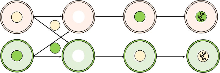
Downloaded from genesdev.cshlp.org on October 1, 2020 - Published by Cold Spring Harbor Laboratory Press
Zhang et al.
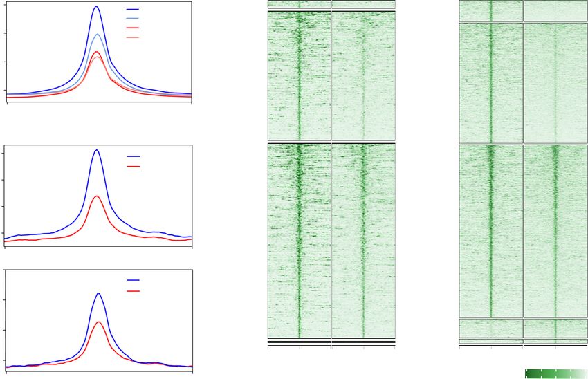
A B Figure 6. Snf2h regulates transcription by
WT GO1 CKO GO1 WT FGO CKO FGO altering chromatin accessibility. (A) Meta-
40 Loss (FC>5, Loss (FC>5,
WT GO1 plot showing the ATAC-seq signals at tran-
WT FGO n=1,992) n=3,096)
30 CKO GO1 scription start sites (TSSs) in WT and CKO
RPKM
CKO FGO
20
growing oocytes (GO1) and fully grown GV
Partial Loss Partial Loss
(2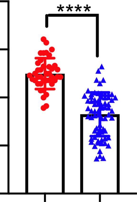
Downloaded from genesdev.cshlp.org on October 1, 2020 - Published by Cold Spring Harbor Laboratory Press

                                                                                    Chromatin remodeling and oocyte meiotic resumption

combined dysregulation of both Cdk1 activation and Cy-             Snf2h CKO mice may prevent activation of key meiotic
clin B2 level can explain the meiotic arrest phenotype of          genes in fully grown oocytes, resulting in meiotic resump-
the CKO oocytes.                                                   tion defect.
   We showed that Ccnb1/2 mRNA overexpression fully
rescued the meiotic arrest phenotype of CKO oocytes                Materials and methods
(Fig. 5). This observation is not surprising considering
the fact that in mitotic cells a high level of Cyclin B can        Mice
lead to a full activation of Cdk1 by activating Cdc25,             Snf2h CKO mice were generated by crossing Zp3 cre mice (Lewan-
which can remove the inhibitory phosphorylation of                 doski et al. 1997) with Snf2hfl/fl mice (Alvarez-Saavedra et al.
Cdk1 (Deibler and Kirschner 2010). Thus, although the              2014). All animal studies were performed in accordance with
high PKA activity can inhibit Cdk1 activation in CKO oo-           guidelines of the Institutional Animal Care and Use Committee
cytes, overexpression of Ccnb1/2 may overcome this in-             at Harvard Medical School.
hibitory effect by activating Cdc25/Cdk1 to achieve a
full rescue of the meiotic arrest phenotype. In contrast, al-      Oocyte collection and in vitro maturation
though Prkar2b and Cdk1AF mRNA overexpression or
                                                                   For fully grown GV oocytes collection, ovaries were dissected
Prkaca siRNA knockdown can reduce PKA-mediated in-
                                                                   from 3- or 6- to 8-wk-old female mice 46–48 h after injection
hibition of Cdk1, MPF activation may be limited by the             with 7.5 international units (IU) of pregnant mare serum gonado-
insufficient level of Cyclin B2. As mentioned earlier,             trophin (PMSG) (Millipore). For non-hormone-stimulated fully
both Ccnb2 mRNA and a Cyclin B2 degradation protector              grown GV oocytes (nFGO), ovaries were dissected from 6- to 8-
Ndc80 are down-regulated in CKO oocytes. Thus, insuffi-            wk-old female mice without PMSG injection. The ovaries were
cient Cyclin B2 cannot be rescued by inhibiting PKA ac-            transferred to M2 medium (Sigma-Aldrich) and punctured with
tivity or increasing Cdk1 activity, which explains why             a 27-gauge needle to release cumulus–oocyte complexes. The cu-
only partial rescue was achieved under these conditions.           mulus cells were gently removed from the cumulus–oocyte com-
                                                                   plexes using a narrow-bore glass pipette. The oocytes were then
                                                                   transferred into α-MEM (Life technologies) supplemented with
Snf2h regulates transcription of meiotic genes by altering         5% FBS (Sigma-Aldrich), 10 ng/mL EGF (Sigma-Aldrich), and 0.2
chromatin accessibility                                            mM 3-isobutyl-1-methylxanthine (IBMX) (Sigma-Aldrich). About
                                                                   30–40 GV oocytes can be obtained from each hormonally primed
Compared with the well-studied nucleosome-positioning              mouse. After culture in MEM + IBMX medium for 1 h, GV oocytes
mechanism, whether Snf2h regulates transcription by al-            with perivitelline space (PVS) were selected for subsequent exper-
tering chromatin accessibility remains largely unknown.            iments as these oocytes are of high meiotic competence (Inoue
In fact, only a couple of studies suggested that Snf2h can         et al. 2007). For the meiotic resumption and in vitro oocyte matu-
alter chromatin accessibility in T cells and ES cells. For         ration (IVM) analyses, GV oocytes were washed at least three
example, it was reported that Snf2h may regulate cytokine          times to remove IBMX before being transferred to IBMX-free α-
gene expression by altering chromatin accessibility in the         MEM medium containing 5% FBS and 10 ng/mL EGF.
murine EL4 T cells (Precht et al. 2010). Recently, Barisic            For growing oocytes collection, ovaries were removed and di-
                                                                   gested in PBS supplemented with 2 mg/mL collagenase (Sigma-
et al. (2019) compared chromatin accessibility landscape
                                                                   Aldrich) and 0.025% trypsin (Sigma-Aldrich) for 30 min at
in WT and Snf2h KO ESCs and suggested that target bind-            37°C. M2 medium (Sigma-Aldrich) was then added to neutralize
ing of a subset of transcription factors may depend on             the digestion mix and oocytes were picked up with a drawn-out
Snf2h-mediated chromatin remodel. Therefore, in vivo               glass pipette. To avoid potential contamination by somatic cells,
evidences are still lacking regarding how Snf2h-mediate            oocytes were washed extensively in clean drops of M2 medium. A
chromatin remodeling can control gene expression.                  stage micrometer was used in combination with an eyepiece ret-
   In this study, using ATAC-seq, we showed that deple-            icle to measure the sizes of oocytes. Mice of postnatal days p7–12
tion of Snf2h resulted in a global decrease in chromatin ac-       and p9–14 were used to collect growing oocytes with diameters of
cessibility during oocyte growth (Fig. 6). Although there is       40–45 µm (from early secondary follicles) and 50–55 µm (from
                                                                   secondary follicles), respectively.
no direct correlation between global RNA abundance and
chromatin accessibility in fully grown GV oocytes, we in-
deed observed a strong correlation between decrease of             mRNA synthesis and microinjection
ATAC-seq signals and transcriptional down-regulation               mRNAs were synthesized by in vitro transcription (IVT) as de-
during oogenesis (Fig. 6; Supplemental Fig. S7). These             scribed previously (Matoba et al. 2014). Briefly, MPF activity sen-
data indicate that Snf2h may function as a chromatin               sor (Addgene plasmid #26064) and full-length cDNA of Ccn1b,
remodeler to facilitate gene expression by increasing chro-        Ccnb2, Prkar2b, and Cdk1AF were cloned into pcDNA3.1 vector
matin accessibility during oocyte growth. Importantly,             with in-fusion cloning kit (Clontech). The constructed plasmids
the key meiotic genes whose dysregulation contribute to            were linearized by XbaI. After purification, the linearized plasmid
                                                                   DNA were used as templates for IVT using mMESSAGE mMA-
the meiotic arrest of Snf2h CKO oocytes, such as Prkar2b,
                                                                   CHINE T7 Ultra kit (Thermo Fisher Scientific). The synthesized
Ccnb2, and Ndc80, had already exhibited reduced chroma-
                                                                   mRNA was dissolved in nuclease-free water and quantified by
tin accessibility at GO1 stage prior to their reduced gene         NanoDrop ND-1000 spectrophotometer (NanoDrop Technolo-
expression in fully grown oocytes. Collectively, these             gies) and stored at −80°C.
data support the notion that Snf2h-mediated promoter ac-             GV oocytes with PVS were injected with ∼10 pL of mRNA with
cessibility might be a prior requirement for meiotic gene          specific concentrations (1.5 µg/µL Ccnb1 mRNA, 1.0 µg/µL
activation. Failure to establish open chromatin at GO1 in          Ccnb2 mRNA, 2.5 µg/µL Prkar2b mRNA, 1.5 µg/µL Cdk1AF

                                                                                                       GENES & DEVELOPMENT          9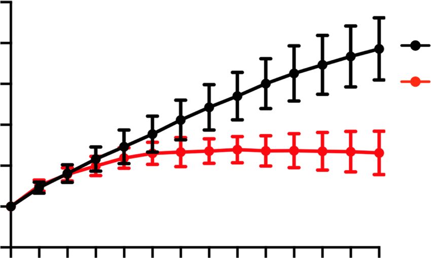
Downloaded from genesdev.cshlp.org on October 1, 2020 - Published by Cold Spring Harbor Laboratory Press

Zhang et al.

mRNA, 1.5 µg/µL MPF activity sensor) using a Piezo-driven mi-          (1/2000 dilution; Sigma T6199), and γH2AX (1/1000 dilution;
cromanipulator (Eppendorf). Injected GV oocytes were cultured          Millipore 05-636) were used in this study.
in MEM + IBMX medium for 3 h to allow mRNA translation and
then transferred into IBMX-free MEM medium for IVM. For
siRNA injection, GV oocytes were injected with 4 µM siPrkaca           FRET imaging and analysis
and arrested in MEM + IBMX medium for 24 h before IVM. Oligo-          GV oocytes were injected with MPF activity sensor mRNA and
nucleotides used in this study are listed in Supplemental Table S3.    cultured in MEM + IBMX medium for 3 h for sufficient mRNA
                                                                       translation. Then oocytes were transferred to IBMX-free MEM
Nucleus exchange experiments in fully grown GV oocytes                 medium in a confocal dish (Green BioResearch LLC). FRET imag-
                                                                       ing was performed as reported (Levasseur et al. 2019) using
GV oocytes nuclear transfer was carried out as described previ-        Screeny (HIM 1023) microscopy with the help of the MicRoN
ously (Inoue et al. 2008). Briefly, GV oocytes were cultured in        team at Harvard Medical School. All quantifications were per-
M2 medium containing 0.2 mM IBMX, 10 mg/mL cytochalasin                formed using ImageJ software.
B (Calbiochem), and 0.1 mg/mL colcemid (Sigma-Aldrich) for
15 min. The nucleus was removed with minimal cytoplasm car-
ryover and fused with an enucleated oocyte using GenomONE -            RNA-seq
CF EX Sendai virus Envelope cell fusion kit (Cosmo Bio HVJ-E).
                                                                       RNA-seq libraries were prepared as previously described (Matoba
The reconstructed oocytes were cultured in MEM + IBMX for
                                                                       et al. 2014). Twenty oocytes at GO1, GO2, and FGO stages from
1 h to ensure complete fusion before IVM experiments.
                                                                       WT and CKO female mice were used for RNA-seq library prepa-
                                                                       ration. Briefly, reverse transcription and cDNA amplification
Western blotting                                                       were performed using SMARTer ultralow input RNA cDNA
                                                                       preparation kit (Clontech) following the manufacturer’s in-
For immunoblot experiments, 120 oocytes were collected and
zona pellucida were removed with acid Tyrode’s solution                structions. Nextera XT DNA library preparation kit (Illumina)
                                                                       was used for cDNA fragmentation, adaptor ligation, and amplifi-
(Sigma-Aldrich). After washing three times with prewarmed 1%
polyvinylalcohol (PVA) (Sigma-Aldrich)/PBS, oocytes in 5 µL of         cation according to the manufacturer’s instructions. Single-end
1%PVA/PBS were transferred using a calibrated glass pipette to         100-bp sequencing was performed on a HiSeq2500 sequencer
                                                                       (Illumina).
15 µL of sample buffer containing 5 µL of 4× NuPAGE LDS sam-
ple buffer (Thermo Fisher Scientific) and 10 µL of sterile filtered
water. The samples were then snap-frozen in dry ice for 5 min          ATAC-seq
and the freeze–thaw procedures were repeated twice before stor-
ing them at −80°C. The samples were heated for 5 min at 90°C           ATAC-seq libraries of growing oocytes and fully grown GV oo-
before loading to SDS-PAGE gels. Western blotting was per-             cytes were prepared as previously described with some modifica-
formed as described previously (Marangos 2016). Primary anti-          tions (Wu et al. 2016). Briefly, 100 fully grown GV nuclei were
bodies against Snf2h (1/1000 dilution; Abcam ab168653),                collected after incubation in M2 medium containing 0.2 mM
Prkar2b (1/50 dilution; Santa Cruz Biotechnology sc-376778),           IBMX, 10 mg/mL cytochalasin B, and 0.1 mg/mL colcemid for
Prkaca (1/1000 dilution; Cell Signaling CST4782), CDK1(Y15)            15 min. After washing with 0.2%BSA/PBS, growing oocytes or
(1/500 dilution; Cell Signaling CST9111), Cyclin B2 (1/1000 dilu-      fully grown GV nuclei were added to 8 µL of tagmentation mix
tion; Abcam ab185622), and a-Tubulin (1/2000 dilution; Sigma-          (33 mM Tris-acetate, 66 mM K-acetate, 10 mM Mg-acetate,
Aldrich T6199) were used in this study.                                16% DMF, 0.02% digitonin) containing 0.5 µL of assembled
                                                                       Tn5/adapter (760 µg/mL unassembled Tn5 enzyme with 0.143
                                                                       vol of 100 µM Tn5MEDS-A/B oligonucleotides). After tagmenta-
Reverse transcription and quantitative PCR analysis
                                                                       tion for 30 min at 37°C, the reaction was stopped with freshly
Twenty oocytes were collected and zona pellucida were removed          made stop buffer (100 mM Tris at pH 8.0, 100 mM NaCl, 0.4%
with acid Tyrode’s solution (Sigma-Aldrich). After being washed        SDS, 40 µg/mL Proteinase K) and incubated for 16 h at 55°C. After
three times with prewarmed 0.2% BSA/PBS, oocytes were sub-             quenching SDS by adding 5 µL of 25% Tween-20, PCR was per-
jected to reverse transcription with the SuperScript III CellsDir-     formed to amplify the library for 16 cycles using the following
ect cDNA synthesis kit according to the manufacturer’s                 PCR conditions: 5 min at 72°C and5 min at 98°C, and thermocy-
instruction. RT-qPCR was performed using Fast SYBR Green               cling for 20 sec at 98°C, 30 sec at 63°C, and 60 sec at 72°C, and
Master Mix (Thermo Fisher Scientific 4385612) using primers            5 min at 72°C. After the PCR reaction, libraries were purified
listed in Supplemental Table S3 for analysis of Prkar2b, Prkaca,       with the 1.6× SPRI beads (Beckman).
Ccnb2, and Ndc80. Relative quantification was performed using
comparative CT method and normalized with Gapdh.
                                                                       RNA-seq analyses

                                                                       Methods for RNA-seq read alignments and differential gene ex-
Immunofluorescence and imaging
                                                                       pression analysis were similar to previously described (Chen
Immunofluorescence assay was carried out as described previous-        and Zhang 2019). The RNA-seq reads were aligned to the
ly (Matoba et al. 2014). Oocytes were fixed and permeabilized in       mm10 reference and the gene annotation file was downloaded
PBS containing 3.7% PFA and 0.2% Triton X-100 for 20 min, and          from GENECODE M20 (https://www.gencodegenes.org/mouse/
then washed with PBS containing 10 mg/mL BSA (PBS/BSA).                release_M20.html). Genes were considered differentially ex-
Oocytes were incubated with primary antibodies overnight at            pressed if they met the following criteria: RPKM > 1, fold change
4°C and secondary antibodies for 1 h at room temperature. The          (FC) > 1.5, and P-value < 0.05. For PCA analyses, raw read counts
oocytes were then mounted on a glass slide in VectaShield anti-        were normalized by DESeq package, and the “prcomp” function
bleaching solution with DAPI (Vector Laboratories). Fluores-           available in R (http://www.r-project.org) was used. Gene ontology
cence was imaged using Zeiss LSM800. Primary antibodies                analyses were performed using DAVID Bioinformatics Resources
against Snf2h (1/200 dilution; Abcam ab168653), α-Tubulin              6.8 (Huang da et al. 2009). Bamcoverage from deeptools (version

10       GENES & DEVELOPMENT
Downloaded from genesdev.cshlp.org on October 1, 2020 - Published by Cold Spring Harbor Laboratory Press
Chromatin remodeling and oocyte meiotic resumption
3.0.2) (Ramírez et al. 2014) was used to generate RNA-seq bigwig package (Supplemental Figs. S4D, S5C,D, S7B; Gu et al. 2016).
tracks with parameters “–skipNonCoveredRegions –binSize 10 ATAC-seq heat maps (Fig. 6B) were generated using Enriched-
–scaleFactor 1/DESeq’s size factor”. Heatmap package (version 1.12.0) (Gu et al. 2018). All genome
browser tracks were visualized in the Integrative Genomic View-
er genome browser (Fig. 6D; Supplemental Figs. S4C, S7D; Robin-
ATAC-seq analyses son et al. 2011).
Low-quality bases and adaptor sequences were trimmed for raw
ATAC-seq reads as described for RNA-seq reads. Quality-
trimmed ATAC-seq reads were aligned to mm10 reference ge- Data availability
nome using bowtie2 (version 2.2.9) (Langmead and Salzberg All RNA-seq and ATAC-seq data sets generated in this study
2012) with default parameters. Following alignment, uniquely are summarized in Supplemental Table S4 and have been depos-
mapped reads (reads without “XS” tag and mapping quality > ited in the Gene Expression Omnibus under accession number
30) were retrieved, and PCR duplicates were removed GSE134279.
using MarkDuplicates command from Picard tools (version
2.8.0; (https://broadinstitute.github.io/picard). ATAC-seq bigwig
tracks were generated using bamcoverage from deeptools (ver-
sion 3.0.2) (Ramírez et al. 2014) with parameters “–binSize 20 Acknowledgments
-e 250 –minMappingQuality 30 –scaleFactor 1 –normalizeUsing
RPKM.” This project is supported by the National Institutes of Health
The remaining read alignments were then subject to peak call- (R01HD092465 to Y.Z. and GM116143 to A.I.S.). Y.Z. is an Inves-
ing using Macs2 (version 2.2.1.20160309) (Zhang et al. 2008) with tigator of the Howard Hughes Medical Institute.
parameters “-g mm –bdg –SPMR –nomodel -q 0.01 –nolambda.” Author Contributions: Y.Z. conceived the project. C.Z. de-
The overlapping peaks from all ATAC-seq libraries were merged signed and performed most of the experiments. Z.C. and Y.L.
using “findOverlapsOfPeaks” function of the ChIPpeakAnno performed sequencing and data analysis. Q.Y. performed ATAC-
Package (version 3.16.1) (Zhu et al. 2010). The reads mapped to seq. X.F. performed Western blot. T.S. and A.I.S. provided the
each peak were counted using “summarizeOverlaps” function Snf2hfl/fl mice. C.Z. and Y.Z. wrote the manuscript.
from the GenomicAlignments package (version 1.18.1) (Lawrence
et al. 2013) and then normalized to RPKM values. The PCA anal-
yses were performed using RPKM values of all of the merged References
peaks with the “prcomp” function available in R (http://www.r-
project.org). To identify peaks with differential signal between Adhikari D, Busayavalasa K, Zhang J, Hu M, Risal S, Bayazit MB,
WT and CKO oocytes, the following criteria were applied: average Singh M, Diril MK, Kaldis P, Liu K. 2016. Inhibitory phosphor-
RPKM > 4, RPKM in each library > 2, and FC > 2 for partial gain or ylation of Cdk1 mediates prolonged prophase I arrest in female
partial loss peaks and FC > 5 for gain or loss peaks. germ cells and is essential for female reproductive lifespan.
The GENCODE M20 annotation file (https://www Cell Res 26: 1212–1225. doi:10.1038/cr.2016.119
.gencodegenes.org/mouse/release_M20.html) was used to Alvarez-Saavedra M, De Repentigny Y, Lagali PS, Raghu Ram EV,
annotate ATAC peaks at promoter regions. The “make- Yan K, Hashem E, Ivanochko D, Huh MS, Yang D, Mears AJ,
TxDbFromGFF” function from the GenomicFeatures package et al. 2014. Snf2h-mediated chromatin organization and
(1.34.6) (Lawrence et al. 2013) was used to load the GTF file, and histone H1 dynamics govern cerebellar morphogenesis and
“annotatePeak” function from ChIPpeakAnno package (version neural maturation. Nat Commun 5: 4181. doi:10.1038/
3.16.1) (Zhu et al. 2010) was used to assign peaks at promoter ncomms5181
regions (< ±2 kb from transcription start sites). Genes with multi- Barisic D, Stadler MB, Iurlaro M, Schübeler D. 2019. Mammalian
ple ATAC-seq peaks at promoter regions were not included for ISWI and SWI/SNF selectively mediate binding of distinct
scatter plots in Supplemental Figure S7C if the peaks show differ- transcription factors. Nature 569: 136–140. doi:10.1038/
ent direction of changes in CKO oocytes. s41586-019-1115-5
Bonnet-Garnier A, Feuerstein P, Chebrout M, Fleurot R, Jan HU,
Debey P, Beaujean N. 2012. Genome organization and epige-
Statistical analyses and graph generation netic marks in mouse germinal vesicle oocytes. Int J Dev
The experiments were randomized and no statistical method was Biol 56: 877–887. doi:10.1387/ijdb.120149ab
used to predetermine sample size. Informed consent was ob- Börner K, Jain D, Vazquez-Pianzola P, Vengadasalam S, Steffen N,
tained from all subjects. Results are given as means ± SEM. Fyodorov DV, Tomancak P, Konev A, Suter B, Becker PB.
Each experiment included at least three independent samples 2016. A role for tuned levels of nucleosome remodeler subunit
(n) or experiments (N). Results for WT and CKO were compared ACF1 during Drosophila oogenesis. Dev Biol 411: 217–230.
by two-tailed unpaired Student’s t-tests. Statistically significant doi:10.1016/j.ydbio.2016.01.039
values were P < 0.05 (∗ ), P < 0.01 (∗∗ ), P < 0.001 (∗∗∗ ), and P < Bornslaeger EA, Mattei P, Schultz RM. 1986. Involvement of
0.0001 (∗∗∗∗ ). Statistical analyses and graph generation were per- cAMP-dependent protein kinase and protein phosphorylation
formed by Prism 8 and Excel 2018. in regulation of mouse oocyte maturation. Dev Biol 114: 453–
Reproducibility between RNA-seq libraries were assessed us- 462. doi:10.1016/0012-1606(86)90209-5
ing Pearson’s r coefficient, which was calculated using “cor” Chen Z, Zhang Y. 2019. Loss of DUX causes minor defects in
function and were visualized using “smoothScatter” function zygotic genome activation and is compatible with mouse de-
available in R (http://www.r-project.org) (Supplemental Figs. velopment. Nat Genet 51: 947–951. doi:10.1038/s41588-019-
S4A, S5A). Reproducibility between ATAC-seq libraries were as- 0418-7
sessed by comparing the enrichment at 5-kb bin resolution for the Clapier CR, Cairns BR. 2009. The biology of chromatin remodel-
entire genome. Gene expression heat maps were generated using ing complexes. Annu Rev Biochem 78: 273–304. doi:10.1146/
the R function “pheatmap” (Fig. 4E) or the ComplexHeatmap annurev.biochem.77.062706.153223
GENES & DEVELOPMENT 11You can also read