Pharmacological and surgical experiments on wing pattern development of Lepidoptera, with a focus on the eyespots of saturniid moths
←
→
Page content transcription
If your browser does not render page correctly, please read the page content below
4         TROP. LEPID. RES., 30(1): 4-19, 2020                                         SOURAKOV & SHIRAI: Effects of heparin on wing pattern

    Pharmacological and surgical experiments on wing pattern development of
         Lepidoptera, with a focus on the eyespots of saturniid moths
                                              Andrei Sourakov1* and Leila T. Shirai2
     1
       McGuire Center for Lepidoptera and Biodiversity, Florida Museum of Natural History, University of Florida, Gainesville, FL 32611, USA; 2 Departamento
     de Biologia Animal, Instituto de Biologia, Universidade Estadual de Campinas, P.O. box 6109, CEP 13083-970 Campinas, SP, Brazil. *corresponding author:
                                                                      asourakov@flmnh.ufl.edu

                                                           Date of issue online: 5 May 2020
      Electronic copies (ISSN 2575-9256) in PDF format at: http://journals.fcla.edu/troplep; https://zenodo.org; archived by the Institutional
                     Repository at the University of Florida (IR@UF), http://ufdc.ufl.edu/ufir; DOI: 10.5281/zenodo.3764163;
                                              Supplementary Materials: DOI: 10.5281/zenodo.3765145
    © The author(s). This is an open access article distributed under the Creative Commons license CC BY-NC 4.0 (https://creativecommons.org/
                                                                  licenses/by-nc/4.0/).

    Abstract: The outstanding diversity of wing color patterns found in Lepidoptera has fascinated humans for centuries, but we know
    little about the common developmental mechanisms that shape this diversity across the order. For instance, the eyespot is a pattern
    element found in numerous lineages that may be separated by over 100 million years of evolution, but whether it is the result of
    homologous developmental mechanisms or convergent evolution remains unclear. Here, we review published data on the effects
    of the medical drug heparin, known to affect wing pattern development in Lepidoptera. We then report on novel experiments
    using this drug with 38 individuals of Antheraea polyphemus and 88 individuals of Automeris io, discussing the commonalities
    and differences between these two species that represent two major lineages within Saturniidae, and between saturniid moths
    and nymphalid butterflies. Lastly, we report observations of localized changes in wing scale color resulting from between-pupae
    transplants of presumed eyespot organizers based on preliminary results involving 18 transplants performed on A. io and Actias
    luna. The latter surgical procedures were accompanied by control cuts, cross-vein disruptions, and point injuries with strong but
    conflicting evidence for wound-induced color patterns.

    Key words: evo-devo, HS-GAGs, insect physiology, metamorphosis, positional information, Saturniidae, wound-induced
    responses

                             INTRODUCTION                                         in Lepidoptera has been discovered in butterflies (Nijhout,
                                                                                  1991; Jiggins, 2017; Sekimura & Nijhout, 2017), although
         Lepidoptera wing color patterns can show striking                        moths were used as the models for early physiological, genetic
    examples of two extremes of variation: from sexual or seasonal                and developmental studies (e.g., Goldschmidt, 1942; Caspari,
    forms so different that they were once classified as different                1949; Williams, 1946). Butterfly wing color variation has
    species, to species converging on similar, or almost identical,               been organized into a scheme of presumably homologous
    morphs. This kind of variation, in addition to the rapid response             pattern elements (Schwantwitsch, 1924), today known as the
    of Lepidoptera wing patterns to natural selection resulting                   Nymphalid Groundplan (reviewed in Nijhout, 1991; Sekimura
    from environmental changes (e.g., industrial melanism in the                  & Nijhout, 2017). Based on their morphology and position,
    peppered moth, Biston betularia Linnaeus, 1758), have produced                pattern elements are divided into three symmetry systems,
    textbook examples of evolution in action. The spectacular color               namely basal, central and border, found respectively at the
    patterns of moths and butterflies have no doubt also contributed              proximal, medial, and distal/marginal regions of the wing.
    to making Lepidoptera one of the few flagship invertebrates                   Pattern elements of other non-nymphalid families have similar
    used for conservation efforts (e.g., New, 1997). In addition                  morphologies at corresponding positions (Martin & Reed,
    to the visual appeal and ecological and evolutionary interest                 2010), and the Nymphalid Groundplan has since been examined
    of Lepidoptera, the order has proved to be experimentally                     in relation to other Lepidoptera (e.g., Gawne & Nijhout, 2019;
    tractable; from Goldschmidt’s (1940) ‘hopeful monsters’ to                    Schachat, 2020).
    pharmacological/surgical manipulations and a working set of                         There are around 17,500 described butterfly species, while,
    modern developmental tools (expression patterns and levels                    overall, Lepidoptera comprises over 15,500 genera and 157,400
    at the gene or genome scale, gene editing with CRISPR-Cas9,                   species (Nieukerken et al., 2011). Despite over 100 million
    etc.), we have at hand a model system to dissect the genetic and              years of divergence between butterflies and moths (Espeland et
    developmental mechanisms behind ecological and evolutionary                   al., 2018; Chazot et al., 2019), it has been postulated that some
    (“eco-evo-devo”) questions.                                                   wing color pattern elements shared by them may be homologous
         The bulk of what is known about developmental changes                    (e.g., Martin & Reed, 2010). However, homology of wing
    that lead to morphological variation with “eco-evo” relevance                 pattern elements throughout Lepidoptera remains a hypothesis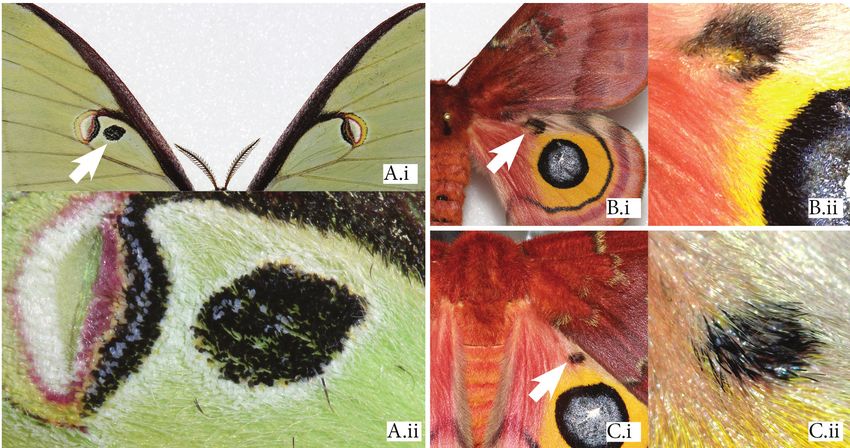
SOURAKOV & SHIRAI: Effects of heparin on wing pattern                              TROP. LEPID. RES., 30(1): 4-19, 2020                5

to be thoroughly tested by the comparison of the developmental      2010; Beldade & Peralta, 2017). In this family, eyespots are
mechanisms behind the formation of such elements between            concentrically organized and are hence known as “bulls-eye”
these groups (de Beer, 1971; Wagner, 1989; Abouheif, 1997;          eyespots. Eyespots are also found in some Saturniidae, but
Weiss & Fullerton, 2000; Young & Wagner, 2011). Attempting          instead of being at the wing margins, they are positioned in
to answer this question would not only increase the phylogenetic    the middle of the wing with the M2-M3 cross-vein in the center.
breadth of comparative insect development studies but would         In Automeris io (Fabricius, 1775) (Fig. 1), this eyespot center
also expand the research tools optimized in butterflies to          is covered with brightly colored, white/UV-reflecting scales,
Lepidoptera clades with important (e.g., behavioral) differences    which are surrounded by concentric circles of iridescent black-
that also have an order of magnitude more species, represented      blue scales mixed with white scales similar to those in the
by over 100 families.                                               center (here referred to as the gray spot), and a black disc. In
     One of the cases supporting possible homology between          other saturniids, the center may be surrounded by a clear (i.e.
butterfly and moth developmental mechanisms is the expression       scale-less) elongated window bordered by yellow scales, and
pattern for two butterfly eyespot genes, Distal-less (Dll) and      the iridescent gray part shifted basally, as in Polyphemus Moth
Engrailed (En), that has been found in the saturniid moths          Antheraea polyphemus. The concentric organization can also
Antheraea polyphemus (Cramer, 1776) and Saturnia pavonia            be greatly reduced, as in the forewing of Luna Moth, Actias
(Linnaeus, 1758) (Monteiro et al., 2006). Nymphalid eyespots        luna (Linnaeus, 1758), where colorful elements surrounding
are serially repeated pattern elements that resemble vertebrate     the cross-vein and the adjacent clear windows are minimal.
eyes, found at the distal region of the wing and hence named        A few saturniids have both medial eyespots and additional
“border ocelli” in the Nymphalid Groundplan. In Nymphalidae,        nymphalid-like distal ones, as, for example, on the forewings of
such as Bicyclus anynana (Butler, 1879) and Junonia coenia          Cecropia Moth, Hyalophora cecropia (Linnaeus, 1758). While
Hübner, 1822, they are the pattern elements for which we            the phenotypic similarity between eyespots of saturniids and
have a lot of knowledge about comparative wing pattern              nymphalids can be striking, convergent evolution is so common
development (e.g., Beldade & Brakefield, 2002; Nijhout,             in insects that one cannot assume a homologous developmental

        Figure 1. A dorsal hindwing eyespot of Automeris io: A. intact hindwing, B. cleared with bleach to show the underlying
        venation; C. close-up of the same eyespot center under (i) white light and (ii) UV light.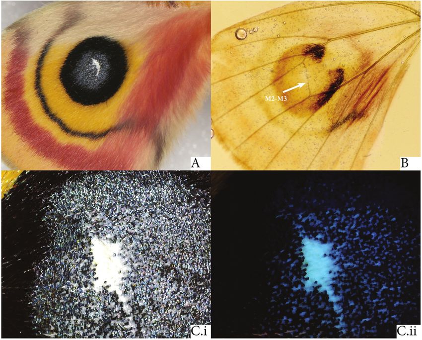
6          TROP. LEPID. RES., 30(1): 4-19, 2020                                       SOURAKOV & SHIRAI: Effects of heparin on wing pattern

    process. Even comparing gene expression in butterfly wings                  axis of the future discal-cell eyespot” (i.e., the eyespot center)
    during the development of a particular pattern element might                of A. polyphemus. Thus, there may be common developmental
    not satisfactorily answer the question of homology, since                   mechanisms underlying eyespot formation despite the different
    homologous eyespots of nymphalids may exhibit dynamic                       locations of medial eyespots in moths and distal eyespots in
    combinations of gene expression during their ca. 90-million                 nymphalids. Additionally, the likelihood of some developmental
    long evolutionary history (Shirai et al., 2012; Oliver et al.,              homology in wing pattern development is supported by the
    2012; see also references above on homology).                               finding that heparin injections can affect wing pattern formation
         Nevertheless, comparative expression patterns have shown               in butterflies and moths when they are performed at the stage
    that Nymphalidae and Saturniidae share at least two genes                   when the previously mentioned genes have been found to be
    associated with eyespot determination (Monteiro et al., 2006).              expressed, the prepupal stage (Sourakov, 2017, 2018b).
    After 2-3 days from the moment the caterpillars began to spin                    Heparin is a highly sulfated form of heparan sulfate
    their cocoon, in the prepupal stage, Monteiro et al. (2006)                 glycosaminoglycans (HS-GAGs), known to interact with
    found Dll and En to be expressed in the center of the eyespot               signaling pathways (Wnt, Hedgehog, Decapentaplegic,
    of S. pavonia, and En in the “line marking the elongated central            Transformation Growth Factor β) from a number of experiments

                                    Dorsal surface      Ventral surface                                 Dorsal surface       Ventral surface
                                 Wild type   Heparin   Wild type   Heparin                           Wild type   Heparin   Wild type   Heparin

                                                                             Glyphodes sibillalis
         Asterocampa clyton
                                                                              3-5 hAP, 10 µg
          5-10 hAP, 30 µg
                                                                              Sourakov 2018b
          Sourakov 2018b ¹
                                                                                                                                Crambidae

         Euphydryas chalcedona*                                              Estigmene acrea*
         10-16 hAP, 20 µg                                                     4-13 hBP, 0.62 mg
         Martin & Reed 2014                                                   Sourakov 2018b

         Junonia coenia*
                                                                              Hypercompe scribonia
          5 hAP, 15 µg
          Serfas & Carroll 2005 2                                             12 hBP, 1.4 mg
                                                                              Sourakov 2018b                                    Erebidae
         Vanessa cardui*
         10-16 hAP, 20 µg
         Martin & Reed 2014                                                   Automeris io
                                                                              10 hAP, 1.5 mg
                                                                              Present study 4

         Limenitis arthemis*
                                                                             Antheraea polyphemus
         8-16 hAP, 20 µg
         Gallant et al. 2014 3                                                8 hAP, 1 mg
                                                                              Present study

         Agraulis vanillae*
         10-16 hAP, 20 µg                                                                                                       Saturniidae
         Martin & Reed 2014 2

                                                                               Heraclides cresphontes
         Heliconius cydno*                                                     2-9 hAP, 0.37 mg
          12-16 hAP, 30 µg                                                     Sourakov 2018b
          Martin et al. 2012

         Heliconius erato*
          12-16 hAP, 30 µg
          Martin et al. 2012                                                   Pterourus troilus
                                                                               5 hAP, 60 µg
         Heliconius sara*
                                                                               Sourakov 2018b
         12-16 hAP, 20 µg
         Martin et al. 2012

                                                        Nymphalidae                                                             Papilionidae
    Figure 2. Schematic summary of all Lepidopteran species injected with heparin (nymphalids at the left and other families at the right), compared
    to the wild-type phenotype (* when it corresponds to an experimental control with such phenotype), for dorsal and ventral wing surfaces, as
    available. The strongest published effect is represented for each species, with corresponding dosage and time of injection (in hours before or after
    pupation, respectively, hBP and hAP), and the reference we used for the schematic color pattern changes, without implying changes in wing size
    or shape. For additional references: ¹ Sourakov, 2018a, ² Sourakov, 2018b, ³ Imhoff, 2016, ⁴ Sourakov, 2017.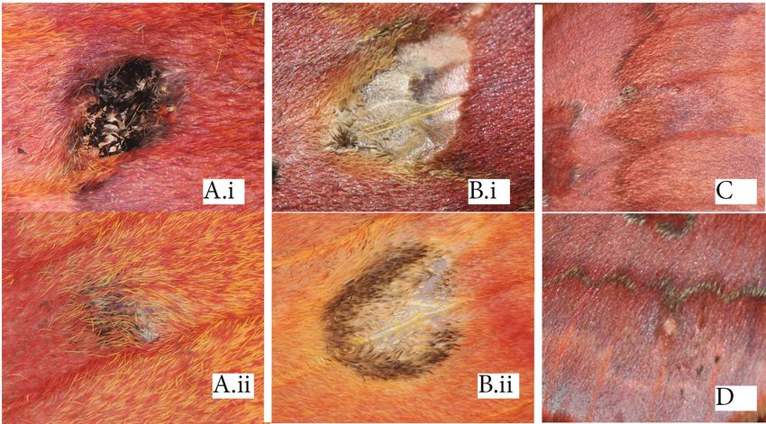
SOURAKOV & SHIRAI: Effects of heparin on wing pattern                                                 TROP. LEPID. RES., 30(1): 4-19, 2020                       7

with mammals (e.g., Bradley & Brown, 1990) and insects, such                      collectively called the “organizer” (the future eyespot center,
as flies (e.g., Reichsman et al., 1996; Baeg et al., 2001; Perrimon               or focus), presumably produce signaling molecules that diffuse
& Häcker, 2004; Selleck, 2000; Nybakken & Perrimon, 2002;                         to and react with surrounding cells, determining their cell
Princivalle & de Agostini, 2002). Developmental disruption                        fate, or color. This method has been pioneered using Common
through pharmacological experiments was pioneered in                              Buckeye (Nijhout, 1980), and has since been applied to other
butterflies with the Common Buckeye, J. coenia; eyespots were                     nymphalid models. However, until the present study, it was
among the target pattern elements (Serfas & Carroll, 2005).                       never attempted with moths, perhaps due to the difficulty in
Such experiments were shown to change Lepidoptera wing                            rearing relevant moth species (e.g., with eyespots) at the scale
color patterns, which include eyespots, as well as leading to                     required by developmental studies.
a complete overwriting of all wing patterns or an expansion
of some of the pattern elements at the expense of others,
depending on the stage of development, the species, and the                                             MATERIAL AND METHODS
dosage (Fig. 2).
      While heparin may be disrupting the action of members                            In the present study, we aimed to investigate the possible
of any of the major signaling pathways, most of which were                        homologous development of Lepidoptera eyespots by using
shown to be expressed during butterfly eyespot development                        pharmacological as well as surgical manipulations in saturniid
(latest review in Beldade & Peralta, 2017, see also Özsu &                        moths. Here, we build on previous research on heparin injections
Monteiro, 2017), the best-studied candidate pathway is Wnt,                       in Automeris io (Sourakov, 2017) by greatly increasing the
specifically secreted ligands Wnt 1 (or wingless, wg) and WntA,                   number of specimens, varying the heparin dosage injected,
which plays a large and diverse role in nymphalid wing color                      and also injecting a single dosage at different developmental
pattern determination (Mazo-Vargas et al., 2017). While other                     stages (time before and after pupation). We also conducted
species, including moths, have been experimented upon with                        heparin injections on Antheraea polyphemus, representing a
heparin and transformed phenotypes have been achieved (e.g.,                      different evolutionary lineage: A. io is placed in Hemileucinae,
Sourakov, 2017, 2018a), it is not yet clear if heparin affects                    while A. polyphemus is in Saturniinae (Regier et al., 2008). We
a color, a pattern element, a symmetry system, or patterns                        discuss the evidence for broad Lepidoptera eyespot homology
associated with a gene or a pathway.                                              based on a review of publications on heparin injections in
      In addition to the pharmacological disruption of                            the order (Fig. 2). Finally, we attempted a limited number of
development, it is also possible to transplant the presumable                     surgical manipulations by ablating eyespot organizers as well
eyespot organizer during the time when signaling is occurring                     as transplanting potential organizers among conspecific pupae
to the same or a conspecific animal but in a different wing                       in A. io and Actias luna. Multiple experiments were conducted
region. The organizer hypothesis proposes that during the                         in the present study, some conclusive, some preliminary. They
development of eyespots, a concentration-dependent, signal-                       are summarized in Table 1 and detailed below.
response mechanism determines different colors by the distance
from the source (Nijhout, 1990; Monteiro et al., 2001; Otaki,                     Heparin injections experiments (H1-H3)
2011). This hypothesis has been validated by two types of                             We obtained eggs of A. io and A. polyphemus in 2017-2018
surgical manipulations: (a) the transplantation of competent                      by keeping female moths caught in Gainesville, Florida (c.
cells which induced ectopic eyespots and (b) cauteries of                         29°38’ N, 82°22’ W) in paper bags/cages. We reared larvae on
these cells which reduced or ablated the eyespot (Nijhout,                        Celtis laevigata Willdenow (Cannabaceae) and Quercus nigra
1980; French & Brakefield, 1995). These competent cells,                          L. (Fagaceae), respectively, at indoor temperatures (around

    Table 1. Experiments performed in the present study. Unmanipulated control individuals are always siblings to the experimental
    (“study”) individuals.
   Experiment Goal                         Manipulation                         Species          # of study    Control 1               Control 2
                                                                                                 individuals
       H1      timing and effect of        heparin injection of larva, prepupa   A. io                  88     >100 unmanipulated      8 phosphate buffer
               heparin injections          and pupa                                                                                    injection
       H2      effect of heparin - pilot   heparin injection of prepupa and pupa A. polyphemus         4       1 unmanipulated
       H3      effect of heparin           heparin injection of prepupae and    A. polyphemus         34       16 unmanipulated        3 water injection
                                           pupa
        T1     necessity of eyespot        cut M2-M3 cross-vein of pupa         A. io                  5       unmanipulated wing of
               organizer                                                                                       each study specimen
        T2     necessity of eyespot        cut M2-M3 cross-vein of pupa         A. luna                2       unmanipulated wing of
               organizer                                                                                       each study specimen
        T3     sufficiency of eyespot      transplant M2-M3 cross-vein tissue   A. io                 18       unmanipulated wing of   3 surgical cuts without
               organizer                   from donor to host pupa                                             each study specimen     any tissue transplanted
        T4     sufficiency of eyespot      transplant M2-M3 cross-vein tissue   A. luna                1       unmanipulated wing of
               organizer                   from donor to host pupa                                             each study specimen
        T5     wound-induced response      needle injury to FW of pupa          A. io                  4       unmanipulated wing of
                                                                                                               each study specimen
        T6     wound-induced response needle injury to HW of pupa               A. io                  1       unmanipulated wing of
                                                                                                               each study specimen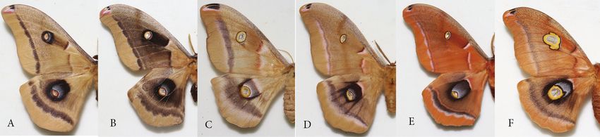
8         TROP. LEPID. RES., 30(1): 4-19, 2020                                 SOURAKOV & SHIRAI: Effects of heparin on wing pattern

                                       A                               B                          C                               D

                                                 E                                        F                                       G

                   Control FW E.i                Heparin FW      G.i             Control HW E.ii              Heparin HW        G.ii

            Figure 3. The gradient of effects of heparin injections on dorsal surfaces of Automeris io (A-D) and Antheraea polyphemus
            (E-G). A. normal pattern (unmanipulated wild-type); B. 5 ul of 30% (ca. 2 mg*) heparin as prepupa within 1 dBP; C. 10 ul
            of 30% (ca. 4 mg*) heparin as prepupa within 1 dBP; D. 4 ul of 27% (1.5mg) heparin as pupa at 10 hAP. E. normal pattern
            (unmanipulated wild-type); F) 5 ul of 16% (ca. 1mg) heparin as prepupa 2 hBP; G. 5 ul of 16% (ca. 1mg) heparin as pupa
            8 hAP. E.i, Gi, close-up of FW eyespot, E.ii, Gii, close-up of HW eyespot. (*some heparin may have been expelled due to
            bleeding). Additional experimental specimens are shown in Figs. S2, S3 of Supplementary Material.

    26⁰C, 65% humidity). Total developmental time during the               Supplementary Material, Table S1. We determined the injection
    larval stage is ca. 2-3 months and 1.5 months, respectively. The       time in relation to pupation by time-lapse photography using a
    prepupal stages last 4-7 days, and adults emerge from pupae            Dinolite camera (AnMo Electronics Co., Taiwan) connected to
    within a month, unless they go into diapause, in which case            a computer, set to take photos every 30 minutes. Sometimes,
    they may take 8-12 month to emerge. Below, we use ‘FW’ for             especially in A. io, the color of pupae was used to determine
    forewing, ‘HW’ for hindwing, ‘D’ for the dorsal wing surface,          the time since pupation using a previously created photographic
    and ‘V’ for the ventral. We refer to times of manipulations in         calibration (see, Supplementary Material in Sourakov, 2017).
    days (d) or hours (h) before pupation (BP) or after pupation           This method can be applied with relative precision up to 5-6
    (AP), using the notation dBP, dAP, hBP, hAP; specific times            hAP. We cut open the A. polyphemus cocoons 3 dBP with the
    are detailed in the Results and Discussion sections and in             top half removed (Fig. S1 of Supplementary Material) and lined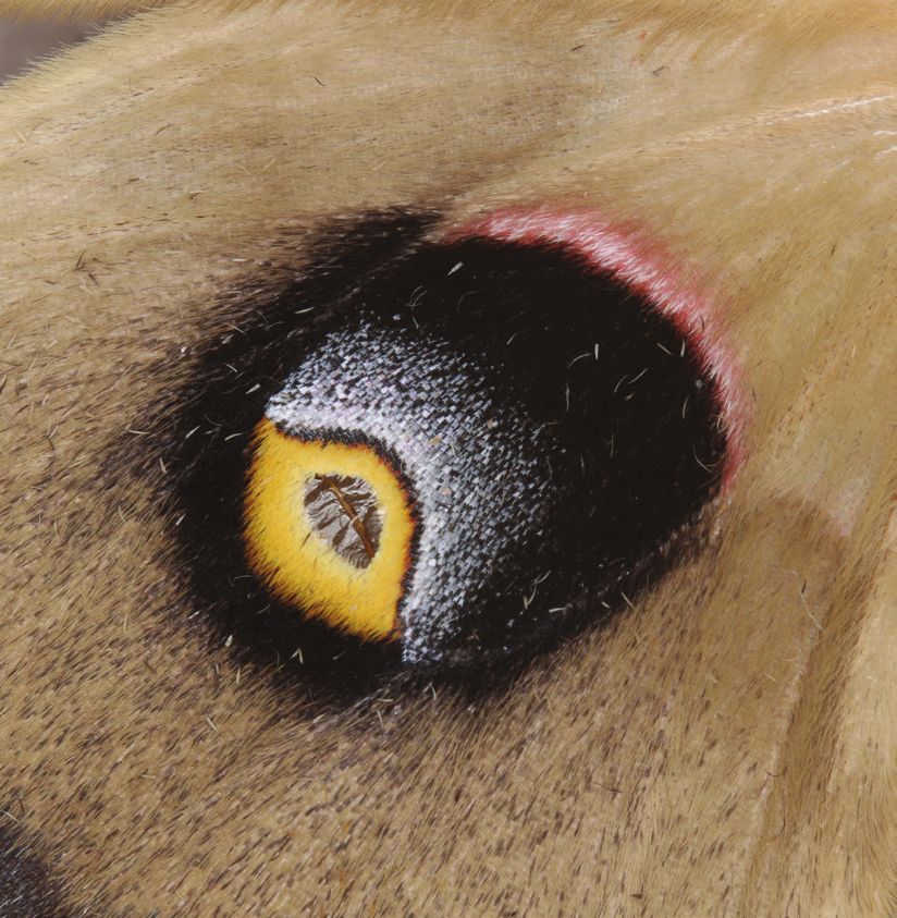
SOURAKOV & SHIRAI: Effects of heparin on wing pattern                                      TROP. LEPID. RES., 30(1): 4-19, 2020                  9

     1. Pupae in modeling clay: A is the host and B the donor.              injected solution was delivered within the wing compartment
                                                                            (Fig. S1B of Supplementary Material). This was easier to do
                                                                            with A. polyphemus, whose pupal wings bulge slightly, than
                                                                            with A. io, whose pupal wings are flush with the surface of the
                                                                            rest of the pupa. In a few cases (indicated in the figure legend
                                                                            of Fig. S3 of Supplementary Material), the injection was made
                                                                            deep through the abdomen. Deep injections can cause rapid
     2. Prepare the host by making a fine incision in the FW through the    melanization and death of the prepupae probably due to a
     cuticle, at the distal half of the wing (we attempted anterior and     punctured gut, so prepupae were injected in the mid-section
     posterior regions). This is the procedure of control individuals.
                                                                            laterally, under the epidermis, avoiding penetrating the gut (Fig.
                                                                            S1A of Supplementary Material).
                                                                                 A. io experiments (H1) were conducted in 2017-2018 and
                                                                            88 individuals from 5 broods were injected with heparin. Three
                                                                            transformed individuals were previously reported in Sourakov
                                                                            (2017), including injections at the prepupal stage within 1 dBP,
                                                                            and they showed an effect similar to that achieved by injecting
                                                                            early pupae at 5 hAP. To determine at which developmental
                                                                            stage heparin begins to affect wing pattern, we injected A.
     3. Cut the cuticle around the entire FW of the donor with fine         io starting at the late larval stage at 11 and 8 dBP, when the
     scissors. Pull out the cuticle with the attached FW, as if lifting a   caterpillar was still feeding. We also injected prepupae after
     car’s trunk, and keep it open. Cut the tissue around the HW M2-        cocoon spinning at 8, 7.5, 7, 5.5, 5, 4, 3, and 2.6 dBP, and pupae
     M3 cross-vein under the stereoscope. Place it in a sterile spatula
     to be transplanted.                                                    until 24 hAP under the same dosage (4 ul of 27%, or 1.5 mg).
                                                                            In order to further investigate the effect of heparin, we added
                                                                            observations on 24 more heparin-injected A. io (with different
                                                                            dosages) that successfully emerged (e.g., Fig. 3 A-D and Fig.
                                                                            S3 of Supplementary Material). Over 100 unmanipulated A.
                                                                            io individuals from the same broods served as unmanipulated
                                                                            ‘controls.’As additional controls, in November 2018, 7 prepupae
                                                                            (1-3 dBP) and 1 pupa (10-15 hAP) of A. io were injected with
                                                                            phosphate buffer and compared to 10 unmanipulated siblings
      4. Gently place the donor HW cross-vein inside the host               (Fig. S4 of Supplementary Material).
      FW incision under the steroscope. Hemolymph is important                   For A. polyphemus (H2, H3), we conducted a pilot study
      to keep the incision moist, but avoid “bleeding.”
                                                                            (H2) in October-November 2017 involving five siblings, four
                                                                            of which were injected with different volumes of 20% heparin
                                                                            (ca. 5 to 20 ul; see figure captions for dosage) and one which
                                                                            was left unmanipulated – four out of five individuals emerged.
                                                                            After observing remarkable wing pattern transformations
                                                                            in the pilot study (Fig. S2 of Supplementary Material), we
                                                                            conducted the second trial (H3) in June-July 2018, injecting 5
                                                                            ul of solution at different concentrations. Injections were made
                                                                            into 34 individuals with 16 left as unmanipulated ‘controls.’ In
                                                                            A. polyphemus, we made prepupal injections (starting from 44
Figure 4. Protocol highlighting the differences in the surgical             hBP) and pupal injections (until 13.5 hAP). To help confirm
procedures performed in saturniids, when compared to standard               that the change in pattern was caused by heparin and not by the
techniques in butterflies (c.f. Brakefield et al., 2009).                   solvent (water) and/or mechanical injury with the needle, in July
                                                                            2019 we injected 5 ul of distilled water into three A. polyphemus
                                                                            pupae (5, 12, and 14 hAP), leaving one unmanipulated (Fig.
up the prepupae in a tray to check their pupation time.                     S5 of Supplementary Material) – three out of four individuals
     Heparin sodium salt (porcine, MP Biomedicals, Inc.) was                emerged. For the second trial with A. polyphemus (H3), 15
diluted in distilled water at different concentrations varying from         unmanipulated and 31 injected pupae or prepupae successfully
3% to 43%. The corresponding amounts of heparin in a certain                emerged: one unmanipulated and three injected individuals
volume of solution delivered by injections are detailed in figure           died as pupae (6% mortality). Heparin concentrations ranged
legends (Fig. 3 and Figs. S2, S3 and Table S1 of Supplementary              from 4% to 16%, and the times of injections in successfully
Material). We injected A. io and A. polyphemus immatures using              emerged individuals varied from 44 hBP until 13.5 hAP (see
either a sterile 0.3 ml hypodermic syringe (volume measured                 Supplementary Materials). Among the three dead experimental
with a micropipette) or with a 10 ul syringe. We injected A.                individuals, one fell outside this range, injected at 21 hAP, and
polyphemus pupae through the cuticle, always attempting to                  the other two were injected shortly after pupation, at 1.5 and 4
keep the needle parallel to the surface of the pupa, so that the            hAP.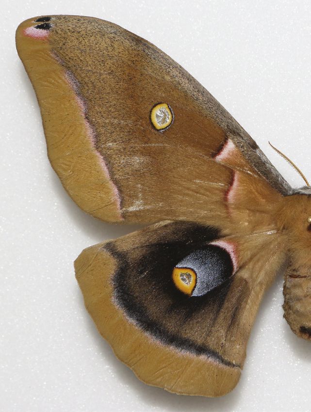
10         TROP. LEPID. RES., 30(1): 4-19, 2020                               SOURAKOV & SHIRAI: Effects of heparin on wing pattern

     Organizer tissue transplanting and disruption experiments            French, 1995). A single transplant in A. luna (T4) was possible
     (T1-T6)                                                              for lack of individuals (we had no control in this species), using
           We conducted surgical manipulations involving between-         a 5 hAP donor to a 10 hAP host, FW to FW transplant.
     pupal transplants of potential eyespot organizers to test for              Additionally (T5, T6), 4 A. io pupae (T5, 2, 4, 4, and 17
     their sufficiency to form eyespots, as well as disrupting such       hAP) received a FW injury with a sterile needle inside the discal
     organizers to test for their necessity in eyespot development.       cell to test if this would lead to a wound-induced response (op.
     We used only the pupal stage since prepupal wings are too            cit.) inside the FW discal cell. An additional individual (T6, 4
     difficult to handle. We did all manipulations with pupae held        hAP) was punctured inside the discal cell, aiming to reach the
     still in modelling clay, following standard techniques (reviewed     HW, through the FW.
     in Brakefield et al., 2009), unless otherwise stated (Fig. 4).             We had at hand a limited sample and we did not have
           Firstly (T1, T2), we disrupted the M2-M3 cross-vein            enough comparable results to statistically treat the data (that
     to determine whether the cross-vein is involved with the             is, enough experimental versus control specimens, different
     formation of the eyespot around it. If the disruption leads to       types of controls, also controlling for donor and host hAP, wing
     loss or reduction of the adult eyespot, this simple surgical         region, wound-induced response, gender, etc.).
     manipulation demonstrates the necessity of the intact cross-vein           We deposited spread experimental specimens in the
     in organizing the future eyespot. We did cross-vein disruptions      collection of the McGuire Center for Lepidoptera and
     in pupae of 5 A. io (T1) (1 to 4 hAP, HW cross-vein) and of          Biodiversity, Florida Museum of Natural History, University
     2 A. luna (T2) (4 hAP, FW cross-vein). Using a sharp sterile         of Florida under MGCL Accession Numbers for the following
     needle, the FW cross-vein was cut through the pupal case, since      voucher specimens: A. io: 286779, 289217-20, 291955-56,
     it was easily visible under the stereoscope. For the HW cross-       291645-6, 292276, 292055-6, 292280, 291957, 292284,
     vein, we cut through the cuticle, the FW, and the peripodial         286790, 291637, 291644, 286791, 292279, 292281, 291648,
     membrane to assess the HW and, under the stereoscope, we             292282-3, 291690, 291953, 291692, 291682-3; A. polyphemus:
     located and cut the HW cross-vein. While we did not have             291924-27, 292961-62, 292083, 292104-111, 292236-254,
     additional individuals of either species to perform control vein     292260-274 (Table S1 of Supplementary Material); and A. luna
     disruptions, the contralateral wing that was left intact served      291928-30. We also used the MGCL collection to assess the
     adequately for comparison.                                           presence of aberrations in wild populations of A. polyphemus,
           Secondly (T3), we transplanted the supposed organizer          for which 797 specimens were examined (see Fig. 5 and Fig. S6
     tissue (1-2 mm2 that included the M2-M3 HW cross-vein) aiming        of Supplementary Material for examples).
     to induce the formation of an ectopic eyespot in the host, which
     would demonstrate that the cross-vein is sufficient to determine                                RESULTS
     the cell fate of host cells (Nijhout, 1980; French & Brakefield,
     1995). Automeris io has a very prominent concentrically              Heparin injection experiments (Table 1, H1-H3)
     organized HW eyespot, so we cut the cuticle around the entire             Heparin injections caused noticeable transformations of the
     FW of donor pupa (4 to 12 hAP) with fine scissors and held           wing patterns in both A. io and A. polyphemus (Fig. 3 and Figs.
     it opened (Fig. 4). We then cut the tissue around the HW M2-         S2 and S3 of Supplementary Material). To illustrate different
     M3 cross-vein (which removes the tissue of both wing surfaces,       degrees of the response to heparin in A. io, the 3 males (one
     dorsal and ventral) and placed it on a sterile spatula to be         unmanipulated control and two experimental prepupae injected
     transplanted. Because of this manipulation, chances of donor         with 5 ul and 10 ul of 30% heparin solution, respectively) from
     survival were low, so we transplanted its tissue to another          Sourakov (2017) are shown alongside a male pupa which was
     (host) individual (as opposed to a transplant within the donor).     injected with 4 ul of 27% solution at 10 hAP (Fig. 3A-D).
     Whenever possible, we used the donor’s right and left HW M2-         The effect of heparin in these three individuals range from a
     M3 cross-veins for two different transplants.                        slight proximal expansion of black scales around the eyespot
           We previously prepared the host pupa (18 individuals of 5 to   to an extreme expansion of black beyond the borders of several
     14 hAP) by making a fine incision in the FW through the cuticle      pattern elements in the HWd. The injection did not seem to
     (1-2 mm), at the distal half of the wing (the responsive region      affect the central white dash nor the gray spot at the center of
     in butterflies, c.f. French and Brakefield, 1995), and we gently     the eyespot and made no effect on the corresponding ventral
     placed the transplanted tissue inside the wing incision. Some        pattern of the HW. However, whenever the FWd responded
     hemolymph was important to keep the cuticle at the incision          to heparin at the discal cell (at the “DII” pattern element) as
     site moist, but we avoided too much hemolymph (“bleeding”)           black finger-like streaks above the venation (Fig. 3C,D), it was
     by removing the excess with paper towel, especially at the thin      always accompanied by a substantial expansion of the black
     cuticular junctions at which the pupal case opens at eclosion. If    disc surrounding the corresponding ventral pattern - the FWv
     this is not taken into consideration, the local melanization due     eyespot (Fig. S3A,B) of Supplementary Material). If such
     to “bleeding” can make the pupal case harder or impossible to        disruption of the FWd did not occur, the FWv eyespot was not
     break out of by the emerging moth. We made control incisions         affected.
     in 3 individuals (5, 10 and 10 hAP) by repeating the procedure            The lowest amount of injected heparin which led to wing
     above without inserting any donor tissue, to check whether there     pattern change occurred when 1 ul of 3% (0.03 mg) was injected
     is wound-induced response due only to damage of the host FW,         at approximately 8 hAP deep into the pupal abdomen (Fig.
     known to occur in butterflies (e.g., Nijhout, 1985; Brakefield &     S3I, J of Supplementary Material). The manner in which the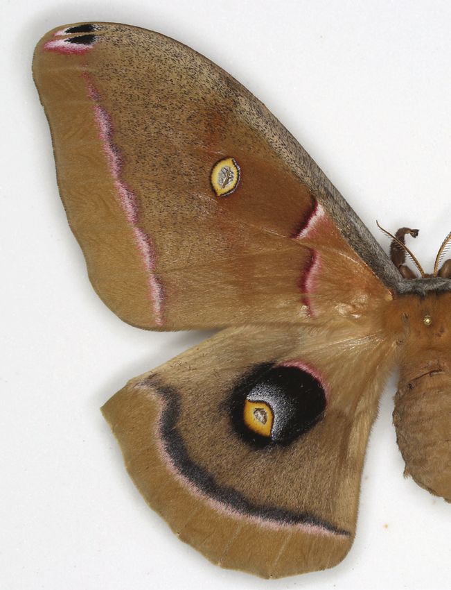
SOURAKOV & SHIRAI: Effects of heparin on wing pattern                              TROP. LEPID. RES., 30(1): 4-19, 2020                  11

injections were made in these two specimens ensured that no          resulted in transformations similar to those caused by heparin
heparin was lost due to bleeding. In the present study, injections   (Figs. S4 and S5 of Supplementary Material). Photographs of
for the timing experiment (Table 1, H1) resulted in specimens        all individuals from A. polyphemus study and the corresponding
with wild-type patterns, with the exception of 1) the individual     injection data (Table S1) can be found in the Supplementary
injected at 3 dBP (specifically, 75 hBP), which showed the           Materials.
expected expansion of the black disc around the HW eyespot,
despite the wings not having fully expanded on one side (Fig.        Organizer tissue transplanting and disruption experiments
S3G of Supplementary Material); and 2) the one injected at 2.6       (Table 1, T1-T6)
dBP (specifically, 63 hBP; Fig. S3K of Supplementary Material),            To explore whether the M2-M3 cross-vein acts as an
where the black disc also expanded, but the formation of the         organizer in A. io, we disrupted this cross-vein in 5 A. io
M2-M3 cross-vein was seemingly aborted, and the center of the        pupae from 1 to 4 hAP (T1) but without any result. While we
eyespot did not form. After pupation, HWd eyespot expansion          clearly observed disrupted, disconnected cross-veins under a
was achieved in specimens injected from 5 to 10 hAP (Fig. 3D,        microscope following the procedure, all resulting adult moths
Fig. S3 of Supplementary Material).                                  were normal and showed an intact cross-vein.
      In 21 of the heparin-injected A. polyphemus (Table 1,                In an attempt to disrupt the M2-M3 cross-vein of a Luna
H2-H3), the black region inside the HWd eyespot changed,             Moth pupa at 4 hAP (T2), the resulting adult had an eyespot
sometimes significantly, resembling responses found in A. io         with normal phenotype, but a wound-induced eyespot-like color
(Fig. 3E-G and Fig. S2 of Supplementary Material). However,          pattern appeared inside the FW discal cell (Fig. 6A), which was
while in A. io the black disc expanded proximally and, with          not observed in the contralateral wing. This novel element,
even further increase in heparin’s effect also expanding distally,   arranged in a circular pattern, is composed of a mixture of black
in A. polyphemus the black disc did not expand as much               and blue scales surrounded by a white border reminiscent of
basally, and certainly not inside the discal cell, as if prevented   the scales surrounding the eyespot basally. The second Luna
from doing so by an invisible border. While in the parental and      Moth in which we disrupted the cross-vein (T2) showed no
unmanipulated sibling groups of A. polyphemus, the proximal          abnormalities, which may be due, as in A. io, to regeneration.
gray region of the eyespot was confined to a narrow semi-                  The eyespot-like response in the A. luna individual (T2,
circle proximal to the yellow eyespot ring (e.g., Fig. 3E and        Fig. 6A), compared with the single Luna Moth transplant where
Fig. S2A of Supplementary Material), in 11 experimental              only white scales appeared at the transplanted site located
specimens, it expanded basally (e.g., Fig. 3F,G and Fig. S2B,C       distally (T4, Fig. 7C), suggests that the proximal (vs. distal)
of Supplementary Material), sometimes touching the proximal          part of the wing may be more responsive to wound-induced
pink contour, widening it into a lighter pink. In the most           color changes. Two additional A. io experimental individuals
extreme case of heparin-induced transformation, in which the         may support this hypothesis, in which black spots appeared
HWd background color became mostly black (e.g., Fig. 3G),            proximally, as a response to injury in the HWd. One of them
the area around the normally-simple FWd eyespot (Fig. 3E.i)          was meant to be a HW vein disruption (at 4 hAP, T1), which
acquired the gray spot pattern and resembled HWd eyespots            was made through the FW; the M2-M3 cross-veins of both wings
(Fig. 3G.i). As in A. io, the central part of the eyespot, here      overlap inside a pupa so veins could be cut on both wings. Only
composed of the scale-less center and yellow ring, does not          a slight scar appeared with little or no scale color changes on
seem to have changed with heparin, even when the strongest           the dorsal and ventral FW, consistent with other control cuts,
effects were observed.                                               but on the HW, while the cross-vein was intact, the black spot
      To understand at what point during development the wing        appeared (Fig. 6B). Following this result, we later tried to
pattern is subjected to change in A. polyphemus, we injected         replicate it by cutting another female pupa roughly at the same
6 pupae before 5 hAP, none of which showed any difference            spot at 4 hAP intentionally reaching the HW (T6, cross-vein
to unmanipulated control siblings, independent of the dosage         was not targeted this time). The result was similar – a local
(2.5, 3.25 and 4 hAP injected 3.7% heparin; 2 individuals of 4       change of scale color to black (Fig. 6C).
hAP injected 5% heparin; and 2 hAP injected 15.7% heparin).                Sixteen of the 18 host individuals of A. io receiving
Individuals injected at 5 hAP varied in their responses to heparin   transplants of HW M2-M3 cross-vein tissue (T3) survived, of
injection, seemingly correlated with the concentration (3.7%         which 6 had holes in their wings and the remaining 10 had
and 5% no effect, and 13% and 25.7% some effect). However,           intact wings. Among the 10 specimens with intact wings, 8
increase in volume at a high concentration (10 ul at 21%) can        demonstrated localized changes in scale color at the transplant
still produce the maximum effect, as shown by an individual at       site: from normally mahogany-brown to black in 5 females (Fig.
5 hAP (Fig. S2C). From 4 to 36 hBP and 9 to 13.5 hAP there           7A), and from normally amber color to brown in 3 males (Fig.
were noticeable changes, but not in a consistent fashion. We         7B). Despite clearly not looking like an eyespot, these changes
observed the strongest effects from injections into prepupae at      do not appear to be due to injury, as the experimental controls
1 to 2 hBP, and 8 to 9 hAP, even at smaller doses of heparin. In     showed no such color change: 1) the control cuts (n = 3, at 5, 5,
five specimens, the formation of the adult HW was completely         and 10-12 hAP) in the FW resulted in visible wounds but never
aborted (this happened when injections were made at 2.5, 4, 5,       showed any color change around the wound (Fig. 8C,D); and
9.5 and 10.5 hAP). None of these specimens showed any signs          2) individuals in which we made wounds inside the FW discal
of transformation in the healthy wings. None of our control          cell (T5) produced no visible effect on the resulting adult moths
water/buffer injections, whether into A. io or A. polyphemus,        (Fig. S7 of Supplementary Material).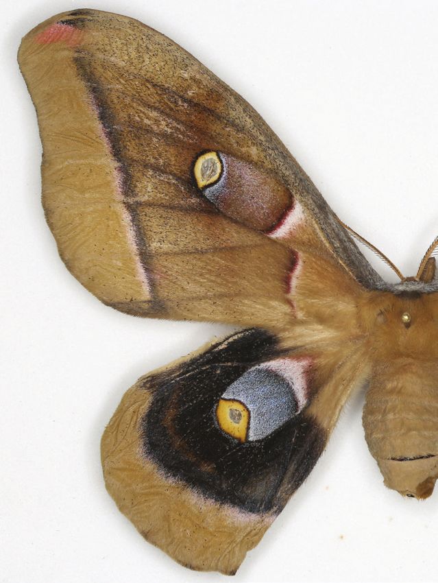
12 TROP. LEPID. RES., 30(1): 4-19, 2020 SOURAKOV & SHIRAI: Effects of heparin on wing pattern
Figure 5. Several unusual Antheraea phenotypes from the MGCL collection including sister species and rare/unique aberrations: A-B. A. oculea:
A. Arizona, Aug 1999 (ex ova); B. Pima Co, Arizona, 7 Aug, 2005, demonstrating melanism around not only HWd but also FWd eyespot (A)
and throughout the wing (B); C-F. A. polyphemus: C. Chicago, Illinois, 3 June 1921, demonstrating an eyespot that lacks distal (within discal
cell) portion of the black disc; D. Salt Lake Co., Utah, 5 Sept 1987 (ex ova) demonstrating HWd black disc expansion along veins; E. Florida,
demonstrating a combination of slight black disc diffusion combined with an enlarged gray spot; F. Quebec, June 2005 (ex ova) demonstrating
a unique aberration in which the clear window and the yellow ring forming the center of the eyespot, but not the black disc, are expanded,
suggesting that the central elements of the eyespot are under different controls from the black disc.
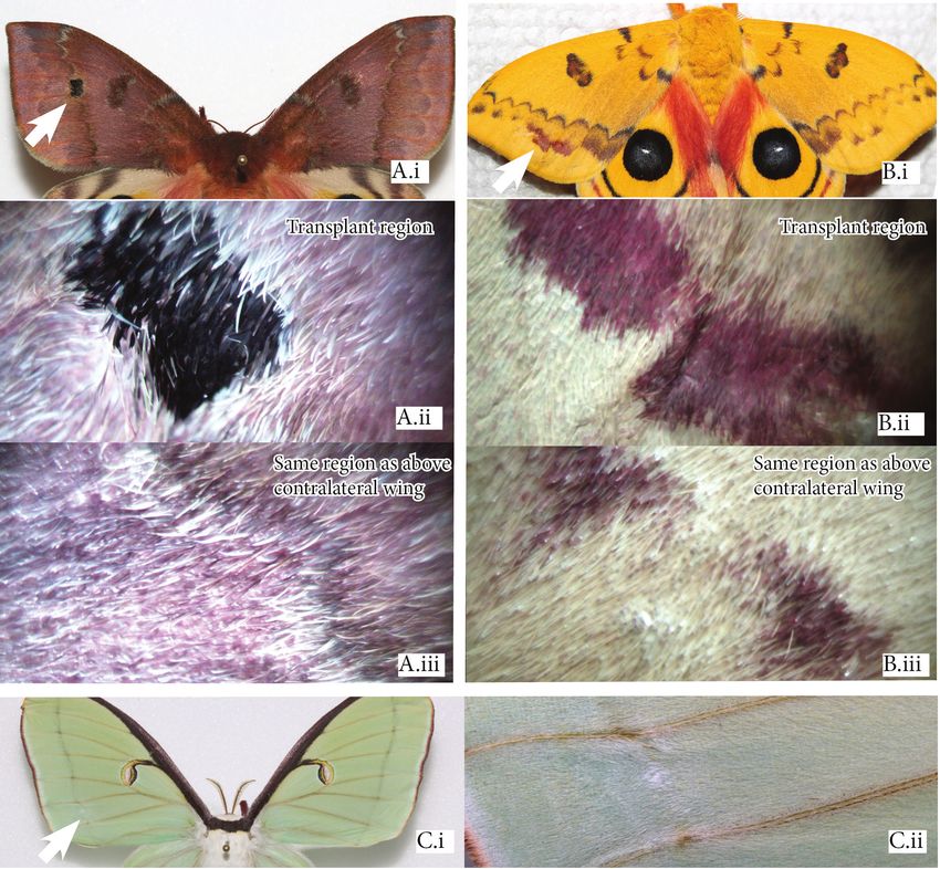
Figure 6. Wound-induced responses in: A. Actias luna: Wound-induced response to a cut inside the FWd discal cell of a pupa
at 4 hAP; and, B,C. Automeris io: Wound-induced response to a cut to the proximal part of HWv of pupae at 4 hAP.
The change in scale color due to transplant sometimes does not alter the production or degradation of signaling
occurred not only on the FWd, but also on the corresponding molecules. Instead, heparin and other HS-GAGs act on the
surface of FWv (Fig. 8A). In one of the transplants (from a extracellular space, binding to signaling molecules and aiding
4 hAP donor into a 9 hAP host) the individual lost the scales the assembly with their receptors (reviewed in Selleck, 2000;
dorsally, but formed a ring of black scales in the corresponding Nybakken & Perrimon, 2002; Princivalle & de Agostini,
spot ventrally (Fig. 8B). 2002; Perrimon & Häcker, 2004). When we inject heparin
into developing Lepidoptera, it overstimulates the signaling
DISCUSSION pathway(s), acting as a “gain-of-function” experiment along
the space into which signaling molecules can act and diffuse.
Eyespot development and heparin influence on wing pattern Specifically, this could mean an enlargement of patterns defined
Signaling molecules in butterfly wing development by these signaling molecules or, as we argue here, a disruption
presumably define the boundaries of pattern elements by of pattern boundaries. The difference between the former and
positional cues achieved through reaction-diffusion, to which the latter is that an enlargement of pattern element should not
cells respond according to threshold-dependent signal levels alter its shape, though the proportions may be skewed, and it
(Nijhout, 1990; Monteiro et al., 2011; Otaki, 2011). Heparin would still resemble the wild-type pattern because boundaries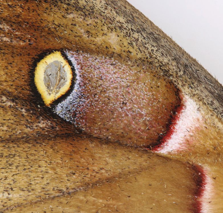
SOURAKOV & SHIRAI: Effects of heparin on wing pattern                                     TROP. LEPID. RES., 30(1): 4-19, 2020                13

       Figure 7. Transplants in Automeris io: A. local change in scale color in 1 of 5 females, resulting from M2-M3 cross-vein transplant
       (7 hAP donor pupa into a 14 hAP host pupa), B. 1 of 3 males where a transplant into the distal part of the wing (8 hAP donor
       into a 8 hAP host) produced local change in color from yellow to brown, expanding the wing pattern element, with (ii vs. iii)
       transplant region under microscope vs. corresponding area on the contralateral wing; and Actias luna: C. FW cross-vein to FW
       distal region transplant (5 hAP donor to a 10 hAP host).

are maintained. In contrast, if it is the disruption of pattern           can be influenced by heparin, not all elements in the system
boundaries that occurs, we should observe a smearing of                   are always affected. There is no consistency in the way a given
the color — that is, a loss of the distinct shape of the pattern          element changes in different species, for example, in eyespots,
element.                                                                  they may expand (saturniids) or disappear (nymphalids), with
     The most extreme heparin-induced color changes so far                some or all rings affected. Tentatively we suggest that a single
achieved in Lepidoptera (Fig. 2) suggest a breakdown of the               component of this pattern element, the eyespot center (when
boundaries in pattern elements, disrupted in different ways               there is one), may, consistently across species, remain unaltered
in different species. A lack of consistency among species is              by heparin injections. Therefore, we suggest that what underlies
seen in: (a) the colors that are affected (such as black, red,            the breakdown of boundaries in a wing pattern element is
orange, or pigment-based versus structural colors), (b) the               the underlying developmental mechanism determining this
color to which a pattern is transformed, (c) the direction in             element. If butterflies and moths have homologous wing
which the pattern element expands (e.g., proximally, distally),           patterns, including the eyespot, we could expect conservation
(d) the type of pattern element (e.g., bands, eyespots), or (e)           of the molecular pathway(s) responsible for determining pattern
the symmetry system of the ‘Nymphalid Groundplan’ that is                 elements. Furthermore, we could also expect that this pathway
disturbed. While pattern elements in every symmetry system                acts by reaction-diffusion, in a concentration-dependent
14 TROP. LEPID. RES., 30(1): 4-19, 2020 SOURAKOV & SHIRAI: Effects of heparin on wing pattern
Figure 8. Additional examples of affected wings (both FWd and FWv) in Automeris io M2-M3 cross-vein transplant (A,B) vs.
control cuts (C,D): A. from a 4 hAP donor pupa into a 7 hAP host pupa, affecting both wing surfaces; and, B. from a 4 hAP donor
pupa into a 9 hAP host pupa, removing scales in both surfaces but generating a larger response in the ventral surface; (i) dorsal,
(ii) ventral; C. Cut made at 5 hAP, D. Cut made at 10-12 hAP.
manner. Thus, the first place to search for candidate pathways is molecules – notice that the eyespot center did not seem to be
among signaling pathways, especially those known to interact affected by heparin, perhaps because it might be insensitive to
with heparin. higher signaling levels since it already has the highest level
It has been suggested, based on associations of expanded of signaling molecules among eyespot rings). However, if
pattern elements in the adult butterfly and Wnt expression in other signaling molecules are detected at the location of Wnt
larval wings, that heparin targets the Wnt pathway (Martin & expression, that could mean that heparin binds to signaling
Reed, 2010, 2014; Martin et al., 2012; Gallant et al., 2014). molecules other than Wnt. Other signaling pathways (e.g.
Although Wnt loss-of-function experiments (such as WntA dpp, Notch, and more recently, Toll) have been implicated in
knock-out using CRISPR-Cas9, Mazo-Vargas et al., 2017; butterfly eyespot development in studies using transcriptomics
or wg knocked-down with RNAi, Özsu et al., 2017), are not (Özsu & Monteiro, 2017).
the exact opposites of gain-of-function experiments (such What remains unexplained from this gain-of-function
as heparin injections), it does appear that Wnt is indeed the experiment, that is, heparin injection presumably increasing
strongest candidate to be the target of heparin in Nymphalidae. the action of members of Wnt pathway, is that some pattern
This conclusion is clearly supported by experiments on Agraulis elements are reduced or are altogether eliminated (Martin &
vanillae, where the effects of WntA transgenic loss-of-function Reed, 2010, 2014; Martin et al., 2012; Gallant et al., 2014).
is complimentary to heparin gain-of-function (Martin & Reed, These happen to be Wnt-negative patterns, but the absence of
2014). Wnt signaling in wild-types does not mean the adult patterns
WntA and wg determine the boundaries of a diverse array should be reduced when injected with a drug that enhances a
of pattern elements in different symmetry systems that have signal they normally lack. Perhaps this reduction or elimination
different colors, which might explain why heparin does not is caused by the systemic action of heparin binding to different
consistently act on a fixed location on the wing, on a single type signaling pathways (not just Wnt), as demonstrated in the
of pattern element, nor on a particular color. If heparin indeed literature for other model organisms. This explanation assumes
acts on members of the Wnt pathway, it seems plausible that that the binding site of the conjugate heparin+signaling
saturniid eyespots are formed through a signaling process, with molecule(s) to extracellular receptors is generic, and that
reaction-diffusion, homologous to that presumably found in signal transduction only occurs when the conjugate binds to
nymphalid eyespots. In future, this potential homology should the Wnt receptor, Frizzled. In pattern elements where reaction-
be examined at the gene level, focusing on WntA and wg which, diffusion does not involve the Wnt pathway (Wnt-negative
in the case of the eyespot of Io and Polyphemus moths, should patterns), receptors would remain clogged, which would lead
be expressed at the location of the black disc (affected by to an absence of their normal signaling levels. Such cells would
heparin) and of the focus (the center of production of signaling receive no developmental instruction (of “cell fate”), causingSOURAKOV & SHIRAI: Effects of heparin on wing pattern TROP. LEPID. RES., 30(1): 4-19, 2020 15
them to adopt the background color (the “default” color) or with the expansion of the black disc on the FWv and changes
the color of a neighboring inductive pattern element, thereby in the “DII” pattern element into black finger-like streaks (as
“disappearing.” seen in Fig. 3C,D), which is even more rare in nature. We are
aware of only a single similar case that appears to be natural,
Heparin injection experiments in a specimen Automeris zugana H. Druce, 1886 collected in
Our experiments demonstrated that eyespots of Saturniidae Colombia in 1967 and illustrated by Lemaire (2002, plate 55).
are affected by heparin (H1-H3), as control injections with Similarly, when searching the collection for aberrations similar
water, buffer and other control solutions performed on the to those caused by heparin in Agraulis vanillae (see Fig. 2),
saturniid species studied here, as well as other species, did not only a single such aberration was found in the 1000 specimens
produce similar effects on wing pattern (e.g., Serfas & Carroll, examined (Sourakov, 2018b). Despite the rarity of these
2005; Sourakov, 2017, 2018). aberrations in nature, the fact that they do occur means that they
It is not easy to judge how much heparin makes its way reflect phenotypic diversity potentially available for selection,
into the organism if there is bleeding from the injection site. and hence such aberrations should be seen as a window into
Hence, it was important, in our opinion, to also make several the mechanisms by which wing pattern diversity may evolve,
injections deep into the pupal abdomen of A. io, where no instead of being treated just as curious artifacts.
bleeding occurred. These injections also demonstrated the
lowest amount (0.03 mg) at which transformation can be How does the timing of injections relate to wing pattern
achieved in an 8 hAP A. io pupa. It is interesting to note that, change?
under this dosage, the wing pattern change (HWd eyespot Based on the time series results (H1), we suggest that
black disc expansion) in the female specimen (e.g., Fig. S3I signaling molecules affected by heparin in A. io are active
of Supplementary Material) is much smaller than in the male from approximately 3 dBP and extend to at least 10 hAP, with
specimen (e.g., Fig. S3J of Supplementary Material). This an inactive period right after pupation (until about 5 hAP).
could be explained by the difference in the body mass of the two While we did not test the whole spectrum of development in A.
individuals: females are at least twice as heavy as males in this polyphemus, heparin had an effect from 1.5 dBP to 13.5 hAP in
species, and the former, in our experience, are much more likely this species, also with the possibility of an inactive period right
to survive higher dosages of heparin. It should be noted that, after pupation.
when designing heparin injection experiments, one should take Injections made at 20 hAP had no effect on the wing patterns
into consideration the relative size of the species and the stage of Buckeye butterflies (Serfas & Carroll 2005), but those made
of development, as can be observed from the dosages used here at the prepupal stage (2.5-20 hBP) by Sourakov (2018b), a stage
and by previous studies (Fig. 2, where some species had mg and hardly investigated in butterflies, did show similar results to the
others ug of injected heparin). Sourakov (2018b) discussed how ones obtained at early pupal stage by Serfas and Carroll (2005).
change of weight during development of immature stages in Other species that showed an effect on wing pattern from
Lepidoptera, together with intraspecific variation, might affect prepupal injections include A. vanillae (1-48 hBP), A. clyton
the results of heparin injections, illustrating the issue with a (1-20 hBP), and tiger moths, E. acrea and H. scribonia (12-
variety of examples. 13 hBP) (Sourakov 2018b). The results of numerous injections
Among the 797 A. polyphemus examined at the MGCL, suggest that heparin injected too early (e.g., 6-9 dBP) has no
which span the entire United States as well as Mexico (see Fig. effect on wing pattern, but that already at 3 dBP heparin may
S6 of Supplementary Material), the gray spot of HWd eyespot is have a significant effect on wing pattern elements. If heparin
expanded in 5.5% of individuals (e.g., Fig. S6C,I,S). However, action is informative about the signaling molecule Wnt, the
the gray spot expansion and black disc expansion together action of this signaling molecule starts in the prepupal stage,
(such as in heparin-injected individuals shown in Fig. 3F and earlier than current data suggest, e.g at 10 hAP for wg (Özsu et
Fig. S2B of Supplementary Material) were only present in 1% al., 2017, and 10.5 hAP for wg protein; see below). Expression
of MGCL specimens (e.g., Fig. 5E). The latter figure shows patterns of Dll and En (that respond to Wnt in Drosophila
cases of unique eyespot aberrations found in MGCL holdings (Arias, 2003)) were found to be active at 3 days after the start of
of Polyphemus Moth, and a larger variety are also illustrated in cocoon-spinning in A. polyphemus (approximately 4 dBP in our
Supplementary Material Fig. S6. experiments) and 2 days after cocoon-spinning in S. pavonia
In Western Polyphemus Moth, Antheraea oculea, formerly (Monteiro et al., 2006). Handling prepupal wings for assays
a subspecies of A. polyphemus, the increase of HWd melanism, of expression patterns is technically difficult, but perhaps
including expansion of the black disc outside of the eyespot, transcriptomics could be a strategy to inspect whether signaling
is typical, although melanization affects both FW and HW molecules are already active in Lepidoptera wings at this stage.
(Fig. 5A,B). It is possible that the phenotypic differences Previous research on imaginal disk development conducted
between the A. polyphemus and A. oculea are based on similar on Buckeye butterflies (Miner et al., 2000) suggests that neither
developmental mechanisms that were affected by heparin in our size nor stage of larval development (days into the final instar
experiment. The genetic difference between them may manifest after molting) are good predictors of the development of the
itself in Wnt genes, as has been recently found in closely related wing disk. A much better predictor turns out to be the rate of
species of Buckeye butterflies (Lalonde & Marcus, 2019). As development, and both starvation and Juvenile Hormone (JH)
for Automeris, in extreme cases, heparin results in a dramatic presence can inhibit wing disk growth. Hence, when conducting
expansion of the black disc of the HWd eyespot combined heparin injection in larvae of a similar developmental stage we16 TROP. LEPID. RES., 30(1): 4-19, 2020 SOURAKOV & SHIRAI: Effects of heparin on wing pattern
may be experimenting on quite different animals from the point of wing scale color in another individual (T3-T4). While one
of view of their wing disk development. Research on the sphinx could argue that melanization could potentially occur in the
moth Manduca sexta (Browder et al., 2001) demonstrates that transplant spot in response to injury, this explanation is not
JH correlates with larval weight and peaks after the larvae pass consistent with the results obtained by us in males of Automeris
the critical weight at which JH secretion ceases. In Sourakov io, where the changed scales are brown, not black (Fig. 7B.ii).
(2018b, Fig 8), a sharp decline in larval/prepupal weight was The single Luna Moth transplant could also serve as evidence
demonstrated for a number of Lepidoptera after they stopped against melanization through injury: the changed scales in the
feeding and began to prepare for pupation (spin silk pads or transplanted site are white, not black (Fig. 7C). If the signal
cocoons). This beginning of weight-loss, according to Browder from the transplanted tissue influenced the FWd inducing
et al. (2011), correlates with the end of JH secretion and localized color changes, that would support the M2-M3 cross-
perhaps gives rise to the stage at which heparin becomes active vein signaling hypothesis. The same distal wing regions did not
as a wing-pattern-altering agent. The role of JH in affecting the react to control cuts or injuries in a similar way. Instead, at most,
ability of heparin to change wing pattern may extend beyond they showed a small scar, but mostly completely regenerated
this point. Nijhout and Wheeler (1982) point out that JH titers (T3-T6). Additionally, based on observations of cross-vein
are affected not only by the activity of the corpora alata disruptions at early stages of pupal development (T1-T2) with
but also by variation in the activity of enzymes that break it not a single change detected, we suggest that A.io wing veins
down, so it is not easy to determine based solely on the larval and membranes might have a high capacity for regeneration.
relative size or days prior to pupation how much JH it has. In However, when injuries were made in other wing areas,
the developmental stages of interest from the point of view of such as the discal cell of A. luna FWd (T2, Fig. 6A) and the
the present experiments (prepupa a day before pupation – early anterior region of A. io HWd (T1, T6; Fig. 6B,C), we observed
pupa), a sharp decline/absence of JH and changes in ecdysone wound-induced responses. These results suggest that the
secretion have been demonstrated (Nijhout and Wheeler, 1982, responsive region for eyespot formation may be within the
p. 115, Fig. 1). Thus, it is reasonable to hypothesize (and discal cell, and not at the distal region of the wing as in the
warrants further investigation) that titers of either or both of butterfly Bicyclus anynana (Brakefield & French, 1995), which
these hormones play a role in making developing Lepidoptera is also the region used in the present study for transplants.
wings sensitive to Wnt signaling and thus to heparin. We thus suggest that future attempts to transplant tissue in
Evidence also continues to grow that while wing patterns Saturniidae should be done in the medial region and within
are sensitive to heparin in late prepupae and early pupae, they the discal cell or, more generally, that transplants should target
may be less responsive immediately after pupation, when the the wing region/symmetry system and wing surface that bear
pupa is still soft and just beginning the tanning process. Could eyespots. This should be tested and the best region optimized
there be a ‘buffer’ time in the transition from prepupa to pupa before performing future transplants with appropriate controls.
during which no signaling occurs? In support of this hypothesis, Surgical manipulations involving between-pupae transplants
during the first hours of pupation (3-6 hAP), no wg expression of potential eyespot organizers show promise for future
was found in the butterfly Bicyclus anynana (when several other exploration of wing pattern formation in saturniid moths,
eyespot genes were found to be expressed, Özsu & Monteiro, including a more in-depth exploration of wound-induced color
2017), the earliest moment wg was ever detected being 10 hAP changes in Lepidoptera.
(Monteiro et al., 2006, Özsu et al., 2017). If one is to compare The 18 inter-pupal transplants conducted on Io Moth
these results to the timing of the hormonal activity in developing required many host pupae and a number of additional donor
Lepidoptera (Nijhout & Wheeler 1982, p. 115, Fig. 1), one can pupae at a similar stage of development. This translates into
hypothesize that the insensitive period to heparin overlaps with a substantial rearing and pupa-monitoring effort, which, in
the insensitive period to JH and low levels of ecdysone. The case of this species, required much time and planning, as Io
idea of a ‘buffer’ time regulated by ecdysone might also relate Moth larvae were raised on a natural hostplant (no artificial diet
to developmental milestones, as demonstrated in Drosophila exists for this species), and larvae develop over period of 60-
wing discs (Oliveira et al., 2014). Milestones are “checkpoint” 90 days. In addition to their long larval development and the
moments, such as at pupariation, when wing and whole-body large amounts of hostplant material needed to rear numerous
development align, ensuring their coordination in the face of saturniid moths, there was a challenge of transplanting the M2-
environmental or physiological variation. Judging by our results, M3 cross-vein from the HW, and not the FW as usually done
a refractory period between 0 to 5 hAP, possibly regulated by in butterflies. In early pupae, the developing FW is attached to
hormones, may exist in Lepidoptera, when no signal-response the cuticle at the time when surgeries are typically made. When
occurs. It would be interesting to evaluate whether the first five this type of manipulation is normally conducted, the donor FW
hours of pupation also serve as a checkpoint in Lepidoptera tissue attached to the cuticle is rotated 180° and scale orientation
wing pattern development. in the adult identifies where the transplanted tissue was placed.
Handling the hardening cuticle (and the “hitchhiked” FW) is
Organizer tissue transplanting and disruption experiments much easier than a loose developing wing tissue, which was the
We showed, though on a limited scale and with the case in our saturniid transplants. More importantly, we left the
central drawback of lacking some experimental controls, donor tissue “floating” on the host wing tissue, anchored in a cut
that transplants of pupal tissue taken from the center of a we had made, which made it impossible to judge, upon the host
developing saturniid eyespot might lead to localized change emergence, whether the tissue at the transplanted site was (a) theYou can also read