SERTRALINE INDUCES DNA DAMAGE AND CELLULAR TOXICITY IN DROSOPHILA THAT CAN BE AMELIORATED BY ANTIOXIDANTS - NATURE
←
→
Page content transcription
If your browser does not render page correctly, please read the page content below
www.nature.com/scientificreports

            OPEN           Sertraline induces DNA damage
                           and cellular toxicity in Drosophila
                           that can be ameliorated by
                           antioxidants
                                             1,3
                           Arpita Jajoo        , Catherine Donlon1,3, Sarah Shnayder1, Michael Levin              1,2
                                                                                                                        & Mitch McVey    1*

                           Sertraline hydrochloride is a commonly prescribed antidepressant medication that acts by amplifying
                           serotonin signaling. Numerous studies have suggested that children of women taking sertraline
                           during pregnancy have an increased risk of developmental defects. Resolving the degree of risk for
                           human fetuses requires comprehensive knowledge of the pathways affected by this drug. We utilized
                           a Drosophila melanogaster model system to assess the effects of sertraline throughout development.
                           Ingestion of sertraline by females did not affect their fecundity or embryogenesis in their progeny.
                           However, larvae that consumed sertraline experienced delayed developmental progression and reduced
                           survival at all stages of development. Genetic experiments showed that these effects were mostly
                           independent of aberrant extracellular serotonin levels. Using an ex vivo imaginal disc culture system,
                           we showed that mitotically active sertraline-treated tissues accumulate DNA double-strand breaks
                           and undergo apoptosis at increased frequencies. Remarkably, the sertraline-induced genotoxicity was
                           partially rescued by co-incubation with ascorbic acid, suggesting that sertraline induces oxidative DNA
                           damage. These findings may have implications for the biomedicine of sertraline-induced birth defects.

                           Serotonin (5-hydroxytryptamine, or 5-HT) is a neurotransmitter that regulates several behaviors in the meta-
                           zoan, such as mood, social behavior, sleep, and food intake1. It is also important for embryogenesis, and con-
                           tributes to oocyte maturation, vascular remodeling, neural crest migration, craniofacial and limb development,
                           left-right asymmetry, cytoskeletal structure, blastomere adhesion, neural patterning, and gastrulation 2–6. Upon
                           release by a presynaptic neuron into the synaptic cleft, serotonin binds 5-HT receptors on the postsynaptic neu-
                           ron and initiates downstream signaling. Signaling is attenuated via reuptake from the synapse into the presynaptic
                           neuron by the serotonin transporter (SERT) or via degradation by monoamine oxidase. Imbalanced serotonin
                           levels are associated with several mental disorders, such as anxiety, depression, post-traumatic stress disorder,
                           and obsessive-compulsive disorder, and this reuptake mechanism is often targeted to restore serotonin levels in
                           patients with these disorders. Selective Serotonin Reuptake Inhibitors (SSRIs) block SERT, thus leading to higher
                           serotonin concentration and longer duration of action in the synapse. Disorders of serotonergic signaling can also
                           contribute to metastatic cell behavior7–9.
                               Sertraline hydrochloride, henceforth referred to as “sertraline” and marketed under the brand name Zoloft ,
                           is a commonly prescribed SSRI for numerous disorders10. Preclinical trials with sertraline in laboratory ani-
                                                                                                                                              ®
                           mals reported no evidence that sertraline is genotoxic11 and a small study using peripheral lymphocytes from
                           human patients found no significant effects of sertraline on numbers of chromosome aberrations12. Despite the
                           laboratory studies that used sertraline to perturb ion channels and a number of cellular signaling pathways 13–21,
                           sertraline currently has Category C designation by the FDA. This has led to its becoming one of the most fre-
                           quently used SSRIs by pregnant women22. An analysis performed in 2015 failed to find significant evidence for
                           harmful effects associated with sertraline use during pregnancy23. However, other studies have demonstrated that
                           sertraline usage during pregnancy is linked to adverse short- and long-term effects in children24, including atrial
                           septal defects25, craniosynostosis26, increased risk of omphalocele27, developmental delays and increased risk of
                           autism in males28, increased risk of clubfoot29, elevated risk of persistent pulmonary hypertension30, decreased

                           1
                           Department of Biology, Tufts University, Medford, MA, USA. 2Allen Discovery Center at Tufts University, Medford,
                           MA, USA. 3These authors contributed equally: Arpita Jajoo and Catherine Donlon. *email: mitch.mcvey@tufts.edu

Scientific Reports |   (2020) 10:4512 | https://doi.org/10.1038/s41598-020-61362-y                                                            1
www.nature.com/scientificreports/ www.nature.com/scientificreports
birth weight31, and preterm births32. These structural phenotypes are in addition to a rich literature discussing
behavioral outcomes of SSRI exposure during gestation 33,34.
The phenomenon of withdrawal (neonatal abstinence syndrome) reveals the profound effects that expo-
sure to SSRIs has on embryos35. Similar to the developmental defects described above, the observed withdrawal
effects present stochastically in the human population, resulting in debate over the relative risks of exposure.
Importantly, regulative developmental mechanisms and physiological as well as genetic heterogeneity of both
mother and fetus result in a variability of phenotypes across the population, as has been seen in developmental
biology studies in animal model systems36. Because different embryos may succumb to or resist perturbation of
specific steps in development, it is critical to comprehensively characterize the means by which sertraline (and
other SSRIs) could impact embryogenesis. Disruption of the known developmental roles of serotonergic sign-
aling has been suggested to be one of the routes of defects in some individuals37–39. However, it is also possible
that sertraline results in genetic, epigenetic, or other cellular abnormalities that affect developmental patterning.
Ethical considerations prevent randomized controlled clinical trials in humans to determine sertraline’s
embryonic effects and risk profile. To circumvent these challenges, model organisms have been utilized to inves-
tigate the effects of sertraline exposure at both cellular and organismal levels. Studies with Xenopus laevis showed
that exposure of tadpoles to sertraline (0.1–10 μg/L) resulted in developmental toxicity40. Furthermore, mice
exposed to low levels (10 μM) of sertraline develop craniofacial abnormalities, due to interference with serotonin
signaling important for normal craniofacial development15.
Drosophila is one of the premiere organisms for studying developmental processes, and is a powerful model
for discovery of mechanisms of relevance to human medicine including cancer, neurobiology, and left-right
asymmetry41–43, all of which are areas where SSRIs have been strongly implicated. The genome of Drosophila
melanogaster contains homologs of ~75% of human disease-related genes44. Drosophila utilize serotonin as a neu-
rotransmitter in the nervous system and as a small molecule in early development45,46 and have serotonin receptor
homologs. Although no monoamine oxidase has been described in Drosophila, their genome does encode a single
serotonin reuptake transporter, named SerT47. Thus, we took advantage of this powerful developmental model
to search for novel mechanisms for sertraline action in development, specifically focusing on its ability to disrupt
genetic integrity.
Only one study has been published investigating the genotoxicity of sertraline in Drosophila. Using an assay
called the somatic mutation and recombination test (SMART), the authors showed that citralopram, another
SSRI, caused dose-dependent genotoxicity, but the results with sertraline were inconclusive48.
To further investigate possible genotoxic effects of sertraline in Drosophila and to identify possible causes for
the sertraline-related birth defects in humans, we performed a series of experiments at larval and adult stages
to determin the impact of sertraline on development and survival. In addition, we incubated larval wing disc
tissues with sertraline to investigate changes in cellular markers related to cell death and DNA damage. While
exposure of female adult flies to sertraline had no effects on egg laying or hatching, larval exposure led to delays
in developmental progression and to impaired viability. Mechanistically, sertraline induced oxidative damage of
DNA, which caused cellular apoptosis in tissues necessary for proper development. Strikingly, the cellular dam-
age induced by sertraline could be rescued by antioxidant supplementation. These results establish sertraline as
detrimental to fly development, elucidate a new mechanism of its developmental toxicity, and for the first time,
provide a potential avenue for mitigation of its adverse effects on embryogenesis that is highly compatible with
human patient use.
Results
Sertraline-treated adult females exhibit no difference in fecundity and embryo hatching fre-
quency. We began by investigating the effects of sertraline ingestion on the quantity and quality of eggs laid
by adult females. Cohorts of eight females 3–4 days of age were fed using capillary feeders in a modified CAFE
apparatus49. Each apparatus had capillaries filled with either a 5% sucrose solution or 5% sucrose with 250 μg/
mL of sertraline, a non-toxic concentration for adult Drosophila48. Although the flies consumed less liquid in the
apparatus that contained sertraline solution, we were successful in exposing the adult females to the drug via this
method (Fig. 1a).
Females that were fed only sucrose or sertraline were then placed into a cage with equal numbers of male flies
and the number of eggs laid by the females in an 18-hour period was counted. Compared to sucrose, we found no
significant difference in fecundity of female flies that drank sertraline-containing solutions (Fig. 1b). On average,
female flies fed plain sucrose laid 1 egg every 2.15 hours, while females fed sucrose with sertraline laid 1 egg every
2.34 hours (p = 0.72).
In addition, there was no significant difference in the number of eggs that hatched between the sertraline-fed
and control flies (Fig. 1c). One- and two-day hatching frequencies for eggs laid by control females were 84% and
91%, compared to frequencies of 78% and 87% for sertraline-treated females (p = 0.11). To determine if sertraline
fed to adult females affected the development of their progeny, we allowed larvae from these eggs to develop into
adulthood. Of more than five hundred progeny examined, no obvious morphological or phenotypic differences
were noted for progeny from the sertraline-treated females. Thus, while we cannot rule out subtle effects on devel-
opment, sertraline-fed females do not appear to produce offspring with obvious morphological defects.
Sertraline-treated larvae display delayed development and decreased survival. Unlike human
development, which takes place in utero and exposes the fetus to sertraline ingested by its mother, Drosophila
embryonic development takes place outside of the mother in an egg that is protected from the environment
through an impermeable vitelline membrane and chorion. Larvae which hatch from the egg continue devel-
opment, paralleling the various stages of organogenesis and remodeling that occur in embryos of all species.
After hatching, Drosophila larvae consume food and grow through three instar stages, each lasting approximately
Scientific Reports | (2020) 10:4512 | https://doi.org/10.1038/s41598-020-61362-y 2www.nature.com/scientificreports/ www.nature.com/scientificreports
a
volume of food consumed (ul)
6
4
2
0
Sucrose Sertraline
b 0.8
Eggs per female per hour
0.6
0.4
0.2
0.0
Sucrose Sertraline
Sucrose
c
Percentage of eggs hatched
100 Sertraline
80
60
40
20
0
1 day AEL 2 days AEL
Figure 1. Sertraline consumption does not impact fecundity or egg hatching frequency in Drosophila. (a)
Amount of liquid consumed by 8 adult female flies during 8 hours from a single capillary tube in a CAFE
apparatus. Each tube contained either 5% sucrose or 250 μg/ml sertraline in 5% sucrose. N = 30, error bars
represent SEM. p < 0.0001, unpaired two-tailed t test. (b) Egg laying calculated as the number of eggs laid by
one female per hour after treatment with either sucrose or 250 μg/mL sucrose + sertraline solution. N = 7,
error bars represent SEM. p = 0.72, unpaired two-tailed t-test. (c) Percentage of eggs hatched one day and two
days after egg laying (AEL) by mothers fed either sucrose solution or 250 μg/ml sertraline solution. N = 5–7
replicates for each condition, error bars represent SEM. p = 0.18 after 1 day and p = 0.11 after 2 days, unpaired
two-tailed t-test.
24 hours. At the end of the third instar stage, they cease eating and enter the wandering third instar stage, which
lasts for several hours and is followed by a cessation of movement and pupariation. During the pupal stage, which
lasts approximately 3–4 days, they undergo metamorphosis and then emerge as adult flies.
We wanted to test whether direct exposure to sertraline might affect Drosophila during its various devel-
opmental stages. Repeated attempts to introduce sertraline directly into eggs drastically reduced the viabil-
ity of the embryos. Therefore, we turned to an established larval feeding protocol, in which larvae consume
sertraline-containing food throughout their development48. First instar larvae derived from an Oregon-R stock
of flies were placed onto food containing different concentrations of sertraline and their rate of development
to various developmental stages was quantified and compared to larvae placed in vehicle-containing food (see
Methods).
We chose the transition to the wandering third instar stage as our first developmental time point. We observed
a dose-dependent effect for development to wandering third instar larvae, with significant delays observed for
the two highest concentrations of sertraline (Fig. 2a). In addition, there was an overall decrease in the survival in
larvae treated with sertraline, with only 33% of first instar larvae treated with the highest sertraline concentration
surviving to third instar stage, compared to 80% for the controls.
Scientific Reports | (2020) 10:4512 | https://doi.org/10.1038/s41598-020-61362-y 3www.nature.com/scientificreports/ www.nature.com/scientificreports
a
Cumulative percentage 1st instar larvae
*** *** *
100 no treatment
1% DMSO
that reach 3rd instar
5 µg/ml sertraline
80
10 µg/ml sertraline
17.5 µg/ml sertraline
60 25 µg/ml sertraline
50 µg/ml sertraline
40
20
0
4 6 8
Days post 1st instar transfer
b
Cumulative percentage 3rd instar larvae
*** *
100 no treatment
1% DMSO
that reach adulthood
5 µg/ml sertraline
80 10 µg/ml sertraline
17.5 µg/ml sertraline
60 25 µg/ml sertraline
50 µg/ml sertraline
40
20
0
9 11 13
Days post 1st instar transfer
Figure 2. Sertraline delays Drosophila development and reduces survival frequency. (a) Cumulative percentage
of wandering third instar larvae following placement of 20–25 Oregon-R first instar larvae into food containing
various concentrations of sertraline at day 0. (b) Cumulative percentage of surviving third instar larvae that
developed to adulthood following placement of first instar larvae into food containing various concentrations
of sertraline at day 0. Each trial lasted for at least 15 days, with no additional changes observed after day 13.
N = 4–7 trials for each treatment, error bars represent SEM. *p < 0.05 and ***p < 0.001 (one-way ANOVA with
Bonferroni correction).
A similar trend was observed for development from third instar larvae to adulthood (Fig. 2b). For this anal-
ysis, we counted the number of adults that eclosed from their pupal cases on successive days and calculated the
percentage of adults relative to the number of total third instar larvae that we obtained for each treatment. The
developmental delay to the mature adult stage for the highest concentrations of sertraline was similar to the delay
seen for the transition from first to third instar larvae, suggesting that the overall delay in development occurred
during the larval stages, when the sertraline-containing food was being consumed. However, we also observed a
further decrease in survival between the third instar and adult stages for the highest concentrations of sertraline.
In the control experiments, over 98% of the wandering third instar larvae survived to adulthood, while only 33%
of the third instar larvae raised in the 50 μg/mL sertraline-containing food successfully pupated and eclosed as
adults. Therefore, sertraline continues to affect Drosophila viability beyond the larval period when it is being
actively ingested.
Sertraline affects Drosophila development independently of serotonin-mediated effects. The
developmental delays and decreased survival caused by exposure to sertraline could be caused by changes in
serotonin levels due to inhibition of the serotonin reuptake mechanism or by genotoxicity of sertraline itself. To
distinguish between these two possibilities, we repeated the larval feeding experiments with flies lacking the sole
serotonin reuptake transporter (hereafter referred to as SerT knockout or SerT −/− flies). We chose a sertraline
concentration of 10 μg/ml, where we previously observed moderate effects on developmental timing, reasoning
that in the SerT knockout flies, any additional developmental delays phenotypes observed in the presence of ser-
traline treatment could be attributed to genotoxic effects of sertraline itself, rather than its effects on the serotonin
transporter. As a control, we measured developmental progression in isogenic flies with an intact SerT gene (see
Methods).
As observed with the Oregon-R flies, there was approximately a 1-2 day developmental delay from first-instar
to wandering third instar larvae in the presence of 10 μg/mL sertraline, for both the SerT knockout and SerT
wild-type stocks (Fig. 3a). Similarly, sertraline-treated flies from both stocks exhibited a delay in development
between the wandering third instar and adult stages (Fig. 3b). While the flies lacking SerT did have a slightly
delayed transition from first to third instar larvae in the absence of sertraline, this developmental delay was
ameliorated by the time the flies reached adulthood. In addition, we observed no increase in sertraline-mediated
lethality in the SerT knockout larvae compared to SerT wild-type larvae. From these data, we conclude that while
a portion of the larval developmental delays caused by sertraline ingestion may be due to changes in serotonin
Scientific Reports | (2020) 10:4512 | https://doi.org/10.1038/s41598-020-61362-y 4www.nature.com/scientificreports/ www.nature.com/scientificreports
a
Cumulative percentage 1st instar larvae
100
SerT +/+ no sertraline
that reach 3rd instar
80 SerT -/- no sertraline
SerT +/+ with sertraline
60 SerT -/- with sertraline
40
20
0
4 5 6 7 8
Days post 1st instar transfer
b
Cumulative percentage 3rd instar larvae
100
SerT +/+ no sertraline
that reach adulthood
80 SerT -/- no sertraline
SerT +/+ with sertraline
60 SerT -/- with sertraline
40
20
0
9 10 11 12 13
Days post 1st instar transfer
Figure 3. The effects of sertraline on development are mostly independent of its effects on serotonin signaling.
(a) Cumulative percentage of wandering third instar larvae following placement of 20–25 first instar larvae into
food containing 10 μg/ml sertraline at day 0. (b) Cumulative percentage of surviving third instar larvae that
developed to adulthood following placement of first instar larvae into food containing 10 μg/ml sertraline at
day 0. Each trial lasted for at least 15 days, with no additional changes observed after day 13. SerT −/− flies lack
the SerT serotonin reuptake channel and SerT +/+ flies possess a wild-type copy of the SerT gene in an isogenic
background. N = 4 replicates for SerT −/− and 3 replicates for SerT +/+, error bars represent SEM.
levels, most of the sertraline-induced delays and lethality is independent of its effects on serotonin signaling.
Thus, we next examined its genotoxic potential.
Sertraline induces apoptosis and DNA damage in wing imaginal discs. In Drosophila, larval imag-
inal disc tissues become adult structures, such as eyes, wings, and antennae, following metamorphosis. The induc-
tion of DNA damage in imaginal disc tissues results in cell death and developmental delays50–52. We hypothesized
that sertraline might induce DNA double-strand breaks, the accumulation of which could result in apoptosis in
larval imaginal discs, developmental delays, and organismal lethality. To test this hypothesis, we measured levels
of DNA damage following sertraline treatment in cultured wing imaginal discs of SerT knockout third instar lar-
vae. We have previously shown that cells in cultured imaginal discs can survive and continue to divide for at least
five hours53. In Drosophila, phosphorylation of the histone H2A variant on serine-137 (γ-H2Av) occurs upon
double-strand break formation and the number of γ-H2Av foci is roughly proportional to the number of breaks 54,55.
Strikingly, a five-hour incubation of wing imaginal discs with 10 μg/mL sertraline resulted in an average three-fold
increase in the number of γ-H2Av foci (Fig. 4a,b). Untreated wing discs displayed 10 ± 11 γ-H2Av foci/micron2,
while sertraline treated discs had an average of 32 ± 20 foci/micron2 (Fig. 4c), indicating that sertraline treatment
induces higher levels of DNA damage in proliferating discs.
To determine whether the elevated levels of DNA damage also lead to increased cell death, we monitored
apoptosis via staining of cleaved death caspase-1 (Dcp-1), which is an effector caspase that promotes degradation
of target proteins during apoptosis56,57. Wing discs cultured in the absence of sertraline displayed expected pat-
terns of apoptosis, with Dcp-1 foci clustered mostly towards the top of the discs (Fig. 4d) and little in the middle
folded “frown” region58. Although the number of apoptotic cells in sertraline treated discs was more variable, on
average the treated discs had three-fold greater numbers of Dcp-1 foci compared to the control discs. Thus, expo-
sure of tissues to sertraline for just a few hours can induce DNA damage in the form of double-strand breaks and
lead to excessive cell death that correlates with a delay in organismal development.
Antioxidant supplementation can ameliorate DNA damage induced by sertraline exposure.
Beyond its extracellular interactions with the serotonin transporter, sertraline can freely diffuse into cells, and its
movement is accelerated by vacuolar proton ATPases59. Although sertraline’s interactions with genetic material
have not been investigated in vivo, sertraline does have affinity for the minor groove of DNA60, indicating its
potential to interact with an organism’s genetic material once inside the cell. Interestingly, serotonin can act as a
pro-oxidant of DNA61 and induce double strand breaks in the presence of copper ions62. Thus, we hypothesized
Scientific Reports | (2020) 10:4512 | https://doi.org/10.1038/s41598-020-61362-y 5www.nature.com/scientificreports/ www.nature.com/scientificreports
Figure 4. Sertraline causes accumulation of DNA double-strand breaks and cell death in wing imaginal discs.
(a and b) Wing imaginal discs dissected from SerT −/− wandering third instar larvae were treated for 5 hours
with DMSO or 10 μg/ml sertraline and stained with antibodies that recognize phosphorylated γ-H2Av. (c)
Quantification of γ-H2Av foci per square micron in control and sertraline treated discs. γ-H2Av foci were
counted and normalized to the area of each wing disc, measured from DAPI-stained images. N = 50 discs for
control and 47 for sertraline treatment, error bars represent SEM. ***p < 0.001 by unpaired t test with Welch’s
correction. (d and e) Wing imaginal discs dissected from SerT −/− wandering third instar larvae, treated with
DMSO or 10 μg/ml sertraline, and stained with antibodies against cleaved Dcp-1. (f) Quantification of the
number of Dcp-1 foci per square micron in control and sertraline-treated discs. Dcp-1 foci were counted and
normalized to the area of each wing disc, measured from DAPI-stained images. N = 22 discs for control and 24
for sertraline treatment, error bars represent SEM. ***p = 0.0005 by unpaired t test with Welch’s correction.
that sertraline, or one of its metabolic breakdown products, might induce oxidative DNA damage that could
eventually lead to the formation of double-strand breaks.
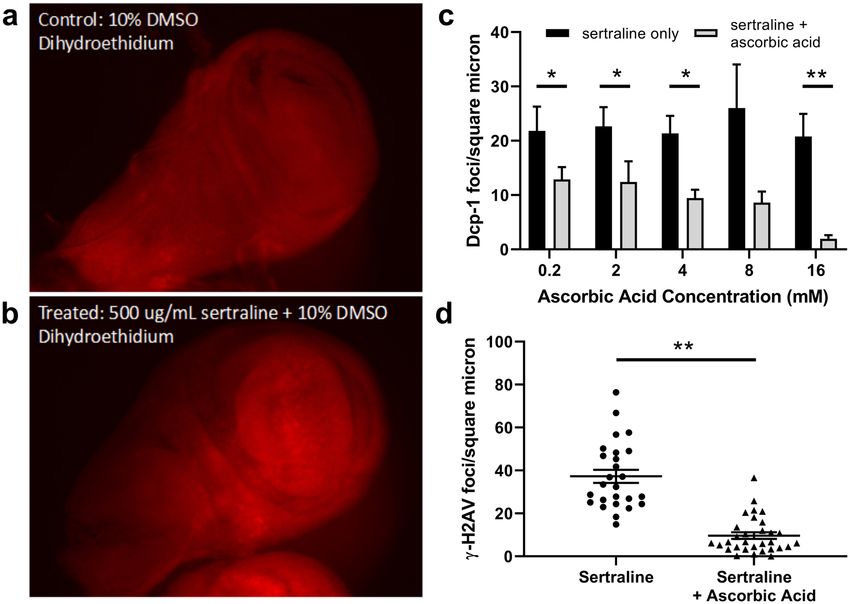
To test whether sertraline might increase the amount of reactive oxygen species, we stained sertraline-treated
SerT knockout wing imaginal discs with dihydroethidium (DHE). DHE is a compound that can diffuse through
cell membranes and produces a red fluorescent ethidium product upon reaction with superoxide radicals63.
Interestingly, we observed more overall DHE staining in sertraline-treated discs than in vehicle-treated controls
(Fig. 5a,b), although the intensity of staining varied in different regions of each disc. Thus, sertraline may increase
the concentration of superoxide radicals in vivo.
If sertraline’s mechanism of toxicity induction is indeed via oxidative damage, we reasoned that supplementa-
tion of an antioxidant might mitigate this toxicity and prevent cell death. To investigate this, we tested the ability
of increasing concentrations of ascorbic acid, a known antioxidant, to affect the sertraline-mediated increase in
apoptotic cell death observed in SerT knockout wing imaginal discs. Indeed, we observed that increasing concen-
trations of ascorbic acid resulted in decreases in cell death associated with sertraline treatment (Fig. 5c). Addition
of 0.2, 4, 8, and 16 mM concentrations of ascorbic acid led to a 1.7-, 2.3-, 3-, and 10-fold decrease in Dcp-1foci/
micron2, respectively, compared to sertraline-treated control discs (Fig. 5c). At a dose of 16 mM, almost no Dcp-1
foci were observed (1.9 foci/micron2, compared to 20.8 foci/micron2 in the sertraline only control).
To determine whether ascorbic acid could also modulate the increase in DNA double-strand breaks observed
with sertraline, we employed immunostaining against γ-H2Av using SerT knockout wing imaginal discs treated
with 10 μg/mL sertraline and 8 mM ascorbic acid. Strikingly, this concentration of ascorbic acid resulted in a
5.9-fold decrease in γ-H2Av foci (6.3 ± 4.9 foci/micron2 in sertraline-treated discs supplemented with ascorbic
acid, compared to 37.5 ± 17.0 foci/micron2 with just sertraline) (Fig. 5d). Thus, antioxidant treatment is capable
of ameliorating both the DNA damage and cell death observed with sertraline exposure.
Antioxidant supplementation rescues delayed development and decreased survival of
sertraline-treated larvae. Based on the immunostaining results, which show that the addition of ascor-
bic acid can largely abolish the negative genotoxic effects of sertraline, we performed development tracking
assays to determine if the beneficial effects extend to the organismal level. First instar SerT knockout larvae were
placed on food containing 50 μg/mL sertraline or 50 μg/mL sertraline + 8 mM ascorbic acid. Remarkably, the
sertraline-mediated developmental delays were decreased when ascorbic acid was included in the food. Addition
of ascorbic acid increased the rate of development of sertraline-treated larvae from first instar to third instar by
2-3 days (Fig. 6a) and from first instar to adulthood by a similar amount of time (Fig. 6b), although progression to
Scientific Reports | (2020) 10:4512 | https://doi.org/10.1038/s41598-020-61362-y 6www.nature.com/scientificreports/ www.nature.com/scientificreports
Figure 5. Addition of ascorbic acid ameliorates sertraline-induced increases in cell death and DNA damage. (a
and b) Wing imaginal discs dissected from SerT −/− wandering third instar larvae were treated for 5 hours with
DMSO or 500 μg/ml sertraline + DMSO and stained with dihydroethidium to indicate presence of superoxide
ions. (c) Relative number of cleaved Dcp-1 foci per square micron in SerT −/− wing imaginal discs treated with
10 μg/ml sertraline plus increasing concentrations of ascorbic acid. Dcp-1 foci were counted and normalized to
the area of each wing disc, measured from DAPI-stained images. For each trial, N ≥ 7 discs for sertraline-only
groups and ≥8 discs for each concentration of ascorbic acid, error bars represent SEM. *p < 0.05, **p < 0.01 by
ANOVA with Bonferroni correction. (d) Quantification of γ-H2Av foci per square micron for discs treated with
10 μg/ml sertraline or 10 μg/ml sertraline plus 8 mM ascorbic acid. N = 26 discs for sertraline and 31 discs for
sertraline + ascorbic acid, error bars represent SEM. **p < 0.01 by unpaired t test with Welch’s correction.
these developmental stages was still slower than that of SerT knockout larvae not exposed to sertraline (compare
Fig. 3 to Fig. 6). In addition, ascorbic acid significantly increased the percentage of sertraline-treated larvae that
survived to adulthood (Fig. 6c). The effect of ascorbic acid on development was sertraline-specific, as inclusion
of 8 mM ascorbic acid in food not containing sertraline did not significantly affect developmental rate from third
instar larvae to adulthood (Fig. 6d). Overall, these data suggest that antioxidants can decrease sertraline-induced
oxidative DNA damage in Drosophila, promoting regular development and survival.
Discussion
Sertraline induces DNA damage and cell death stochastically, mimicking its pattern of effects
on human development. Because sertraline is one of the most commonly prescribed antidepressants22, a
better understanding of the mechanistic basis behind the birth defects sporadically observed in children of moth-
ers taking the drug is needed. Here, we utilized a Drosophila model system to determine the effects that sertraline
may have on metazoan development. Our initial attempts to test the effects of feeding sertraline to females had no
apparent impact, as we failed to see any obvious morphological defects in the embryos, larvae, or adult progeny
derived from these females. Because most of the egg development and patterning in these females took place prior
to sertraline exposure, this result is perhaps not surprising. Future work using quantitative morphometrics and
transcriptional, proteomic, and epigenetic markers will be needed to delineate potential additional phenotypes
that may not have been seen in our analysis.
We then turned to a larval feeding protocol, which exposes the developing organism to a physiologically rel-
evant amount of sertraline during a critical stage of development. Although all larval tissues are likely exposed
to the drug, we focused on imaginal discs as the target tissue. In Drosophila larvae, these discs contain rapidly
dividing diploid cells that are preserved during metamorphosis and eventually form many of the external struc-
tures of the adult fly. When cultured ex vivo, cells within the discs continue to rapidly proliferate and maintain
DNA damage checkpoints, making them a good model for early cell divisions in human embryos. The concentra-
tion of sertraline in the imaginal disc assays was within an order of magnitude of the estimated bioavailability of
sertraline in humans64. Discs treated ex vivo with sertraline experienced high levels of oxidative stress, increased
DNA damage, and heightened levels of apoptosis. Because these cellular processes are likely to be associated with
Scientific Reports | (2020) 10:4512 | https://doi.org/10.1038/s41598-020-61362-y 7www.nature.com/scientificreports/ www.nature.com/scientificreports
a b
Cumulative percentage 3rd instar larvae
Cumulative percentage 1st instar larvae
100
100 sertraline sertraline
sertraline +
that reach adulthood
sertraline + 80
that reach 3rd instar
80 ascorbic acid ascorbic acid
60
60
40 40
20 20
0 0
4 5 6 7 8 9 10 11 10 11 12 13 14
Days post 1st instar transfer Days post 1st instar transfer
c d
Cumulative percentage 3rd instar larvae
100
** 100
water
80 ascorbic acid
that reach adulthood
% adult survival
80
60
60
40
40
20
20
0 0
Sertraline Sertraline 8 9 10 11 12
+ Ascorbic Acid Days post 1st instar transfer
Figure 6. Addition of ascorbic acid rescues developmental and survival defects caused by larval exposure to
sertraline. (a) Cumulative percentage of wandering third instar larvae following treatment of SerT −/− first
instar larvae with 50 μg/ml of sertraline or 50 μg/ml sertraline + 8 mM ascorbic acid. N = 4 independent
trials, error bars represent SEM. (b) Cumulative percentage of eclosed adults (number of adults/number of
third instar larvae) following treatment of SerT −/− first instar larvae with 50 μg/ml sertraline or 50 μg/ml
sertraline + 8 mM ascorbic acid. N = 4 independent trials, error bars represent SEM. (c) Total percentage of first
instar larvae that survived to adulthood in the presence of 50 μg/ml sertraline or 50 μg/ml sertraline + 8 mM
ascorbic acid. N = 4 independent trials, **p = 0.0025 by unpaired T test with Welch’s correction. (d) Cumulative
percentage of eclosed adults (number of adults/number of third instar larvae) following treatment of SerT −/−
first instar larvae with water or 8 mM ascorbic acid. N = 4 independent trials, error bars represent SEM.
mutagenesis, we anticipate that our findings may have implications for human developmental processes that
occur in the presence of sertraline.
Notably, we observed that these effects are highly variable, paralleling the stochastic nature of
sertraline-correlated birth defects in humans. For example, while the overall effect of sertraline ingestion by
larvae was a general slowing of development, some sertraline-treated larvae developed at the same rate as
untreated larvae. A possible explanation for this variability could be that larvae consumed different amounts of
drug-containing food, resulting in variability in drug concentration between individuals. However, the amount of
sertraline-induced DNA damage and apoptosis in wing imaginal discs also varied widely, with the distributions
for both appearing to be almost bimodal (Fig. 4). The reasons for this are not known, but because the Drosophila
in our experiments were genetically homogenous, the variability is likely due to stochastic differences in cellular
responses to sertraline and not to genetic differences. This represents an exciting model system for future studies
of variability and non-genetic heterogeneity, as physiological variability and stochasticity are now an important
aspect of areas of developmental biology65 as well as cancer biology7,66.
Prior research has been mixed with regards to the genotoxic potential of sertraline. Pre-clinical trials did not
find evidence for genotoxicity in mice, rats, rabbits, or dogs11 and a single human study that measured chromo-
some aberrations by cytogenetic staining showed no significant effect of sertraline12. In contrast, other inves-
tigations demonstrated that sertraline treatment induces apoptosis in human cancer cell lines67, inhibits cell
proliferation, and decreases cellular viability68,69. The only other sertraline study in Drosophila showed muta-
genicity at certain concentrations, but a dose-response effect was not observed48. Our study supports a model in
which highly proliferative cells may be particularly sensitive to sertraline exposure, possibly including certain cell
populations in a developing fetus.
Partial rescue of sertraline-mediated effects by antioxidant supplementation. While a consid-
erable literature on the risks associated with SSRIs exists, the debate has been largely about weighing risks to the
mother (by avoiding SSRI use) vs. risks to the fetus (via exposure). Optimal care of both patient populations, as
well as insight into molecular mechanisms, thus hinge on the identification of potential treatments that could
reverse deleterious effects of SSRI therapies. To our knowledge, no prior studies have addressed the issue of how
sertraline-induced damage could be prevented; we thus focused on validating one potential candidate approach
to this problem, identified via the mechanistic information obtained in our study.
Scientific Reports | (2020) 10:4512 | https://doi.org/10.1038/s41598-020-61362-y 8www.nature.com/scientificreports/ www.nature.com/scientificreports
Based on the increased numbers of DNA double-strand breaks in sertraline-treated imaginal discs, we hypoth-
esized that sertraline might induce oxidative damage, the repair of which could result in DNA single-strand
breaks that are converted to double-strand breaks in replicating cells. We observed a decrease in DNA damage
and apoptotic cell markers in sertraline-treated wing imaginal discs in the presence of the antioxidant ascorbic
acid. In addition, larvae that ate food containing sertraline and ascorbic acid developed faster and had increased
survival compared to larvae that ingested only sertraline. Together, these findings support a model that sertraline
promotes genotoxicity through the induction of oxidative damage, which may have a greater (but not exclusive)
effect on the viability of rapidly dividing cells.
It is important to note that while ascorbic acid decreased the cellular markers of sertraline-induced DNA dam-
age to a level observed in larvae not exposed to sertraline, the developmental progression of larvae consuming
sertraline and ascorbic acid was still slower than those of larvae not consuming sertraline. Thus, sertraline may
have additional detrimental effects on cells, independent of its ability to induce DNA damage and possibly linked
to an increased concentration of extracellular serotonin. Indeed, the observation that larvae lacking the serotonin
reuptake channel develop slower and have slightly reduced survival compared to normal larvae (Fig. 3) supports
this idea.
In conclusion, the results of our studies reveal an unanticipated genotoxic effect of sertraline that may partially
explain the increased rate of birth defects in children of pregnant women taking the drug. They also suggest that
potential negative effects of sertraline may be mitigated by endogenous antioxidant enzymes and/or supplemen-
tation with antioxidant compounds – a regime that does not utilize drugs with additional potential side effects,
and thus is a very low-risk, likely high patient-compliance approach for optimizing fetal health.
Methods
Drosophila stocks and mutants. All experiments were carried out using stocks of Drosophila melano-
gaster that were maintained at 25 °C on a 12hr light:12hr dark cycle. Oregon-R (Bloomington stock number
25211) and SerT knockout (Bloomington stock number 36004) stocks were obtained from the Bloomington
Drosophila Stock Center. The SertT knockout (y1 w*; Mi[MIC]SerTMI02578) has a Minos-Mediated Integration
Cassette (MiMIC) transposon70, containing stop codons in all three reading frames, inserted into intron 3 of the
SerT gene. RT-PCR was used to verify the lack of functional SerT transcript. Flies isogenic to the SerT knockout
strain, but with an intact SerT gene, were created through excision of the SerTMI02578 transposon by crossing to
flies carrying the Minos transposase (w1118; nocSco/SM6a, P{hs/MiT-2.4}, Bloomington stock 24613). Precise exci-
sion of the transposon was verified by Sanger sequencing.
Capillary feeder (CAFE) assays. Eight, 3-to-4-day-old, adult female flies were placed into a CAFE appara-
tus49 with capillary tubes containing either 5% sucrose or 5% sucrose + 250 μg/mL sertraline for 8 hours at 25 °C.
Control CAFEs did not contain any flies. After 4 hours, capillary tubes were filled with additional solution. The
volume of liquid lost due to evaporation in the control capillary tubes was calculated and subtracted from the
volume of liquid lost in capillary tubes present in the fly-containing CAFEs. This allowed for calculation of the
amount of liquid consumed by the flies, while also accounting for evaporation.
Measurement of adult female fecundity and egg-hatching frequency. Cohorts of eight female
flies were fed sucrose or sertraline using the CAFE assay49, then anesthetized using carbon dioxide gas and placed
into cages capped with grape juice agar plates with equal numbers of male flies. Embryos were collected for
18 hours and the numbers of hatched and unhatched embryos were counted 48 hours after removal of the females.
Hatching frequencies were calculated based on at least three trials. The first instar larvae that hatched from these
eggs were then transferred to cornmeal medium so that they could be followed to adulthood to observe possible
abnormalities. Statistical analysis for fecundity and hatching frequency was performed using two-tailed, unpaired
t-tests.
Preparation of food. Drosophila food was prepared by mixing Jazz-Mix (Fisher Scientific) with the man-
ufacturer recommended amount of water and boiling for 5 minutes. Sertraline solution was prepared at concen-
trations of 50–500 µg/mL in 1% dimethyl sulfoxide (DMSO) solution and incubated at 55 °C until fully dissolved.
For the larval feeding experiments, 500 µL of the solution was added to 4.5 mL of cooled (~55 °C) liquid food for
final concentrations of 5–50 µg/mL sertraline. Control food was prepared by adding 500 μL of 1% DMSO solution
without sertraline to 4.5 mL of cooled liquid food. For the antioxidant experiments, ascorbic acid was dissolved
in water and added directly to the cooled liquid food.
Developmental stage tracking. Flies were placed into collection cages with grape juice agar plates and
yeast paste to collect embryos. The agar plates were removed after two hours and incubated at 25 °C overnight.
20–25 first instar larvae were individually transferred into vials containing the appropriate food solution within
four hours of hatching. Transfers were done with a metal probe, being careful not to injure the larvae. These vials
were kept at 25 °C, and the number of wandering third instar larvae (based on size and wandering behavior),
pupae, and eclosed adults was tallied every 12–24 hours, depending on the experiment. Statistical analysis was
performed using a one-way ANOVA with Bonferroni correction.
Quantifying DNA damage and apoptosis. Wing imaginal discs were dissected from wandering
third instar larvae71,72 and incubated in 100 µL of 0.7% NaCl solution containing 20% fetal bovine serum with
either 0.02% dimethyl sulfoxide (DMSO) or 10 µg/mL sertraline in 0.02% DMSO for 5 hours at 25 °C. For meas-
urement of ascorbic acid’s effects on DNA damage and apoptosis, 10 µg/mL sertraline solution was compared
to 10 µg/mL sertraline solution containing increasing concentrations of ascorbic acid (0.2, 4, 8, and 16 mM) in
separate trials. Treated discs were fixed with 1.48% formaldehyde for 30 minutes and incubated overnight with
Scientific Reports | (2020) 10:4512 | https://doi.org/10.1038/s41598-020-61362-y 9www.nature.com/scientificreports/ www.nature.com/scientificreports
a 1:500 dilution of primary antibody anti-γ-H2Av (Rockland Inc.) or 1:100 dilution of Dcp-1 (Cell Signaling
Technology). After 2 hours of incubation with secondary antibody solution (1:1000 goat anti-Rabbit IgG
Rhodamine Red conjugated (Invitrogen) and 50 μg/mL DAPI in blocking solution), discs were mounted on
™
microscope slides in 30 µL VECTASHIELD droplets and imaged with a Zeiss Z-stacking microscope using
a 40X objective. 10–15 individual image slices from a Z-stack of the entire width of each wing disc were decon-
volved using ZenPro image processing and stacked into one extended depth of field for each channel. A DsRed
filter was used to visualize γ-H2Av or Dcp-1 foci, while a DAPI filter was used to visualize individual cell nuclei.
ImageJ was used to quantify the number of γ-H2Av or Dcp-1 foci. Background in DsRed images was reduced
until only foci were visible using intermodes auto-thresholding73, one of the most stringent means of histogram
thresholding available, to ensure that background signal did not falsely inflate foci counts. All particles larger than
4 square microns were counted as foci. DAPI images were used to determine the area of each wing disc tested.
γ-H2Av and Dcp-1 foci were normalized to wing disc area.
Measurement of reactive oxygen species formation. The formation of reactive oxygen species was
measured through dihydroethidium (DHE) staining. Sertraline solution was prepared at a concentration of
500 µg/mL in 10% DMSO and compared to 10% DMSO in picopure water as a negative control. Imaginal wing
discs were dissected from third instar larvae and incubated in culture media containing each solution for 5 hours
at 25 °C as described. Discs were incubated in 30 µM DHE solution (Thermo Fisher Scientific) for 6 mins pro-
tected from light, fixed in 7% formaldehyde for 5 minutes, washed with PBS, and immediately imaged using the
RFP filter on a Zeiss Z-stacking microscope.
Data availability
The datasets generated during and/or analyzed during the current study are available from the corresponding
author on reasonable request.
Received: 5 July 2019; Accepted: 26 February 2020;
Published: xx xx xxxx
References
1. Sghendo, L. & Mifsud, J. Understanding the molecular pharmacology of the serotonergic system: using fluoxetine as a model. J.
Pharm. Pharmacology 64, 317–325, https://doi.org/10.1111/j.2042-7158.2011.01384.x (2012).
2. Levin, M., Buznikov, G. A. & Lauder, J. M. Of minds and embryos: left-right asymmetry and the serotonergic controls of pre-neural
morphogenesis. Developmental Neurosci. 28, 171–185, https://doi.org/10.1159/000091915 (2006).
3. Vitalis, T. & Parnavelas, J. G. The role of serotonin in early cortical development. Dev. Neurosci. 25, 245–256, https://doi.
org/10.1159/000072272 (2003).
4. Gaspar, P., Cases, O. & Maroteaux, L. The developmental role of serotonin: news from mouse molecular genetics. Nat. Rev. Neurosci.
4, 1002–1012 (2003).
5. Schroeter, S. & Blakely, R. D. Drug targets in the embryo. Studies on the cocaine- and antidepressant-sensitive serotonin transporter.
Ann. NY. Acad. Sci. 801, 239–255 (1996).
6. Shuey, D. L., Yavarone, M., Sadler, T. W. & Lauder, J. M. Serotonin and morphogenesis in the cultured mouse embryo. Adv. Exp. Med.
Biol. 265, 205–215 (1990).
7. Lobikin, M. et al. Serotonergic regulation of melanocyte conversion: A bioelectrically regulated network for stochastic all-or-none
hyperpigmentation. Sci. Signal. 8, ra99, https://doi.org/10.1126/scisignal.aac6609 (2015).
8. Lobikin, M., Chernet, B., Lobo, D. & Levin, M. Resting potential, oncogene-induced tumorigenesis, and metastasis: the bioelectric
basis of cancer in vivo. Phys. Biol. 9, 065002, https://doi.org/10.1088/1478-3975/9/6/065002 (2012).
9. Blackiston, D., Adams, D. S., Lemire, J. M., Lobikin, M. & Levin, M. Transmembrane potential of GlyCl-expressing instructor cells
induces a neoplastic-like conversion of melanocytes via a serotonergic pathway. Dis. Model. mechanisms 4, 67–85, https://doi.
org/10.1242/dmm.005561 (2011).
10. Trevino, L. A., Ruble, M. W., Trevino, K., Weinstein, L. M. & Gresky, D. P. Antidepressant Medication Prescribing Practices for
Treatment of Major Depressive Disorder. Psychiatr. Serv. 68, 199–202, https://doi.org/10.1176/appi.ps.201600087 (2017).
11. Davies, T. S. & Kluwe, W. M. Preclinical toxicological evaluation of sertraline hydrochloride. Drug. Chem. Toxicol. 21, 521–537,
https://doi.org/10.3109/01480549809002220 (1998).
12. Bozkurt, G. et al. Clastogenicity of selective serotonin-reuptake inhibitors. Mutat. Res. 558, 137–144, https://doi.org/10.1016/j.
mrgentox.2003.11.005 (2004).
13. Scheffel, U., Kim, S., Cline, E. J. & Kuhar, M. J. Occupancy of the serotonin transporter by fluoxetine, paroxetine, and sertraline: in
vivo studies with [125I]RTI-55. Synap. 16, 263–268 (1994).
14. Yavarone, M. S., Shuey, D. L., Tamir, H., Sadler, T. W. & Lauder, J. M. Serotonin and cardiac morphogenesis in the mouse embryo.
Teratology 47, 573–584, https://doi.org/10.1002/tera.1420470609 (1993).
15. Shuey, D. L., Sadler, T. W. & Lauder, J. M. Serotonin as a regulator of craniofacial morphogenesis: site specific malformations
following exposure to serotonin uptake inhibitors. Teratology 46, 367–378 (1992).
16. Kalyoncu, N. I., Ozyavuz, R. & Karaoglu, S. Sertraline inhibits the contractile responses to noradrenaline, KCl and electrical field
stimulation of rat isolated vas deferens. J. Auton. Pharmacol. 19, 365–369 (1999).
17. Ohno, Y., Hibino, H., Lossin, C., Inanobe, A. & Kurachi, Y. Inhibition of astroglial Kir4.1 channels by selective serotonin reuptake
inhibitors. Brain Res. 1178, 44–51, https://doi.org/10.1016/j.brainres.2007.08.018 (2007).
18. Reddy, K. K. et al. The antidepressant sertraline downregulates Akt and has activity against melanoma cells. Pigment. Cell melanoma
Res. 21, 451–456, https://doi.org/10.1111/j.1755-148X.2008.00481.x (2008).
19. Wang, G. K., Mitchell, J. & Wang, S. Y. Block of persistent late Na+ currents by antidepressant sertraline and paroxetine. J. Membr.
Biol. 222, 79–90, https://doi.org/10.1007/s00232-008-9103-y (2008).
20. Amit, B. H. et al. Proapoptotic and chemosensitizing effects of selective serotonin reuptake inhibitors on T cell lymphoma/leukemia
(Jurkat) in vitro. Eur. neuropsychopharmacology: J. Eur. Coll. Neuropsychopharmacology 19, 726–734, https://doi.org/10.1016/j.
euroneuro.2009.06.003 (2009).
21. Huang, C. J. et al. Effect of the antidepressant sertraline on Ca2+ fluxes in Madin-Darby canine renal tubular cells. J. Recept. Signal.
Transduct. Res. 29, 342–348, https://doi.org/10.3109/10799890903295135 (2009).
22. Wichman, C. et al. Recent trends in selective serotonin reuptake inhibitor use in pregnancy. J. Clin. Psychopharmacol. 28, 714–716
(2008).
Scientific Reports | (2020) 10:4512 | https://doi.org/10.1038/s41598-020-61362-y 10www.nature.com/scientificreports/ www.nature.com/scientificreports
23. Reefhuis, J. et al. Specific SSRIs and birth defects: Bayesian analysis to interpret new data in the context of previous reports. BMJ 351,
h3190, https://doi.org/10.1136/bmj.h3190 (2015).
24. Shen, Z. Q. et al. Sertraline use in the first trimester and risk of congenital anomalies: a systemic review and meta-analysis of cohort
studies. Br. J. Clin. Pharmacol. 83, 909–922, https://doi.org/10.1111/bcp.13161 (2017).
25. Pedersen, L. H., Henriksen, T. B., Vestergaard, M., Olsen, J. & Bech, B. H. Selective serotonin reuptake inhibitors in pregnancy and
congenital malformations: population based cohort study. BMJ 339, b3569, https://doi.org/10.1136/bmj.b3569 (2009).
26. Bérard, A., Zhao, J.-P. & Sheehy, O. Sertraline use during pregnancy and the risk of major malformations. Am. J. Obstet. Gynecol.
212, 795.e791–795.e712, https://doi.org/10.1016/j.ajog.2015.01.034 (2015).
27. Louik, C., Lin, A. E., Werler, M. M., Hernández-Díaz, S. & Mitchell, A. A. First-Trimester Use of Selective Serotonin-Reuptake
Inhibitors and the Risk of Birth Defects. N. Engl. J. Med. 356, 2675–2683, https://doi.org/10.1056/NEJMoa067407 (2007).
28. Harrington, R. A., Lee, L.-C., Crum, R. M., Zimmerman, A. W. & Hertz-Picciotto, I. Prenatal SSRI Use and Offspring With Autism
Spectrum Disorder or Developmental Delay. Pediatrics, peds. 2013-3406, https://doi.org/10.1542/peds.2013-3406 (2014).
29. Yazdy, M. M., Mitchell, A. A., Louik, C. & Werler, M. M. Use of Selective Serotonin-Reuptake Inhibitors during Pregnancy and the
Risk of Clubfoot. Epidemiol. 25, 859–865, https://doi.org/10.1097/EDE.0000000000000157 (2014).
30. Chambers, C. D. et al. Selective Serotonin-Reuptake Inhibitors and Risk of Persistent Pulmonary Hypertension of the Newborn. N.
Engl. J. Med. 354, 579–587, https://doi.org/10.1056/NEJMoa052744 (2006).
31. Oberlander, T. F., Warburton, W., Misri, S., Aghajanian, J. & Hertzman, C. Neonatal Outcomes After Prenatal Exposure to Selective
Serotonin Reuptake Inhibitor Antidepressants and Maternal Depression Using Population-Based Linked Health Data. Arch. Gen.
Psychiatry 63, 898–906, https://doi.org/10.1001/archpsyc.63.8.898 (2006).
32. Suri, R. et al. Effects of Antenatal Depression and Antidepressant Treatment on Gestational Age at Birth and Risk of Preterm Birth.
Am. J. Psychiatry 164, 1206–1213, https://doi.org/10.1176/appi.ajp.2007.06071172 (2007).
33. Gentile, S. The safety of newer antidepressants in pregnancy and breastfeeding. Drug. safety: an. Int. J. Med. Toxicol. drug. experience
28, 137–152 (2005).
34. Andalib, S. et al. Maternal SSRI exposure increases the risk of autistic offspring: A meta-analysis and systematic review. Eur.
Psychiatry 45, 161–166, https://doi.org/10.1016/j.eurpsy.2017.06.001 (2017).
35. Levinson-Castiel, R., Merlob, P., Linder, N., Sirota, L. & Klinger, G. Neonatal abstinence syndrome after in utero exposure to
selective serotonin reuptake inhibitors in term infants. Arch. pediatrics Adolesc. Med. 160, 173–176, https://doi.org/10.1001/
archpedi.160.2.173 (2006).
36. McDowell, G., Rajadurai, S. & Levin, M. From cytoskeletal dynamics to organ asymmetry: a nonlinear, regulative pathway underlies
left-right patterning. Philos. Trans. R. Soc. Lond. B Biol. Sci. 371, 20150409 (2016).
37. Hernandez-Diaz, S. & Levin, M. Alteration of bioelectrically-controlled processes in the embryo: a teratogenic mechanism for
anticonvulsants. Reprod. Toxicol. 47, 111–114, https://doi.org/10.1016/j.reprotox.2014.04.008 (2014).
38. Fukumoto, T., Kema, I. P. & Levin, M. Serotonin signaling is a very early step in patterning of the left-right axis in chick and frog
embryos. Curr. Biol. 15, 794–803 (2005).
39. Fukumoto, T., Blakely, R. & Levin, M. Serotonin transporter function is an early step in left-right patterning in chick and frog
embryos. Dev. Neurosci. 27, 349–363 (2005).
40. Conners, D. E., Rogers, E. D., Armbrust, K. L., Kwon, J. W. & Black, M. C. Growth and development of tadpoles (Xenopus laevis)
exposed to selective serotonin reuptake inhibitors, fluoxetine and sertraline, throughout metamorphosis. Environ. Toxicol. Chem. /
SETAC 28, 2671–2676 (2009).
41. Sen, A. & Cox, R. T. Fly Models of Human Diseases: Drosophila as a Model for Understanding Human Mitochondrial Mutations
and Disease. Curr. Top. Dev. Biol. 121, 1–27, https://doi.org/10.1016/bs.ctdb.2016.07.001 (2017).
42. Coutelis, J. B., Petzoldt, A. G., Speder, P., Suzanne, M. & Noselli, S. Left-right asymmetry in Drosophila. Semin Cell Dev Biol (2008).
43. Cheng, L., Baonza, A. & Grifoni, D. Drosophila Models of Human Disease. Biomed. Res. Int. 2018, 7214974, https://doi.
org/10.1155/2018/7214974 (2018).
44. Pandey, U. B. & Nichols, C. D. Human disease models in Drosophila melanogaster and the role of the fly in therapeutic drug
discovery. Pharmacol. Rev. 63, 411–436, https://doi.org/10.1124/pr.110.003293 (2011).
45. Colas, J. F., Launay, J. M. & Maroteaux, L. Maternal and zygotic control of serotonin biosynthesis are both necessary for Drosophila
germband extension. Mech. Dev. 87, 67–76 (1999).
46. Colas, J. F., Launay, J. M., Vonesch, J. L., Hickel, P. & Maroteaux, L. Serotonin synchronises convergent extension of ectoderm with
morphogenetic gastrulation movements in Drosophila. Mech. Dev. 87, 77–91 (1999).
47. Thurmond, J. et al. FlyBase 2.0: the next generation. Nucleic Acids Res. 47, D759–D765, https://doi.org/10.1093/nar/gky1003 (2019).
48. Gurbuzel, M., Oral, E., Kizilet, H., Halici, Z. & Gulec, M. Genotoxic evaluation of selective serotonin-reuptake inhibitors by use of
the somatic mutation and recombination test in Drosophila melanogaster. Mutat. Res. 748, 17–20, https://doi.org/10.1016/j.
mrgentox.2012.06.004 (2012).
49. Ja, W. W. et al. Prandiology of Drosophila and the CAFE assay. Proc. Natl Acad. Sci. USA 104, 8253–8256, https://doi.org/10.1073/
pnas.0702726104 (2007).
50. Poodry, C. A. & Woods, D. F. Control of the developmental timer forDrosophila pupariation. Roux Arch. Dev. Biol. 199, 219–227,
https://doi.org/10.1007/BF01682081 (1990).
51. Stieper, B. C., Kupershtok, M., Driscoll, M. V. & Shingleton, A. W. Imaginal discs regulate developmental timing in Drosophila
melanogaster. Dev. Biol. 321, 18–26, https://doi.org/10.1016/j.ydbio.2008.05.556 (2008).
52. Wells, B. S. & Johnston, L. A. Maintenance of imaginal disc plasticity and regenerative potential in Drosophila by p53. Developmental
Biol. 361, 263–276, https://doi.org/10.1016/j.ydbio.2011.10.012 (2012).
53. Khodaverdian, V. Y. & McVey, M. in Fast Detection of DNA Damage Methods in Molecular Biology 203-211 (Humana Press, New
York, NY, 2017).
54. Madigan, J. P., Chotkowski, H. L. & Glaser, R. L. DNA double-strand break-induced phosphorylation of Drosophila histone variant
H2Av helps prevent radiation-induced apoptosis. Nucleic Acids Res. 30, 3698–3705 (2002).
55. Rogakou, E. P., Boon, C., Redon, C. & Bonner, W. M. Megabase chromatin domains involved in DNA double-strand breaks in vivo.
J. Cell Biol. 146, 905–916, https://doi.org/10.1083/jcb.146.5.905 (1999).
56. Sarkissian, T., Timmons, A., Arya, R., Abdelwahid, E. & White, K. Detecting apoptosis in Drosophila tissues and cells. Methods 68,
89–96, https://doi.org/10.1016/j.ymeth.2014.02.033 (2014).
57. Song, Z., McCall, K. & Steller, H. DCP-1, a Drosophila cell death protease essential for development. Sci. 275, 536–540 (1997).
58. Verghese, S. & Su, T. T. Drosophila Wnt and STAT Define Apoptosis-Resistant Epithelial Cells for Tissue Regeneration after
Irradiation. PLoS Biol 14, https://doi.org/10.1371/journal.pbio.1002536 (2016).
59. Chen, J., Korostyshevsky, D., Lee, S. & Perlstein, E. O. Accumulation of an Antidepressant in Vesiculogenic Membranes of Yeast Cells
Triggers Autophagy. PLoS ONE 7, https://doi.org/10.1371/journal.pone.0034024 (2012).
60. Dorraji, P. S. & Jalali, F. Investigation of the interaction of sertraline with calf thymus DNA by spectroscopic methods. J. Braz. Chem.
Soc. 24, 939–945, https://doi.org/10.5935/0103-5053.20130123 (2013).
61. Rehmani, N., Farhan, M. & Hadi, S. M. 4. DNA Binding and its Degradation by the Neurotransmitter Serotonin and its Structural
Analogues Melatonin and Tryptophan: Putative Neurotoxic Mechanism. Journal of Molecular and Genetic Medicine 10 (2016).
62. Hadi, N., Malik, A., Azam, S., Khan, N. U. & Iqbal, J. Serotonin–Cu(II)-mediated DNA cleavage: mechanism of copper binding by
serotonin. Toxicol. Vitro 16, 669–674, https://doi.org/10.1016/S0887-2333(02)00083-8 (2002).
Scientific Reports | (2020) 10:4512 | https://doi.org/10.1038/s41598-020-61362-y 11You can also read