Leaving the dark side? Insights into the evolution of luciferases - Preprints.org
←
→
Page content transcription
If your browser does not render page correctly, please read the page content below
Preprints (www.preprints.org) | NOT PEER-REVIEWED | Posted: 4 March 2021                   doi:10.20944/preprints202103.0144.v1

                 Leaving the dark side? Insights into the evolution of luciferases

                   Jérôme Delroisse 1*, Laurent Duchatelet 2, Patrick Flammang 1, Jérôme Mallefet 2*

             1
              Biology of Marine Organisms and Biomimetics Unit, Research Institute for Biosciences,
             Université de Mons (UMONS), Mons, Belgium

             2
              Marine Biology Laboratory, Earth and Life Institute, University of Louvain (UCLouvain),
             Louvain-la-Neuve, Belgium

             * Correspondence:
             Dr. Jérôme Delroisse
             Jerome.Delroisse@umons.ac.be
             Prof. Jérôme Mallefet
             Jerome.Mallefet@uclouvain.be

             Keywords: bioluminescence, molecular evolution, photoproteins, photobiology

             Abstract
             Bioluminescence – i.e., the emission of visible light by living organisms - is defined as a biochemical
             reaction involving, at least, a luciferin substrate, an oxygen derivative, and a specialised luciferase
             enzyme. In some cases, the enzyme and the substrate are durably associated and form a photoprotein.
             While this terminology is educatively useful to explain bioluminescence, it gives a false idea that all
             luminous organisms are using identical or homologous molecular tools to achieve light emission. As
             usually observed in biology, the reality is more complicated. To date, 11 different luciferins have
             indeed been discovered, and several non-homologous luciferases lato sensu have been identified
             which, all together, confirms that bioluminescence emerged independently multiple times in
             evolution. While some phylogenetically related organisms may use non-homologous luciferases (e.g.,
             at least four convergent luciferases found in Pancrustacea), it has also been observed that
             phylogenetically distant organisms may use homologous luciferases (e.g., parallel evolution observed
             in some cnidarians, tunicates and echinoderms that are sharing a homologous luciferase-based
             system). The evolution of luciferases then appears puzzling. The present review takes stock of the
             diversity of known “bioluminescent proteins”, their evolution and potential evolutionary origins. A
             total of 134 luciferase and photoprotein sequences have been investigated (from 75 species and 11
             phyla), and our analyses identified 12 distinct types – defined as a group of homologous
             bioluminescent proteins. These analyses indicated that genes coding for luciferases and photoproteins
             have potentially emerged as new genes or have been co-opted from ancestral non-
             luciferase/photoprotein genes. In this latter case, the homologous gene’s co-options may occur
             independently in phylogenetically distant organisms.

            © 2021 by the author(s). Distributed under a Creative Commons CC BY license.
Preprints (www.preprints.org) | NOT PEER-REVIEWED | Posted: 4 March 2021 doi:10.20944/preprints202103.0144.v1
State of the art
Bioluminescence, i.e., the biochemical production of visible light by living organisms, is a widespread
feature in the tree of life. Bioluminescent species are found, so far, in at least 700 genera belonging to
a large variety of evolutionary lineages (Haddock et al., 2010; Widder, 2010; Lau and Oakley, 2020, for
review) such as bacteria, dinoflagellates, arthropods, molluscs, annelids, echinoderms, urochordates
or vertebrates (but only in fishes, e.g., Davis et al., 2016; Claes et al., 2009). Most luminous species -
around 80% - inhabit marine habitats (Hastings, 1983; Herring, 1987; Haddock et al., 2010; Widder,
2010).
From a molecular perspective, bioluminescence is the product of a luciferin substrate’s
oxidation catalysed by a luciferase enzyme. The electronically excited oxyluciferin emits light as it
relaxes to the ground state. In some cases, the luciferase and the luciferin are associated in a single
unit, the so-called photoprotein. In photoprotein systems, the substrate/enzyme complex may require
additional cofactors to be functionally active (Shimomura 2012). The general luminescence reaction is
ubiquitous in all known luminescent organisms however this ability independently emerged multiple
times in the tree of life: more than 94 times according to the recent literature (Hasting, 1983; Wilson
and Hastings, 1998; Haddock et al., 2010; Davis et al., 2016; Lau and Oakley, 2020). Several luciferins -
i.e., 11 different molecules identified so far - and luciferases have indeed been described in a large
variety of taxa (Herring, 1987; Haddock et al., 2010; Lau and Oakley, 2020, for review). Around 100
different species have been substantially described using biochemical approaches (Supplementary
Table S1). While the majority of investigated luminous species use a luciferase/luciferin system (at
least 75 species, Supplementary Table S1), photoproteins have been described in around 25 species
(i.e., in cnidarians, ctenophores, annelids, molluscs, crustaceans, echinoderms and fishes,
Supplementary Table S1) (Shimomura, 1986, 2008, 2012).
Luciferases are generally considered as “taxon-specific” (Shimomura, 2012; Haddock et al.,
2010). Besides, phylogenetically related organisms may sometimes rely on a non-homologous enzyme
for the photogenesis supporting the convergent evolution of bioluminescence. Therefore, there is no
common luminous ancestor for all bioluminescent species. The luminescent systems have different
origins, resulting in highly diverse systems involving different molecular actors and different associated
morphological and anatomical structures and different types of control mechanisms (Haddock et al.,
2010). It is strongly suggested that the multi-convergent evolution of bioluminescence demonstrates
the existence of intense selective pressures in support of the emergence of bioluminescence
mechanisms during organism evolution (Haddock et al., 2010). In that view, the acquisition of the
ability to emit light could be seen as an “evolutionary fast and easy process” during evolution (Haddock
et al., 2010).Preprints (www.preprints.org) | NOT PEER-REVIEWED | Posted: 4 March 2021 doi:10.20944/preprints202103.0144.v1
Based on the unpredictable emergence of luminescent species throughout evolution, and the
necessity for oxygen in luminescence reactions, it has been speculated that bioluminescence might
have evolved to eliminate oxygen or reactive oxygen species from the organism (Wilson and Hastings
2013, 2013; Timmins et al., 2001). Bioluminescence would then be derived from defence mechanisms
against free-radicals, i.e., coopted from an oxygen detoxifying mechanism to a light-related
communication type (Selliger, 1975). Wilson and Hastings argue that bioluminescence evolved in
response to low oxygen levels during the time between the evolutionary emergence of photosynthesis
on earth (the so-called “great oxidation event” that occurred around 2 billion years ago) and the
Cambrian explosion (around 500–550 million years ago). According to Wilson and Hastings, all
bioluminescence systems "consume" oxygen and could therefore be considered primary oxygen
detoxification strategies, with light simply considered a secondary by-product. Bioluminescence would
have then acquired a different functional role when antioxidant pathways, such as those involving
superoxide dismutases and catalases, became widespread with increasing oxygen levels. Valiadi et al.
(2013) stated this hypothesis mainly based on the bioluminescence systems of bacteria and fireflies,
but it is largely plausible for other bioluminescent organisms. In cell cultures, coelenterazine (i.e., the
most common luciferin in the marine environment) has been shown to reduce the death of fibroblasts
exposed to oxidative stress (Rees et al., 1998). Coelenterazine is detected not only in luminescent
organs but is also found in the digestive tract and hepatopancreas of several luminous and non-
luminous decapods, cephalopods and fishes (Shimomura, 1987; Mallefet and Shimomura, 1995;
Thomson et al., 1997; Rees et al., 1998; Duchatelet et al., 2019). These observations support an anti-
oxidative function of this kind of compound and luciferins might then be antioxidant molecules
emitting light as a by-product of their reactive oxygen scavenging chemical activity (Rees et al., 1998;
Haddock et al., 2010). The presence of common light-emitting luciferins in luminous but also is non-
luminous organisms (i.e., ecological notion of luciferin reservoir) led to the hypothesis of the “luciferin
dietary acquisition” (Shimomura, 2012; Haddock et al., 2010): luminous organisms acquired their
luciferin through their food, and those molecules can transit via the food chain (i.e., a predator can
retrieve the luciferin produced by its prey) (demonstrated in some species: Barnes et al., 1973; Frank
et al., 1984; Thompson et al., 1987; Warner and Case, 1980; Haddock et al., 2001; Mallefet et al., 2020).
The same luciferin can then be found in phylogenetically distant organisms (e.g., coelenterazine is
found in at least nine phyla). This “oxygen defence” hypothesis has also been adapted for luciferases
that might initially be antioxidative enzymes secondarily co-opted in luciferases (Wilson and Hastings,
2013, Haddock et al., 2001). The hypothesis has the advantage to explain the widespread occurrence
of bioluminescence in organisms. However, the general idea that luminescence might have evolved to
eliminate oxygen stress is over-simplistic and has been disproved by the discovery of severalPreprints (www.preprints.org) | NOT PEER-REVIEWED | Posted: 4 March 2021 doi:10.20944/preprints202103.0144.v1
luciferases which are not homologous to antioxidative enzymes but rather derived, i.e., coopted, from
unrelated enzymes (Viviani, 2002; Loening et al., 2006; Müller et al., 2009; Delroisse et al., 2017).
Understanding the evolution of bioluminescence is challenging because various biological
processes were shown to be synergically involved in the emergence of bioluminescence (e.g., substrate
dietary acquisition, gene cooption, potential horizontal gene transfers, …) (e.g., Loening et al., 2006;
Bessho-Uehara et al., 2020; Viviani, 2002; Delroisse et al., 2017). Recently, a remarkable example of
dietary enzyme acquisition has been described in the predator fish Parapriacanthus for which not only
the luciferin was shown to be recovered from the ostracod prey, but also the functionally active
luciferase (Bessho-Uehara et al., 2020).
Methodological approach
Luciferase and photoprotein protein sequences were retrieved from NCBI (Supplementary Table S1).
The global dataset was analysed using a sequence-similarity-based clustering approach based on
BLASTp e-values and using the CLANS software (Frickey and Lupas 2004). Based on the CLANS
clustering, pairwise sequence identity and similarity were calculated from trimmed multiple sequence
alignments of each luciferase/photoprotein subset (defined as a group of potentially homologous
luciferases/photoproteins) using SIAS web tool (http://imed.med.ucm.es/Tools/sias.html).
Molecular domain prediction was performed using the Hidden Markov Model of Simple
Modular Architecture Research Tool (SMART, http://smart.embl-heidelberg.de) (Figure 2). Molecular
weight values (determined by biochemical approaches, not by in silico analyses) were collected from
the literature.
All figures presented in the manuscript were edited using Adobe Illustrator 2020 (v24.3.0).
Diversity and similarity among known bioluminescent proteins
To illustrate the homology status of known bioluminescent proteins, luciferase and photoprotein
sequences were retrieved from public databases and analysed using a sequence-similarity-based
clustering approach based on BLASTp E-values. Sequence similarity searching is typically performed
using BLAST. It is the most widely used and most reliable strategy for characterising newly determined
sequences. Sequence similarity searches can identify “homologous” proteins by detecting excess
similarity corresponding to the statistically significant similarity that reflects common ancestry
(Pearson, 2013).
In total, 136 sequences of luciferases and photoproteins (from 75 species and 11 phyla), were
collected in the context of the present review. In parallel, we generated the list of knownPreprints (www.preprints.org) | NOT PEER-REVIEWED | Posted: 4 March 2021 doi:10.20944/preprints202103.0144.v1
bioluminescent proteins, including those for which no sequences are available, yet (Supplementary
Table S1). To our knowledge, it is the most complete repertoire of known luciferases and
photoproteins (however, the idea of generating an exhaustive set is certainly utopian).
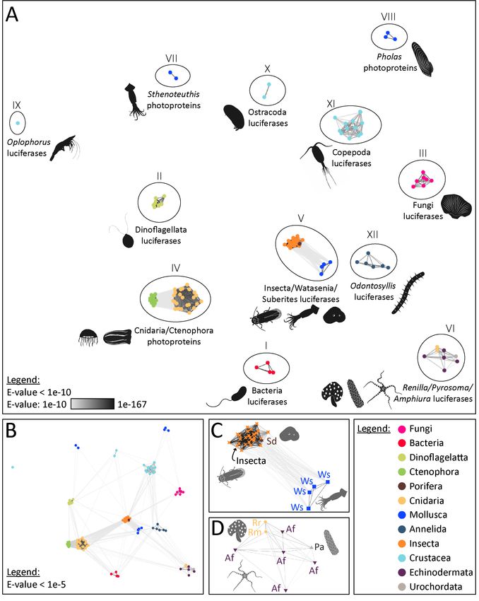
Our analyses highlighted the presence of 12 distinct types of bioluminescent proteins, defined
as clusters of homologous bioluminescent proteins based on an E-value threshold of 1e-10 (Fig. 1). In
the following sections, we will discuss each of these photoprotein/luciferase clusters from an
evolutionary perspective.Preprints (www.preprints.org) | NOT PEER-REVIEWED | Posted: 4 March 2021 doi:10.20944/preprints202103.0144.v1
Figure 1. Visualisation of the similarity of known bioluminescent proteins. (A) Sequence-similarity-based
clustering approach based on BLASTp e-values (CLANS software) using known luciferases and photoproteins
(E-value threshold of 1e-10). (B) Same analysis with an E-value threshold of 1e-.5 (C) Focus on the Insecta-
type luciferase cluster (Group V) that also contains luciferases of Watasenia scintillans and Suberites
domuncula (E-value threshold of 1e-10). (D) Focus on the Renilla-type luciferase cluster (Group VI) containing
Pyrosoma atlanticum and Amphiura filiformis candidate luciferases (E-value threshold of 1e-10). The names
and accession numbers of the sequences are referenced in the Supplementary Table S1.Preprints (www.preprints.org) | NOT PEER-REVIEWED | Posted: 4 March 2021 doi:10.20944/preprints202103.0144.v1
Figure 2. Molecular domains of known bioluminescent proteins. Domain detected within the 12 types of
luciferase/photoprotein types using Hidden Markov Model of Simple Modular Architecture Research Tool
SMART. For each cluster of luciferases/photoproteins, all available sequences were tested. Only one
representative sequence is shown for each cluster. Bacteria-type luciferase: Vibrio fischeri (Q6VFQ4),
Dinoflagellata-type luciferase: Pyrocystis lunula (AAL40677), Fungi-type luciferase: Panellus stipticus
(BBH43512), Aequora-type photoprotein: Mnemiopsis leidyi (AFK83786), Insecta-type luciferase:
Arachnocampa luminosa (AXM90651), Renilla-type luciferase: Renilla reniformis (CAA01908),
Sthenoteuthis-type photoprotein: Sthenoteuthis oulalaniensis (BAH89068), Pholas-type photoprotein:
Pholas dactylus (CAA10292), Oplophorus-type photoprotein: Oplophorus gracilirostris (BAB13776),
Ostracoda-type luciferase: Vargula hilgendorfii (AAB86460), Copepoda-type luciferase: Metridia pacifica
(BAY00656), Odontosyllis-type luciferase: Odontosyllis octodentata (BBG43629). Signal peptides (in red)
were specifically detected in all known secreted luciferases.
Supplementary Table S1. List of known bioluminescent systems based on biochemical and molecular
data (including the sequence references used for the CLANS analysis presented in Figure 1).
Supplementary File S2. References used to generate the list of known bioluminescent systems based
on biochemical and molecular data (Supplementary Table S1).Preprints (www.preprints.org) | NOT PEER-REVIEWED | Posted: 4 March 2021 doi:10.20944/preprints202103.0144.v1
Bacteria luciferases (Group I), the most ancient type of bioluminescent proteins shared by all
luminous bacteria
At least 28 luminous bacteria species are known, and they are all distributed into seven genera
belonging to three families of Gammaproteobacteria (e.g., Vibrio harvey, Photobacterium sp) (Tanet
et al. 2020). Symbiotic associations with luminescent bacteria have also been described in teleost fish
and squids (Dunlap and Kita-Tsukamoto, 2006) and suggested in other organisms (e.g., Taylor et al.,
1983; Mackie and Bone, 1978; Duchatelet et al., 2020 and Tessler et al., 2020 recently challenged these
hypotheses, respectively). Multiple bacteria species have been identified in bioluminescent symbiotic
associations: Aliivibrio fischeri, A. logei, Photobacterium leiognathi, P. phosphoreum, P. kishitanii, P.
mandapamemsis, Candidatus Enterovibrio luxaltus, Candidatus E. escacola, Candidatus Photodesmus
katoptron, Candidatus P. blepharon… (Boettcher and Ruby, 1990; Dunlap and Kita-Tsukamoto, 2006;
Ast et al., 2007; Dunlap et al., 2007; Kaeding et al., 2007; Hendry et al., 2014, 2018; Freed et al., 2019)
and studies suggested that unidentified species could also be involved (Haygood and Distel, 1993). In
all described bacterial luminescence cases, the luciferin is a reduced riboflavin phosphate (FMNH2)
which is oxidised in association with a long-chain aldehyde, oxygen, and a luciferase. The bacterial
luciferase is described as a flavin-dependent monooxygenase and is composed of two different but
homologous subunits: the α subunit is the catalytic core, and the β subunit is crucially required for
maintaining the catalytic function of the α subunit.
Up to now, all investigated bacteria are sharing a common type of luciferase (observed as the
“Bacteria-type luciferases (Group I)” cluster in our meta-analyses, see Fig. 1). It is interesting to note
that similar bacterial luciferase-like encoding gene has been detected in Archaea (identity superior to
45%) and Fungi (identity superior to 35%), although more functional information is missing for these
groups. Also, no homologous sequences have been found in metazoans (J.D. personal observations).
Dinoflagellata luciferases (Group II), a common type of luciferase for all Dinoflagellates with unclear
evolutionary origins
Dinoflagellates are the most commonly encountered luminescent organisms in coastal environments,
and at least 29 luminous species have been discovered (Sweeney 2012). Marine bioluminescence of
dinoflagellates is stimulated by hydrodynamic turbulence generated by predators or waves (Latz et al.,
1994, 2004; Rohr et al., 1995; Latz and Rohr, 1999, 2005).
Dinoflagellate luciferin is thought to be derived from chlorophyll and has a very similar
structure (Dunlap et al., 1981; Topalov and Kishi, 2001). A modified form of this luciferin is also foundPreprints (www.preprints.org) | NOT PEER-REVIEWED | Posted: 4 March 2021 doi:10.20944/preprints202103.0144.v1
in herbivorous euphausiid shrimps, indicating a probable dietary link for the luciferin acquisition
(Shimomura, 1980, 1995).
The dinoflagellate luciferase contains three homologous domains, and each domain is known
to be enzymatically active and to participate in the bioluminescence reaction (Liming et al., 1997). The
crystal structure of one of the domains (D3) in its inactive form was solved by Schultz et al. (Schultz et
al., 2005). All luminous dinoflagellates investigated until now are sharing a homologous luciferase type
(Dinoflagellata-type luciferase cluster (Group II) in Fig. 1).
Evolutionary origin of dinoflagellate luciferase remains elusive, and no exact homologous
sequences have been detected in non-dinoflagellate organisms however a structural similarity has
been found with fatty-acid-binding proteins (FABPs) (lipocalin family) present in metazoans (Schultz et
al., 2005).
Fungi luciferases (Group III), a common type of luciferase for all Fungi
Approximately 100 fungi species from the order Agaricales emit light using a standard luciferase-
luciferin system (Oliveira et al., 2012). Although fungal bioluminescence’s ecological role is not fully
understood, there is evidence that it might be used to attract spore-dispersing insects (Oliveira et al.,
2015).
Fungal bioluminescence was known to utilise molecular oxygen, a specific reduced luciferin,
which was recently identified as 3-hydroxyhispidin (a product of oxidation of the simple plant and
fungal metabolite hispidin (Purtov et al., 2015)) and a luciferase (Airth and McElroy, 1959, Oliveira et
al., 2009).
Our analyses confirm that all investigated luminous Fungi share a common luciferase type
(Group III, Fig. 1). Unlike all other described bioluminescent proteins, Fungi luciferases are
characterised by the presence of a transmembrane domain (Fig. 2).
Ctenophora and Medusozoa photoproteins (Group IV), the first described photoprotein-type
Bioluminescence is well represented in ctenophores and cnidarians. More than 90 % of known
planktonic genera of ctenophores can produce light while no luminous benthic species have been
identified (Haddock and Case, 1995). In cnidarians, bioluminescence is found in both benthic and
planktonic species. Luminous hydrozoans include both hydromedusae (e.g., the species Aequorea
Victoria in which GFP was discovered, Prasher et al. 1985, 1992; Shimomura 2005) and siphonophores
(e.g., 91% of known planktonic siphonophore genera are luminous) (Haddock et al., 2010). Two orders
of scyphozoans contain luminous members: the Coronatae (e.g., Atolla sp., Periphylla sp.) and thePreprints (www.preprints.org) | NOT PEER-REVIEWED | Posted: 4 March 2021 doi:10.20944/preprints202103.0144.v1
Semaeostomeae (e.g., Pelagia noctiluca, Phacellophora sp., Poralia sp.) (Haddock & Case 1999,
Haddock et al., 2010).
All investigated ctenophores and cnidarians use coelenterazine as their light-emitting
substrate. All investigated ctenophores and most investigated medusozoans (Cnidaria) species use
calcium-activated proteins. The specific case of Periphylla sp, however, will be discussed in the section
“Other groups of luciferases or photoproteins” as the species has been shown to use a luciferase
system. In addition, the case of the anthozoans appears to be different, as well, and will be tackled in
the section “The Renilla-type luciferase (Group VI)” as the octocorals Renilla sp., and most probably
other sea pansies and sea pens (Bessho-Uehara et al. 2020), are using a different and non-homologous
luciferase system.
Ctenophore photoproteins share around 20-25% sequence identity and around 40-45%
sequence similarity with known hydromedusan cnidarian photoproteins. Mnemiopsis photoproteins
share 85% to 91% sequence identity with other ctenophore photoproteins (i.e., Beroe and Bolinopsis).
Within all hydromedusan photoproteins, there is around 60% to 94% sequence identity (Schnitzler et
al. 2012).
Ctenophoran and cnidarian photoproteins are functionally related to coelenterazine-binding
proteins from Renilla, sarcoplasmic calcium-binding protein from the marine worm Nereis diversicolor
and calmodulin proteins (Schnitzler et al., 2012).
Schnitzler et al. proposed a metazoan-wide phylogeny for the “Aequora-type photoprotein”
gene family. They identified photoprotein-like genes in non-luminescent taxa (i.e., the poriferan
Amphimedon and the cnidarian Nematostella), and demonstrated that the gene family likely arose at
the base of the Metazoa (Schnitzler et al. 2012). Calcium-binding photoprotein genes (i.e., coding for
a functional photoprotein) may have evolved independently from a homologous gene found in
ctenophores, cnidarians, and non-luminous sponges (Prasher, McCann and Cormier 1985, Tsuji et al.
1995, Schnitzler et al. 2012). The emergence of photoproteins in cnidarians and ctenophores could
then appear as an example of parallel evolution of conserved and homologous genes.
Insecta luciferases (Group V), a luciferase type also found in the cephalopod Watasenia scintillans
and the sponge Suberites domuncula.
All luminous insects (i.e., Coleoptera with around 2300 luminous species (Li et al. 2021); Diptera with
a lower specific diversity but not yet exhaustively evaluated to the best of our best knowledge) share
a unique homologous luciferase-type, and the insect proto-luciferase is known to derive from Acyl-
CoA ligase enzymes that have another primary metabolic function (Viviani, 2002). These enzymes arePreprints (www.preprints.org) | NOT PEER-REVIEWED | Posted: 4 March 2021 doi:10.20944/preprints202103.0144.v1
members of the ANL superfamily of adenylating enzymes. This superfamily consists of various
enzymes, in addition to the Insecta-type luciferase, such as long-chain fatty acid Co-A ligases and
acetyl-CoA synthetases as well as other closely related synthetases and a plant auxin-responsive
promoter family. The name ANL derives from three subfamilies - Acyl-CoA synthetases, the NRPS
adenylation domains, and the Luciferase enzymes. Members of this superfamily catalyse the initial
adenylation of a carboxylate to form an acyl-AMP intermediate, followed by a second partial reaction,
most commonly forming a thioester (Gulick, 2009). While the homology between all insect luciferase
genes is apparent (Fig. 1), the evolutionary origin of bioluminescence in this group still appears very
complicated and recent studies supported independent emergences of bioluminescence and parallel
evolution of luciferases in fireflies, click beetles and Diptera (Fallon et al., 2018; Watkins et al., 2018).
Several authors suggested that the high abundance of ancestral gene duplications in this gene family,
and as a result the associated closely-related enzymatic activities, served as “raw materials for the
selection of new adaptive catalytic functions” (Weng, 2014, Fallon et al., 2018).
The insect luciferin-luciferase system uses the firefly-type luciferin (which should be called the
“insect”-type luciferin) and requires ATP as a cofactor.
Surprisingly, homologous luciferases to the Insecta-type luciferase were also described in the
marine squid Watasenia scintillans (Cephalopod, Mollusca) and the sponge Suberites domuncula
(Porifera) (Fig. 1). The firefly squid Watasenia scintillans emits intense blue bioluminescence from
photophores located at the tip of two of its arms. Within the photophore, luciferases are specifically
organised in microcrystals, and these proteins are catalysing the bioluminescent reaction using ATP
and the coelenterazine disulfate luciferin. Watasenia luciferases (i.e., several related proteins were
pinpointed: wsluc1–3) share around 20% sequence identity with firefly luciferases, which produce light
using ATP and a different substrate, the firefly luciferin (Gimenez et al., 2016). In W. scintillans, the
luciferase expression profile is precisely matching with the luminous patterns, but additional functional
studies would be necessary to confirm the bioactivity of the predicted luciferase.
Müller et al. suggested that an Insecta-type luciferase (acetyl-CoA synthetase) was involved in
the bioluminescence of the sponge Suberites domuncula (Müller et al., 2009; Wiens et al., 2010). The
authors showed that tissue extracts produce light that was detected using sensitive films in the dark.
Then, the luciferase protein was immunodetected within the tissue (and shown to be associated with
the spicules). Finally, the recombinant sponge luciferase produced in E. coli was shown to be bioactive.
Surprisingly, the marine sponge Suberites is then possibly using a firefly luciferase homolog but also
the firefly luciferin. Additional data would be required to confirm the presumed involvement of the
insect-type luciferase in ecologically relevant light emission in the poriferan Suberites domuncula. The
records of luminescence in Porifera are extremely limited and the clear status of intrinsic
bioluminescence in these organisms still needs to be confirmed. Martini et al., (2020) recentlyPreprints (www.preprints.org) | NOT PEER-REVIEWED | Posted: 4 March 2021 doi:10.20944/preprints202103.0144.v1
described the first reliable observation of bioluminescence in a deep-sea sponge paving the way to a
better understanding of bioluminescence in these organisms.
It appears clear from the above examples that enzyme of the ANL superfamily enzymes have
independently evolved in distant species to produce light using unrelated substrates (Gimenez et al.,
2016). It represents a striking example of parallel evolution
The Renilla-type luciferase (Group VI), a luciferase type also found in the brittle star Amphiura
filiformis and the tunicate Pyrosoma atlanticum
The luminescence of the sea pansy Renilla reniformis, a shallow-water soft coral (octocoral) that
displays blue-green bioluminescence upon mechanical stimulation, has been intensively studied since
the luciferase has been cloned and sequenced in 1991 (Lorenz et al., 1991). The Renilla luciferase
enzyme catalyses coelenterazine oxidation leading to bioluminescence. The Renilla-type luciferase
shows a characteristic alpha/betahydrolase fold (Marchler-Bauer et al., 2003). It is found to have a
high level of tertiary structure similarity and to be homologous to bacterial haloalkane dehalogenases
which are primarily hydrolase enzymes cleaving a carbon-halogen bond in halogenated compounds
(Hynkova et al., 1999, Loening et al., 2006). Horizontal gene transfers that are known to play critical
roles in the evolutionary acquisition of novel traits in eukaryotes (Boto, 2014), have been suspected to
explain the high similarity of the Renilla-type luciferase compared to bacterial haloalkane
dehalogenases (Loening et al., 2006; Delroisse et al., 2017).
Several other luminous anthozoans are found within the octocorals (Alcyonaria) in shallow
sandy bottoms (e.g., Ptilosarcus, Pennatula), and in the deep sea (e.g., Stylatula, Halipterus,
Anthomastus). A recent work performed on diverse sea pen species strongly suggests the use of a
conserved luciferase throughout the lineage (families Isididae, Alcyoniidae, Umbellulidae,
Funiculinidae, Kophobelemnidae and Protoptilidae) (Bessho-Uehara et al., 2020). In parallel,
bioluminescent species have also been found in hexacorals (e.g., bamboo corals), but their
bioluminescence’s biochemistry remains unexplored.
The Renilla-type luciferase was recently identified in the brittle star Amphiura filiformis
(Echinodermata, Ophiuroidea). In this species, which emits a blue luminescence at the level of the arm
spines (Delroisse et al., 2017a), coelenterazine is the luciferin and is acquired via a dietary pathway
(Mallefet et al., 2020). The predicted A. filiformis luciferase, highly similar to the Renilla luciferase (up
to 47% of identity, up to 69% of similarity) (Fig. 1), would constitute the unique example of luciferase
described so far in echinoderms. Amphiura luciferase has been detected specifically in the animal’s
spine photocytes, which constitutes a strong indication of its photogenesis implication (Delroisse et
al., 2014, 2017a,b). However, given the expression of Renilla luciferase-like proteins in non-luminousPreprints (www.preprints.org) | NOT PEER-REVIEWED | Posted: 4 March 2021 doi:10.20944/preprints202103.0144.v1
echinoderms, this hypothesis must be confirmed by the recombinant expression of the A. filiformis
protein sequence to verify its luciferase activity.
It has been suggested that the haloalkane-dehalogenase function constitutes the metazoan
ancestral state, which shifted to luciferase in cnidarians (lineage of Renilla) and brittle stars (lineage of
A. filiformis) (Loening et al. 2006; Delroisse et al. 2017). Haloalkane dehalogenases were presumably
co-opted in luciferases in these two specific lineages. In A. filiformis, the apparent late duplications of
luciferase-like genes could suggest both functions’ co-occurrence. Renilla sp and A. filiformis would
then possess a similar and homologous luciferase to catalyse the photogenous reaction. Delroisse et
al. (2017) hypothesised that a co-emergence happened between these two luminous systems using
the same compounds under similar environmental pressure. The ecological similarities between the
Renilla (R. mulleri, R. reniformis and potentially luminous sea-pens in general) and A. filiformis, such as
the benthic position on loose sediment and the suspension-feeding strategy, would presumably permit
to acquire coelenterazine from planktonic organisms from a “dietary way”. The predation pressure
would positively select the emergence of the bioluminescence function endowing these slow-moving
organisms with an efficient anti-predation strategy.
Similar to what was observed in the brittle star A. filiformis, a Renilla-like luciferase has also
been found in the luminous tunicate Pyrosoma atlanticum (Fig. 1). Immunodetections of the luciferase
have been performed within the Pyrosoma tissues. In parallel, in vitro expression and functional testing
of the protein confirmed the enzyme’s bioactivity (Tessler et al., 2020). The hypothesis of the Renilla-
like luciferase involved in the bioluminescence of P. atlanticum has recently been questioned (Berger
et al. 2021).
The Sthenoteuthis-type photoprotein or symplectin (Group VII)
The flying squid Sthenoteuthis oualaniensis is characterised by a light organ on its mantle. The light
organ contains thousands of small granules, in which a photoprotein exists as the active form (Kuse,
2014; for review). The species emits light using the oxidation of the dehydro-coelenterazine luciferin
substrate by the so-called symplectin photoprotein enzyme (Fujii et al., 2002). The 60-kDa symplectin
photoprotein was extracted and characterised (Fujii et al., 2002).
Sequence analyses revealed no sequence similarity to known bioluminescent proteins (Fig. 1)
but the significant similarity to the carbon-nitrogen hydrolase domain found in mammalian biotinidase
and vanin (pantetheinase) (Fujii et al., 2002).
Warren et al. recently explored the phylogenetic distribution of these enzymes, grouped in the
symplectin/pantetheinase protein family, in metazoans (Warren et al., 2017). These authors suggested
that symplectins may have multiple functions including hydrolase activity (Warren et al., 2017).Preprints (www.preprints.org) | NOT PEER-REVIEWED | Posted: 4 March 2021 doi:10.20944/preprints202103.0144.v1
The Pholas-type photoprotein, or pholasin (Group VIII), a system only described in the bivalve Pholas
dactylus (Bivalvia, Mollusca)
Pholas dactylus, the common glowing piddock, is a famous luminescent organism because Dubois, who
discovered the general luciferin-luciferase reaction back in the 19th century, was specifically working
on this species (Dubois, 1889). The biochemistry of the Pholas bioluminescence was studied in depth
in the seventies (Henry and Michelson, 1973; Michelson, 1978). The term Pholasin was initially
dedicated to the luciferin before Pholasin was confirmed to be a photoprotein (Henry and Michelson,
1973; Robert et al., 1987; Kuse, 2020).
Dunstan et al. (2000) cloned the gene coding for the Pholasin apoprotein. These authors
compared the amino acid sequence with known proteins present in the public databases and more
specifically with the sequences of other cloned bioluminescent proteins (available at that time). A small
region of similarity was found between the recombinant protein and the putative luciferin-binding
sites of Vargula luciferase and Renilla luciferin-binding protein. However, these sites are very small
and do not inform on the potential homology status of these proteins. Our analyses indicated that
Pholas photoproteins have no clear homology with other known bioluminescent proteins (Fig. 1).
The Oplophorus-type luciferase (Group IX), a system only confirmed in Oplophorus gracilirostris
(Decapoda, Pancrustacea)
This cluster only contains the luciferase of the deep-sea shrimp Oplophorus gracilirostris. O.
gracilirostris secretes a luminous blue cloud from the basal part of its antennae when disturbed
(Shimomura et al. 1978). Similar behaviours are observed in various luminescent decapod shrimps
including the genera Heterocarpus, Systellaspis and Acanthephyra (Harvey, 1952). However, the
involvement of an Oplophorus-type luciferase in the light emission has only been confirmed in O.
gracilirostris.
The Oplophorus luciferase catalyses the oxidation of coelenterazine. The enzyme consists of
two subunits (19kDa and 35 kDa), but the smaller subunit is the only one to have a catalytic activity
while the 35 kDa protein is thought to have a role in the stabilisation of the catalytic unit. The 19 KDa
protein of Oplophorus luciferase is the smallest known catalytic component having a luciferase
function (Inouye et al., 2000).
Oplophorus luciferase presents no homology with other known bioluminescent proteins.Preprints (www.preprints.org) | NOT PEER-REVIEWED | Posted: 4 March 2021 doi:10.20944/preprints202103.0144.v1
Ostracoda-type luciferase (Group X), a common type of luciferase for all Ostracods
Around 150 ostracod species from the family Cypridinidae (out of about 300 species) can produce light.
These organisms use bioluminescence for defence or to create courtship displays. All investigated
luminous ostracods use the same luciferin and homologous enzymes to produce light (Harvey, 1924).
Luminous ostracods synthesise their luciferin from the amino acids tryptophan, isoleucine, and
arginine. This luciferin, called Vargulin or Cypridina-type luciferin as it was initially found in the
ostracods Vargula and Cypridina, is also the one used by the midshipman fish Porichthys sp. A clear
dietary link has been established, and fish are losing their ability to luminesce until they are fed with
luciferin-containing food. The luminescent fish Parapriacanthus ransonneti obtains its luciferin but also
its luciferase enzyme from bioluminescent ostracod preys (Bessho-Uehara et al., 2020).
Previous research indicated that cypridinid luciferases evolved independently of other
luciferases. These enzymes have specific features such as a signal peptide leading to protein secretion
outside of the cell, two Von Willebrand Factor-D domains (VWD), multiple disulphide bonds between
conserved cysteines and post-translational N-linked glycosylation (Fig. 2; Hunt et al., 2017; Inouye &
Sahara, 2008; Nakajima et al., 2004; Oakley, 2005; Mitani et al., 2017; Yasuno et al., 2018).
Ostracoda-type luciferases have no homology with other known bioluminescent proteins (Fig.
1).
Copepoda-type luciferase (Group XI), a common type of luciferase for all Copepods
Some marine copepods emit a bright blue light using a classical luciferase-luciferin system based on
the coelenterazine substrate. It is a case of secreted bioluminescence, and the simple oxidation
reaction do not require any additional cofactors. Copepod luciferases are small secreted proteins of
around 18-24 kDa. The luciferases from the copepods Gaussia princeps and Metridia longa have been
cloned and used as bioluminescent reporters in various applications (Thouand and Robert, 2014).
Our analyses confirm that copepod luciferases do not share sequence or structural similarity
with other identified bioluminescent proteins, including other coelenterazine-dependent luciferases
(e.g., Oplophorus-type luciferase, Renilla-type luciferase) (Fig. 1).
The Odontosyllis-type luciferase (Group XII)
The Group XII cluster only contains the luciferase of the luminous annelid worms of the genus
Odontosyllis. The bioluminescent systems of O. enopla and O. octodentata have been partly
characterised. The luciferin of O. enopla has been partially purified, showing that light emission
requires the presence of magnesium, molecular oxygen, and crude luciferase. Its chemical structure,Preprints (www.preprints.org) | NOT PEER-REVIEWED | Posted: 4 March 2021 doi:10.20944/preprints202103.0144.v1
however, has not yet been determined (Shimomura et al. 1963; Trainor, 1979). Odontosyllis luciferases
have no homology with other known bioluminescent proteins (Fig. 1).
Interestingly, Deheyn and Latz proposed that a photoprotein may be involved in the
bioluminescence of the species O. phosphorea (Deheyn and Latz, 2009). If this assumption is
confirmed, it will imply the presence of two convergent bioluminescent protein types (a luciferase and
a photoprotein) in the genus Odontosyllis (Deheyn and Latz, 2009).
Other groups of luciferases or photoproteins
There are many luminous organisms in which, although no luciferase sequence is available, crucial
biochemical information indicate that the bioluminescent proteins involved do not correspond to any
of the 12 luciferase types described above. It suggests that different groups might be described in the
future.
Conversely to the other medusozoans (see section “Ctenophora and Medusozoa photoproteins
(Group IV), the first described photoprotein-type”), Periphylla periphylla has been depicted as using a
luciferase rather than a photoprotein for its light emission. Shimomura and Flood (1998) described two
types of luciferase catalysing the luminous reaction - i.e., luciferase-L (32 kDa) and luciferase-O (75
kDa) using coelenterazine as substrate-, occurring in the jellyfish marginal exumbrella photocytes and
eggs, respectively (Shimomura and Flood, 1998; Shimomura et al., 2001). The bioluminescent system
of Periphylla suggests the emergence of convergent bioluminescent protein in medusozoans, with at
least two different systems: the luciferases observed in Periphylla and the photoproteins observed in
several Medusozoa species.
In annelids, there is a wide diversity of chemical reactions and kinetics recognised among the
different luminous species (Aida and Gruber, 2017, for review). A 300-kDa heterotrimeric Cu2+
metalloprotein has been identified as the luciferase of the Megascolecidae earthworm Diplocardia
longa (Bellisario and Cormier, 1971; Bellisario et al., 1972; Ohtsuka et al., 1984; Oba et al., 2016) and
a 65-kDa polynoidin photoprotein is used in Polynoidae scale worms (Nicolas et al., 1982; Bassot, 1987;
Bassot and Nicolas, 1995; Martin and Plyuscheva, 2009). The chemistry of bioluminescence in
parchment tubeworms has been mainly studied in Chaetopterus variopedatus (Shimomura and
Johnson, 1966; Anctil, 1979; Martin and Anctil, 1984; Zinner, 1986; Shimomura, 2012; Branchini et al.,
2013; Deheyn et al., 2013; Rawat and Deheyn, 2016, Mirza et al.,2020). The luminous system includes
a photoprotein (Shimomura and Johnson, 1966; Shimomura, 2012). In the holopelagic Tomopteridae,
the bioluminescence system is thought to be a membrane-bound photoprotein tightly associated with
small particles (Shimomura, 2012; Francis et al., 2014). The independent emergence of multiple types
of bioluminescent enzymes is evident in the phylum Annelida (Verdes and Gruber, 2017).Preprints (www.preprints.org) | NOT PEER-REVIEWED | Posted: 4 March 2021 doi:10.20944/preprints202103.0144.v1
In echinoderms, despite the relatively common occurrence of luminous species, only two
ophiuroid species, Amphiura filiformis and Ophiopsila californica have been investigated
biochemically. The former luminesces with a luciferin-luciferase system stricto sensu (see above)
whereas the latter emits light with a photoprotein system (Shimomura, 1984, 1986, 2012; Mallefet et
al., 2013, 2020). A high diversity of physiological luminescence control mechanisms has been described
in these organisms. Species from the same genus (e.g., O. californica and O. aranea) can sometimes
exhibit different control mechanisms of the photogenous reaction (Mallefet, 2009 for review).
For a considerable number of studied bioluminescent organisms, the luminous system is
partially or totally unknown. For the majority of decapods or bony fishes, for example, only one part
of the system (i.e., the luciferin) is known and the luciferases or photoproteins remain unknown
(Shimomura, 2012; see Table S1). Some studies only show cross-reactivity between extracts of closely
related, or even phylogenetically distant species, to determine the type of luciferin used (Shimomura,
2012). Most often, no data on the luciferase or photoprotein sequence, activity, specificity is available
for species that are difficult to collect and fragile, such as those found in deep oceanic strata.
Therefore, efforts still need to be made to discover bioluminescent systems unknown to date. As a
further example, lanternfish, dragonfish and viperfish luminous systems are demonstrated to use
coelenterazine as luciferin, while no luciferase/photoprotein was determined to date (Tsuji and
Haneda, 1971; Mallefet and Shimomura, 1995; Duchatelet et al., 2019). Similarly, shark
bioluminescence system remains totally enigmatic, even if attempts were performed to decipher the
bioluminescent compound in the lanternshark Etmopterus spinax (Renwart and Mallefet, 2013). Cross-
reactivity with known luciferin failed to trigger light production, and preliminary search for luciferase
homologues within the available transcriptomic data did not yield any results suggesting the
involvement of an unknown bioluminescent system in luminous sharks (Renwart and Mallefet, 2013;
Delroisse et al., 2018; Delroisse et al., 2021).
Discussion
Bioluminescence evolution is often used as a striking illustration of convergent evolution in life
history. While it appears clear that many extant bioluminescent systems have evolved independently
on earth, the number of fully characterised bioluminescent proteins is still minimal, and the evolution
of these light-emitting luciferases and photoproteins remains mostly enigmatic. While the diversity of
luciferins involved in bioluminescent systems is rather well evaluated (Lau and Oakley, 2020, for
review), the diversity of bioluminescent proteins is, without a doubt, mostly under-evaluated.
It is essential to clarify that the present review does not illustrate the evolutionary history of
bioluminescence because it only focused on visualising the bioluminescence protein homology across
the tree of life. Knowing the luciferase evolutionary history is not enough to explain thePreprints (www.preprints.org) | NOT PEER-REVIEWED | Posted: 4 March 2021 doi:10.20944/preprints202103.0144.v1
bioluminescence’s evolutionary history. The reality may indeed be more complex, and other substrates
of bioluminescence likely possess different evolutionary histories from luciferases (as presented by
Fallon et al., 2008 in the case of luminous insects). As illustrated in Lau and Oakley, 2020,
understanding how bioluminescence emerged in living organisms requires the investigation of all
potential substrates of bioluminescence including luciferin biosynthetic pathways or dietary
acquisition pathways, luciferases, bioluminescence control, … While bioluminescence can be
convergent at one biological level, the convergence may not be found at other levels (Lau and Oakley,
2020).
Twelve distinct bioluminescent protein types are currently described
Multiple types of luciferases emerged convergently in the tree of life (Fig. 3). Based on the
currently available sequence data, our meta-analyses suggest that at least 12 non-homologous
bioluminescent protein types (i.e., three types of photoproteins, nine types of luciferases) appeared
independently during Evolution. Our analyses confirmed that luciferases/photoproteins appear
relatively lineage-specific (e.g., all described luminous bacteria share a common and homologous
luciferase type). To cite Lau and Oakley (2020), “most known bioluminescent proteins exhibit wide
molecular diversity and are not homologous across distantly related taxa, which suggest that most
origins of bioluminescent proteins are the result of convergent, but not parallel, evolution”. However,
our analyses also highlighted that, in several cases, a similar system – i.e., homologous enzymes – could
be used by phylogenetically distant organisms: Group IV photoproteins are shared by ctenophores and
medusozoans, Group V luciferases are shared by insects, the cephalopod Watasenia scintillans and,
putatively, the sponge Suberites domuncula; Group VI luciferases are shared by the sea pansy Renilla
sp, the tunicate Pyrosoma atlanticum and the brittle star Amphiura filiformis. While enzymes appear
to be homologous within all precited Groups (IV, V, VI), it also appears that they have been
independently co-opted into luciferases in these distant lineages. In short, in these examples of parallel
molecular evolution, the proteins are homologous, but their luciferase function is not. However, as
exemplified by Tyler (1988), homology should apply most appropriately to the structural features, not
their functions.Preprints (www.preprints.org) | NOT PEER-REVIEWED | Posted: 4 March 2021 doi:10.20944/preprints202103.0144.v1
Figure 3. Bioluminescence tree of life annotated with the corresponding bioluminescent protein Group.
Distribution of terrestrial and aquatic organisms is based on Lau et al. 2020 and Haddock et al. 2006.
Phylogenetic tree based on Giribet and Edgecombe, 2020.
Bioluminescent proteins could be bifunctional enzymes
The evolution of bioluminescence in insects is thought to have emerged from the activity of ancestral
fatty acyl-CoA synthetase (ACS) enzymes present in all insects. Beetle luciferases share high sequence
identity with these enzymes and often retain ACS activity. Besides, some ACS enzymes from non-
luminous insects can catalyse bioluminescence from synthetic D-luciferin analogues (Adams et al.,
2020).Preprints (www.preprints.org) | NOT PEER-REVIEWED | Posted: 4 March 2021 doi:10.20944/preprints202103.0144.v1
The annelid polynoidin is also present in non-luminescent scale worms suggesting that
bioluminescence might have originated from a non-related mechanism (in this case: quenching of
superoxide radicals) (Martin and Plyuscheva, 2009).
The case of Renilla-type luciferase, characterising the brittle star A. filiformis, was
investigated in detail based on genomic and transcriptomic data. While the Renilla-type luciferase
appears to be specifically expressed in the photocytes in the luminous brittle star, it was also
highlighted the that similar Renilla-type luciferases are present in non-luminous echinoderms,
raising exciting questions on the evolution of bioluminescence in echinoderms. In the sea-urchin
Strongylocentrotus purpuratus, a Renilla-type luciferase protein (DspA) was recently studied and
identified as the first biochemically characterised haloalkane dehalogenase of non-microbial origin
(Fortova et al., 2013). Nagata et al. (1999) noted homology between the luciferase from R. reniformis
and some microbial hydrolases catalysing the removal of halogens from aliphatic hydrocarbons, the
so-called haloalkane dehalogenases. Both enzymes share the conserved catalytic triad of residues.
Microbial haloalkane dehalogenase (Shingomonas sp) shares high sequence identity (42%) and
similarity (62%) with the sequence of Renilla luciferase. This similarity is somewhat surprising
considering that haloalkane dehalogenases are hydrolases and Renilla luciferase is an oxygenase. It
seems therefore that the “luciferase-like” proteins could have kept the original microbial function of
haloalkane dehalogenases, at least, in sea urchins. Fortova et al. (2013) and Delroisse et al. (2017) also
reported the absence of light emission after coelenterazine addition indicating the absence of a
luciferase function for this enzyme in the sea-urchin S. purpuratus and in the sea-star Asterias rubens,
respectively.
Symplectins, from the cephalopod Sthenoteuthis that are derived from pantetheinase
enzymes, also contain active site residues involved in pantetheinase catalysis suggesting that these
photoproteins may have multiple functions including hydrolase activity (Warren et al., 2017).
These examples of functional shift indicate that luciferases did not necessarily derive from
ancestral oxygenases and that luciferase may retain the ancestral function and be bifunctional in some
cases. Outside of the bioluminescence field, this observation has already been reported for oxygenases
(Chen et al., 2005). Cooptions of genes non-related to monooxygenases into luciferases are predicted
for the Groups IV (Ctenophora/Cnidaria photoproteins), V (Insecta/Watasenia/Suberites luciferases)
and VI (Renilla/Pyrosoma/Amphiura luciferases) luciferases that emerged from calmodulin, acyl-CoA
ligase and bacterial haloalkane dehalogenase enzymes, respectively. However, determining the
ancestral function of a protein is difficult, and these enzymes might have been functioning as
oxygenases long before the emergence of their non-oxygenase activity...Preprints (www.preprints.org) | NOT PEER-REVIEWED | Posted: 4 March 2021 doi:10.20944/preprints202103.0144.v1
Conclusion
Evolution (and natural selection, in particular) often promotes evolutionary innovation by co-opting
preexisting genes for new functions, and gene duplication is known to facilitate this process (Hoffmann
et al., 2010). Here we emphasise that multiple bioluminescent proteins potentially appeared during
evolution by the independent emergences of new genes or by the cooption of existing genes with an
ancestral function unrelated to bioluminescence (i.e., convergent evolution). In this latter case,
cooption might have occurred independently across the tree of life (i.e., parallel evolution) leading to
homologous light-emitting systems in non-related luminous organisms. As already suggested, our
findings suggest that co-option may be an underappreciated process underpinning protein
neofunctionalisation” (Casewell, 2017).
“This example of convergent evolution of protein function provides an impressive demonstration of
the ability of natural selection to cobble together complex design solutions by tinkering with different
variations of the same basic protein scaffold” (Hoffmann et al., 2010).
Author contributions
J.D. performed analyses and wrote the first draft of the manuscript. L.D. participated in the data
collection from the literature. All authors participated in discussions and revised the final manuscript.
P.F. and J.M. supervised the work.
Acknowledgements
This study is a contribution from the ‘Centre Interuniversitaire de Biologie Marine’ (CIBIM). J.D., J.M.
and P.F. are, respectively, postdoctoral fellow, Research Associate, and Research Director of the Fund
for Scientific Research of Belgium (F.R.S-FNRS). L.D. is postdoctoral researcher at the University of
Louvain. This work is supported by the F.R.S.-FNRS PDR project “Glow & See” (T.0169.20) awarded to
the University of Louvain (Marine Biology Laboratory) and the University of Mons (Biology of Marine
Organisms and Biomimetics Laboratory).Preprints (www.preprints.org) | NOT PEER-REVIEWED | Posted: 4 March 2021 doi:10.20944/preprints202103.0144.v1
References
Adams Jr, S. T., & Miller, S. C. (2020). Enzymatic promiscuity and the evolution of bioluminescence.
The FEBS journal. 287(7), 1369-1380.
Airth, R. L. and McElroy, W. D. (1959). Light emission from extracts of luminous fungi. J. Bacteriol.
77, 249–250.
Ast, J.C., Cleenwerck, I., Engelbeen, K., Urbanczyk, H., Thompson, F.L., De Vos, P., Dunlap, P.V.
(2007). Photobacterium kishitanii sp. nov., a luminous marine bacterium symbiotic with deep-sea
fishes. Int. J. Syst. Evol. Microbiol., 57(9), 2073-2078. https://doi.org/10.1099/ijs.0.65153-0
Berger, A., Blackwelder, P., Frank, T., Sutton, T. T., Pruzinsky, N. M., Slayden, N., & Lopez, J. V.
(2021). Microscopic and Genetic Characterization of Bacterial Symbionts With Bioluminescent
Potential in Pyrosoma atlanticum. Front. Mar. Sci. 8: 606818.
https://doi.org/10.3389/fmars.2021.606818
Bessho-Uehara, M., Francis, W. R. and Haddock, S. H. D. (2020). Biochemical characterization of
diverse deep-sea anthozoan bioluminescence systems. Mar. Biol. 167, 114.
https://doi.org/10.1007/s00227-020-03706-w
Bessho-Uehara, M., Yamamoto, N., Shigenobu, S., Mori, H., Kuwata, K. and Oba, Y. (2020).
Kleptoprotein bioluminescence: Parapriacanthus fish obtain luciferase from ostracod prey. Sci. Adv.
6(2), eaax4942. https://doi.org/10.1126/sciadv.aax4942
Boettcher, K.J. and Ruby, E.G. (1990). Depressed light emission by symbiotic Vibrio fischeri of the
sepiolid squid Euprymna scolopes. J. Bacteriol., 172(7), 3701-3706.
https://doi.org/10.1128/jb.172.7.3701-3706.1990
Boto, L. (2014). Horizontal gene transfer in the acquisition of novel traits by metazoans. Proc. R.
Soc. B, Biol. Sci. 281(1777), 20132450. https://doi.org/10.1098/rspb.2013.2450
Casewell, N. R. (2017). Evolution: Gene co-option underpins venom protein evolution. Current
Biology, 27(13), R647-R649.
Chen, Y. H., Wang, C. C., Greenwell, L., Rix, U., Hoffmeister, D., Vining, L. C., ... & Yang, K. Q. (2005).
Functional analyses of oxygenases in jadomycin biosynthesis and identification of JadH as a
bifunctional oxygenase/dehydrase. Journal of Biological Chemistry. 280(23), 22508-22514.
https://doi.org/10.1074/jbc.M414229200
Claes, J. M. and Mallefet, J. (2009). Bioluminescence of sharks: First synthesis. In: Meyer Rochow,
V. (Ed). Bioluminescence in Focus – A Collection of Illuminating Essays, pp. 51–65. Research Signpost,
Kerala, India.
Davis, M. P., Sparks, J. S. and Smith, W. L. (2016). Repeated and widespread evolution of
bioluminescence in marine fishes. PLoS One. 11, e0155154.
https://doi.org/10.1371/journal.pone.0155154
Deheyn, DD, Latz, MI. 2009. Internal and secreted bioluminescence of the marine polychaete
Odontosyllis phosphorea (Syllidae). Invertebr Biol. 128:31–45.
Delroisse, J., Duchatelet, L., Flammang, P., & Mallefet, J. (2018). De novo transcriptome analyses
provide insights into opsin-based photoreception in the lanternshark Etmopterus spinax. PLoS one,
13(12), e0209767. https://doi.org/10.1371/journal.pone.0209767
Delroisse, J., Duchatelet, L., Flammang, P., & Mallefet, J. (submitted in Frontiers in Marine Science,
Marine Megafauna, in revision). Photophore distribution and enzymatic diversity within the
photogenic integument of the cookie cutter shark Isistius brasiliensis (Chondrichthyes: Dalatiidae).You can also read