Modifying an Implant: A Mini-review of Dental Implant Biomaterials - Bio Integration
←
→
Page content transcription
If your browser does not render page correctly, please read the page content below
Mini Review
Modifying an Implant: A Mini-review
of Dental Implant Biomaterials
Oliver K. Semisch-Dieter1, Andy H. Choi1 and Martin P. Stewart1,*
1School of Life Sciences,
Abstract
Faculty of Science, University
Dental implants have been used as far back as 2000BC, and since then have developed into highly sophisti-
of Technology Sydney, Ultimo,
cated solutions for tooth replacement. It is becoming increasingly important for the materials used in dental
implants to exhibit and maintain favorable long-term mechanical, biological and more recently, aesthetic NSW, Australia
properties. This review aims to assess the biomaterials used in modern dental implants, introducing their
properties, and concentrating on modifications to improve these biomaterials. Focus is drawn to the promi-
nent biomaterials, titanium (Ti) and zirconia due to their prevalence in implant dentistry. Additionally, novel
coatings and materials with potential use as viable improvements or alternatives are reviewed. An effective *Correspondence to:
dental biomaterial should osseointegrate, maintain structural integrity, resist corrosion and infection, and not Martin P. Stewart
cause systemic toxicity or cytotoxicity. Current materials such as bioactive glass offer protection against bio- E-mail: martin.stewart@uts.
film formation, and when combined with a titanium–zirconium (TiZr) alloy, provide a reliable combination edu.au
of properties to represent a competitive alternative. Further long-term clinical studies are needed to inform
the development of next-generation materials. Received: September 7 2020
Revised: December 7 2020
Keywords Accepted: January 4 2021
Biocompatibility, biomaterials, dental implant, titanium, zirconia. Published Online: March 19
2021
Significance Statement Available at:
Biomaterials have become essential for modern implants. A suitable implant biomaterial integrates into the https://bio-integration.org/
body to perform a key function, whilst minimizing negative immune response. Focusing on dentistry, the use
of dental implants for tooth replacement requires a balance between bodily response, mechanical structure
and performance, and aesthetics. This mini-review addresses the use of biomaterials in dental implants with
significant comparisons drawn between Ti and zirconia. Attention is drawn to optimizing surface modifi-
cation processes and the additional use of coatings. Alternatives and novel developments are addressed,
providing potential implications of combining biomaterials to form novel composites that combine and
synergize the benefits of each material.
Introduction situation, and optimizing the clinically rel-
evant performance of that therapy” [2].
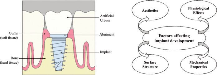
Biomaterials are crucial for modern med- The application of biomaterials as a
ical applications, and in the last several dental implant is shown in Figure 1A; an
decades, these materials have been refined endosseous fixture is used to replace the
and improved for applications seen in root and support a dental prosthesis. Dental
dentistry, orthopedics, drug delivery, and implants have become an evidence-based
cardiology. Candidate materials include treatment for the replacement of missing
polymers, metals (and their alloys), cera- teeth. Selecting a biomaterial for a den-
mics, composites (e.g. carbon fiber), and tal implant requires additional considera-
glass. For clinical use, the material must tions for biological, mechanical, and aes-
not cause any adverse or damaging effects thetic properties. Modern dental implants
to the patient [1]. This concept is known need to feature effective osseointegration
as biocompatibility and was described by and maintain long-term stability of their
Williams as, “the ability of a biomaterial favorable properties to maintain both the
to perform its desired function with respect implant’s structure and the integrity of sur-
to a medical therapy, without eliciting any rounding hard and soft tissue [3, 4].
undesirable local or systemic effects in the This review aims to assess the current
recipient or beneficiary of that therapy, but state of biomaterials (Ti, zirconia, and
generating the most appropriate beneficial novel materials) used in dental implants.
cellular or tissue response to that specific Materials are assessed on their attributes,
12 BIOI 2021, Vol 2, No. 1, 12–21
https://bio-integration.org doi: 10.15212/bioi-2020-0034
© 2021 The Authors. Creative Commons Attribution 4.0 International LicenseMini Review
BIOI 2021
A B
Figure 1 (A) Illustrates a cross-sectional view of a dental implant and the surrounding oral tissue. (B) Selecting the appropriate material to
use for the implant requires considerations of implant durability, cell adhesion, and physiological implications for implant biocompatibility, and
aesthetic concerns for patient satisfaction.
characteristics, comparison of strengths and weaknesses, deterring biofilm formation. Current surface topographies
biocompatibility, and new advancements in addressing any include anodization, sandblasting, and polishing, however,
issues or concerns. conclusive evidence on which specific surface topographies
are optimal and why remains undetermined [9].
Titanium Alternative alloys
Ti is widely used for medical applications and is currently It is known that Ti can form alloys to solve reactivity concerns
the most commonly used material for dental implants. The [1]. Vanadium (V) and aluminum (Al) are common alloying
popularity is in part due to the combination of its tensile elements for Ti (seen in Ti6Al4V alloy). Individually each
strength, biocompatibility, ability to osseointegrate, density, element has shown potential adverse effects in high concen-
corrosion resistance, and inert properties. Pure Ti, while trations, causing carcinogenicity and neurotoxicity [10–12].
being safe for most clinical use, has been observed to form However, reports on V also demonstrate antidiabetic effects
an accumulation of Ti ions around the implant site. While and resulted in reduced weight gain and gastrointestinal dis-
no material is entirely bioinert, such ions released from an comfort [1, 13]. Attempts are being made to replace these
implant that fails to osseointegrate may induce proinflam- alloying elements with less toxic elements such as niobium
matory responses from sensitization. Such failure occurs (Nb), tantalum (Ta), and palladium (Pd), to improve biocom-
when there is a lack of biomechanical stability due to inad- patibility; however, Ti6Al4V remains a common choice [14].
equate osseointegration. Stability can be promoted by mod- When comparing a Ti alloy to surgical stainless steel and
ifying the implant’s physiochemical properties and surface cobalt alloys, the superior quality of Ti is its corrosion resist-
topography. Observations from failed Ti implants show an ance. The ability for Ti alloys to withstand corrosive environ-
increased lymph node Ti ion concentration by 7.4–9 times ments and develop an oxide layer with a shorter repassiva-
and lung Ti ion concentration by 2.2–3.8 times when com- tion time compared to other metal alloys, aids in preventing
pared to a successfully integrated implant [5, 6] (Table 1). a release of ions which can potentially be toxic (cobalt) or
Ti dental implants have been occasionally observed to result cause allergic reactions (nickel, found in stainless steel) [15].
in an allergic reaction. However, the exact mechanism for Ti A unique alloy, TiZr has shown clinical success in the past
ion release and systemic effects remains unclear [7]. decade. Under the trade name Roxolid, Institut Straumann
Surface modifications of Ti implants are performed to AG, Basel, Switzerland (a 15% Zr, 85% Ti composition), the
improve osseointegration and modulate cell adhesion. The TiZr alloy was designed for dental implants, becoming a via-
largest post-operative issue with such implants is biofilm-in- ble alternative to zirconia and other Ti-based implants [16].
duced mucositis, inflammation of mucous membranes, TiZr offers enhanced osseointegration compared to Ti6Al4V
which can further develop into peri-implantitis, the inflam- and pure Ti and avoids the use of toxic alloying elements.
mation of the gum and the surrounding bone structure. The Additionally, corrosion resistance can potentially reduce
first generation of surface modification was developed in problems associated with peripheral Ti ion concentrations
the 1960s, where machined Ti (smooth surface) was used [17–19]. A recent study observed that TiZr retains the ability
to promote osseointegration by leaving a small degree of for surface enhancement without compromising the mechan-
roughness. Second generation surface modifications have ical strength of the implant [20]. While TiZr dental implants
been widely available since the 1990s and improve on the do not offer the aesthetic qualities of zirconia, they demon
first generation by creating a microscopically rough surface strate highly preferable qualities among clinically available
topography. Finally, the current third generation (also known Ti alloy options.
as bioactive surfaces) maintains similar roughness to the sec- TiNb is another alloy with favorable properties and
ond generation and are modified chemically or topographi- provides a low elastic modulus similar to human cortical
cally to promote osseointegration [8]. Most surface topogra- bone. It is clinically significant as it reduces the issue of
phies minimize vertical deviation to 1 μm or less, effectively stress concentration between the implant and bone, thus
Oliver K. Semisch-Dieter et al.: Modifying an Implant in Dental Implant Biomaterials 13Mini Review
14
Table 1 Summary of in vivo and in vitro studies assessing clinically relevant factors of Ti and zirconia dental implants
Authors Study type Description Conclusion
Wang et al. (2019) In vitro Investigating macrophage and fibroblasts behavior at the Surface composition (Ti vs ZLA vs Ti-Zi) had little effect on macrophage or
[5] bone–implant interface of commercial implants fibroblast behavior, while hydrophilicity promoted lower inflammation and
better soft tissue integration
El-Hadad et al. In vitro and in Evaluating in vitro and in vivo performance of functionally FG Ti implants (Ti6Al4V) demonstrated the best in vitro cytocompatibility and
(2018) [24] vivo graded (FG) biomaterials of calcium phosphate/Ti alloys in vivo systemic toxicity and osseointegration results
Roehling et al. In vitro Comparing biofilm formation on zirconia and Ti implants Zirconia presented a significant reduction in biofilm and plaque formation
(2017) [45] compared to Ti implant surfaces
Siddiqui et al. In vitro Assessing the effect of bacterial and mechanical fatigue Mechanical loading and colonizing bacteria did not induce significant
(2019) [46] on zirconia implants degradation or damage. The authors consider zirconia a suitable alternative
to Ti implants
Langhoff et al. In vivo: sheep Comparing the osseointegration of modified Ti and All implants achieved successful osseointegration, with zirconia reaching
(2008) [47] model zirconia implant surfaces slightly higher initial bone-implant contact (BIC). Overall, there were no
significant differences in BIC
Schwarz et al. In vitro Assessing the effect of Ti and zirconia particles on Both Ti and zirconia particles reduced osteoblast and fibroblast cell viability,
(2019) [58] osteoblast and fibroblast activity, and cytokine expression with no proinflammatory effect. The findings do require in vivo validation
Sánchez et al. In vitro Assessing biofilm formation on hydroxyapatite, Ti and Biofilm formed irrespective of implant material, while only the morphology
(2014) [77] zirconia implant surfaces and structure of the biofilms were different
Rosentritt et al. In vitro Investigating fracture resistance and implant failure Bonded/screwed zirconia implants displayed lower fracture resistance
(2014) [78] compared to two-piece Ti and one-piece zirconia implants
Rottmar et al. In vitro Investigating the blood interaction, and resulting Thrombogenicity was the highest on zirconia surfaces compared to Ti. It
(2019) [79] osseointegration, at the bone-implant interface of was suggested that a higher thrombogenicity promotes osseointegration,
microstructure Ti and zirconia implants. however, similarly micro-structured Ti maintained good osseointegration
potential
Zeller et al. (2020) In vivo Investigation of biofilm formation on gold and silver metal PEEK, Zirconia and TiZr materials demonstrated similar biofilm formation
[80] alloys, PEEK, zirconia, and TiZr alloy implants. on commercially available implants. Only gold and silver-based materials
showed reduced biofilm growth
Herrmann et al. In vivo Examining early and mature biofilms on Ti and zirconia Zirconia showed significantly higher total bacterial cell counts, but could be
(2020) [81] implants at different roughness. due to surface roughness. Biofilm formation and total bacterial cell counts
could be assumed to be caused by surface roughness
Kubasiewicz-Ross In vivo: pig Studying the osseointegration of commercially available Overall zirconia displayed similar levels of osseointegration, measured
et al. (2018) [82] model Ti and zirconia implants through BIC. Differences between implant material yielded no statistically
important differences
Oliver K. Semisch-Dieter et al.: Modifying an Implant in Dental Implant Biomaterials
BIOI 2021Mini Review
BIOI 2021
promoting osseointegration. This alloy improves on other [29]. Long-term studies are required to translate this techno-
functional aspects compared to pure Ti, possessing higher logy for clinical application, and cost will also need to be
toughness, hardness, and corrosion resistance. TiNb has considered for commercial implementation.
been proposed as a suitable alternative for replacing failed Continued research in coatings has led to novel improve-
hard tissue [21, 22]. ments on hydroxyapatite (HAp) for Ti6Al4V implants. In
situ synthesis of HAp-based nanocomposites including chi-
tosan and polycaprolactone/fluoride (PCL/F) substitution
Recent trends aim to utilize the advantages of HAp with promoting the
formation of bone-like apatite at the bone–implant interface,
Recent trends in the field point toward the utility of novel while minimizing the brittleness of HAp. A sol–gel method
ideas such as: (1) the development of implant coatings, and and alkali-treated Ti is used to provide a homogenous, crack-
(2) the incorporation of alternative elements (e.g. iodine (I) free coating that does not suffer from any delamination. Both
and strontium (Sr)) to support Ti implants. The next gen- nanocomposites required a minimum 10 wt.% of HAp to
eration of therapeutic implants may incorporate nano-en- provide the desired results with the respective composite [30,
gineered surface topographies. The use of anodized titania 31]. Research for chitosan/HAp identified improved cellular
(titanium dioxide, TiO2) nanotubes (TNT) on the surface response at lower HAp content (10 wt.%), with higher con-
of Ti implants shows promising results. These NTs offer tent (50 and 60 wt.%) providing improved surface adhesion
advantages in topography, mechano-transduction, drug strength, roughness, and hydrophilicity [30]. It was noted
release, and mediating toxicity. The TNTs mimic the topo- with PCL/F that HAp content up to 30 wt.% improved sim-
graphical scale of surrounding native tissues to significantly ilar surface properties, and higher corrosion resistance [31].
upregulate protein absorption. The bone remodeling caused Both coatings meet the required performance of bioactive
by mechanical strain stimulates cells at the bone–implant coatings and show potential for future clinical application.
interface to promote initial attachment and continued adhe- An alternative material to Ti is Ta. Ta is a useful bio-
sion. TNTs that are capable of local drug release potentially material due to being highly bioactive and holds similar
allow for direct targeting of the implant site and a reduction properties to Ti regarding corrosion resistance and biocom-
in toxicity compared to systemic administration. Together patibility. The improvement over Ti is the optimization of
these factors contribute to influence cellular functions and the osseointegration process. However, it is a high-density
immunomodulatory responses towards dental implants [23, material which is expensive to manufacture. Therefore, com-
24]. Compared to conventional Ti implants, TNTs have been bining the properties of Ti and Ta, research has focused on
observed to orchestrate osteogenesis [25]. developing Ta oxide films. Preliminary research observed
A recent development in surface modifications is plasma promising results, improving wear resistance to microbial
immersion ion implantation which provides a uniform appli- and chemical degradation. Improved osseointegration was
cation over complex surface geometries, while reducing also observed with increased osteoblastic activity and mor-
overall cost and processing time. The in vitro testing of an phology. Finally, the study did not indicate any increased
oxygen plasma immersion ion implantation (OPIII) to a Ti bacterial cell adhesion when compared to conventional Ti
implant identified improvements in protein expression and implants [32].
corrosion resistance [26]. The use of OPIII is capable of Adding alternative elements is not a new concept, and
producing a bioactive oxide film which was determined to continuing with the idea of surface treatment, the addition
improve in vitro cell differentiation and in vivo osseointegra- of Sr is a possibility. Historically Sr is biologically known
tion. It is undetermined to what degree the process optimizes for promoting osteoblast activity and inhibiting osteo-
the implant’s surface properties (topography, wettability, and clast activity. Oral administration of Sr may cause adverse
roughness), however, the most current research has observed effects; therefore, surface incorporation (on Ti implants) has
no significant alteration [27]. In its current state, OPIII has been preliminarily observed to increase early bone osseoin-
a good potential to become a viable surface modification tegration [33]. Another potential element is I, an essential
through further research. element with antiseptic properties and may be a useful bio-
Additionally, an in vivo study demonstrated the ability to material for dental implants. A previous study indicated that
use macrophage polarization via NTs to augment osseoin- I-supported Ti implants in orthopedic applications substan-
tegration [28]. The property of local drug release is another tially reduced the incidence of inflammation. The enhanced
significant advantage of this technology. While some implants exhibited cytocompatibility and increased antimi-
attempts have been made to incorporate antibiotics into NTs, crobial activity [34].
these traditional or organic antibiotics pose the risk of resist- Another approach to modification is altering the implant
ance and are limited by selective toxicity. This has resulted design; the use of porous Ti has been researched to improve
in attention towards inorganic antibacterial agents, such as integration into hard tissue. The sponge-like framework also
silver. Using silver as a coating has shown strong antimi- allows for antibacterial agents to be directly incorporated
crobial effects, however, it poses biocompatibility issues. into the implant surface or via degradable drug carriers [35].
Studies are testing the effect of loading silver nanoparticles Currently challenges around optimization of the structure
onto TNTs. These TNTs use a reducing agent to disperse sil- and high manufacturing cost are a barrier to implementation
ver nanoparticles from the coating and can aid in patient care of this technology in the clinic [36].
during the initial postoperative risk of infection. Clinical A recent study has improved on the capability of porous
safety and molecular mechanism still require further studies Ti through using micropore channels in Ti implants to
Oliver K. Semisch-Dieter et al.: Modifying an Implant in Dental Implant Biomaterials 15Mini Review BIOI 2021
preserve alveolar height without additional growth factors. observe the role of oral temperature and pH fluctuation on
The canine in vivo model provides evidence in minimizing zirconia implants.
alveolar bone loss, while promoting cell viability, prolif- The success of an implant is reliant on the degree of
eration, and osteogenic differentiation [37]. Natural bone biomaterial osseointegration. Much research in surface
has periodontal ligament fibers which apply the required modifications has been conducted to enhance implant sta-
mechanical stimulation to maintain the correct height of bility and bone apposition, however few optimal alterations
alveolar bone. Traditional dental implants lack this function have been identified. The use of surface topography for Ti
due to a direct osseointegration at the bone–implant inter- plays a similar role in zirconia implants; however, pro-
face [38]. The horizontal traction of the micropores simu- ducing modified surfaces is more complicated than in Ti
lates the periodontal ligament fibers which provide a poten- due to a potential compromise of biomechanical stability.
tial clinical opportunity for higher quality dental implants. Furthermore, the current developments in surface modi-
Overall, Ti offers many unique opportunities to be alloyed fications for zirconia do not have clinical applications as
and altered. Thus, novel permutations are continually being seen in novel surface topographies of Ti dental implants.
invented and designed to improve the material for more Many clinicians remain skeptical of the osseointegrative
effective clinical use. properties of zirconia. Improvement of surface proper-
ties has taken two main approaches; (1) via optimization
of surface roughness, and (2) through the application of a
coating. Traditional surface modifications follow the first
Zirconia approach, which include air particle abrasion, acid etching,
and polishing. However, these methods introduce risk of
Dental implants made from the bioceramic zirconia (zir- surface and internal damage, possibly leading to fractur-
conium dioxide; ZrO2) are a non-metal alternative to Ti ing. An effective roughening process would make zirconia
implants that address aesthetic concerns. Widely available as implants more viable, as optimal roughness demonstrates
partially stabilized zirconia (PSZ), it provides a transforma- a similar capacity for osseointegration compared to rough-
tive toughening of the biomaterial [39]. Additionally, zirco- ened Ti implants [3, 49].
nia advantageously displays a significant reduction in plaque
affinity and proinflammatory responses in surrounding soft
tissue [40]. Recent trends in surface
Compared to Ti implants, similar or better results from modifications
zirconia have been observed in in vitro cell culture experi-
ments. These results however are not directly comparable, Following the second approach to surface modifications,
probably due to inconsistencies in influential variables HAp resembles the chemical composition of bone and teeth,
of cell type, incubation conditions, and surface proper- and has been suggested as a bioactive coating for zirconia
ties. Surface modifications for both implant materials implants. The advantage is stimulating osseointegration
are well characterized and contribute a significant effect through the supply of calcium and phosphate ions [50].
on the bone–implant interface. Nanoporous zirconia sur- Research has indicated that HAp-coated metallic implants
faces have indicated promise for improved cell growth as heal faster and attach more readily and completely to the
observed by larger cell size and higher cell count compared surrounding tissue [51]. While design of the coating’s prop-
to polished zirconia and surface modified Ti implants (acid erties would need to be optimized, transitioning this coat-
etched and sandblasted) [41, 42]. Assessing the biocom- ing to zirconia may also prove difficult, because of potential
patibility of zirconia, the cytotoxicity is comparable to bioactivity of calcium phosphate coatings [52–55]. A novel
Ti nanoparticles. High concentrations of zirconia nano- implementation of etching (selective infiltration via hydro-
particles inhibit osteogenic differentiation and may induce fluoric acid and a molten glass coating) has been found to
necrotic or apoptotic effects on tissue. Proinflammatory maintain surface chemistry and alter the roughness for effec-
effects for ziconia have not been reported. Research is thus tive osseointegration [56, 57]. Other novel surface modifi-
limited and requires further study to rule out potential bio- cations include biofunctionalization via an adhesive peptide
compatibility issues [43, 44]. (arginine–glycine–aspartate, fibronectin, or collagen), and
The most significant flaw of zirconia is an aging phe- laser treatment. Laser-treated zirconia exerted no surface
nomenon known as low-temperature degradation (LTD) contamination and promoted fibroblast attachment, an effect
which transforms the tetragonal surface back to the initial attributed to improved surface wettability [58, 59]. The
monoclinic structure in the presence of water or a humid immobilized peptide in biofunctionalization enhances the
environment [45, 46]. Zirconia surfaces have shown lower biocompatibility of the implant through extracellular matrix
bacterial adhesion compared to Ti; however, these bacteria regulation, as peptides such as fibronectin encourage cell
are still able to cause deterioration of zirconia surfaces [47]. adhesion and bone formation [60, 61]. While being effec-
Currently, no research has been able to adequately study tive for in vitro treatments, these modifications are clinically
the effect of oral bacteria on LTD. One study attempted limited by a lack of supporting research and the question of
to identify a correlative link between oral bacteria and longevity remains open [58].
mechanical fatigue; however, it was unable to arrive at A promising approach to improving bioactivity of zirco-
any conclusive evidence [48]. The majority of LTD comes nia without a substantial compromise of mechanical proper-
from manufacturing flaws. Further studies are required to ties is through a gradated design and composite outer layer.
16 Oliver K. Semisch-Dieter et al.: Modifying an Implant in Dental Implant BiomaterialsMini Review
BIOI 2021
While the calcium phosphate composite has contraindica-
tions in the literature, the gradated design aids to ensure no
Bioactive glass
coating detachment after implantation which otherwise may
The use of bioactive glass has long been considered for its
result in implant failure [62].
capability to bond to soft and hard tissue. It is commonly
Bioactive surface coatings, such as HAp, can result in
used as a reinforcement material in HAp-coatings on metal-
delamination of the coating from the implant after a long
lic implants to improve mechanical properties and osseoin-
loading time. Control of the coating thickness is a strategy
tegration [67]. A recent study has proposed the use of bio-
to mitigate this issue. Simple and repeatable methods of arc
active glass as a coating to reduce the incidence of bacterial
induction or plasma spraying are used to provide accurate
infections which result in implant failure. The bioglass was
thickness control and maintain the required chemical effect.
synthesized with up to 2% silver (in weight), to aid in anti-
Plasma anodization and fluoridation are alternative methods
bacterial properties. The addition of ≥0.5% silver prevented
to control surface energy on zirconia implants. Diode laser
the growth of common Gram-negative and positive bacteria
application can be used to clean implant surfaces in the cases
(Staphylococcus aureus and Escherichia coli) [68]. Long-
of peri-implantitis. Erbium: YAG and carbon dioxide lasers
term structural stability and additional bacteria types specific
cannot be recommended due to their penetrative, and surface
to the oral environment, such as Streptococcus salivarius,
altering properties, respectively. Modification from long-
need to be further examined.
term exposure to fluoride has shown enhanced contact of the
bone-implant interface [63].
Another interesting surface modification includes selec- Polymer materials
tive infiltration etching (SIE), which creates a nanoporous
surface to facilitate cell growth and attachment. The tech- The use of fiber reinforced composites (FRC) have been
nique involves molten glass infiltration to alter the surface used extensively within the broader fields of science and
grain of the zirconia implant and promote grain reorgani- engineering. FRC implants exhibit durable mechanical
zation. It is followed by acid etching to dissolve the glass, performance and elasticity, with in vivo studies indicating
which results in a few microns of porous surface material. equal biocompatibility and osseointegration compared to
This is advantageous by maintaining structural integrity and pure Ti. A large benefit over conventional materials is the
creating an ideal surface for osseointegration [64]. ability to be used as a fixed implant that is mouldable in
The use of plasma immersion ion implantation has also situ. The implant creates a strong adhesion to underlaying
been researched for zirconia implants. Similarly to Ti, the tooth substrate, transferring stress based on fiber direction.
method implanted surface level functional groups without Further novel approaches have been made by embedding the
compromising the structural integrity of the material. The FRC surface with previously mentioned bioactive glass to
nitrogen-containing functional groups promoted early stage improve the osseointegration process [69, 70].
osseointegration in vitro. The proliferation and osteogenic Another polymer of interest is a thermo plastic, polyeth-
properties provide evidence for a foundational method that eretherketone (PEEK). PEEK composites can be reinforced
will require future in vivo investigations [65]. with glass fiber or carbon fiber to replicate the biomechanics
Zirconia remains a viable alternative to Ti implants. While of human cortical bone, potentially decreasing bone loss and
not as mechanically robust, zirconia implants satisfy a need improving osseointegration. Compared to pure Ti implants,
in the consumer market for aesthetic and non-metal alterna- PEEK implants have a lower fracture resistance, however,
tives. Current research is developing improvements for the they suffer from decreased stress shielding. Clinical evi-
bone–implant interface to improve successful osseointegra- dence is currently lacking to determine if polymer compos-
tion and adapt to structural limitations. ites will replace common Ti and zirconia implants [71, 72].
A potential biopolymer that can be considered for future
directions are collagen hydrogels, which as a coating could
promote osseointegration. The use of a naturally occurring
Other alternatives polymer may provide favorable properties in terms of bio-
compatibility and lower cytotoxicity, due to having a low
immune response. Collagen hydrogels benefit from being
Graphene capable of simulating the oral microenvironment and are
easy to form through self-assembly [73]. This polymer
The highly published carbon allotrope, graphene, has the requires further research in the potential introduction of col-
potential for many bioapplications, including dental implants. lagen hydrogels into the bone-implant interface to promote
Graphene offers reliable functionality through mechanical implant osseointegration.
strength and stability. Early research of graphene identi-
fied issues related to long-term biocompatibility in an oral
environment and the possibility of oxidative debris which Magnesium composite
can induce cytotoxicity. Current attempts at applications are
limited by manufacturing difficulties and cost effectiveness Magnesium-based materials are not new to medical applica-
[66]. However, future applications may deploy graphene tions and often degrade during the healing process [74]. The
as a nanotube coating alternative to the TNTs mentioned in use of alloyed magnesium raises concerns with ion release
Titanium – Recent Trends. from degradation, which may cause systemic toxicity [75, 76].
Oliver K. Semisch-Dieter et al.: Modifying an Implant in Dental Implant Biomaterials 17Mini Review BIOI 2021
Studies recognize the use of powder metallurgy as a prom- on the use of Ti and zirconia, our discussion expands on the
ising approach to the development of magnesium-based bio- properties of these biomaterials and novel modifications that
composites [83, 84]. Metallic or ceramic additions, such as Ti, can be implemented to improve their utility. Alternative bio-
zirconia, or zinc oxide in the metallurgy process allows for the materials are also introduced; however, analysis is limited
composite matrix to be reinforced for high mechanical perfor- due to lack of extensive clinical and laboratory data. This
mance [85–88]. Using fluorapatite, calcium, or rare-earth ele- review seeks to provide guidance for continued research into
ments such as dysprosium or gadolinium enhances corrosion modifying Ti and zirconia-based dental implants. While it
resistance [86–92]. Through utilizing a bioactive, bioinert, takes account of current clinical implications of novel mod-
and mechanically stable composition, a modern magnesium ifications, this is not the focus of the review. Future reviews
composite has the potential to become a viable alternative. can address the clinical management of a wider range of den-
Studies are still required to investigate if the composite can tal implants as a complement to the current review.
outperform alternative zirconia forms or Ti alloys. In modern dentistry there is a growing demand for
improvements in mechanical, bioactive, and aesthetic
properties of tooth replacement implants. Furthermore, the
Ceramic composites success of dental implants depends on their capability for
osseointegration, corrosion resistance, infection preven-
Combinations of alumina (Al2O3) and zirconia (ZrO2 have
tion, and durability against degradation. Current commer-
been explored due to their high stiffness and toughness,
cially available biomaterials exhibit a unanimous capability
however, they are limited by high cost and complex manu-
for osseointegration, however, they suffer from limitations
facturing methods. The addition of TiO2 has been researched
in their clinical application. Ti-based materials offer supe-
to improve production through a less complex sintering pro-
rior mechanical properties (such as tensile strength and
cess and enhanced bioactivity at the bone–implant interface.
stability) that achieve reduced implant failure and longer
The resulting alumina-zirconia-titania ceramic has been
lifespan, while zirconia focuses on satisfying aesthetic pur-
optimized into a low-cost manufacturing process with high
poses by mimicking natural tooth-like color. Surface topog-
mechanical strength. Compared to zirconia, aging resistance
raphies will remain an essential aspect of implants in opti-
has been improved, showing no signs of LTD in 40 years of
mizing cell adhesion for improving osseointegration and
simulated clinical use [93].
reducing biofilm formation [9]. The importance of main-
The inclusion of titania has promoted studies into the
taining mechanical and aesthetic concerns is supported
ceramic’s biocompatibility. An increased wettability of the
by López-Píriz et al., who also agree that the impact of
ceramic surface produced a positive correlation with cell
peri-implantitis from biofilm formation is another signif-
viability, with no significant cytotoxic effect. While a higher
icant cause of implant failure in modern dental implants
content of titania improved wettability, DNA damage was
[4]. Bacterial colonization is reduced by novel biomaterials
associated with the composite containing >10wt. % TiO2
such as a bioactive glass coating, which prevents growth on
[94]. The proposed implant contains less titania than the gen-
the implant surface. Additionally, the use of certain coat-
otoxic concentration, providing enhanced surface properties
ings induces a similar effect to surface topographies for cell
and suitability for future in vivo trialing.
and tissue adhesion, while providing additional mechanical
support and corrosion resistance. Corrosion will remain an
issue as no material is entirely bioinert. Novel materials
Conclusion and outlook and compositions such as graphene and polymers require
additional long-term studies to observe potential effects
This review aims to provide an accessible overview of the on stability and biocompatibility [66, 71]. The utilization
use of biomaterials in implant dentistry. Focusing primarily of bioactive glass as a coating for polymer and metallic
Table 2 Main characteristics and future direction of biomaterials for dental implants. Materials are ordered by appearance
Benefits Concerns Anticipated future directions
Ti Biocompatible, fatigue and Low wear resistance, Alloys (e.g. TiZr), surface
corrosion resistant, and high potential Ti ion cytotoxicity modification (topography and
mechanical strength [1, 5, 6] [7–10] coatings) [23, 25, 27, 35, 36]
zirconia Natural tooth-like color, wear- Brittle, biofilm formation, Novel methods for surface
resistant, biocompatible [39, 40] cytotoxicity [45, 57, 58] topography modification [54–56]
Ta High bioactivity, similar High density, difficult to Ta oxide film coating [34]
mechanical properties to Ti manufacture
Graphene High mechanical strength and Biocompatibility, production Nanotube coating [66]
stability efficacy and cost
PEEK Simulate cortical bone, reduced Low fracture resistance Further research for clinical
effects of stress shielding evidence [71, 72]
Magnesium Biocompatible [74] Low corrosion resistance, Refining composition of
composite hydrogen evolution from alloy/composite to enhance
degradation [75, 76] mechanical strength and
bioactivity [83–92]
18 Oliver K. Semisch-Dieter et al.: Modifying an Implant in Dental Implant BiomaterialsMini Review
BIOI 2021
implants presents an effective improvement for osseointe- ions (such as V and Al) from the composition, adequate
gration [70]. mechanical strength, and surface modification capability
Future directions for implants are likely to retain the [16]. For example, a productive future direction may be
use of Ti and zirconia, based on patient preference for an acid-etched TiZr dental implant with a bioactive glass
mechanical vs aesthetic and metal vs non-metal (Table 2). coating. In summary, dental implants are well-established
Zirconia used in a PSZ form should undergo further but require continued measurement and optimisation of
development in surface modifications via coatings or biocompatibility and material properties to improve the
innovations in altered topography to maintain its posi- technology.
tion as a competitive alternative to Ti. Improved surface
properties will enhance adhesion to soft and hard tissues
and inhibit bacterial colonization [53]. Ti currently offers
the most versatility in design for alloying, surface modi- Conflicts of interest
fication, and coating adhesion. A TiZr implant may be the
most promising alloy due to the lack of potentially toxic The authors declare no conflict of interest.
References
[1] Kaur M, Singh K. Review on titanium and titanium based [13] Srivastava AK. Anti-diabetic and toxic effects of vanadium com-
alloys as biomaterials for orthopaedic applications. Mater Sci pounds. Mol Cell Biochem 2000;206:177-82. [PMID: 10839208
Eng C 2019;102:844-62. [PMID: 31147056 DOI: 10.1016/j. DOI: 10.1023/a:1007075204494]
msec.2019.04.064] [14] Matsuno H, Yokoyama A, Watari F, Uo M, Kawasaki T. Bio-
[2] Williams DF. On the mechanisms of biocompatibility. Bioma- compatibility and osteogenesis of refractory metal implants,
terials 2008;29:2941-53. [PMID: 18440630 DOI: 10.1016/j. titanium, hafnium, niobium, tantalum and rhenium. Bioma-
biomaterials.2008.04.023] terials 2001;22:1253-62. [PMID: 11336297 DOI: 10.1016/
[3] Roehling S, Schlegel KA, Woelfler H, Gahlert M. Zirconia com- s0142-9612(00)00275-1]
pared to titanium dental implants in preclinical studies – A systematic [15] Solar RJ, Pollack SR, Korostoff E. In vitro corrosion testing of tita-
review and meta-analysis. Clin Oral Implants Res 2019;30:365-95. nium surgical implant alloys: An approach to understanding tita-
[PMID: 30916812 DOI: 10.1111/clr.13425] nium release from implants. J Biomed Mater Res 1979;13:217-50.
[4] López-Píriz R, Cabal B, Goyos-Ball L, Fernández A, Bartolomé JF, [PMID: 429392 DOI: 10.1002/jbm.820130206]
et al. Current state-of-the-art and future perspectives of the three [16] Grandin HM, Berner S, Dard M. A review of titanium zirconium
main modern implant-dentistry concerns: aesthetic requirements, (TiZr) alloys for use in endosseous dental implants. Materials
mechanical properties, and peri-implantitis prevention. J Biomed 2012;5:1348-60. [DOI: 10.3390/ma5081348]
Mater Res A 2019;107:1466-75. [PMID: 30786152 DOI: 10.1002/ [17] Ikarashi Y, Toyoda K, Kobayashi E, Doi H, Yoneyama T, et al.
jbm.a.36661] Improved biocompatibility of titanium–zirconium (Ti–Zr) alloy:
[5] Wang Y, Zhang Y, Sculean A, Bosshardt DD, Miron RJ. Mac- tissue reaction and sensitization to Ti–Zr alloy compared with pure
rophage behavior and interplay with gingival fibroblasts cultured on Ti and Zr in rat implantation study. Materi Trans 2005;46:2260-7.
six commercially available titanium, zirconium, and titanium-zirco- [DOI: 10.2320/matertrans.46.2260]
nium dental implants. Clin Oral Investig 2019;23:3219-27. [PMID: [18] Khan MA, Williams RL, Williams DF. Conjoint corrosion and wear
30415441 DOI: 10.1007/s00784-018-2736-z] in titanium alloys. Biomaterials 1999;20:765-72. [PMID: 10353659
[6] Frisken KW, Dandie GW, Lugowski S, Jordan G. A study of tita- DOI: 10.1016/s0142-9612(98)00229-4]
nium release into body organs following the insertion of single [19] Naganawa T, Ishihara Y, Iwata T, Koide M, Ohguchi M, et al.
threaded screw implants into the mandibles of sheep. Aust Dent J In vitro biocompatibility of a new titanium-29niobium-13tan-
2002;47:214-7. [PMID: 12405460 DOI: 10.1111/j.1834-7819.2002. talum-4.6zirconium alloy with osteoblast-like MG63 cells. J
tb00331.x] Periodontol 2004;75:1701-7. [PMID: 15732874 DOI: 10.1902/
[7] Delgado-Ruiz R, Romanos G. Potential causes of titanium parti- jop.2004.75.12.1701]
cle and ion release in implant dentistry: a systematic review. Inter- [20] Cordeiro JM, Faverani LP, Grandini CR, Rangel EC, da Cruz NC,
nat J Mol Sci 2018;19:3585. [PMID: 30428596 DOI: 10.3390/ et al. Characterization of chemically treated Ti-Zr system alloys
ijms19113585] for dental implant application. Mater Sci Eng C 2018;92:849-61.
[8] Ivanovski S. Osseointegration – the influence of implant surface. [PMID: 30184814 DOI: 10.1016/j.msec.2018.07.046]
Ann R Australas Coll Dent Surg 2010;20:82. [PMID: 22046744] [21] Bai Y, Deng Y, Zheng Y, Li Y, Zhang R, et al. Characterization, cor-
[9] Dhaliwal JS, Rahman NA, Knights J, Ghani H, de Albuquerque rosion behavior, cellular response and in vivo bone tissue compati-
Junior RF. The effect of different surface topographies of titanium bility of titanium–niobium alloy with low Young’s modulus. Mater
implants on bacterial biofilm: a systematic review. SN Appl Sci Sci Eng C 2016;59:565-76. [PMID: 26652409 DOI: 10.1016/j.
2019;1:615. [DOI: 10.1007/s42452-019-0638-6] msec.2015.10.062]
[10] Flaten TP. Aluminium as a risk factor in Alzheimer’s disease, [22] El-Hadad S, Safwat EM, Sharaf NF. In-vitro and in-vivo, cytotox-
with emphasis on drinking water. Brain ResBull 2001;55:187-96. icity evaluation of cast functionally graded biomaterials for dental
[PMID: 11470314 DOI: 10.1016/s0361-9230(01)00459-2] implantology. Mater Sci Eng C 2018;93:987-95. [PMID: 30274137
[11] Ress NB, Chou BJ, Renne RA, Dill JA, Miller RA, et al. Carcino- DOI: 10.1016/j.msec.2018.09.003]
genicity of inhaled vanadium pentoxide in F344/N rats and B6C3F1 [23] Gulati K, Hamlet MS, Ivanovski S. Tailoring the immuno-re-
mice. Toxicol Sci 2003;74:287-96. [PMID: 12773761 DOI: sponsiveness of anodized nano-engineered titanium implants. J
10.1093/toxsci/kfg136] Mater Chem B 2018;6:2677-89. [PMID: 32254221 DOI: 10.1039/
[12] Shaw CA, Petrik MS. Aluminum hydroxide injections lead to c8tb00450a]
motor deficits and motor neuron degeneration. J Inorg Bio- [24] Yeo I-SL. Modifications of dental implant surfaces at the micro- and
chem 2009;103:1555-62. [PMID: 19740540 DOI: 10.1016/j. nano-level for enhanced osseointegration. Materials 2020;13:89.
jinorgbio.2009.05.019] [PMID: 31878016 DOI: 10.3390/ma13010089]
Oliver K. Semisch-Dieter et al.: Modifying an Implant in Dental Implant Biomaterials 19Mini Review BIOI 2021
[25] Bjursten LM, Rasmusson L, Oh S, Smith GC, Brammer KS, Jin [42] Hempel U, Hefti T, Dieter P, Schlottig F. Response of human bone
S. Titanium dioxide nanotubes enhance bone bonding in vivo. J marrow stromal cells, MG-63, and SaOS-2 to titanium-based den-
Biomed Mater Res A 2010;92A:1218-24. [PMID: 19343780 DOI: tal implant surfaces with different topography and surface energy.
10.1002/jbm.a.32463] Clin Oral Implants Res 2013;24:174-82. [PMID: 22092368 DOI:
[26] Yang CH. Oxygen plasma immersion ion implantation treat- 10.1111/j.1600-0501.2011.02328.x]
ment enhances the human bone marrow mesenchymal stem cells [43] Ben-Nissan B, Choi AH. Sol–gel production of bioactive nanocoat-
responses to titanium surface for dental implant application. Clin ings for medical applications. Part I: an introduction. Nanomedicine
Oral Implants Res 2015;26:166. [PMID: 24313899 DOI: 10.1111/ 2006;1:311-9. [PMID: 17716161 DOI: 10.2217/17435889.1.3.311]
clr.12293] [44] Choi AH, Ben-Nissan B. Sol-gel production of bioactive nanocoat-
[27] Chen C-S, Chang J-H, Srimaneepong V, Wen J-Y, Tung O-H, et al. ings for medical applications. Part II: current research and devel-
Improving the in vitro cell differentiation and in vivo osseointegra- opment. Nanomedicine 2007;2:51-61. [PMID: 17716190 DOI:
tion of titanium dental implant through oxygen plasma immersion 10.2217/17435889.2.1.51]
ion implantation treatment. Surf Coat Technol 2020;399:126125. [45] Chevalier J. What future for zirconia as a biomaterial? Bio-
[DOI: 10.1016/j.surfcoat.2020.126125] materials 2006;27:535-43. [PMID: 16143387 DOI: 10.1016/j.
[28] Ma Q-L, Zhao L-Z, Liu R-R, Jin B-Q, Song W, et al. Improved biomaterials.2005.07.034]
implant osseointegration of a nanostructured titanium surface via [46] Lawson S. Environmental degradation of zirconia ceramics. J Eur
mediation of macrophage polarization. Biomaterials 2014;35:9853- Ceram Soc 1995;15:485-502. [DOI: 10.1016/0955-2219(95)00035-S]
67. [PMID: 25201737 DOI: 10.1016/j.biomaterials.2014.08.025] [47] Roehling S, Astasov-Frauenhoffer M, Hauser-Gerspach I, Brais-
[29] Gunputh UF, Le H, Handy RD, Tredwin C. Anodised TiO2 nano- sant O, Woelfler H, et al. In vitro biofilm formation on titanium and
tubes as a scaffold for antibacterial silver nanoparticles on titanium zirconia implant surfaces. J Periodontol 2017;88:298-307. [PMID:
implants. Mater Sci Eng C 2018;91:638-44. [PMID: 30033297 27712464 DOI: 10.1902/jop.2016.160245]
DOI: 10.1016/j.msec.2018.05.074] [48] Siddiqui DA, Sridhar S, Wang F, Jacob JJ, Rodrigues DC. Can oral
[30] Ansari Z, Kalantar M, Soriente A, Fasolino I, Kharaziha M, et al. bacteria and mechanical fatigue degrade zirconia dental implants
In-situ synthesis and characterization of chitosan/hydroxyapatite in vitro? ACS Biomater Sci Eng 2019;5:2821-33. [DOI: 10.1021/
nanocomposite coatings to improve the bioactive properties of acsbiomaterials.9b00223]
Ti6Al4V substrates. Materials 2020;13:3772. [PMID: 32859071 [49] Langhoff JD, Voelter K, Scharnweber D, Schnabelrauch M, Schlot-
DOI: 10.3390/ma13173772] tig F, et al. Comparison of chemically and pharmaceutically mod-
[31] Ansari Z, Kalantar M, Kharaziha M, Ambrosio L, Raucci MG. ified titanium and zirconia implant surfaces in dentistry: a study
Polycaprolactone/fluoride substituted-hydroxyapatite (PCL/FHA) in sheep. Int J Oral Maxillofac Surg 2008;37:1125-32. [PMID:
nanocomposite coatings prepared by in-situ sol–gel process for den- 18977118 DOI: 10.1016/j.ijom.2008.09.008]
tal implant applications. Prog Org Coat 2020;147:105873. [DOI: [50] Pae A, Lee H, Noh K, Woo Y-H. Cell attachment and proliferation
10.1016/j.porgcoat.2020.105873] of bone marrow-derived osteoblast on zirconia of various surface
[32] Beline T, da Silva JHD, Matos AO, Azevedo Neto NF, de Almeida treatment. J Adv Prosthodont 2014;6:96-102. [PMID: 24843393
AB, et al. Tailoring the synthesis of tantalum-based thin films for DOI: 10.4047/jap.2014.6.2.96]
biomedical application: characterization and biological response. [51] Pardun K, Treccani L, Volkmann E, Streckbein P, Heiss C, et al.
Mater Sci Eng C 2019;101:111-9. [PMID: 31029304 DOI: Mixed zirconia calcium phosphate coatings for dental implants:
10.1016/j.msec.2019.03.072] tailoring coating stability and bioactivity potential. Mater Sci
[33] Lin G, Zhou C, Lin M, Xu A, He F. Strontium-incorporated titanium Eng C 2015;48:337-46. [PMID: 25579931 DOI: 10.1016/j.
implant surface treated by hydrothermal reactions promotes early msec.2014.12.031]
bone osseointegration in osteoporotic rabbits. Clin Oral Implants [52] Song Y-G, Cho I-H. Characteristics and osteogenic effect of
Res 2019;30:777-90. [PMID: 31104360 DOI: 10.1111/clr.13460] zirconia porous scaffold coated with β-TCP/HA. J Adv Prost-
[34] Shirai T, Shimizu T, Ohtani K, Zen Y, Takaya M, et al. Antibacterial hodont 2014;6:285-94. [PMID: 25177472 DOI: 10.4047/
iodine-supported titanium implants. Acta Biomater 2011;7:1928- jap.2014.6.4.285]
33. [PMID: 21115142 DOI: 10.1016/j.actbio.2010.11.036] [53] Soon G, Pingguan-Murphy B, Lai KW, Akbar SA. Review of
[35] Sternberg K, Petersen S, Grabow N, Senz V, Meyer zu Schwabedis- zirconia-based bioceramic: surface modification and cellu-
sen H, et al. Implant-associated local drug delivery systems based lar response. Ceram Int 2016;42:12543-55. [DOI: 10.1016/j.
on biodegradable polymers: customized designs for different medi- ceramint.2016.05.077]
cal applications. Biomedi Tech 2013;58:417-27. [PMID: 23979120 [54] Aboushelib MN, Kleverlaan CJ, Feilzer AJ. Selective infiltra-
DOI: 10.1515/bmt-2012-0049] tion-etching technique for a strong and durable bond of resin
[36] Pałka K, Pokrowiecki R. Porous titanium implants: a review. Adv cements to zirconia-based materials. J Prosthet Dent 2007;98:379-
Eng Mater 2018;20:1700648. [DOI: 10.1002/adem.201700648] 88. [PMID: 1802182 DOI: 10.1016/S0022-3913(07)60123-1]
[37] Yin S, Zhang W, Tang Y, Yang G, Wu X, et al. Preservation of [55] Aboushelib MN, Osman E, Jansen I, Everts V, Feilzer AJ. Influ-
alveolar ridge height through mechanical memory: a novel dental ence of a nanoporous zirconia implant surface of on cell viabil-
implant design. Bioact Mater 2021;6:75-83. [PMID: 32817915 ity of human osteoblasts. J Prosthodont 2013;22:190-5. [PMID:
DOI: 10.1016/j.bioactmat.2020.07.015] 23432766 DOI: 10.1111/j.1532-849X.2012.00920.x]
[38] Ikar M, Grobecker-Karl T, Karl M, Steiner C. Mechanical stress [56] Hao L, Lawrence J. Effects of CO2 laser irradiation on the wet-
during implant surgery and its effects on marginal bone: a literature tability and human skin fibroblast cell response of magnesia par-
review. Quintessence Int 2020;51:142-50. [PMID: 31781692 DOI: tially stabilised zirconia. Mater Sci Eng C 2003;23:627-39. [DOI:
10.3290/j.qi.a43664] 10.1177/0885328204043546]
[39] Al-Amleh B, Lyons K, Swain M. Clinical trials in zirconia: a sys- [57] Ye M, Shi B. Zirconia nanoparticles-induced toxic effects in oste-
tematic review. J Oral Rehabil 2010;37:641-52. [PMID: 20406352 oblast-like 3T3-E1 cells. Nanoscale Res Lett 2018;13:353. [PMID:
DOI: 10.1111/j.1365-2842.2010.02094.x] 30402719 DOI: 10.1186/s11671-018-2747-3]
[40] Akagawa Y, Hosokawa R, Sato Y, Kamayama K. Comparison [58] Schwarz F, Langer M, Hagena T, Hartig B, Sader R. Cytotoxicity
between freestanding and tooth-connected partially stabilized zir- and proinflammatory effects of titanium and zirconia particles.
conia implants after two years’ function in monkeys: a clinical and Int J Implant Dent 2019;5:25. [PMID: 31286286 DOI: 10.1186/
histologic study. J Prosthet Dent 1998;80:551-8. [PMID: 9813805 s40729-019-0178-2]
DOI: 10.1016/s0022-3913(98)70031-9] [59] Hao L, Lawrence J. Laser surface treatment of magnesia partially
[41] Hempel U, Hefti T, Kalbacova M, Wolf-Brandstetter C, stabilized zirconia for enhanced human skin fibroblast cell response.
Dieter P, et al. Response of osteoblast-like SAOS-2 cells to J Laser Appl 2004;16:55-64. [DOI: 10.2351/1.1642634]
zirconia ceramics with different surface topographies. Clin [60] Rubinstein AI, Sabirianov RF, Namavar F. Enhanced cell growth
Oral Implants Res 2010;21:174-81. [PMID: 19709059 DOI: by nanoengineering zirconia to stimulate electrostatic fibronectin
10.1111/j.1600-0501.2009.01797.x] activation. Nanotechnology 2014;25:065101.
20 Oliver K. Semisch-Dieter et al.: Modifying an Implant in Dental Implant BiomaterialsMini Review
BIOI 2021
[61] Wilson CJ, Clegg RE, Leavesley DI, Pearcy MJ. Mediation of bio- anterior application. J Dent 2014;42:1019-26. [PMID: 24699071
material–cell interactions by adsorbed proteins: a review. Tissue DOI: 10.1016/j.jdent.2014.03.010]
Eng 2005;11:1-18. [PMID: 15738657 DOI: 10.1089/ten.2005.11.1] [79] Herrmann H, Kern J-S, Kern T, Lautensack J, Conrads G, et al.
[62] Faria D, Pires JM, Boccaccini AR, Carvalho O, Silva FS, et al. Devel- Early and mature biofilm on four different dental implant materials:
opment of novel zirconia implant’s materials gradated design with an in vivo human study. Clin Oral Implants Res 2020;31:1094-104.
improved bioactive surface. J Mech Behav Biomed Mater 2019;94:110- [PMID: 32871610 DOI: 10.1111/clr.13656]
25. [PMID: 30884280 DOI: 10.1016/j.jmbbm.2019.02.022] [80] Zeller B, Stöckli S, Zaugg LK, Astasov-Frauenhoffer M, Haus-
[63] Stübinger S, Homann F, Etter C, Miskiewicz M, Wieland M, et al. er-Gerspach I, et al. Biofilm formation on metal alloys, zirconia
Effect of Er:YAG, CO2 and diode laser irradiation on surface prop- and polyetherketoneketone as implant materials in vivo. Clin Oral
erties of zirconia endosseous dental implants. Lasers Surg Med Implants Res 2020;31:1078-86. [DOI: 10.1111/clr.13654]
2008;40:223-8. [PMID: 18366074 DOI: 10.1002/lsm.20614] [81] Rottmar M, Müller E, Guimond-Lischer S, Stephan M, Berner S,
[64] Aboushelib MN, Salem NA, Taleb ALA, El Moniem NMA. Influ- et al. Assessing the osteogenic potential of zirconia and titanium
ence of surface nano-roughness on osseointegration of zirconia surfaces with an advanced in vitro model. Dent Mater 2019;35:74-
implants in rabbit femur heads using selective infiltration etching 86. [PMID: 30424918 DOI: 10.1016/j.dental.2018.10.008]
technique. J Oral Implant 2013;39:583-90. [PMID: 21905894 DOI: [82] Kubasiewicz-Ross P, Hadzik J, Dominiak M. Osseointegration of
10.1563/AAID-JOI-D-11-00075] zirconia implants with 3 varying surface textures and a titanium
[65] Guo S, Liu N, Liu K, Li Y, Zhang W, et al. Effects of carbon and implant: a histological and micro-CT study. Adv Clin Exp Med
nitrogen plasma immersion ion implantation on bioactivity of zirco- 2018;27:1173-9. [PMID: 29912481 DOI: 10.17219/acem/69246]
nia. RSC Adv 2020;10:35917-29. [DOI: 10.1039/D0RA05853J] [83] Witte F, Ulrich H, Rudert M, Willbold E. Biodegradable magnesium
[66] Tahriri M, Del Monico M, Moghanian A, Tavakkoli Yaraki M, Tor- scaffolds: part 1: appropriate inflammatory response. J Biomed
res R, et al. Graphene and its derivatives: opportunities and chal- Mater Res A 2007;81A:748-56. [PMID: 17390368 DOI: 10.1002/
lenges in dentistry. Mater Sci Eng C 2019;102:171-85. [PMID: jbm.a.31170]
31146988 DOI: 10.1016/j.msec.2019.04.051] [84] Zheng YF, Gu XN, Xi YL, Chai DL. In vitro degradation and cyto-
[67] Choi AH, Ben-Nissan B, Matinlinna JP, Conway RC. Current per- toxicity of Mg/Ca composites produced by powder metallurgy.
spectives: calcium phosphate nanocoatings and nanocomposite Acta Biomater 2010;6:1783-91. [PMID: 19815098 DOI: 10.1016/j.
coatings in dentistry. J Dent Res 2013;92:853-9. [PMID: 23857642 actbio.2009.10.009]
DOI: 10.1177/0022034513497754] [85] Hassan SF, Gupta M. Effect of nano-ZrO2 particulates reinforce-
[68] Gavinho SR, Prezas PR, Ramos DJ, Sá-Nogueira I, Borges JP, et al. ment on microstructure and mechanical behavior of solidification
Nontoxic glasses: Preparation, structural, electrical and biologi- processed elemental Mg. J Compos Mater 2007;41:2533-43. [DOI:
cal properties. Int J Appl Ceram Technol 2019;16:1885-94. [DOI: 10.1177/0021998307074187].
10.1111/ijac.13243] [86] Lei T, Tang W, Cai S-H, Feng F-F, Li N-F. On the corrosion behav-
[69] Ballo AM, Cekic-Nagas I, Ergun G, Lassila L, Palmquist A, et al. iour of newly developed biodegradable Mg-based metal matrix
Osseointegration of fiber-reinforced composite implants: Histolog- composites produced by in situ reaction. Corros Sci 2012;54:270-7.
ical and ultrastructural observations. Dent Mater 2014;30:e384-95. [DOI: 10.1016/j.corsci.2011.09.027]
[PMID: 25182369 DOI: 10.1016/j.dental.2014.08.361] [87] Rashad M, Pan F, Tang A, Lu Y, Asif M, Hussain S, et al. Effect of
[70] Vallittu PK. An overview of development and status of fiber-re- graphene nanoplatelets (GNPs) addition on strength and ductility of
inforced composites as dental and medical biomaterials. Acta magnesium-titanium alloys. J Magnes Alloys 2013;1:242-8. [DOI:
Biomater Odontol Scand 2018;4:44-55. [PMID: 29707613 DOI: 10.1016/j.jma.2013.09.004]
10.1080/23337931.2018.1457445] [88] Umeda J, Kawakami M, Kondoh K, Ayman E-S, Imai H. Micro-
[71] Mishra S, Chowdhary R. PEEK materials as an alternative to titanium structural and mechanical properties of titanium particulate
in dental implants: a systematic review. Clin Implant Dent Relat Res reinforced magnesium composite materials. Mater Chem Phys
2019;21:208-22. [PMID: 30589497 DOI: 10.1111/cid.12706] 2010;123:649-57. [DOI: 10.3390/app8112012]
[72] Panayotov IV, Orti V, Cuisinier F, Yachouh J. Polyetherether- [89] Grillo CA, Alvarez F, de Mele MAFL. Cellular response to rare earth
ketone (PEEK) for medical applications. J Mater Sci Mater Med mixtures (La and Gd) as components of degradable Mg alloys for
2016;27:118. [PMID: 27259708 DOI: 10.1007/s10856-016-5731-4] medical applications. Colloids Surf B Biointerfaces 2014;117:312-
[73] Xue P-P, Yuan J-d, Yao Q, Zhao Y-Z, Xu H-L. Bioactive factors-im- 21. [PMID: 24667077 DOI: 10.1016/j.colsurfb.2014.02.030]
printed scaffold vehicles for promoting bone healing: the potential [90] Razavi M, Fathi MH, Meratian M. Microstructure, mechanical
strategies and the confronted challenges for clinical production. properties and bio-corrosion evaluation of biodegradable AZ91-FA
BIO Integration 2020;1:37-54. [DOI: 10.15212/bioi-2020-0010] nanocomposites for biomedical applications. Mater Sci Eng A
[74] Ali M, Hussein MA, Al-Aqeeli N. Magnesium-based composites 2010;527:6938-44. [DOI: 10.1016/j.msea.2010.07.063]
and alloys for medical applications: a review of mechanical and [91] Yang L, Huang Y, Feyerabend F, Willumeit R, Kainer KU, et al.
corrosion properties. J Alloys Compd 2019;792:1162-90. [DOI: Influence of ageing treatment on microstructure, mechanical
10.1016/j.jallcom.2019.04.080] and bio-corrosion properties of Mg–Dy alloys. J Mech Behav
[75] Hassan SF, Gupta M. Effect of type of primary processing on the Biomed Mater 2012;13:36-44. [PMID: 22842274 DOI: 10.1016/j.
microstructure, CTE and mechanical properties of magnesium/ jmbbm.2012.04.007]
alumina nanocomposites. Compos Struct 2006;72:19-26. [DOI: [92] Zhang S, Zhang X, Zhao C, Li J, Song Y, et al. Research on an Mg–
10.1016/j.compstruct.2004.10.008] Zn alloy as a degradable biomaterial. Acta Biomater 2010;6:626-40.
[76] Kirkland NT, Staiger MP, Nisbet D, Davies CHJ, Birbilis N. [PMID: 19545650 DOI: 10.1016/j.actbio.2009.06.028]
Performance-driven design of Biocompatible Mg alloys. JOM [93] Camposilvan E, Leone R, Gremillard L, Sorrentino R, Zarone F,
2011;63:28-34. [DOI: 10.1007/s11837-011-0089-z] et al. Aging resistance, mechanical properties and translucency of
[77] Sánchez MC, Llama-Palacios A, Fernández E, Figuero E, Marín different yttria-stabilized zirconia ceramics for monolithic dental
MJ, et al. An in vitro biofilm model associated to dental implants: crown applications. Dent Mater 2018;34:879-90. [PMID: 29598882
structural and quantitative analysis of in vitro biofilm formation DOI: 10.1016/j.dental.2018.03.006]
on different dental implant surfaces. Dent Mater 2014;30:1161-71. [94] Khaskhoussi A, Calabrese L, Currò M, Ientile R, Bouaziz J,
[PMID: 25110288 DOI: 10.1016/j.dental.2014.07.008] et al. Effect of the compositions on the biocompatibility of new
[78] Rosentritt M, Hagemann A, Hahnel S, Behr M, Preis V. In vitro alumina–zirconia–titania dental ceramic composites. Materials
performance of zirconia and titanium implant/abutment systems for 2020;13:1374. [PMID: 32197510 DOI: 10.3390/ma13061374]
Oliver K. Semisch-Dieter et al.: Modifying an Implant in Dental Implant Biomaterials 21You can also read