Therapeutic Cancer Vaccination With a Peptide Derived From the Calreticulin Exon 9 Mutations Induces Strong Cellular Immune Responses in Patients ...
←
→
Page content transcription
If your browser does not render page correctly, please read the page content below
ORIGINAL RESEARCH
                                                                                                                                          published: 26 February 2021
                                                                                                                                       doi: 10.3389/fonc.2021.637420

                                              Therapeutic Cancer Vaccination
                                              With a Peptide Derived From the
                                              Calreticulin Exon 9 Mutations
                           Edited by:
                               Bing Xu,
                                              Induces Strong Cellular Immune
               Xiamen University, China

                      Reviewed by:
                                              Responses in Patients With
         Giuseppe Alberto Palumbo,
           University of Catania, Italy
                                              CALR-Mutant Chronic
                     Garima Pandey,
 Moffitt Cancer Center, United States          Myeloproliferative Neoplasms
                *Correspondence:
               Mads Hald Andersen             Jacob Handlos Grauslund 1†, Morten Orebo Holmström 1†, Nicolai Grønne Jørgensen 1,
     mads.hald.andersen@regionh.dk            Uffe Klausen 1, Stine Emilie Weis-Banke 1, Daniel El Fassi 2,3, Claudia Schöllkopf 2,
     †
      These authors have contributed          Mette Borg Clausen 2, Lise Mette Rahbek Gjerdrum 4, Marie Fredslund Breinholt 5,
       equally to this work and share         Julie Westerlin Kjeldsen 1, Morten Hansen 1, Steffen Koschmieder 6, Nicolas Chatain 6,
                       first authorship        Guy Wayne Novotny 2, Jesper Petersen 2, Lasse Kjær 7, Vibe Skov 7, Özcan Met 1,8,
     ‡
      These authors have contributed          Inge Marie Svane 1, Hans Carl Hasselbalch 7‡ and Mads Hald Andersen 1,8*‡
       equally to this work and share
                                              1 National Center for Cancer Immune Therapy (CCIT-DK), Department of Oncology, Copenhagen University Hospital, Herlev,
                     senior authorship
                                              Denmark, 2 Department of Hematology, Copenhagen University Hospital, Herlev, Denmark, 3 Department of Medicine,
                                              Copenhagen University, Copenhagen, Denmark, 4 Department of Pathology, Zealand University Hospital, Roskilde, Denmark,
                     Specialty section:       5 Department of Pathology, Copenhagen University Hospital, Herlev, Denmark, 6 Department of Hematology, Oncology,
           This article was submitted to      Hemostaseology and Stem Cell Transplantation, Faculty of Medicine, RWTH Aachen University, Aachen, Germany,
             Hematologic Malignancies,        7 Department of Hematology, Zealand University Hospital, Roskilde, Denmark, 8 Institute for Immunology and Microbiology,
                 a section of the journal     Copenhagen University, Copenhagen, Denmark
                   Frontiers in Oncology

         Received: 03 December 2020
           Accepted: 18 January 2021          Background: The calreticulin (CALR) exon 9 mutations that are identified in 20% of
          Published: 26 February 2021         patients with Philadelphia chromosome negative chronic myeloproliferative neoplasms
                            Citation:         (MPN) generate immunogenic antigens. Thus, therapeutic cancer vaccination against
                Handlos Grauslund J,
                                              mutant CALR could be a new treatment modality in CALR-mutant MPN.
     Holmström MO, Jørgensen NG,
Klausen U, Weis-Banke SE, El Fassi D,         Methods: The safety and efficacy of vaccination with the peptide CALRLong36 derived
           Schöllkopf C, Clausen MB,
        Gjerdrum LMR, Breinholt MF,
                                              from the CALR exon 9 mutations was tested in a phase I clinical vaccination trial with
             Kjeldsen JW, Hansen M,           montanide as adjuvant. Ten patients with CALRmut MPN were included in the trial and
          Koschmieder S, Chatain N,
                                              received 15 vaccines over the course of one year. The primary end point was evaluation of
    Novotny GW, Petersen J, Kjær L,
            Skov V, Met Ö, Svane IM,          safety and toxicity of the vaccine. Secondary endpoint was assessment of the immune
   Hasselbalch HC and Andersen MH             response to the vaccination epitope (www.clinicaltrials.gov identifier NCT03566446).
                   (2021) Therapeutic
   Cancer Vaccination With a Peptide          Results: Patients had a median age of 59.5 years and a median disease duration of 6.5
 Derived From the Calreticulin Exon 9
                                              years. All patients received the intended 15 vaccines, and the vaccines were deemed safe
    Mutations Induces Strong Cellular
      Immune Responses in Patients            and tolerable as only two grade three AE were detected, and none of these were
          With CALR-Mutant Chronic            considered to be related to the vaccine. A decline in platelet counts relative to the
       Myeloproliferative Neoplasms.
            Front. Oncol. 11:637420.
                                              platelets counts at baseline was detected during the first 100 days, however this did not
     doi: 10.3389/fonc.2021.637420            translate into neither a clinical nor a molecular response in any of the patients.

Frontiers in Oncology | www.frontiersin.org                                         1                                     February 2021 | Volume 11 | Article 637420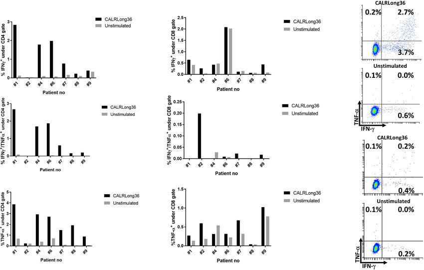
Handlos Grauslund et al.                                                                                                     Vaccination in CALR-Mutant MPN

                                              Immunomonitoring revealed that four of 10 patients had an in vitro interferon (IFN)-g
                                              ELISPOT response to the CALRLong36 peptide at baseline, and four additional patients
                                              displayed a response in ELISPOT upon receiving three or more vaccines. The amplitude of
                                              the immune response increased during the entire vaccination schedule for patients with
                                              essential thrombocythemia. In contrast, the immune response in patients with primary
                                              myelofibrosis did not increase after three vaccines.
                                              Conclusion: Therapeutic cancer vaccination with peptide vaccines derived from mutant
                                              CALR with montanide as an adjuvant, is safe and tolerable. The vaccines did not induce
                                              any clinical responses. However, the majority of patients displayed a marked T-cell
                                              response to the vaccine upon completion of the trial. This suggests that vaccines
                                              directed against mutant CALR may be used with other cancer therapeutic modalities to
                                              enhance the anti-tumor immune response.
                                              Keywords: myeloproliferative neoplasms, cancer immune therapy, cancer vaccines, neo-antigen, calreticulin

INTRODUCTION                                                                            Several lines of evidence show that the immune system is
                                                                                     severely dysregulated in MPN (13–15). Cytokine levels and the
The Philadelphia chromosome-negative chronic myeloproliferative                      immune phenotype in patients differ from those in healthy
neoplasms (MPN) are chronic blood cancers originating in the                         donors. This immune derangement is likely partially driven by
hematopoietic stem cells in the bone marrow (1, 2). MPN have                         the driver mutations identified in MPN, as they all affect the
overlapping signs and symptoms and thus may be difficult to                           JAK-STAT signaling pathway which is of paramount
distinguish from one another. Essential thrombocythemia (ET)                         importance in the regulation of the immune-system (16). The
and polycythemia vera (PV) are the non-advanced MPN, with                            mutational landscape in MPN is highly homogeneous, as
patients presenting elevated levels of peripheral blood cells, whereas               approximately 90% of patients harbor a mutation in the
primary myelofibrosis is the advanced disease characterized by bone                   Janus kinase-2 (JAK2), myeloproliferative leukemia virus
marrow fibrosis, and patients often present with cytopenia.                           (MPL), or calreticulin (CALR) gene. The JAK2V617F
Prefibrotic or early myelofibrosis (PreMF) was recently                                mutation has been identified in 98% of patients with PV and
acknowledged as a distinct MPN with patients presenting with                         in 50% of patients with ET and PMF (17, 18). The MPL
early signs of myelofibrosis (3). Allogeneic hematopoietic stem cell                  mutations are identified in 5–10% of patients with ET and
transplantation (alloHSCT) remains the only curative treatment for                   PMF (19), and the CALR exon 9 mutations are found in 20–
MPN, and as patients with ET and PV may live for several decades                     25% of patients with ET and PMF (20, 21). Whereas the JAK2
(4, 5) alloHSCT, which has a high treatment-related mortality, is not                and MPL mutations generate single-amino-acid substitutions
used for the treatment of these diseases. AlloHSCT is used for                       in their respective proteins, the CALR deletion or insertion
younger and fit patients with PMF (6), but as the majority of                         mutations result in frameshift mutations that generate a novel
patients with PMF are of older age, alloHSCT is not used routinely                   mutant C-terminus different from the wild-type (wt) CALR C-
for these patients. Due to the elevated blood cell counts and the                    terminus (20, 21). Interestingly, peptides derived from the
presence of the MPN-driver mutations per se, patients with ET and                    mutant C-terminus are recognized by peripheral blood
PV have a dramatically increased risk of thromboembolic episodes                     mononuclear cells (PBMCs) isolated both from patients with
and hemorrhage. Hence, the treatment for ET and PV aims to lower                     CALR-mutant (CALRmut) MPN and from healthy donors (22,
this risk with cytoreductive therapy (7) such as hydroxyurea (HU),                   23), and T cells isolated from patients with CALRmut MPN
anagrelide (ANA), and pegylated interferon alpha (IFN-a). HU is a                    recognize and kill autologous CALRmut cells in a CALRmut-
weak chemotherapeutic agent that reduces peripheral blood cell                       dependent manner (22). These data have established that the
counts, whereas ANA is solely used to reduce the platelet count.                     CALR mutations encode tumor-specific antigens (TSAs) that
IFN-a is a cytokine that in the past has been used to treat several                  are recognized by patient T cells.
cancers, such as multiple myeloma, chronic myelogenous leukemia                         Studies on other TSAs have shown that they may be targeted
(CML), malignant melanoma and renal cancer (8) but in general                        by the immune system (24), and clinical trials have used this in
with a low-rate of success except partly for CML (9). In MPN, IFN-                   the setting of therapeutic cancer vaccination, where vaccination
a can induce major molecular remissions, complete hematological                      with peptides derived from the TSA is aimed at inducing/
responses (10, 11), and even normalization of bone marrow                            enhancing the tumor-specific immune response (25). The first
architecture in some patients (11). As IFN-a is an                                   TSAs targeted by therapeutic cancer vaccines arose from RAS
immunostimulatory cytokine, it has been speculated that one of                       mutations, which are the most common somatic mutations in
its mechanisms of action is to normalize the immune environment                      human cancer (26). Initial studies showed that patient T cells
in patients with MPN (12, 13).                                                       respond to stimulation in vitro with epitopes derived from

Frontiers in Oncology | www.frontiersin.org                                      2                                 February 2021 | Volume 11 | Article 637420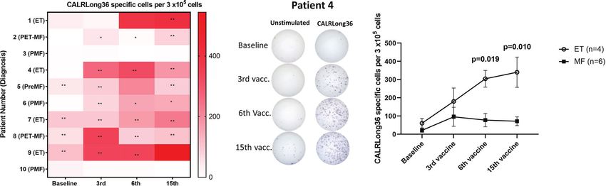
Handlos Grauslund et al.                                                                                          Vaccination in CALR-Mutant MPN

mutant RAS (27, 28), and that these T cells can kill HLA-                   Vaccine Composition and Treatment
matched RAS-mutant cancer cells (29). These results spurred                 Schedule
the first therapeutic cancer vaccines using TSA-derived peptides,            Each vaccine was composed of 200 µg CALRLong36, a 36-amino
where vaccination against mutant KRAS induced or enhanced an                acid peptide (RMRRMRRTRRKMRRKMSPARPRTS
immune response specific to the KRAS mutation-derived TSA                    CREACLQGWTEA) spanning the entire mutant C-terminus
(30). Several clinical vaccination trials testing vaccination against       created by the most frequent CALR mutations. The peptide
mutant KRAS have shown a survival benefit to patients who                    was provided by Polypeptide (Strasbourg, France). The peptide
develop an immune response to the vaccination antigen (31–33),              was dissolved in 500 µL sterile water and mixed with 500 µL
thus establishing the potential of TSA-specific therapeutic                  montanide ISA-51 just prior to administration. The vaccine was
cancer vaccines.                                                            administered subcutaneously, and patients were vaccinated with
   Given the high immunogenicity of the CALRmut-derived                     a total of 15 doses, with the first six doses administered every
TSA, we initiated a phase I clinical vaccination trial with a               second week and the final nine doses every fourth week. A Gant
CALRmut-derived peptide in 10 patients with CALRmut MPN.                    chart of the vaccination schedule is provided in Supplementary
Safety and tolerability were the primary end points, attainment of          Figure 2.
immune response to the CALRmut TSA was the secondary end
point, and clinical response was the tertiary end point. The                Evaluation of Adverse Events and
vaccine was safe and tolerable, and although the vaccines did not           Clinical Response
induce any clinical responses, they induced marked alterations in           Adverse events (AE) were assessed according to the Common
the immune phenotype and immune response to the                             Terminology Criteria for Adverse Events (CTCAE) version
vaccination antigen.                                                        4.03. Before receiving the first vaccination, each patient
                                                                            underwent a full medical examination and had an
                                                                            electrocardiogram recorded. This was repeated before each
SUBJECT, MATERIALS AND METHODS                                              of the first three vaccinations, and then before the 7th and after
                                                                            the 15th vaccination. Biochemical tests on peripheral blood
Study Design                                                                were performed before each vaccination. The patients
This was a phase I clinical vaccination trial testing the safety and        underwent a full bone marrow examination and analysis by
immunogenicity of vaccination with the CALRLong36 peptide                   next-generation sequencing (NGS) performed on
with montanide as an adjuvant. Patients from the Departments                peripheral blood prior to the first vaccination and at end of
of Hematology at Herlev and Gentofte Hospital, Copenhagen,                  study. The clinical response was evaluated according to the
Denmark, as well as Zealand University Hospital, Roskilde,                  response criteria described by the European LeukemiaNet
Denmark were enrolled in the trial between August 30th, 2018                (34, 35).
and February 17th 2019 with the last vaccination administered
January 14th 2020. The study was conducted in accordance with               Evaluation of the CALRmut Allelic Burden
the Helsinki Declaration, and Good Clinical Practice                        and Next-Generation Sequencing
recommendations. All participants provided written informed                 PCR detection of CALR exon 9 frameshift mutations was
consent before enrollment in the trial. The protocol was                    performed as described by Klampfl et al. (20). Primers used
approved by the Ethics Committee of the Zealand Region,                     are: CALR Forward: 5′-Fam-GGCAAGGCCCTGAGGTGT-3′,
the National Board of Health, and the Danish Data Protection                CALR Reverse: 5′-GGCCTCAGTCCAGCCCTG-3′. The PCR
Agency, and registered at www.clinicaltrials.gov (NCT03566446;              product was run on a ABI3500 Genetic Analyzer (Thermo
date of registration June 25th, 2018). One patient from whom we             Fisher Scientific) and mutated CALR allele burden calculated
wished to isolate PBMC after the end of the study provided                  as 100*AUC (area under the curve) for mutated CALR/(AUC for
informed consent for this in accordance with an approval from               mutated + AUC wildtype).
the Ethics Committee of Zealand Region (approval number                         Next generation sequencing was performed on a custom
SJ-456).                                                                    made panel of 42 genes designed to capture mutations
    As therapeutic cancer vaccines have not been tested earlier in          implicated in myeloid malignancies (ASXL1, BCOR, BRAF,
patients with MPN, we had no basis for any power calculations.              CALR, CEBPA, CBL, CSF3R, CUX1, DNMT3A, ETV6, EZH2,
Based on our previous experience with phase I clinical                      FLT3, GATA2, GNAS, IDH1, IDH2, JAK2, KIT, KRAS, MLL3,
vaccination trials, we chose to include 10 patients in the trial.           MPL, MYD88, NF1, NFE2, NOTCH1, NPM1, NRAS, PHF6,
Main inclusion criteria were a diagnosis of ET, PreMF, or PMF               PPM1D, PTPN11, RB1, RIT, RUNX1, SETBP1, SF3B1, SH2B3,
according to the WHO criteria, verified mutation in exon 9 of                SRSF2, STAG2, TET2, TP53, U2AF1, ZRSR2). Genomic DNA
CALR, and Eastern Cooperative Oncology Group performance                    was purified from peripheral blood at baseline and 12 months
status of ≤2. A full list of the inclusion and exclusion criteria can       after the first vaccination of 10 CALR mutated MPN patients.
be found in Supplementary Figure 1. Patients were allowed                   Libraries were prepared using the Nextera Flex for Enrichment
concurrent treatment with IFN-a, HU, or ANA in any                          protocol (Illumina® Inc, San Diego, CA, USA), and 2 × 150 bp
combination, but no other anti-neoplastic or anti-MPN                       paired-end sequencing was done on the NextSeq 500 platform
treatments were allowed.                                                    (Illumina® Inc, San Diego, CA, USA). Quality control of all

Frontiers in Oncology | www.frontiersin.org                             3                               February 2021 | Volume 11 | Article 637420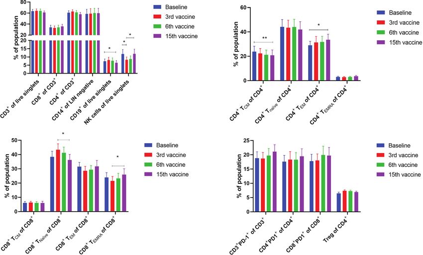
Handlos Grauslund et al.                                                                                       Vaccination in CALR-Mutant MPN

sequencing runs was assessed using the Illumina Sequencing              IFN-g ELISPOT Assay
Analysis Viewer (SAV) software. CLC Genomics Workbench                  In order to assess the immune response against CALRLong36, we
v.12.0.3 software was applied to align sequencing data to the           used IFN-g ELISPOT assays performed on PBMCs as described
human reference genome (GRCh37/hg19) and to call variants               in detail earlier (37). In short, PBMCs were thawed and
from the mapped reads. Annotation and filtering of variants              stimulated with CALRLong36 and then stimulated with IL-2
were performed using the VarSeq™ v.2.2.1 software (Golden               (120 U/mL) the next day. The cells were incubated for 14 days in
Helix, Inc., Bozeman, MT, USA). Variants with coverage
Handlos Grauslund et al. Vaccination in CALR-Mutant MPN
DFR analyses which were performed using the statistical analysis nine of ten patients at any time. Most common systemic reaction
program R. was flu like symptoms in five patients. Two grade 3 AE were
registered: One vasovagal reaction during bone marrow biopsy at
the end of study, and one patient experienced hypertension before
the first vaccine was administered. One autoimmune reaction was
RESULTS registered (see below) but this reaction was not deemed to be
Patient Characteristics triggered by the vaccines.
Eleven patients were included and evaluated by examination of the
bone marrow biopsies and blood samples prior to initiation of Clinical Response and Variations in
vaccinations. Due to compliance issues, one patient was excluded, CALRmut Variant Allele Frequency
and thus 10 patients continued in the trial. All patients received the As the CALR mutations mainly affect the megakaryocytic
planned 15 vaccine doses over the course of one year. The male: lineage, and patients present with marked thrombocythemia,
female ratio was 5:5, the median age at inclusion was 59.5 years we expected that the vaccinations would induce a decrease in
(range 41-73 years), and the median duration of disease was 6.5 platelet counts. Looking at each patient individually, we did not
years (range 2–26 years) (Tables 1 and 2). At study entry, four demonstrate any decrease in platelet counts (Figure 1A). Of
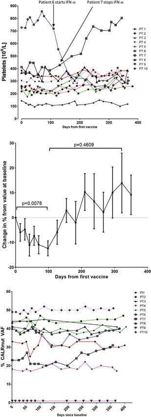
patients had ET, three patients PMF, two patients post-essential note, one patient (patient 6), who did not receive any therapy at
thrombocythemia MF (PET-MF) and one patient prefibrotic/early inclusion in the trial, was started on IFN-a after 7th vaccination
myelofibrosis (PreMF). All but one patient had received MPN- due to high platelet counts. Another patient (patient 7) had to
directed therapy including HU, ANA, or INF-a prior to stop IFN-a after 8 vaccinations due to elevated liver enzymes and
enrollment. Of the nine treated patients, eight were treated with concomitant development of smooth muscle antibodies (SMA).
INF-a and one with ANA at the start of vaccination. This one Before and during the first vaccines, the patient developed
patient treated with ANA had received IFN-a earlier, however IFN- increasing levels of alanine aminotransferase (ALAT) below
a had been withdrawn due to side effects. Median platelet count was upper normal limit. After 5 vaccinations the level of ALAT
283 × 109/l (range 145 × 109/l–728 × 109/l), median hemoglobin reached a maximum of 241 U/l (upper normal limit 70 U/L).
count was 7.95 mmol/l (range 6.5 mmol/l–9.2 mmol/l), and median SMA were detected in the blood, and examination of archived
leucocyte count was 4.9 × 10⁹/l (range 3.3 × 10⁹/l–10.5 × 109/l). samples taken at study inclusion prior to vaccine initiation
Four patients presented with splenomegaly on clinical examination. showed low levels of SMA. After IFN-a was stopped the
Four patients had the type 1 CALR-mutation, one patient a type-1- patient continued his vaccinations as planned and normalized
like CALR-mutation and five had the type 2 CALR-mutation. The his liver function.
median CALRmut VAF was 40.5% (range, 1–49%). We detected a relative decrease in platelet count during the first
100–120 days after the first vaccination that was statistically
Adverse Events and Safety Profile significant (Figure 1B). Platelet levels increased thereafter to a
All patients experienced AE and these were both local and systemic level above baseline levels, but this increase was not statistically
(Table 3). No vaccination related AE ≥ grade 3 were observed. The significant (Figure 1B). We expected to identify a decrease in the
most common reactions were injection site specific with grade 2 in CALRmut variant allele frequency (VAF) in peripheral blood. The
CALRmut VAF showed some fluctuations over time, which is also
TABLE 1 | Patient characteristics. our experience in our normal clinical setting. However, the
CALRmut VAF displayed no substantial decrease nor increase in
Sex Female n= 5, Male n=5 any of the patients (Figure 1C) even in the patient that displayed a
Age at inclusion in years, median (min–max) 59.5 (41-73) CALRmut VAF of only 1% at baseline. As described, patients
Duration of disease in years, median (min– 6.5 (2-26) underwent a full bone marrow examination at baseline and at
max) end of study. No morphological or cytological changes were
Diagnosis ET n=4 detected in patients during the vaccination schedule. Analysis of
PMF N=3
PreMF n=1
additional mutations quantified by NGS showed that seven of 10
PET-MF n=2 patients harbored additional mutations at baseline and three
Treatment Pegylated Interferon-alpha n=8 patients only had a CALR mutation with no additional mutations
Anagrelide n=1 (Table 4). However, at end of study one of these three patients had
No Treatment n=1 developed an additional mutation. Of note, among the additional
Platelet count at inclusion, median (min-max) 283x109/l (145x109/l–728x109/l)
Hemoglobin at inclusion, median (min-max) 7.95 mmol/l (6.5mmol/l–
mutations identified by NGS, several genes of importance in
9.2mmol/l) myeloid cancers such as ASXL1, TET2, EZH2, SF3B1, and
Leukocyte count at inclusion, median (min- 4.9x10⁹/l (3.3x10⁹/l–10.5x109/l) DNMT3A were detected. Three patients displayed a mutation in
max) TP53. The majority of patients did not experience marked
Splenomegaly at inclusion n=4
alterations in mutant VAF as measured by NGS. However,
CALR-mutation type Type 1 (52 bp del) n=5
Type 2 (5 bp ins) n=5
patient 6 showed a decrease in DNMT3A (20% VAF to 12%
% CALRmut VAF at inclusion, median (min- 40.5% (1% - 49%) VAF), whereas ASXL1 increased from 0.66% to 2.1%. Patient 8
max) was the only patient who demonstrated a marked expansion of a
Frontiers in Oncology | www.frontiersin.org 5 February 2021 | Volume 11 | Article 637420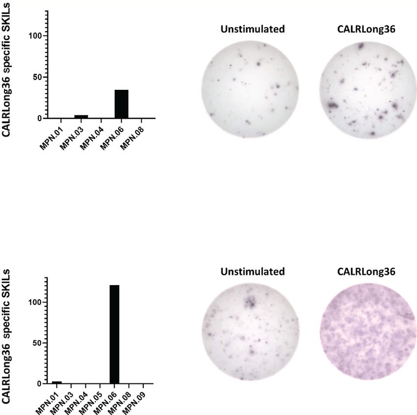
Handlos Grauslund et al. Vaccination in CALR-Mutant MPN
TABLE 2 | Individual patient data for all patients in the trial.
Patient number 1 2 3 4 5 6 7 8 9 10
Age (years) 73 70 61 61 43 52 41 57 67 58
Sex Female Female Male Female Male Male Male Female Female Male
Duration of disease (years) 7 18 26 2 22 3 4 14 3 6
Diagnosis ET PET-MF PMF ET PreMF PMF ET PET-MF ET PMF
Treatment at inclusion Pegylated Anagrelide Pegylated Pegylated Pegylated None Pegylated Pegylated Pegylated Pegylated
interferon- interferon- interferon- interferon- interferon- interferon- interferon- interferon-
alpha alpha alpha alpha alpha alpha alpha alpha
Splenomegaly at inclusion (Y/N) N N Y N Y Unknown N Y N Y
CALR mutation type Type 1 (52 Type 2(5 Type 1 (52 Type 2(5 Type 2(5 Type 2(5 Type 1 (52 Type 2(5 Type 1 (52 Type 1 (52
bd del) bp ins) bd del) bp ins) bp ins) bp ins) bp del) bp ins) bp del) bp del)
CALRmut VAF at inclusion 33% 43% 44% 18% 41% 41% 23% 40% 1% 49%
Immune response (Y/N) Y Y N Y Y Y Y Y Y N
subclone as GNAS mutant VAF increased from 0.99% to 10% vaccination antigen using IFN-g ELISPOT. We detected a
during the vaccination schedule. Additionally, patient 4 developed a DFR2x-defined immune response in four of 10 patients at
new STAG2-mutant subclone and patient 10 developed a BCOR- baseline (Figure 2A). Interestingly, the vaccinations induced
mutant subclone during the vaccination schedule. immune responses in several patients that did not show an
immune response at baseline: Seven of 10 patients displayed at
Immune Response to the Vaccination least a DFR-defined response after three vaccinations, six of 10
Antigen in Peripheral Blood had at least a DFR-defined response after six vaccinations, and
Mononuclear Cells seven of 10 had at least a DFR-defined response at end of study
Patient PBMC isolated at baseline and after three, six, and 15 (Figure 2A). The amplitude of the responses increased over time
vaccinations were analyzed for immune responses against the (Figure 2B), but interestingly the response in patients with MF
TABLE 3 | Adverse events registered during the trial.
Adverse event Number of patients Grade 1 Grade 2 Grade 3
Injection site reactions 10 1 9 0
Flu like symptoms 5 4 1 0
Headache 3 3 0 0
Infection 7 6 1 0
Pruritus 2 2 0 0
Vomiting 1 1 0 0
Mucositis 2 2 0 0
Hot flashes 2 2 0 0
Nausea 2 2 0 0
Myalgia 3 3 0 0
Fever without neutropenia 1 1 0 0
Fatigue 5 2 3 0
Diarrhea 2 2 0 0
Chills 2 1 1 0
Coughing 2 2 0 0
Arthralgia 2 2 0 0
Anorexia 1 0 1 0
ALAT derangement 1 0 1 0
Vertigo 2 1 1 0
Vasovagal reaction 1 0 0 1
Tinnitus 1 1 0 0
Palpitations 1 2 0 0
Pain in extremity 1 1 0 0
Malaise 1 1 0 0
Hypertension 1 0 0 1
Gastrointestinal disorder—other 1 1 0 0
Fall 1 1 0 0
Epistaxis 1 1 0 0
Bruising 1 0 1 0
Allergic rhinitis 2 1 0 0
Atrial fibrillation 1 1 1 0
Acute coronary syndrome 1 1 0 0
Abdominal pain 1 0 1 0
Frontiers in Oncology | www.frontiersin.org 6 February 2021 | Volume 11 | Article 637420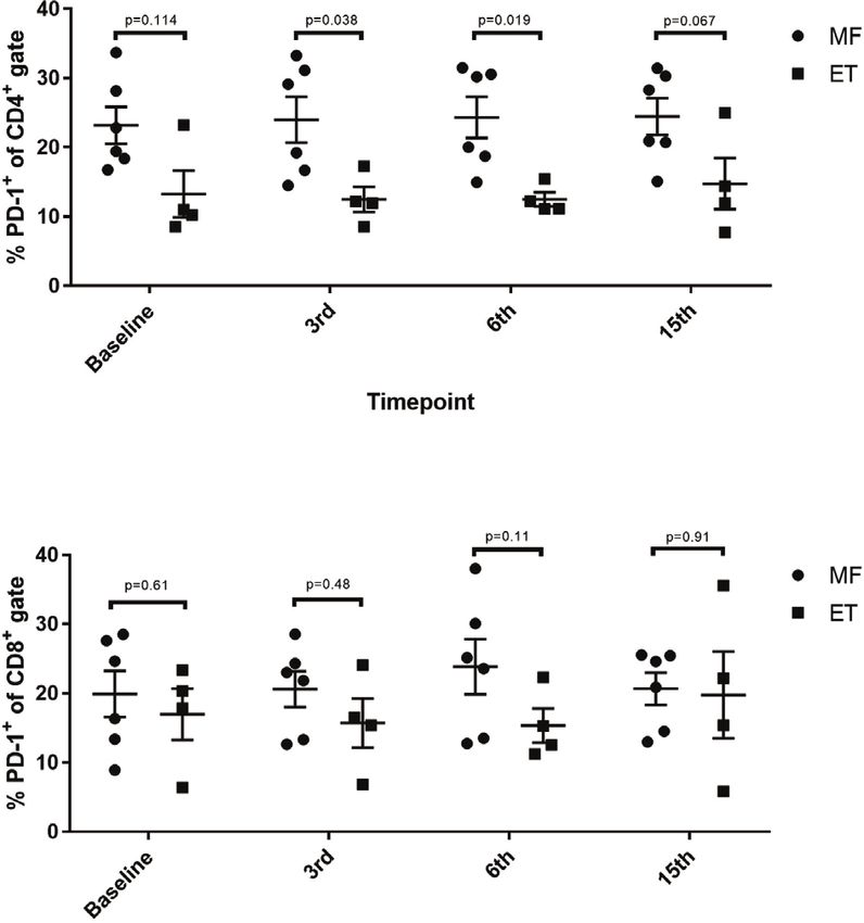
Handlos Grauslund et al.                                                                                                     Vaccination in CALR-Mutant MPN

                                                                                       did not increase after the third vaccination, whereas the
                                                                                       amplitude of the response in patients with ET increased during
       A
                                                                                       the entire vaccination schedule (Figure 2C). The difference in
                                                                                       response amplitude between patients with ET and PMF were
                                                                                       statistically significant at 6th vaccination and at the end of the
                                                                                       study (Figure 2C). Two patients (patients 3 and 10), both of
                                                                                       whom had PMF, did not attain an immune response at all during
                                                                                       the vaccination schedule.
                                                                                           The phenotype of the cytokine-producing cells was evaluated
                                                                                       using intracellular cytokine staining (ICS). From the eight
                                                                                       patients who showed an immune response to CALRLong36 in
                                                                                       ELISPOT, we analyzed PBMC isolated at the time point where
                                                                                       the response in ELISPOT was deemed to be highest. For patients
                                                                                       3 and 10, who did not display an immune response in ELISPOT,
                                                                                       we chose to analyze PBMC isolated after 15 vaccinations. Not
                                                                                       surprisingly, we did not identify a response in patients who did
                                                                                       not show a response in ELISPOT. Additionally, patient 5 had a
       B
                                                                                       very low fraction of CD3+ T cells, making us unable to gate on a
                                                                                       satisfactory number of T cells for our analysis. However, of the
                                                                                       remaining seven patients, six displayed a CD4+ T-cell response to
                                                                                       stimulation with CALRLong36 (Figures 3A, C), and two patients
                                                                                       displayed a CD8+ T-cell response (Figures 3B, D). Patient 7
                                                                                       demonstrated both a CD4+ and a CD8+ T-cell response, whereas
                                                                                       patient 2 demonstrated only a CD8+ T-cell response. Patient 2
                                                                                       provided informed consent for us to isolate PBMC at several time
                                                                                       points after the end of study and showed a sustained CD8+ T-cell
                                                                                       response to CALRLong36 even at 37 and 49 weeks after the last
                                                                                       vaccination (Supplementary Figure 5).

                                                                                       Delayed-Type Hypersensitivity and
                                                                                       Response in Skin-Infiltrating Lymphocytes
                                                                                       Ten patients received intradermal injections with CALRLong36
                                                                                       as described in Materials and Methods. Of note, none of the
       C
                                                                                       patients demonstrated an induration at the site of intradermal
                                                                                       injections. In order to investigate if the injections could have
                                                                                       induced a migration of CALRLong36-specific T cells to the
                                                                                       injection site, 10 patients were subjected to a punch biopsy at
                                                                                       the injection site. Punch biopsies were cultured as described
                                                                                       previously (39) to expand specific T cells in the biopsy.
                                                                                       Surprisingly, even though none of the patients displayed an
                                                                                       induration at the injection sites, we were able to expand skin-
                                                                                       infiltrating lymphocytes (SKILs) from five patients by culturing
                                                                                       with low dose IL-2 (100 U/mL) and SKILs from seven patients by
                                                                                       culturing with high dose IL-2 (6,000 U/mL). The SKILs were
                                                                                       tested in IFN-g ELISPOT for response to CALRLong36. Only
                                                                                       patient 6 displayed a response (Figure 4); the responses in the
                                                                                       remaining patients were either absent or the background was too
                                                                                       high (Supplementary Figure 6).

 FIGURE 1 | Variation in platelet counts and CALRmut variant allele frequency          Phenotype of Peripheral Blood
 (VAF) during the trial. (A) Platelet counts (109/L) for each patient during the
 trial. (B) To get a better impression of the cumulative change in platelet
                                                                                       Mononuclear Cells During the
 counts, the relative change in platelet counts from baseline was calculated for       Vaccination Schedule
 each patient. Each dot represents the mean change in platelet count in                Alterations in the composition of immune cells in peripheral
 percent relative to the platelet count at baseline. Error bars depict the             blood were analyzed during the vaccination schedule. No
 standard error of the mean. Statistical testing was performed with the
                                                                                       alterations were identified in the number of T cells in general,
 Wilcoxon signed-rank test. (C) Changes in the CALRmut VAF over time.
                                                                                       nor in the numbers of CD4+ and CD8+ T cells. Neither did we

Frontiers in Oncology | www.frontiersin.org                                        7                               February 2021 | Volume 11 | Article 637420
Handlos Grauslund et al. Vaccination in CALR-Mutant MPN
TABLE 4 | Mutant variant allele frequency (VAF) of CALR and additional CD8+ naïve T cells (Tnaïve) during the vaccination schedule,
mutations identified by next generation sequencing.
and levels of Tnaïve only returned to baseline levels at the end of
Patient Mutations at baseline (% Mutations at end of study (% the study period (Figure 5C). Conversely, the number of CD8+
ID VAF) VAF) TEMRA decreased after the first three vaccinations, after which it
increased to reach the levels at baseline (Figure 5C). Levels of
1 CALR (15) CALR (15)
2 CALR (37) CALR (39)
PD-1 on both T cells in total and in CD4+ and CD8+ T cells
3 CALR (24) CALR (26) remained constant, as did the levels of regulatory T cells (Treg)
TP53 (1) TP53 (0.5) (Figure 5D). As PD-1 expression is increased on T cells in
4 CALR (15) CALR (14) patients with MPN (40, 41) we sought to analyze differences in
STAG2 (1.5)
PD-1 expression between ET- and PMF-patients. Most
5 CALR (32) CALR (32)
SF3B1 (0.22) SF3B1 (1.6) interestingly, we showed that at the third and sixth
6 CALR (34) CALR (30) vaccination, CD4+ T cells from patients with MF expressed
DNMT3A (20) DNMT3A (12) significantly higher levels of PD-1 compared with patients with
EZH2 (3.4) EZH2 (5) ET (Figure 6A). A difference in expression levels was also
TP53 (0.84) TP53 (1.2)
detected at baseline and at the 15 th vaccination, but the
ASXL1 (0.66) ASXL1 (2.1)
7 CALR (14) CALR (17) difference was not statistically significant (Figure 6A). On the
NF1 (1.3) NF1 (1.2) other hand, expression of PD-1 by CD8+ T cells did not differ
8 CALR (35) CALR (35) between patients with ET and PMF (Figure 6B). No significant
GNAS (0.99) GNAS (10) changes were identified in the different NK cell subsets (Figure
9 CALR (1) CALR (1)
TP53 (1.02) TP53(1.4)
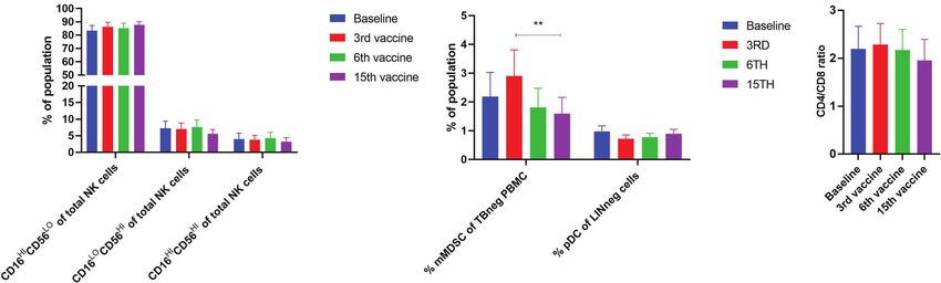
7A). Of note, levels of monocytic myeloid-derived suppressor
TET2 (1.4) TET2(1.4) cells (mMDSC) increased initially but decreased to a level lower
10 CALR (31) CALR (33) than that at baseline (Figure 7B), and the CD4+/CD8+ T-cell
SF3B1 (43) SF3B1 (45) ratio remained constant throughout the study (Figure 7C).
BCOR (1.5)
detect any changes in the levels of CD14+ monocytes. B cells DISCUSSION
increased slightly during the study, but returned to baseline levels
at the end of the trial. NK cells decreased initially, but towards In order to attain a clinical response using cancer immune therapy,
the end of the study they reached their level at baseline (Figure it is of utmost importance that the tumor cells express
5A). Interestingly, we identified a statistically significant decrease immunogenic antigens that may be targeted by specific T cells
in the number of circulating CD4+ central memory T cells (TCM) (42). The RAS mutations generate several immunogenic neo-
in combination with a statistically significant increase in the antigens that are targets of specific T cells, and therapeutic
number of CD4+ effector memory T cells (TEM) (Figure 5B). cancer vaccines targeting these neo-antigens have shown a
Changes in the CD8+ T-cell fraction were not dramatic, but we survival benefit in patients with an immune response to the
detected a somewhat paradoxical increase in the number of vaccine epitope (33, 43). Vaccination of AML patients with
A B C
FIGURE 2 | Immune responses to CALRLong36 in IFN-g ELISPOT. (A) Heat map depicting the responses to CALRLong36 in patient peripheral blood mononuclear
cells at each time point for all patients. The number of CALRLong36-specific cells was calculated by subtracting the mean spots in the control wells from the mean
spots in the peptide-stimulated wells. The analysis was only performed in duplicates for patient 1 and 5 at the 6th vaccination and for patient 9 at the 15th
vaccination, which prevented us from performing statistical analysis of the results. All other experiments were performed in triplicates. * Indicates a statistically
significant response according to the DFR-rule. ** Indicates a statistically significant response according to the DFR(2x)-rule (38). (B) Representative image from
(A, C) ELISPOT responses over time in patients with essential thrombocythemia (ET) and patients with myelofibrosis (MF) with each dot representing the mean of
normalized spots. Error bars depict the standard error of the mean. Statistical testing was performed using the Mann-Whitney test.
Frontiers in Oncology | www.frontiersin.org 8 February 2021 | Volume 11 | Article 637420Handlos Grauslund et al. Vaccination in CALR-Mutant MPN
A B C
D
FIGURE 3 | Responses against CALRLong36 identified by intracellular cytokine staining in patients with a response in ELISPOT. (A) Responses in CD4+-gated T
cells with IFN-g secreting cells (top), IFN-g/TNF-a secreting cells (middle). and TNF-a secreting cells (bottom). (B) Responses in CD8+-gated T cells with IFN-g
secreting cells (top), IFN-g/TNF-a secreting cells (middle), and TNF-a secreting cells (bottom). (C) An example of a CD4+ T-cell response. (D) An example of a CD8+
T-cell response.
immunogenic tumor-associated antigens such as Wilms tumor CALR mutations have been shown to be early mutations
antigen-1 (WT1) and NY-ESO have also demonstrated a survival residing in the highly undifferentiated HSC (21), a strong
benefit for patients with an immune response (44, 45). The NPM1 CALRmut-specific immune response in patients may be of
mutations identified in a substantial proportion of patients with concern, as a strong immune response could potentially
AML generate highly immunogenic antigens (46), and it has been eradicate all CALRmut HSC in the patients, and thus, result in
shown that patients with an intact immune response to NPM1- bone marrow failure. Only a few side effects were identified in our
derived neo-antigens have significantly longer overall survival study, with only two grade 3 AE. One patient developed
compared with patients without a response (47). As such, the hypertension during the trial, but this occurred before the
existence of a neo-antigen-specific immune response is believed to administration of the first vaccine, thus it is impossible that the
be highly important in attaining a therapeutic effect. In recent vaccines elicited this AE. Another patient experienced a vasovagal
years, research has mainly focused on the potentially large number episode just prior to the end-of-study bone marrow biopsy. The
of patient-specific, mutated neoantigens. They comprise the episode could be explained simply by the fact that bone marrow
predicted products of the numerous mutations revealed by biopsies are highly uncomfortable and somewhat painful for the
exome sequencing of primary tumors, and it was demonstrated patients. Apart from these, no severe AE (SAE) were observed. The
that it is not only the number of mutations that predict outcome most frequent AE registered in this trial, infection, fatigue and
but also the immunogenicity of the antigens generated by the myalgia, are complaints that are common in patients with MPN.
somatic mutations (48). The injection-site reactions experienced by all patients and the flu-
We have shown earlier that patients with CALRmut MPN like symptoms experienced by 50% of the patients are likely
harbor T cells specific to neo-antigens derived from the mutant reactions to the montanide adjuvant in the vaccine. The
CALR C-terminus. However, patient T-cell responses are less occurrence of these AE could actually be regarded as a beneficial
frequent and weaker than the responses identified in healthy sign, as they indicate that a proper inflammatory response to the
donors. This could indicate that patients with CALRmut MPN vaccine was induced. In conclusion, this vaccination trial adds
harbor an exhausted CALRmut-specific immune response, and we weight to the notion that cancer vaccines only very rarely induce
speculated that therapeutic cancer vaccination with a CALRmut- SAE (49). The patient that experienced a transient increase in
derived peptide could enhance the tumor-specific immune ALAT showed evidence of increased SMA even before study entry,
response in patients and lead to a clinical response. As the thus this AE is not believed to be induced by the vaccines.
Frontiers in Oncology | www.frontiersin.org 9 February 2021 | Volume 11 | Article 637420Handlos Grauslund et al. Vaccination in CALR-Mutant MPN
A B
C D
FIGURE 4 | Responses in skin-infiltrating lymphocytes (SKILs) against CALRLong36. SKILs were harvested as described in the Materials and Methods section and
were then expanded in either high-dose IL-2 (6,000 U/mL) culture medium or low-dose IL-2 (100 U/mL) culture medium. Cells were harvested and analyzed by IFN-g
ELISPOT for a response against CALRLong36. (A) Normalized numbers of cells specific to CALRLong36 in SKILs from patient 1, 3, 4, 6 and 8 cultured in low dose
IL-2. (B) Response in SKILs from patients 6 expanded in low dose IL-2. (C) Normalized numbers of cells specific to CALRLong36 in SKILs from patient 1, 3, 4, 5, 6,
8 and 9 cultured in high dose IL-2. (D) Response in SKILs from patients 6 expanded in high dose IL-2.
Given the immunogenicity of the CALR mutations, we number 6 was not receiving any treatment at inclusion, but
expected the vaccines to induce a clinical response in patients. after seven vaccinations it was decided to start treatment with
As the CALR mutations mainly affect the megakaryocytic lineage IFN-a (45 µg weekly) due to a high platelet count. As IFN-a is a
(50–53) and patients with CALRmut MPN display higher platelet highly immunostimulatory cytokine, we believed that the
counts than their JAK2V617F+ counterparts (54, 55), we addition of IFN-a to the vaccines would induce a molecular
expected that a clinical response would be identified by a response. Patient number 9 displayed a very low CALRmut
decline in platelet counts. Indeed, we detected a decline in VAF of only 1%. Earlier reports on therapeutic cancer vaccines
platelet counts during the first 100 days, after which the suggest that patients with a low tumor burden are more likely to
numbers increased. Importantly, apart from patient 6 and 7, benefit from therapy (56, 57). Since patient 9 had a relatively
no patients had any adjustments in their MPN-directed therapy low tumor burden we believed that the low number of tumor
during the trial. Thus, this cannot explain the changes detected in cells would not impede the tumor-specific immune response.
platelet counts. A potential cause of the decline in platelets is a Despite the low tumor burden in this patient, the vaccines did
tumor-specific immune response that targets the megakaryocytes not induce a clinical nor molecular response. However, it is
and thus reduces platelet production. Importantly, the intriguing to consider, whether the lack of responses might be
subsequent increase in platelet counts coincided with the attributed to the additional mutations in both TP53 and TET2,
conversion of biweekly vaccinations into monthly vaccinations. rendering the CALR-mutant clone resistant to immune
To our belief, the decline in platelet counts during the first period mediated clearing.
of time could be explained by a tumor specific T-cell response Our results clearly demonstrate that a lack of clinico-
and certainly needs further investigation in future trials. hematological and molecular response to the CALRmut-
The decline in platelet counts did not translate into a decline vaccine is not explicitly due to incompetent CALRLong36-
in the CALRmut VAF. In daily practice the VAF oscillates over specific immune responses. Thus, eight patients displayed
time but largely remains constant, which was also recorded in strong immune responses to CALRLong36. Of these eight
our cohort. Theoretically, some patients were more likely to patients, four showed an immune response at baseline that was
display a response to the vaccines than others. Thus, patient later enhanced by the vaccines. In the remaining four patients
Frontiers in Oncology | www.frontiersin.org 10 February 2021 | Volume 11 | Article 637420Handlos Grauslund et al. Vaccination in CALR-Mutant MPN
A B
C D
FIGURE 5 | Phenotyping of peripheral blood mononuclear cells (PBMC) and T cells by fluorescence-activated cell sorting. (A) Quantification of the mean PBMC
subsets in peripheral blood of vaccinated patients. (B) Quantification of CD4+ central memory T cells (TCM), CD4+ naïve T cells (Tnaïve), CD4+ effector memory T cells
(TEM), and CD4+ effector memory T cells re-expressing CD45RA (TEMRA). (C) Quantification of CD8+ central memory T cells (TCM), CD8+ naïve T cells (Tnaïve), CD8+
effector memory T cells (TEM), and CD8+ effector memory T cells re-expressing CD45RA (TEMRA). (D) Expression levels of PD-1 on T cells and CD4+ and CD8+ T
cells, and number of regulatory T cells (Treg). Statistical testing was performed using the Wilcoxon signed-rank test. Bars represent standard error of the mean.
* denotes p ≤ 0.05, ** denotes p ≤ 0.01.
who displayed an immune response to CALRLong36, the Earlier reports have shown that the majority of CALRmut-
response was absent at baseline but induced during the course specific immune responses identified are CD4+ T-cell responses
of the trial. Two patients, both of whom have PMF, did not (22, 23, 41, 61). In this trial, six of seven patients showed a CD4+
display an immune response against the vaccine. Interestingly, T-cell response, and one of these displayed a CD8+ T-cell
we found that the immune response in patients with ET response as well. One patient displayed only a CD8+ T-cell
increased during the vaccination course, in contrast to patients response, and this response was sustained even after cessation
with PMF in whom the amplitude of the immune response of vaccination. Earlier trials have shown that a sustained tumor-
remained stable after the first three vaccinations. We believe that specific CD8+ T-cell response is important in attaining a clinical
this could be due to the more severe immune dysfunction in response (62), and the apparent lack of CD8+ T-cell responses
patients with PMF, and the results are on par with our earlier could explain the lack of clinical results. However, the low
results showing that patients with ET have more frequent frequency of CD8+ T-cell responses could also be due to our
responses than patients with PMF (23, 58, 59). Recently it was experimental setup during the ICS, as the cells are only allowed
demonstrated that monocyte-derived dendritic cells (moDC) to incubate with the peptide for 5 h, thus leaving only a little time
from patients with PMF display lower levels of costimulatory for antigen-presenting cells to process and present possible CD8+
molecules compared with healthy-donor moDC, and that these epitopes. Arshad et al. showed that the CALR mutations inhibit
moDC have inferior priming potential (60). This could explain the presentation of peptides by human leukocyte antigen (HLA)-
the weaker responses identified in patients with PMF. In I molecules (63), as the CALR protein is an important chaperone
contrast, T cells from patients with CALRmut MPN are more in the assembly of the peptide:HLA-I complex. The low number
prone to activation, and Treg from CALRmut patients display a of CD8+ T-cell responses identified could thus also be explained
lower inhibitory potential than Treg from CALRwt patients (60), by the inability of patient antigen-presenting cells to process and
which should counteract the deficiency in moDC. We present high-quality HLA-I-restricted epitopes. We do therefore
hypothesized that the differences in immune responses might find it noteworthy that two of the seven patients analyzed
be partially explained by the occurrence of additional mutations, showed a CALRmut-specific CD8+ T-cell response.
however we did not identify any association between lack of Earlier peptide vaccination trials have evaluated the immune
immune response and additional mutations identified by NGS. response not only by measuring response in PBMC but also by
Frontiers in Oncology | www.frontiersin.org 11 February 2021 | Volume 11 | Article 637420Handlos Grauslund et al. Vaccination in CALR-Mutant MPN
tested in IFN-g ELISPOT assays and only one patient displayed a
response to CALRLong36. The apparent lack of DTH in our
A
patient cohort is striking. First, because the antigen is highly
immunogenic, and second because the majority of patients
receiving intradermal injections with antigen in other trials
show a DTH. As no patients demonstrated a DTH, we
speculate that this was due to a general feature, either of
CALRLong36 that prevents it from being processed and
presented by dermal dendritic cells, or of MPN patients in
general, who might harbor a deficiency in the dermal immune
system that renders the dermal immune cells hyporesponsive to
stimuli. Our current trial testing vaccinations with epitopes
derived from programmed death ligand (PD-L)-1 and arginase
(ARG)-1 (NCT04051307) will shed further light on this
B question. Of note, patients with psoriasis, an autoimmune skin
disease primarily driven by T cells (66), are at an elevated risk of
developing PMF (67), which could indicate that the dermal
immune system in at least patients with PMF is dysregulated.
As therapeutic cancer vaccines generally do not induce any
systemic effects, a marked alteration in the immune phenotype of
patients was not expected. However, as patients with mutated
CALR are heterozygous for the mutations, and the mutant VAF
reaches almost 50% in all patients, essentially all cells of myeloid
origin are believed to carry the mutation. Additionally, we have
shown that T and B cells may also harbor the mutation (68). As
FIGURE 6 | Changes in levels of PD-1 expression in CD4+ and CD8+ T cells
in patients with essential thrombocythemia (ET) and primary myelofibrosis
such, T-cell mediated targeting of the CALR mutations has the
(PMF) during the vaccination schedule. (A) PD-1 expression on CD4+ T cells. potential to have a noticeable effect on the phenotype of
(B) PD-1 expression on CD8+ T cells. Statistical testing was performed using circulating immune cells. Alterations in the phenotype of
the Mann-Whitney test. Bars represent standard error of the mean. effector or suppressor immune cells could also be expected. We
identified an increase in the number of CD4+ TEM, whereas
measuring the immune response in SKILs and the occurrence of CD4+ TCM levels declined. We believe that these data are highly
a DTH induration from the vaccination antigen after interesting, as they could imply that the immune system reaches
intradermal injection. Collectively, patients with tumor-specific a more effector-like phenotype as TCM differentiate into TEM,
T cells at the DTH site have a greater chance of a clinical which are more active in cytotoxic killing of tumor cells (69, 70).
response to vaccination (64, 65). Strikingly, no patients CD8+ Tnaïve increased initially, and then declined. The decline
displayed a DTH induration in this trial. We performed punch might be explained by the priming of Tnaïve by the vaccine, while
biopsy at the site of the intradermal injection in all patients and the subsequent increase in CD8+ TEMRA might be explained by
were able to grow SKILs from eight patients. These SKILs were the sustained presentation of antigen to T cells that subsequently
A B C
FIGURE 7 | Phenotyping of NK cells and other cells fractions in PBMC by fluorescence-activated cell sorting. (A) Levels of different NK cell subsets. (B) Alterations
in the levels of monocytic MDSC (mMDSC) and plasmacytoid dendritic cells (pDC). (C) Quantification of the CD4+/CD8+ ratio during the trial. Statistical testing was
performed using the Wilcoxon signed-rank test. Bars represent standard error of the mean. ** denotes p ≤ 0.01.
Frontiers in Oncology | www.frontiersin.org 12 February 2021 | Volume 11 | Article 637420Handlos Grauslund et al. Vaccination in CALR-Mutant MPN
terminally differentiate and become hyporesponsive. Riley et al. from the CALR exon 9 mutations induces T-cell responses
monitored the phenotype of circulating immune cells in patients specific to the vaccination antigen, this does not translate into
with JAK2V617F+ PV treated with IFN-a extensively and a molecular response. Thus, future trials employing therapeutic
identified an increase in Treg numbers in these patients (71). cancer vaccinations against mutant CALR should aim to
Another study demonstrated an expansion of CD56hi NK cells in combine the vaccines with other treatments. This should be
patients receiving IFN-a for more than a year (72). We did not highly feasible as the vaccination is well tolerated. One obvious
identify any changes in the levels of Tregs nor in the numbers of treatment to combine with a vaccine would be IFN-a. IFN-a
NK cell subsets, but the total number of NK cells declined after enhances the Th1 immune response (8) and, specifically in MPN,
the first vaccines, after which the levels increased to baseline the expression of genes related to antigen processing and
levels after 15 vaccinations. As noted above, the main CALRmut presentation (78). However, the majority of patients in our
cells in the PBMC fraction are monocytes, and a clinical response trial were receiving IFN-a, and we cannot rule out that the
to therapy would likely be reflected by decreasing numbers of dose administered is too low to enhance the effect of the vaccine.
CD14+ monocytes in the PBMC fraction, but such changes were Immune-checkpoint inhibitors (ICPI) have demonstrated strong
not identified. Monocytic myeloid-derived suppressor cells clinical potential in several solid cancers and in the treatment of
(mMDSC) are cells of myeloid origin that are highly Hodgkin lymphoma (79), and the potential of using ICPI to treat
immunosuppressive (73). Patients with MPN have elevated MPN is highly intriguing. A recent report showed that treatment
levels of mMDSC in their peripheral blood (74), which could of CALRmut MPN patients with ICPI induces an immune
impede the tumor-specific immune response. Patients response in patient PBMC stimulated in vitro with both the
experienced a small increase in peripheral blood mMDSC peptide and ICPI (41), adding impetus to the notion that ICPI
followed by a decrease, a phenomenon also identified in can be used to enhance a neo-antigen-specific immune response
CD19+ B cells. Neither of these were highly significant and to in patients with CALRmut MPN. However, data on the clinical
our thinking are not alterations reflecting the effect of the effect of ICPI are yet to be reported. Considering the immense
vaccinations. Earlier studies of the expression of PD-1 on T number of adverse events observed following treatment with
cell in patients with MPN have shown that PD-1 expression is ICPI, caution should be used when treating patients with non-
increased on patient T cells compared to healthy donor T cells advanced MPN, as several less toxic treatment modalities are at
(40, 41). We did not have a healthy donor cohort for comparison hand for these patients. It has been shown that patients with
in this study, but compared our subjects’ levels with the normal CALRmut PMF have better overall survival after alloHSCT
levels identified in earlier reports (40, 41). Looking at PBMC compared with patients with CALRwt PMF (80). This could be
from CALRmut patients only, Cimen-Bozkus et al. found that explained by recognition and clearing of residual CALRmut cells
19.2% of CD3+ T cells were PD-1+, which is comparable to our by donor T cells. In the setting of alloHSCT, donor lymphocyte
results (41). The PD-1 expression on CD4+ and CD8+ T-cells are infusion (DLI) can be used for patients who do not attain
also comparable to our results, and in another report analyzing complete remission after alloHSCT. DLI acts by enhancing the
PBMC from both JAK2V617F+ and CALRmut patients, the graft-versus-leukemia effect and has shown its potency in the
numbers of PD-1+ CD4+ and CD8+ T-cells were even higher treatment of PMF (81). Interestingly, a patient with NPM1-
(40). Of note, both reports show that patient T cells have mutant AML was brought into remission by DLI, and after
significantly higher expression of PD-1 compared to healthy- engraftment the authors detected NPM1mut-specific T-cell
donor T cells, and several additional exhaustion markers such as responses by ELISPOT (82). One probable mechanism beyond
CTLA-4 have also been shown to be enhanced in T cells from the therapeutic effect of the DLI may be that NPM1mut-specific
CALRmut patients (41). T cells in the infusion product have recognized and killed
As the majority of responses to mutant CALR in both patients residual AML blasts. Vaccination of HSCT donors before
and healthy donors are CD4+ T cell responses (23, 41, 61), and harvest of the DLI product could potentially enhance the
mutant CALR is expressed on the surface of mutant cells (53), number of CALRmut-specific T cells in the infusion product,
one would expect the occurrence of CALRmut specific thus increasing the likelihood of DLI-mediated clearing of
antibodies. However, the occurrence of such antibodies has not residual CALRmut cells. Another highly active compound in
been reported. Of note, CALRmut cells secrete mutant CALR to MPN is ruxolitinib which is a very potent anti-inflammatory
the extracellular compartment (75, 76), and patients with PMF drug that inhibits JAK1-2 signaling. Ruxolitinib decreases
exhibit elevated levels of circulating CALR compared to healthy constitutional symptoms and reduces splenomegaly in a
donors (77). This could explain the apparent lack of antibodies majority of patients (83). However, combination of ruxolitinib
specific to mutant CALR, as circulating mutant CALR could and vaccines is not believed to induce clinico-hematological
neutralize the formed antibodies making these undetectable and responses in patients since ruxolitinib, due to its JAK1-2
additionally impede on the anti-tumor effect of the antibodies. inhibiting properties, attenuates the functionality of dendritic
Other possible effects of circulating mutant CALR could be that cells, NK cells and T cells (84–86). Hence, the combination of
the mutant protein induces tolerance in specific T cells due to ruxolitinib with cancer immune therapeutic modalities is not
chronic antigen stimulation. believed to result in clinical responses.
One of the main conclusions from this trial is that even As noted above, several studies have shown that patients with
though therapeutic cancer vaccination against an antigen derived MPN harbor an increased number of MDSC in peripheral blood,
Frontiers in Oncology | www.frontiersin.org 13 February 2021 | Volume 11 | Article 637420Handlos Grauslund et al. Vaccination in CALR-Mutant MPN
and patient T cells express elevated amounts of PD-1 in translate into a clinical response as no patients experienced
conjunction with elevated amounts of PD-L1 (40). Together, any improvement in their disease status. Since the vaccines
these factors render the immune environment in MPN highly have a good safety profile, we suggest combining the vaccine
immunosuppressive, which could also explain the lack of with other immune therapeutic modalities in order to induce a
response to the vaccines. As mentioned, one way to clinical response.
circumvent this immune suppression would be ICPI. However,
it has recently been reported that the immune system itself
harbors autoreactive anti-regulatory T cells that are able to kill
regulatory immune cells through the recognition of regulatory
DATA AVAILABILITY STATEMENT
proteins and enzymes (87). T cells specific to important The data sets presented in this article are not readily available
immunoregulatory molecules such as PD-L1, PD-L2, because Danish Law and the General Data Protection Rules
indoleamine 2,3-dioxygenase (IDO), and ARG-1/2 have been prohibits this. Requests to access the data sets should be directed
described (88–92), and vaccination with IDO- and PD-L1- to molecular biologist VS at vihs@regionsjaelland.dk.
derived epitopes have already shown clinical benefit to patients
with stage IV non-small-cell lung cancer and metastatic
melanoma (93, 94). As patients with MPN have increased
levels of several of these regulatory proteins, we believe that ETHICS STATEMENT
induction of an anti-regulatory immune response through
The studies involving human participants were reviewed and
vaccination with one or several anti-regulatory T cell epitopes
approved by Zealand Region Ethics Committee. The patients/
will enhance the anti-regulatory immune responses which
participants provided their written informed consent to
ultimately will lower the immune suppression in patients (95).
participate in this study.
In combination with CALRmut-specific therapeutic cancer
vaccination, these anti-regulatory vaccines have the potential to
induce a clinical response, especially as patients with MPN
harbor T cells specific to both PD-L1- and ARG-1-derived AUTHOR CONTRIBUTIONS
epitopes (58, 59). This finding spurred us to initiate a phase I/
II vaccination trial testing the combination of PD-L1- and ARG- JHG conducted the trial, analyzed the data, and wrote the
1-derived epitopes in patients with ET and PV (NCT04051307). manuscript. MOH concieved the project, performed
Immune tolerance might be another barrier to the vaccines experiments, analyzed data, and wrote the manuscript. NJ
having an effect. Patients with MPN may live for decades with analyzed data. UK analyzed data. SW-B performed
their disease. As chronic antigen stimulation has been shown to experiments and analyzed the data. CS and MC recruited,
induce tolerance in antigen-specific T cells, we speculate that treated patients, and analyzed data. LG and MB performed
specific T cells in patients with CALRmut MPN are exhausted experiments and analyzed data. JK analyzed data. MH
and the effect of vaccines is simply not enough to revert this analyzed data and provided vital reagent. SK and NC analyzed
tolerance. Recently, a Danish population-based study showed data. GN, JP, LK, and VS performed experiments, analyzed data,
that healthy donors may harbor CALR exon 9 mutations with a and wrote the manuscript. ÖM analyzed data and provided vital
low VAF without showing any signs of MPN (96). Thus, we reagents. IS analyzed data and provided vital reagent. HH and
believe that CALRmut MPN may evolve due to immune escape MHA conceived the project, analyzed data, and wrote the
after prolonged exposure of CALRmut antigens to T cells, and manuscript. All authors contributed to the article and
speculate that therapeutic cancer vaccination at a very early approved the submitted version.
stage, if possible the preclinical stage, will induce a clinical effect.
Of note, CALRmut healthy donors identified in the study by
Cordua et al. harbor CALRmut-specific immune responses,
supporting the notion that these individuals hold the FUNDING
CALRmut HSC at bay through T-cell-mediated elimination of This study was supported in part by Kræftens Bekæmpelse grant
mutant cells (97). Thus, we suggest early up-front vaccination number R149-A10159-16-S47 and a pre-seed grant from the
therapy in order to minimize the risk of T-cell exhaustion and Novo Nordisk Foundation.
concurrently induce a T-cell response when the tumor burden is
still relatively low. Although therapeutic cancer vaccination in
healthy individuals is controversial, it is a feasible option given
the low frequency of serious AE identified in this trial. ACKNOWLEDGMENTS
In conclusion, this clinical phase I vaccination trial with an
epitope derived from the CALR exon 9 mutations showed that We thank laboratory technician Merete Jonassen for her
the vaccine is safe and tolerable. The vaccines either induced or excellent assistance in performing the immunomonitoring for
enhanced an existing CALRmut-specific immune response in the this study. We thank laboratory technicians Sandra Ullitz Færch,
majority of patients. However, the immune responses did not Barbara Thaysen and Bettina Johansen for isolation of peripheral
Frontiers in Oncology | www.frontiersin.org 14 February 2021 | Volume 11 | Article 637420You can also read