Serum Health Biomarkers in African and Asian Elephants: Value Ranges and Clinical Values Indicative of the Immune Response - MDPI
←
→
Page content transcription
If your browser does not render page correctly, please read the page content below
animals
Article
Serum Health Biomarkers in African and Asian
Elephants: Value Ranges and Clinical Values
Indicative of the Immune Response
Katie L. Edwards 1, *,† , Michele A. Miller 2 , Jessica Siegal-Willott 3 and Janine L. Brown 1
 1   Center for Species Survival, Smithsonian Conservation Biology Institute, 1500 Remount Road,
     Front Royal, VA 22630, USA; BrownJan@si.edu
 2   Department of Science and Innovation-National Research Foundation Centre of Excellence for Biomedical
     Tuberculosis Research, South African Medical Research Council Centre for Tuberculosis Research,
     Division of Molecular Biology and Human Genetics, Faculty of Medicine and Health Sciences,
     Stellenbosch University, Cape Town 8000, South Africa; miller@sun.ac.za
 3   Department of Wildlife Health Sciences, Smithsonian Institution’s National Zoological Park and
     Conservation Biology Institute, Washington, DC 20008, USA; siegalwillottj@si.edu
 *   Correspondence: k.edwards@chesterzoo.org
 †   Current address: North of England Zoological Society, Chester Zoo, Caughall Road,
     Upton-by-Chester CH2 1LH, UK.

 Received: 11 August 2020; Accepted: 25 September 2020; Published: 27 September 2020

 Simple Summary: Biomarkers are biological molecules found in the blood or other fluids or tissues
 that can indicate normal or abnormal processes or disease. Developing tools to measure biomarkers
 that indicate immune function and establishing concentrations observed within a species is an
 important first step in their use for managing health and understanding disease processes. Here we
 report assays, observed value ranges, and concentrations during illness or injury for seven immune
 biomarkers measured in the serum of African and Asian elephants under human care. Concentrations
 were variable in both clinical and non-clinical samples, but all seven biomarkers were elevated in
 at least one case and most increased in response to routine vaccination in a single Asian elephant.
 These tools provide an exciting avenue for monitoring health status and helping diagnose and treat
 health problems in wildlife species, like elephants.

 Abstract: Serum biomarkers indicative of inflammation and disease can provide useful information
 regarding host immune processes, responses to treatment and prognosis. The aims of this study were to
 assess the use of commercially available anti-equine reagents for the quantification of cytokines (tumor
 necrosis factor-alpha (TNF-α), interferon-gamma (IFN-γ), interleukins (IL) 2, 6, and 10) in African
 (Loxodonta africana, n = 125) and Asian (Elephas maximus, n = 104) elephants, and alongside previously
 validated anti-human reagents for acute-phase proteins (serum amyloid A and haptoglobin), calculate
 species-specific biomarker value ranges. In addition, we used opportunistically collected samples to
 investigate the concentrations of each biomarker during identified clinical cases of illness or injury,
 as a first step to understanding what biomarkers may be useful to managing elephant health. Immune
 biomarkers were each elevated above the calculated species-specific value ranges in at least one
 clinical case, but due to variability in both clinical and non-clinical samples, only serum amyloid
 A was significantly higher in clinical compared to non-clinical paired samples, with tendencies for
 higher TNF-α and IL-10. We also detected increased secretion of serum amyloid A and all five
 cytokines following routine vaccination of a single Asian elephant, indicating that these biomarkers
 can be beneficial for studying normal immune processes as well as pathology. This study indicates
 that assays developed with commercial reagents can be used to quantify health biomarkers in wildlife
 species and identifies several that warrant further investigation to elucidate immune responses to
 various pathologies.

Animals 2020, 10, 1756; doi:10.3390/ani10101756                               www.mdpi.com/journal/animals
x 2 of 23
Animals 2020, 10, 1756 2 of 19
Acute phase proteins; acquired immunity; cytokines; Elephas maximus; ELISA; equine;
unity; Loxodonta africana; reference
Keywords: Acuteintervals; serum chemistries
phase proteins; acquired immunity; cytokines; Elephas maximus; ELISA; equine;
innate immunity; Loxodonta africana; reference intervals; serum chemistries
on
biomarkers indicative1. Introduction
of immune function are important health assessment tools in
icine [1–5] and studies Serum of biomarkers
laboratoryindicative
and domesticated
of immune functionanimal are species
important[6–11].health assessment tools in human
ts of acute-phasemedicine
proteins[1–5] (APPs) and cytokines
and studies of laboratory aid andin domesticated
detecting pathologies,
animal species [6–11]. Measurements of
ng disease processes and susceptibility,
acute-phase proteins (APPs) monitoring diseaseaid
and cytokines progression,
in detectingand assessingunderstanding disease processes
pathologies,
f treatments. APPsandform an integral part
susceptibility, of the acute-phase
monitoring response,and
disease progression, contributing
assessing to thethe
efficacy of treatments. APPs form
ne system. Changes in APPspart
an integral haveofbeen observed inresponse,
the acute-phase cases of inflammation,
contributing toinfection,
the innate immune system. Changes in
ess and trauma, playing
APPs have a role
beeninobserved
promoting healing,
in cases and restoring
of inflammation, homeostasis
infection, neoplasia, [12].stress and trauma, playing a role
ation, APPs are generally present
in promoting in negligible
healing, amounts
and restoring and characterized
homeostasis [12]. Prior bytothe speed APPs are generally present in
activation,
production [7]. With some, amounts
negligible a rapid, high magnitude response
and characterized by the speed is associated
and scale with acute [7]. With some, a rapid, high
of production
y events. Others ofmagnitude
moderate concentration remain elevated
response is associated with acute for more prolonged
inflammatory periods,
events. Others of moderate concentration
th chronic inflammation. Thus, the
remain elevated forevaluation
more prolonged of APPs in serum
periods, enableswith
associated interpretation
chronic inflammation. Thus, the evaluation
al progression of of inflammatory
APPs in serum responses,
enables and the potential
interpretation of the to clinical
distinguish between
progression of inflammatory responses, and
acute conditions the[13].potential
Furthermore, the relatively
to distinguish between rapid response
chronic of theconditions
and acute acute-phase [13]. Furthermore, the relatively
kes measuring APP rapidconcentrations
response of the useful for identifying
acute-phase response makessub-clinical disease
measuring APP before
concentrations useful for identifying
are manifested [12]. APPs have
sub-clinical been before
disease measured in a signs
clinical variety areofmanifested
wildlife species
[12]. APPsin recent
have been measured in a variety of
ng Asian [14–16] and a single
wildlife African
species elephant
in recent years, [17] to assessAsian
including the response
[14–16] and to pathologies
a single African elephant [17] to assess the
hant endotheliotropic herpesvirus
response to pathologies(EEHV), such pododermatitis, trauma, and herpesvirus
as elephant endotheliotropic infection. In(EEHV), pododermatitis, trauma,
rum amyloid A (SAA) is considered
and infection. to be a major
In elephants, serumAPP, amyloid increasing
A (SAA)rapidly in response
is considered to be a major APP, increasing rapidly
mmation, whereasinhaptoglobin
response to (HP) acute responds
inflammation, morewhereas
moderately and may(HP)
haptoglobin be reflective
responds more moderately and may be
nic inflammation [15].
reflective of more chronic inflammation [15].
es are protein mediators of the
Cytokines immune
are protein response,
mediators of theassociated with recruiting,
immune response, associated with recruiting, proliferating,
activating, differentiating,
activating,and otherwise regulating
differentiating, and otherwise immune cells. immune
regulating Cytokines may
cells. be
Cytokines may be pro-inflammatory,
atory, secreted at the beginning
secreted at theofbeginning
an immune of response,
an immune or anti-inflammatory,
response, or anti-inflammatory,secreted secreted to downregulate
late the immune response
the immune and response
prevent over-activation. There are numerous
and prevent over-activation. There are cytokines
numerous cytokines associated with
ith cell-mediated,cell-mediated,
humoral, and innate and
humoral, immune innateresponses, which can
immune responses, be highly
which can be highly informative regarding
egarding immuneimmuneactivation and progression
activation and progression in response to a to
in response variety of pathologies.
a variety of pathologies. Typically, Th1 cytokines such
1 cytokines such as IFN-IFN-ɣ,, IL-2, and and TNF-α
TNF-α stimulate
stimulate cell-mediated
cell-mediatedimmunity immunityto tohelp
helpcombat intracellular pathogens
cellular pathogens (e.g., viruses), whereas Th2 cytokines, including IL-10 and
(e.g., viruses), whereas Th2 cytokines, including IL-10 and IL-6, promote humoral immune responses, IL-6,
moral immune responses,
targetingtargeting
extracellular extracellular
pathogens pathogens (e.g., extracellular
(e.g., extracellular bacteria and bacteria
parasites). Indeed, the combination
). Indeed, the combination
of cytokines produced can reflect the type and stage of the immunethe
of cytokines produced can reflect the type and stage of response [18], as well as provide
ponse [18], as well as provide
prognostic prognostic
information information
regarding regardingorthe
the likelihood likelihoodofordisease [19,20]. Some cytokine
progression
of disease [19,20]. mRNAs
Some cytokine
have been mRNAs have been
characterized characterized
in Asian elephants in[21,22]
Asian and elephants
studied previously [23–27]; however,
tudied previouslymeasures
[23–27]; of however, measures of circulating protein concentrations
circulating protein concentrations through different pathological processes are lacking,
rent pathological asprocesses
are typicalare values
lacking, as are typical
observed withinvalues
a speciesobserved within atospecies
or population better interpret results.
n to better interpret results.
Several health problems affect elephants both in situ and ex situ that could benefit from a better
health problems affect elephants both
understanding in situ anddisease
of underlying ex situ that could benefit
processes from a better
and improved tools for detection and monitoring.
ng of underlying disease
Elephant processes and improved
endotheliotropic tools for
herpesvirus detection and
hemorrhagic diseasemonitoring.
(EEHV HD) affects almost one in four
otheliotropic herpesvirus hemorrhagic
Asian elephant calves borndisease (EEHV
in zoos HD) [28],
globally affectsas almost one in four
well as African elephants [17,29,30], and in captive
nt calves born in zoos globally [28], as well as African elephants
and wild populations in Asia [31–37]. One proposed hypothesis [17,29,30], and in captive
for the severity of EEHV HD could be
pulations in Asia [31–37]. One proposed naïve
that immunologically hypothesis
calvesfor failthe
to severity
mount an ofeffective
EEHV HD could response to keep up with viral
immune
unologically naïvereplication
calves fail [38].
to mountA better understanding of how the host immune with
an effective immune response to keep up system responds to the virus would
on [38]. A better understanding
therefore be beneficial of howand thecould
host immune
help assess system responds
the efficacy to the virus
of treatment and novel vaccination options, once
ore be beneficial and
theycould
become help assess the
available. efficacy are
Elephants of treatment and novel
also susceptible vaccination
to infection with Mycobacterium tuberculosis, the
they become available. Elephants
same causative are as
agent also
thatsusceptible
for humanto infection with
tuberculosis (TB).Mycobacterium
Around 10% of elephants currently residing in
he same causativeNorth
agentAmerica
as that for human
have testedtuberculosis
positive for (TB).
TB, withAroundcases 10%
also of elephants
identified in Europe [39,40], Australasia [41],
ding in North America have tested positive for TB, with cases also identified in Europe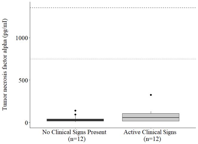
human medicine [1–5] and studies of laboratory an
Measurements of acute-phase proteins (APPs) and c
understanding disease processes and susceptibility, moni
the efficacy of treatments. APPs form an integral part of the
innate immune system. Changes in APPs have been obs
Animals 2020, 10, 1756 3 of 19
neoplasia, stress and trauma, playing a role in promoting
Prior to activation, APPs are generally present in negligibl
Africa [42,43], and Asia [44–51]. Although active disease can be and scale ofvia
detected production
culturing trunk[7]. With some, a rapid, high ma
secretions,
it is not possible to diagnose subclinical infection with currentinflammatory
methodologies.events.Understanding
Others of moderate the concentration rem
associated with chronic
host immune response to infection could yield important information regarding the transition from inflammation. Thus, the evaluatio
of efficacy
subclinical to active infection, susceptibility to disease, and the the clinical progression
of various treatments. of inflammatory responses, a
In addition to infectious disease, biomarkers indicativechronic and acuteand
of inflammation conditions
immune[13]. Furthermore, the rela
function
would improve the ability to detect and manage other commonresponsepathologies.makes measuring
A recent surveyAPP concentrations useful f
of elephant
health in North America highlighted gastrointestinal issues,clinical signs and
skin lesions are manifested
wounds, lameness [12]. APPsandhave been measur
foot lesions, eye issues, tusk and sulcus injuries, and dental disorders as prevalent pathology types [52]. African elephan
years, including Asian [14–16] and a single
Similarly, Miller et al. [53] highlighted injuries, parasitism,such as elephantdisease,
gastrointestinal endotheliotropic
and infectiousherpesvirus (EEHV), p
disease as being responsible for elephant morbidity and mortalityelephants, serum
in Asia. Allamyloid A (SAA) isare
these pathologies considered to be a m
to acute inflammation,
associated with inflammation and immune activation, and therefore tools to help with sub-clinical whereas haptoglobin (HP) respond
detection, diagnosis and treatment would benefit overall elephantof more chronic
health. inflammation
To-date, published[15]. reference
ranges only exist for two APPs, SAA and HP, in Asian elephants [16]; Cytokines
none haveare protein
yet been mediators
established for of the immune
proliferating,
African elephants. The goal of this study was to establish and validate assaysactivating,
for several differentiating,
other candidate and otherwise re
biomarkers using commercially available reagents and calculate pro-inflammatory,
reference intervalssecreted for at the beginning
African and of an immun
to downregulate the immune
Asian elephants in North American zoos. Although reference interval calculations were conducted response and prevent over-
using standard methodology [54], we hereafter refer to theseassociated with cell-mediated,
as species-specific value ranges because humoral, and innate im
underlying health issues without overt clinical signs cannotinformative
be ruled out. regarding immune
Specifically, activation and progressio
we analyzed
APPs (SAA and HP) and cytokines (tumor necrosis factor-alpha Typically,
(TNF-α), Th1interferon-gamma
cytokines such as(IFN- IFN-ɣ,),IL-2, and TNF-α
interleukins 2 (IL-2), 6 (IL-6) and 10 (IL-10)) and compared combat
values tointracellular
individualspathogens
exhibiting (e.g., clinicalviruses), whereas
promote humoral immune
signs of illness or injury, or prior to death. To add to the current literature available, we also report responses, targeting extracellu
serum chemistry value ranges from the same population, representing a large sample-set analyzed in of cytokines prod
and parasites). Indeed, the combination
the same laboratory. immune response [18], as well as provide prognostic
progression of disease [19,20]. Some cytokine mRNAs ha
2. Materials and Methods [21,22] and studied previously [23–27]; however, measu
through different pathological processes are lacking, as ar
2.1. Subjects, Sample Collection, and Assessment of Health Status
or population to better interpret results.
Single serum samples were obtained from 229 elephants housed Several health problems
at 69 institutions in North affect elephants both in situ a
America.
Subjects included 125 African (18 male, 107 female) and 104 Asianunderstanding
(18 male, 86 of female)
underlying disease
elephants agedprocesses and impr
Elephant endotheliotropic
4 to 65 years. Additional serum samples were collected opportunistically from elephants herpesvirus
with activehemorrhagic disea
clinical pathology at the time of sample collection (n = 10) and Asian
prior toelephant = 10),born
death (ncalves and from in zoostheglobally
same [28], as well as A
individuals when no clinical signs were present, a minimumand wild
of one populations
month before or in after
Asia the[31–37]. One proposed hypo
clinical
sample. Weekly serum samples collected prior to and following beroutine
that immunologically
vaccination withnaï ve calves
tetanus toxoidfail to mount an eff
(1 mL intramuscularly) and Imrab 3 (1 mL intramuscularly)viral werereplication
also collected[38].from
A better
a femaleunderstanding
Asian of how the h
elephant, aged 21 years. Active clinical cases and conditions would
presenttherefore be beneficial
at necropsy and could help assess the ef
were determined
by the attending veterinarian or pathologist. Blood samplesoptions, once they
were collected becometoavailable.
according phlebotomy Elephants are also su
protocols at each institution, typically from an ear vein while tuberculosis, the same
the elephant wascausative agent as that for human tu
under behavioral
restraint. After being allowed to clot at room temperature forcurrently
1 h, serum residing in North and
was separated America
frozen have
at tested positive f
◦
−20 C before shipment to the Smithsonian Conservation Biology Institute for analysis. This research
was approved by the Smithsonian National Zoo (NZP-ACUC #11-10, #15-03, and #18-18) and where
applicable, was reviewed and approved by participating zoo research and animal care committees.
2.2. Acute Phase Protein Analysis
SAA and HP were measured using an RX Daytona automated clinical chemistry analyzer (Randox
Industries-US Ltd., Kearneysville, WV, USA). Commercially available reagents, calibrators, and two-level
controls were used (Eiken Chemical Co. Ltd., Tokyo, Japan and Tridelta Tri-DD, Boonton, NJ, USA,
respectively). The technical ranges were 0.1 to 500 mg/L and 0.01 to 2.5 mg/mL, respectively. The analyzer
was subject to routine quality control measurements throughout the study, with normal and elevated
controls for each analyte maintained within 2 standard deviations (SD) of the respective lot-specific
target value. Samples were typically analyzed neat, but some with HP above the technical range were
diluted 1:5 or 1:10 in calibrator diluent as needed.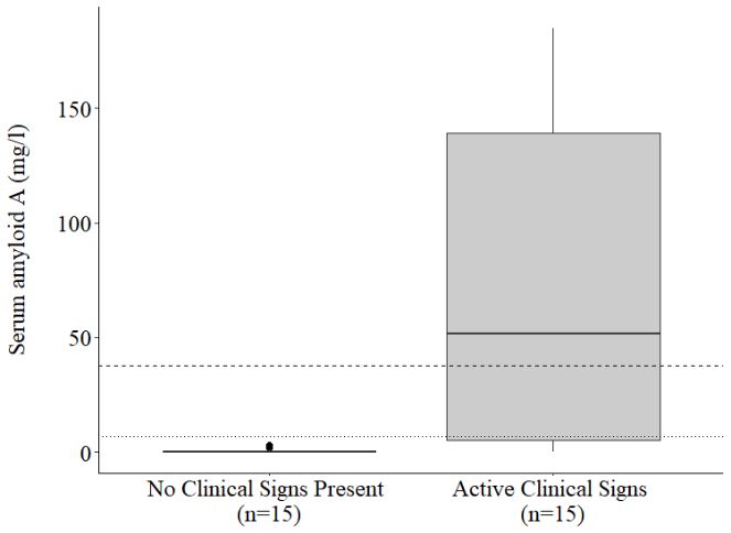
2020, 10, x Animals 2020, 10, 1756 2 of 23 4 of 19
ords: Acute phase proteins; acquired immunity; cytokines; Elephas maximus; ELISA; equine;
immunity; Loxodonta
2.3.africana;
Cytokine reference
Enzymeintervals; serum chemistries
Immunoassays
TNF-α was measured using an equine TNF-α enzyme immunoassay (EIA) (Invitrogen ESS0017;
Thermo Fisher Scientific, Frederick, MD, USA) according to the manufacturer’s instructions (Table 1).
duction In brief, anti-equine TNF-α coating antibody was diluted in carbonate-bicarbonate buffer (0.2 M,
10, x pH 9.4),ofand 100 µL addedare to important
each well health of a 96-well microtiter 2 of (Costar,
23
rum biomarkers indicative immune function assessment tools inplate Corning Life Sciences,
medicine [1–5] and Tewkesbury,
studies of MA, USA).and
laboratory Following
domesticatedincubation animalatspeciesroom temperature
[6–11]. overnight, coating antibody
s: Acute of
rements phase proteins;proteins
acute-phase acquired(APPs) immunity; and cytokines;aid Elephas maximus; ELISA; equine;
solution was aspirated, andcytokines
wells were blocked in detecting with apathologies,
Dulbecco’s phosphate-buffered saline (D-PBS;
munity; Loxodonta
tanding disease africana;susceptibility,
processes reference intervals; serum chemistries and assessing
8 mMand Na2 HPO4 , 2 mMmonitoring KH2 PO4 ,disease 0.14 Mprogression,
NaCl, 10 mM KCl, pH 7.4.) solution containing 4% bovine
cacy of treatments. APPs form an integral part of the acute-phase response, contributing to the
serum albumin (BSA) and 5% sucrose, for a minimum of 1 h. Blocking buffer was aspirated and 50 µL
immune system. Changes in APPs have been observed in cases of inflammation, infection,
standards, controls, or samples added in duplicate, before incubation for 1 h at room temperature
sia, stress and trauma, playing a role in promoting healing, and restoring homeostasis [12].
otion while
activation, APPs are generally shaking
present at in
500 RPM. Recombinant
negligible equine TNF-α
amounts and characterized by the standards
speed were serially diluted in reagent
ale of production [7].diluent With some, (4% aBSA rapid,in high
D-PBS, pH 7.4),response
magnitude and additionally
is associateddiluted with acute to provide high and low concentration
mmatory
biomarkers indicative
events. Others control
of moderateof immune
samples. function
Serum
concentration wasare
remain important
typically
elevated runmore
for health
neat or assessment
diluted
prolonged up to
periods, tools1:20inin reagent diluent as needed.
ted with chronic
edicine [1–5] inflammation.
and studies
Plates were Thus,
of
then the evaluation
laboratory
washed and
three of times
APPs in serumTween
domesticated
(0.05% enables TM
animal interpretation
-20species
in D-PBS, [6–11].
pH 7.4), before 100 µL per well of
clinical progression
ents of acute-phase of inflammatory
proteins
anti-equine TNF-α responses,
(APPs) and
and antibody
detection the potential
cytokineswas to
aidadded distinguish
in detecting between
and incubated pathologies,
for a further 1 h at room temperature
and disease
acute conditions [13]. Furthermore,
ding processes
while and
shaking at 500 the
susceptibility, RPM. relatively
monitoring
Following rapid
a response
disease
further of the acute-phase
progression,
three washes, and 100 assessing
µL per well streptavidin-horseradish
se makes measuring APP concentrations useful for identifying sub-clinical disease before
of treatments. APPs form an(diluted
peroxidase integral 1:400 part of inthe acute-phase
reagent diluent)response,
was added contributing
and incubated to thefor 30 min at room temperature
signs are manifested [12]. APPs have been measured in a variety of wildlife species in recent
mune system. Changes
ncluding Asian [14–16] while ina single
andshaking
APPsAfrican have
at 500 been
RPM.
elephant
observed
After in wash
[17]atofinal
assess
cases of inflammation,
step,
the response 100toµL infection, substrate solution was added
of chromogenic
pathologies
stress and trauma, playing
per well, a role
incubated in promoting
in the dark healing,
for 20
s elephant endotheliotropic herpesvirus (EEHV), pododermatitis, trauma, and infection. In min andat restoring
room homeostasis
temperature, stopped [12].with 100 µL stop solution and
ivation, APPs
nts, serum amyloid A are generally
absorbance present
measured
(SAA) is considered in negligible
to at
be 450
a major amounts
nm APP, and
with increasing
a reference characterized
of 570innm.
rapidly by
response the speed
of production [7].
e inflammation, Withhaptoglobin
whereas some, a rapid, high magnitude
(HP) responds more moderatelyresponse andismayassociated
be reflective with acute
e chronic
ory events.inflammation [15].
Others of moderate Information for remain
Table 1. concentration cytokineelevated
enzyme immunoassays
for more prolonged validated for African and Asian elephants.
periods,
ytokines
with chronic are protein mediators
inflammation. Thus,of the immune response,
the evaluation of APPsassociated
in serum with enables recruiting,
interpretation Antibody Concentration
ating, activating, differentiating,
Cytokine and otherwise regulating immune Reagents
Supplier cells. Cytokines mayStandard be Range
ical progression of inflammatory responses, and the potential to distinguish between Capture Detection
lammatory, secreted at the beginning of an immune response, or anti-inflammatory, secreted
d acute conditions [13]. Tumor Furthermore,
necrosis the relatively rapid response of the acute-phase
nregulate the immune response and preventThermo over-activation. There are numerous cytokines
makes
ted with measuring APP
cell-mediated, concentrations
factor-alpha
humoral, useful
andScientific/Invitrogen
innate for identifying
immune responses,ESS0017 sub-clinical
which can bedisease before
3.9–1000
highly pg/mL * 1:100 1:100
(TNF-α)
ns
ativeareregarding
manifested [12].activation
immune APPs have and been measured
progression in in a variety
response to a of wildlife
variety of species in recent
pathologies.
Interferon-gamma
uding
ly, Th1Asian [14–16]
cytokines suchand a single
as (IFN-
IFN-ɣ, ) IL-2, andR&D
African TNF-α Systems
elephant stimulate[17] to Duoset
assess
cell-mediated theDY1586
response
immunity to 15.6–4000 pg/mL *
to pathologies
help 0.4 µg/mL 0.4 µg/mL
intracellular pathogens Interleukin
(e.g.,
phant endotheliotropic herpesvirus (EEHV), 1β
viruses), whereas Th2 cytokines,
pododermatitis, including IL-10 and IL-6,
R&D Systems Duosettrauma,
DY3340 and infection. 125–8000 pg/mL In 0.8 µg/mL 0.15 µg/mL
e humoral immune (IL-1β)targeting extracellular pathogens (e.g., extracellular bacteria
serum amyloid A responses,
(SAA) is considered
Interleukin 2 to be a major APP, increasing rapidly in response
rasites). Indeed, the combination of cytokines R&Dproduced
Systems can reflect the type and stage of15.6–4000
may bethe
Duoset DY1613 pg/mL * 2.0 µg/mL 0.2 µg/mL
lammation, whereas haptoglobin (IL-2) (HP) responds more moderately and reflective
e response [18], as well as provide
Interleukin 4 prognostic information regarding the likelihood or
ronic inflammation [15]. R&D Systems Duoset DY1809 31.2–2000 pg/mL 0.8 µg/mL 0.8 µg/mL
ssion of disease [19,20]. Some cytokine mRNAs have been characterized in Asian elephants
(IL-4)
ines are protein mediators
and studied previouslyInterleukin of
[23–27]; 6however, the immune
measures response, associated with recruiting,
R&D Systems of circulating protein concentrations
AF1886/BAF1886/1886-EL 0.1–25 ng/mL * 0.4 µg/mL 0.4 µg/mL
ng, activating, (IL-6)
differentiating,
h different pathological processes and otherwise
are lacking, as areregulating
typical values immuneobserved cells.
withinCytokines
a speciesmay be
Interleukin 10
matory,
ulation to secreted at theresults.
better interpret beginning
(IL-10) of an R&Dimmune
Systems response,Duoset or anti-inflammatory,
DY1605 secreted
0.078–20 ng/mL * 0.4 µg/mL 0.1 µg/mL
veral health
gulate problems response
the immune affect elephantsand both in situ
prevent and ex situ that There
over-activation. could benefit
are from a better
numerous cytokines
* Assay sensitivity was increased throughout the course of assay development, so samples at the lower end of
tanding of underlying disease
with cell-mediated, humoral, processes
detection may and and improved
innate
be higher thanimmune tools
the final for given
detection
responses,
range and monitoring.
here.which can be highly
nt endotheliotropic herpesvirus hemorrhagic disease
e regarding immune activation and progression in response to a variety (EEHV HD) affects almost oneofinpathologies.
four
lephant calves born in zoos globally [28], as well as African elephants [17,29,30], and in captive
Th1 cytokines such as IFN- IFN-ɣ,, IL-2, and IL-10 TNF-αwere stimulate
measured cell-mediated
using equine immunity
Duosets to(R&D
help Systems, Inc., Minneapolis,
ld populations in Asia [31–37]. One proposed hypothesis for the severity of EEHV HD could
racellular pathogens
immunologically naï MN, (e.g.,
USA)fail
ve calves
viruses),
according
to mount to
whereas
an modified
Th2 cytokines,
effective immune manufacturer’s including
response toinstructions
IL-10 and IL-6,
keep up with(Table 1). In brief, goat anti-equine
umoral immune
plication [38]. A better responses,
coating targeting
antibodyofwas
understanding extracellular
howdiluted
the hostin pathogens
phosphate-buffered
immune (e.g.,
system responds extracellular
saline
to the(PBS;virusbacteria
137 mM NaCl, 2.7 mM KCl, 8.1 mM
tes). Indeed,
therefore the combination
be beneficialNaand2 HPO could of
4 , 1.5
help cytokines
mM KHthe
assess 2 PO produced
4 , pH 7.4),
efficacy of can and reflect
treatment 100and thenovel
µL type
was and stage
added
vaccinationto each of well
the of a 96-well microtiter plate
esponse
, once they [18],
becomeas well
available.
(Costar). as provide
Elephants
Following prognostic
areincubation information
also susceptible to infection
at room regarding
temperature the
with Mycobacterium likelihood
overnight, or
the coating antibody solution was
nlosis, the same [19,20].
of disease causative agent
Some as that
cytokine for human
mRNAs tuberculosis
have been (TB). Around
characterized 10% inof
aspirated, and plates were washed three times with wash buffer (0.05% Tween TM -20 in PBS, pH 7.4) elephants
Asian elephants
ly residing in
d studied previously North America have however, tested positive for TB, with cases also identified inconcentrations
Europe
and [23–27];
then blocked with ameasures 4% BSA, 5% of circulating
sucrose PBSprotein solution for a minimum of 1 h. Blocking buffer was
fferent pathological processes
aspirated and are50lacking,
µL standards,as are typical
controls, values observed
and samples within
added in aduplicate,
species before incubation for 2 h at
on to better interpret results.
room temperature while shaking at 500 RPM. Recombinant equine standards and control samples were
al health problemsdiluted affect elephants
in 50% fetal both in situserum
bovine and ex(FBS)situ that could benefit
in reagent diluent from (1% BSA a betterin PBS, pH 7.4). Serum samples
ding of underlyingwere disease processes and improved tools for detection and
typically run neat or diluted up to 1:20. Plates were then washed three times before 100 µL per monitoring.
ndotheliotropic herpesvirus hemorrhagic
well of biotinylated goat disease
anti-equine (EEHV HD) affects
detection antibody almost
(dilutedone in reagent
four diluent without FBS) was
hant calves born in zoos globally [28], as well as African elephants [17,29,30], and in captive
opulations in Asia [31–37]. One proposed hypothesis for the severity of EEHV HD could
munologically naïve calves fail to mount an effective immune response to keep up with
ation [38]. A better understanding of how the host immune system responds to the virus
efore be beneficial and could help assess the efficacy of treatment and novel vaccination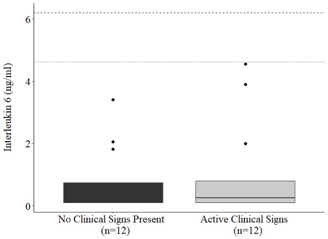
Animals 2020, 10, 1756 5 of 19
added and incubated for a further 2 h at room temperature while shaking at 500 RPM. Following a
further three washes, 100 µL per well streptavidin-horseradish peroxidase (diluted 1:200 in reagent
diluent without FBS) was added and incubated in the dark for 20 min at room temperature. After a final
wash step, 100 µL of substrate solution (high kinetic TMB peroxidase substrate, Moss, Inc., Pasadena,
MD, USA) was added per well, incubated in the dark at room temperature, stopped with 50 µL of stop
solution (1N HCl), and absorbance measured at 450 nm with a reference of 570 nm. Anti-equine IL-1β
and IL-4 Duoset antibodies (Table 1) also showed good cross-reactivity with both African and Asian
elephant serum following a similar protocol except for dilution of detection antibody in 2% FBS in
reagent diluent. However, intermittent issues with elevated background prevented the measurement
of IL-1β and IL-4 for the remainder of this study. IL-6 was measured using goat anti-equine antibodies
Animals 2020, 10, x
(Table 1) with a protocol similar to that described for Duosets, except that standards and controls
were diluted in reagent diluent without FBS. All EIAsAcute
Keywords: were biochemically
phase proteins;validated
acquiredaccording
immunity;tocytokines;
the Elephas ma
manufacturer’s recommendations prior to innate
the start of the study by performing spike and recovery and
immunity; Loxodonta africana; reference intervals; serum chemistries
linearity assessments with elephant serum [55,56]. Inter-assay coefficients of variation were maintained
below 15% for high and low concentration controls on all assays.
2.4. Serum Chemistries
1. Introduction
Twenty-two serum analytes (Alanine aminotransferase, albumin, alkaline phosphatase, aspartate
Serum biomarkers indicative of immune function are important healt
aminotransferase, bilirubin, calcium, carbon dioxide, chloride, cholesterol, creatine kinase, creatinine,
human medicine [1–5] and studies of laboratory and domesticated a
gamma glutamyl transferase, glucose, iron, lactate dehydrogenase, magnesium, phosphorus, potassium,
Measurements of acute-phase proteins (APPs) and cytokines aid in d
sodium, total protein, triglycerides, and urea nitrogen) were measured in each sample using a
understanding disease processes and susceptibility, monitoring disease prog
Dimension® Xpand Plus automated clinical chemistry analyzer (Siemens Medical Solutions USA, Inc.,
the efficacy of treatments. APPs form an integral part of the acute-phase respo
Malvern, PA, USA). The analyzer was subject to routine quality control measurements throughout the
innate immune system. Changes in APPs have been observed in cases of in
study, with two-level controls maintained within manufacturer specifications.
neoplasia, stress and trauma, playing a role in promoting healing, and resto
2.5. Value Range Calculation Prior to activation, APPs are generally present in negligible amounts and char
and scale of production [7]. With some, a rapid, high magnitude response is
Value ranges for African and Asian elephants under
inflammatory events.human
Others of care were calculated
moderate for each
concentration remain elevated for m
serum biomarker according to reference interval guidelines from the American Society for Veterinary
associated with chronic inflammation. Thus, the evaluation of APPs in serum
Clinical Pathology [54]. Value ranges for serum
of the chemistry parameters of
clinical progression were generated using
inflammatory the robust
responses, and the potential to
method, and immune biomarkers using the nonparametric method, all using the “referenceIntervals”
chronic and acute conditions [13]. Furthermore, the relatively rapid respon
package [57] in R statistical software [58],response
version 3.6.1.
makes Outlying
measuring valuesAPPwere identified using
concentrations Cook’s
useful for identifying sub-
distance and were removed prior to calculation;
clinical all value
signs are ranges represent
manifested [12]. 95%
APPsofhave
the population
been measured and in a variety of w
are reported with 90% confidence intervals. years, including Asian [14–16] and a single African elephant [17] to assess the r
such as elephant endotheliotropic herpesvirus (EEHV), pododermatitis, tra
2.6. Statistical Analyses
elephants, serum amyloid A (SAA) is considered to be a major APP, increasi
Each of the 29 analytes (2 APP, 5 cytokine,
to acuteand 22 serum chemistry)
inflammation, used for determining
whereas haptoglobin value more moderately
(HP) responds
ranges were compared by species (125 Africanof more and 104 Asian)
chronic using a Mann–Whitney
inflammation [15]. Wilcoxon test.
A Wilcoxon signed-rank test was used to compare Cytokines eachareof the sevenmediators
protein immune biomarkers (2 APP, response, associ
of the immune
5 cytokine) in individuals that exhibited clinical signs ofactivating,
proliferating, illness to control samples taken
differentiating, from the same
and otherwise regulating immune c
individual when no clinical signs were exhibited. All analyses
pro-inflammatory, were conducted
secreted in R [58],
at the beginning of version
an immune3.6.1,response, or anti-i
with alpha set to 0.05. to downregulate the immune response and prevent over-activation. There ar
associated with cell-mediated, humoral, and innate immune responses,
3. Results informative regarding immune activation and progression in response to a
Concentrations of SAA (p < 0.001), HP (p < 0.001),
Typically, Th1 TNF-α
cytokines(p =such as IFN-ɣ,
0.011), IFN- (p = 0.025),
IL-2, and TNF-α
and IL-2stimulate cell-medi
(p = 0.029) were higher in Asian elephants, with IL-6 (p = 0.464) and IL-10 (p = 0.139) not differing cytokines, incl
combat intracellular pathogens (e.g., viruses), whereas Th2
between species (Table 2). Value ranges promote
calculated humoral
for APPs immune responses,
and cytokines are targeting
presentedextracellular
in Table 2. pathogens (e.g.
A summary of APP and cytokine concentrationsand parasites). Indeed, the
in individuals combination
with of cytokines
active clinical signs of produced
injury can reflect the
immune response [18], as well as provide
or illness is shown in Table 3, and around the time of death in Table 4. SAA was elevated above prognostic information regard
progression
species-specific value ranges in 10/12 clinical of disease
cases (Table 3) and[19,20]. Some cytokine
5/10 individuals mRNAsup
at or leading have
to been characteriz
death (Table 4), with the highest concentrations (251.82 mg/L) observed in an African elephant with of circulating
[21,22] and studied previously [23–27]; however, measures
through different pathological processes are lacking, as are typical values obs
or population to better interpret results.
Several health problems affect elephants both in situ and ex situ that cou
understanding of underlying disease processes and improved tools for dete
Elephant endotheliotropic herpesvirus hemorrhagic disease (EEHV HD) affe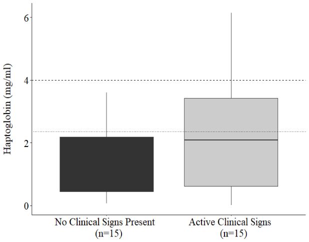
Animals 2020, 10, 1756                                                                            6 of 19

       bronchopneumonia. Overall, SAA was higher in individuals with active clinical signs of illness or
       injury compared to the same individuals when no clinical signs were apparent (p = 0.004; Figure 1a).
       HP was elevated in 5/12 clinical cases (Table 3) and 2/10 individuals leading up to death (Table 4),
      with the highest concentrations observed in an Asian elephant that had become recumbent and unable
       to rise. Average HP concentrations did not differ between individuals with clinical signs compared to
Animalsthose
        2020, without
              10, x   (Figure 1b) (p = 0.477).                                                       10 of 23

                              (a)                                                (b)

                              (c)                                                (d)

                              (e)                                                (f)
                                                  Figure 1. Cont.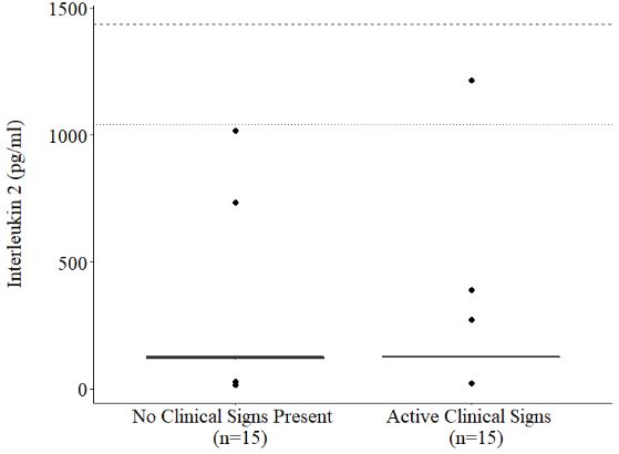
1. Introduction
Serum biomarkers indicative of immune function are important health assessment tools in
human medicine [1–5] and studies of laboratory and domesticated animal species [6–11].
MeasurementsAnimals 2020, 10, 1756 proteins (APPs) and cytokines aid in detecting pathologies,
of acute-phase 7 of 19
(e)
understanding disease processes and susceptibility, monitoring disease progression, and assessing (f)
the efficacy of treatments. APPs form an integral part of the acute-phase response, contributing to the
innate immune system. Changes in APPs have been observed in cases of inflammation, infection,
neoplasia, stress and trauma, playing a role in promoting healing, and restoring homeostasis [12].
Prior to activation, APPs are generally present in negligible amounts and characterized by the speed
and scale of production [7]. With some, a rapid, high magnitude response is associated with acute
inflammatory events.
AnimalsOthers
2020, 10,ofx moderate concentration remain elevated for more prolonged periods, 11 of 23
associated with chronic inflammation. Thus, the evaluation of APPs in serum enables interpretation
of the clinical progression
Figure 1.of inflammatory
Paired concentrations responses, and the potential
of serum biomarkers (a) SAA (pto= 0.004),
distinguish
(b) HP,between
(c) TNF-α (p = 0.021),
chronic and acute conditions [13]. Furthermore, the relatively rapid response of the
(d) TNF-α outliers removed, (e) IFN-ɣ, (f) IL-2, (g) IL-6, and (h) IL-10 in individuals with or without acute-phase
response makes measuring APP signs
active clinical concentrations
present. Theuseful for identifying
calculated upper limitssub-clinical disease before
of the species-specific value ranges are
clinical signs are manifested
denoted by [12]. APPs(E.m.)
dashed haveorbeen
dotted measured in a variety
(L.a.) horizontal lines. of wildlife species in recent
years, including Asian [14–16] and a single African elephant [17] to assess the response to pathologies
Cytokine concentrations
such as elephant endotheliotropic (g) were
herpesvirus elevated
(EEHV), above the species-specific
pododermatitis, trauma, and value
(h) rangesIn
infection. in several clinical
elephants, serum cases
amyloid(TableA 3), (SAA)including pododermatitis,
is considered to be a major systemic
APP,infection,
increasingacute rapidly lameness,
in response ventral edema, and
tusk Figure 1. Paired
infection. concentrations
Overall, TNF-α of serum
was higher biomarkers
in (a) SAAwith
individuals (p = active
0.004), (b) HP, (c)
clinical TNF-α
signs of (p = 0.021),
illness or injury
to acute inflammation, whereas haptoglobin (HP) responds more moderately and may be reflective
(d) TNF-α outliers removed, (e) IFN-γ, (f) IL-2, (g) IL-6, and (h) IL-10 in individuals with or without
compared
of more chronic inflammation [15]. to when no clinical signs were apparent (p = 0.021), IL-10 tended to be higher during active
active clinical signs present. The calculated upper limits of the species-specific value ranges are denoted
Cytokines clinical
are proteincases (pmediators
= 0.059), whereasof theIFN-ɣ,immune IL-2, and IL-6 didassociated
response, not differ significantly
with recruiting, despite higher mean
by dashed (E.m.) or dotted (L.a.) horizontal lines.
concentrations in clinical cases (Figure 1). One female
proliferating, activating, differentiating, and otherwise regulating immune cells. Cytokines may be African elephant, representing two clinical
pro-inflammatory, cases (tusk at
secreted
Cytokine injury and infection),
the beginning
concentrations had TNF-α
of anelevated
were immune concentrations
response,
above considerably
or anti-inflammatory,
the species-specific value ranges higher
secreted than all
in several other
clinical
to downregulate individuals
the immune tested.
responseWhen samples
and prevent from this individual
over-activation. were
There excluded
are
cases (Table 3), including pododermatitis, systemic infection, acute lameness, ventral edema, and tusk numerous from the paired
cytokines comparison,
associated with TNF-α
infection. in
cell-mediated,clinical
Overall, samples
humoral,
TNF-α wasonlyand tended
higher innate to be higher
immune
in individuals with than non-clinical
responses,
active which
clinical signs samples
can be highly
of illness (p
or =injury
0.068). In many
compared
tocases,
whencytokines
informative regarding clinicalwere
immune
no signsonly
weremildly
activation and
apparent elevated and
(p = 0.021),
progression did
in response
IL-10not exceed
to ato
tended the
variety upper
be higher value range
of pathologies.
during activeor were cases
clinical below
the detection
= 0.059), whereas
Typically, Th1(pcytokines limit
such as IFN-ɣ, of the assays;
IL-2, and
IFN- , IL-2, in
andIL-6 no
TNF-α cases
did not were
stimulate all seven
cell-mediated
differ significantly biomarkers
immunity
despite elevated
highertomean concurrently.
helpconcentrations By
contrast,
combat intracellular
in clinical routine
pathogens
cases (Figure vaccination
(e.g., 1).
viruses),of a female
whereas
One female African Asian elephant
Th2elephant,
cytokines, against tetanus
including IL-10
representing and rabies
and IL-6,
two clinical cases (tusk injuryan
stimulated
promote humoral increase
and immune in SAA
infection), hadand
responses,TNF-αalltargeting
cytokines (Figure
extracellular
concentrations 2). Concentrations
considerablypathogens higher (e.g.,were
than allall below
extracellular
other assay detection
bacteria
individuals at nine
tested. When
and
and parasites).samples two
Indeed,from days prior
the combination to
this individual vaccination
of cytokines but
were excluded increased
produced fromcan from 7
the reflect (SAA),
paired the 12 (IFN-γ,
type and TNF-α
comparison, IL-2), or
stage ofinthe 19 (TNF-α, IL-10)
clinical samples
immune response daystended
only post-vaccination.
[18], astowell as provide
be higher IL-6 non-clinical
than showed
prognostic a samples
similar
information pattern
(p = 0.068). to that
regarding the
In many of likelihood
IFN-γ andorIL-2,were
cases, cytokines but only
peak
concentrations
progression ofmildly
disease [19,20]. could
Some only be
cytokine extrapolated,
mRNAs have so instead
been are presented
characterized
elevated and did not exceed the upper value range or were below the detection limit of the in as optical
Asian density
elephants (OD).
assays;Descriptive
[21,22] and studied previously
in no cases were statistics
[23–27]; and calculated
however,
all seven measures
biomarkers value ofranges
elevated for 22 protein
circulating
concurrently. serum biochemistriesroutineare
concentrations
By contrast, providedofin
vaccination
a Table
through different female 5.Asian
Compared
pathological elephant to previously
processes published
are lacking,
against tetanus asand data [59],
are rabies
typical values value
stimulated ranges
observed
an increasein ourinstudy
within aSAA wereall
species
and narrower
cytokines for
albumin,
or population (Figure alkaline
to better2).interpret phosphatase, bilirubin, calcium, cholesterol (Asians
results. were all below assay detection at nine and two days prior to vaccination but
Concentrations only), creatinine, gamma
glutamyl
Several health
increasedproblems transferase,
from affect
7 (SAA), glucose
elephants
12 (IFN-γ,(Africans
both in situ
IL-2), only),
or and iron,
ex
19 (TNF-α, situmagnesium,
that could
IL-10) days potassium,
benefit from atotal
post-vaccination. protein,
better and urea
IL-6 showed a
nitrogen.
understandingsimilar
of underlying Except for
disease alkaline
processesphosphatase,
and improved aspartate
tools aminotransferase,
for detection
pattern to that of IFN-γ and IL-2, but peak concentrations could only be extrapolated, so instead and creatinine,
monitoring. carbon dioxide,
and
Elephant endotheliotropic
are glucose,herpesvirus
presented alloptical
as other biochemistries(OD). differed
hemorrhagic
density disease significantly
(EEHV HD) affects between species
almost one(p in
≤ 0.04).
four
Asian elephant calves born in zoos globally [28], as well as African elephants [17,29,30], and in captive
and wild populations in Asia [31–37]. One proposed hypothesis for the severity of EEHV HD could
be that immunologically 24 naïve calves fail to mount an effective immune 4response to keep up with
viral replication [38]. A better understanding of how the host immune system responds to the virus
Serum amyloid A (mg/l)
Haptoglobin (mg/ml)
would therefore be beneficial
18 and could help assess the efficacy of treatment
3 and novel vaccination
options, once they become available. Elephants are also susceptible to infection with Mycobacterium
tuberculosis, the same causative
12 agent as that for human tuberculosis (TB).2Around 10% of elephants
currently residing in North America have tested positive for TB, with cases also identified in Europe
6 1
0 0
-10 0 10 20 30 40 -10 0 10 20 30 40
(a) (b)
Figure 2. Cont.Animals 2020, 10, 1756 8 of 19
Animals 2020, 10, x 12 of 23
20 4000
Interferon gamma (pg/ml)
Tumor necrosis factor alpha
15 3000
(pg/ml)
10 2000
5 1000
0 0
-10 0 10 20 30 40 -10 0 10 20 30 40
(c) (d)
200 4
Interleukin 2 (pg/ml)
150 3
Interleukin 6 (OD)
100 2
50 1
0 0
-10 0 10 20 30 40 -10 10 30
(e) (f)
1.6
Interleukin 10 (ng/ml)
1.2
0.8
0.4
0.0
-10 0 10 20 30 40
(g)
Figure 2.
Figure 2. Response
Response toto tetanus
tetanus and
and rabies
rabies vaccination
vaccination inin an
an adult
adult female
female Asian
Asian elephant,
elephant, aged
aged 21
21 years:
years:
(a) SAA,
(a) SAA, (b)
(b) HP,
HP,(c)
(c)TNF-α,
TNF-α,(d)
(d)IFN-γ,
IFN-γ,(e)
(e)IL-2,
IL-2,(f)
(f)IL-6,
IL-6,and
and(g)
(g)IL-10.
IL-10. Dashed
Dashed line
line denotes
denotes the
the day
day of
of
vaccination, day
vaccination, day 0.
0.
Descriptive statistics and calculated value ranges for 22 serum biochemistries are provided
in Table 5. Compared to previously published data [59], value ranges in our study were narrower
for albumin, alkaline phosphatase, bilirubin, calcium, cholesterol (Asians only), creatinine, gamma
glutamyl transferase, glucose (Africans only), iron, magnesium, potassium, total protein, and urea
nitrogen. Except for alkaline phosphatase, aspartate aminotransferase, creatinine, carbon dioxide, and
glucose, all other biochemistries differed significantly between species (p ≤ 0.04).nflammatory events. Others of moderate concentration remain elevated for more prolonged periods,
ssociated with chronic inflammation. Thus, the evaluation of APPs in serum enables interpretation
f the clinical progression of inflammatory responses, and the potential to distinguish between1. Introduction
hronic and acute conditions [13]. Furthermore, the relatively rapid response of the acute-phase Serum biomarkers indicative of immune function are important health assessment tools in
esponse makes measuring APP concentrations useful for identifying sub-clinical disease beforehuman medicine [1–5] and studies of laboratory and domesticated animal species [6–11].
linical signs are manifested [12].10,
Animals 2020, APPs
1756have been measured in a variety of wildlife species in recentMeasurements of acute-phase proteins (APPs) and cytokines aid in detecting pathologies, 9 of 19
ears, including Asian [14–16] and a single African elephant [17] to assess the response to pathologiesunderstanding disease processes and susceptibility, monitoring disease progression, and assessing
uch as elephant endotheliotropic herpesvirus (EEHV), pododermatitis, trauma, and infection. Inthe efficacy of treatments. APPs form an integral part of the acute-phase response, contributing to the
lephants, serum amyloidTable 2. Descriptive
A (SAA) is considered statistics
to be and calculated
a major value ranges
APP, increasing (with in
rapidly 90% confidence
response intervals,
innate immune CI)system.
for serum acute-phase
Changes in APPs proteins
have and
beencytokines
observedfrom 125 African
in cases (L.a.) and infection,
of inflammation,
o acute inflammation, whereas104 Asian (E.m.) elephants
haptoglobin under human
(HP) responds care.
more moderately and may be reflectiveneoplasia, stress and trauma, playing a role in promoting healing, and restoring homeostasis [12].
f more chronic inflammation [15]. Prior to activation, APPs areagenerally present in negligible amounts and characterized by the speed
Analyte Species Mean SD Median Minimum Maximum N Value Range Lower CI b Upper CI
Cytokines are protein mediators of the immune response, associated with recruiting,and scale of production [7]. With some, a rapid, high magnitude response is associated with acute
roliferating, activating, differentiating, and otherwiseL.a.
SAA (mg/L) regulating4.16 immune cells.26.25 Cytokines
0.10 may be0.10 251.82 123 0.10–6.91 - 6.38–8.38
inflammatory events. Others of moderate concentration remain elevated for more prolonged periods,
* E.m. 16.04 47.05 1.84 0.10 231.92 98 0.10–37.62 - 23.98–53.25
ro-inflammatory, secreted at the beginning of an immune response, or anti-inflammatory, secretedassociated with chronic inflammation. Thus, the evaluation of APPs in serum enables interpretation
L.a. 1.36 0.74 1.40 0.19 5.45 124 0.21–2.35 0.18–0.23 2.25–2.40
o downregulate the immune response HP (mg/mL)
and prevent over-activation.
* E.m. 1.96There are 1.57numerous 1.83cytokines0.19
of the clinical progression100of inflammatory
10.50 0.24–4.00 responses, and the potential
0.18–0.29 to distinguish between
2.95–4.93
ssociated with cell-mediated, humoral, and innateL.a. immune 311.89responses,1783.69
which can15.60 be highly15.60
chronic and17,381.84
acute 123 [13]. 15.60–748.10
conditions Furthermore, the -
relatively 309.31–1031.38
rapid response of the acute-phase
TNF-α (pg/mL)
nformative regarding immune activation and progression * E.m. in response
336.35 to a1249.48
variety of 25.29
pathologies.15.60
response makes10,484.78
measuring 101APP concentrations
15.60–1355.83 useful- for identifying 1319.30–1929.39
sub-clinical disease before
ypically, Th1 cytokines such as IFN-ɣ, IL-2, and TNF-α L.a. stimulate745.73 2094.26immunity
cell-mediated 62.50 to help62.50 19,176.99 122 62.50–3565.07 - 2424.93–4021.17
IFN- (pg/mL) clinical signs are manifested [12]. APPs have been measured in a variety of wildlife species in recent
* E.m. 3564.55 16,761.97 62.50 62.50 124,117.26 102 62.50–13,317.40 - 6888.22–22,342.10
ombat intracellular pathogens (e.g., viruses), whereas L.a.
Th2 cytokines,
293.91
including
866.31
IL-10
125.00
and IL-6, years, including
125.00 Asian [14–16]
8651.83 123 and 125.00–1043.61
a single African elephant - [17] to assess the response to pathologies
762.66–1444.99
romote humoral immune responses, IL-2 (pg/mL)
targeting extracellular pathogens
* E.m. 309.88 (e.g., extracellular
1303.44 125.00bacteria such as elephant
125.00 12,770.00 endotheliotropic
96 herpesvirus (EEHV),- pododermatitis,
125.00–1438.85 trauma, and infection. In
1096.83–2499.38
nd parasites). Indeed, the combination of cytokines produced L.a. can reflect the6.15
1.85 type and 0.39
stage of the0.39
elephants, 51.59 amyloid116
serum A (SAA) is0.39–4.63
considered to be - major APP, 4.55–7.04
a increasing rapidly in response
IL-6 (ng/mL)
mmune response [18], as well as provide prognostic E.m.information 2.02 regarding 7.37 the likelihood
0.39 or0.39 56.15
to acute inflammation, 93
whereas 0.39–6.20 (HP) responds
haptoglobin - 2.38–8.92 and may be reflective
more moderately
rogression of disease [19,20]. Some cytokine L.a. 0.92 1.85 0.31 0.31 16.04 120 0.31–2.73 - 1.39–3.53
IL-10 (ng/mL) mRNAs have been characterized in Asian elephantsof more chronic inflammation [15].
E.m. 4.25 30.39 0.31 0.31 303.27 99 0.31–18.00 - 17.04–32.16
21,22] and studied previously [23–27]; however, measures of circulating protein concentrations Cytokines are protein mediators of the immune response, associated with recruiting,
a Number of samples used for value range calculation after outlier removal. b For all biomarkers except HP, the lower end of the calculated value range is the limit of detection so no lower
hrough different pathological processes are lacking, as are typical values observed within a speciesproliferating, activating, differentiating, and otherwise regulating immune cells. Cytokines may be
CI could be calculated. * Species has higher concentrations at p < 0.05 level.
r population to better interpret results. pro-inflammatory, secreted at the beginning of an immune response, or anti-inflammatory, secreted
Several health problems affect elephants both in situ and ex situ that could benefit from a betterto downregulate the immune response and prevent over-activation. There are numerous cytokines
Table 3. Acute-phase protein and cytokine concentrations in African (L.a) and Asian (E.m) elephants with active clinical cases. Numbers in bold exceed the upper end
nderstanding of underlying disease processes and improved tools for detection and monitoring.associated with cell-mediated, humoral, and innate immune responses, which can be highly
of the calculated value range for that species.
lephant endotheliotropic herpesvirus hemorrhagic disease (EEHV HD) affects almost one in fourinformative regarding immune activation and progression in response to a variety of pathologies.
Asian elephant calves born in zoos globally [28], as well as African elephants Age [17,29,30], and inSAA captiveTypically,
HP Th1 cytokines TNF-αsuch as IFN-ɣ,
IFN- IL-2, andIL-2 TNF-α stimulateIL-6 cell-mediated
IL-10 immunity to help
Species Clinical Event Sex
nd wild populations in Asia [31–37]. One proposed hypothesis for the severity of
(Years) EEHV HD couldcombat
(mg/L) intracellular
(mg/mL) pathogens (pg/mL)
(pg/mL) (e.g., viruses),(pg/mL)
whereas Th2(ng/mL)
cytokines, including
(ng/mL) IL-10 and IL-6,
e that immunologically naïve calves E.m fail to mount an effective immune
Pododermatitis 49 responseF to keep 10.00 up withpromote 2.36humoral immuneYou can also read