Mesenchymal Bmp3b expression maintains skeletal muscle integrity and decreases in age-related sarcopenia
←
→
Page content transcription
If your browser does not render page correctly, please read the page content below

The Journal of Clinical Investigation                                                                                                                                 RESEARCH ARTICLE

Mesenchymal Bmp3b expression maintains skeletal
muscle integrity and decreases in age-related sarcopenia
Akiyoshi Uezumi,1 Madoka Ikemoto-Uezumi,1 Heying Zhou,1 Tamaki Kurosawa,1 Yuki Yoshimoto,1 Masashi Nakatani,2
Keisuke Hitachi,3 Hisateru Yamaguchi,4 Shuji Wakatsuki,5 Toshiyuki Araki,5 Mitsuhiro Morita,6 Harumoto Yamada,6
Masashi Toyoda,7 Nobuo Kanazawa,8 Tatsu Nakazawa,9 Jun Hino,10 So-ichiro Fukada,11 and Kunihiro Tsuchida3
Muscle Aging and Regenerative Medicine, Tokyo Metropolitan Institute of Gerontology (TMIG), Tokyo, Japan. 2Faculty of Rehabilitation and Care, Seijoh University, Tokai, Japan. 3Division for Therapies
1

against Intractable Diseases, Institute for Comprehensive Medical Science (ICMS), Fujita Health University, Toyoake, Japan. 4Department of Medical Technology, School of Nursing and Medical Care, Yokkaichi
Nursing and Medical Care University, Yokkaichi, Japan. 5Department of Peripheral Nervous System Research, National Institute of Neuroscience, National Center of Neurology and Psychiatry, Tokyo, Japan.
6
    Department of Orthopaedic Surgery, Fujita Health University, Toyoake, Japan. 7Vascular Medicine, TMIG, Tokyo, Japan. 8Department of Surgery, Tokyo Metropolitan Geriatric Hospital and Institute of
Gerontology (TMGHIG), Tokyo, Japan. 9Seibo Hospital, Tokyo, Japan. 10Department of Biochemistry, National Cerebral and Cardiovascular Center Research Institute, Osaka, Japan. 11Project for Muscle Stem
Cell Biology, Graduate School of Pharmaceutical Sciences, Osaka University, Osaka, Japan.

                Age-related sarcopenia constitutes an important health problem associated with adverse outcomes. Sarcopenia is closely
                associated with fat infiltration in muscle, which is attributable to interstitial mesenchymal progenitors. Mesenchymal
                progenitors are nonmyogenic in nature but are required for homeostatic muscle maintenance. However, the underlying
                mechanism of mesenchymal progenitor–dependent muscle maintenance is not clear, nor is the precise role of mesenchymal
                progenitors in sarcopenia. Here, we show that mice genetically engineered to specifically deplete mesenchymal progenitors
                exhibited phenotypes markedly similar to sarcopenia, including muscle weakness, myofiber atrophy, alterations of fiber types,
                and denervation at neuromuscular junctions. Through searching for genes responsible for mesenchymal progenitor–dependent
                muscle maintenance, we found that Bmp3b is specifically expressed in mesenchymal progenitors, whereas its expression
                level is significantly decreased during aging or adipogenic differentiation. The functional importance of BMP3B in maintaining
                myofiber mass as well as muscle-nerve interaction was demonstrated using knockout mice and cultured cells treated with
                BMP3B. Furthermore, the administration of recombinant BMP3B in aged mice reversed their sarcopenic phenotypes. These
                results reveal previously unrecognized mechanisms by which the mesenchymal progenitors ensure muscle integrity and
                suggest that age-related changes in mesenchymal progenitors have a considerable impact on the development of sarcopenia.

Introduction                                                                                                ferentiate into other mesenchymal lineages, and they have also
Sarcopenia, the loss of skeletal muscle mass and strength, con-                                             been demonstrated to be the origin of fibrosis (7, 8) and hetero-
stitutes an important health problem associated with adverse                                                topic ossification (9, 10) in muscle.
outcomes such as disability, poor quality of life, and even death.                                               In addition to their roles in the development of disease
A decline in muscle strength precedes the loss of muscle mass in                                            pathology in the muscles, mesenchymal progenitors play an
older adults (1), suggesting decreased muscle quality as a caus-                                            important role in muscle regeneration. Regeneration of adult
al factor of sarcopenia. Fat infiltration is a notable change that                                          myofibers depends entirely on muscle stem cells called satellite
affects muscle quality with advancing age. Older and frail indi-                                            cells. Satellite cells are located between the basal lamina and
viduals demonstrate increased fat infiltration in the muscles (2,                                           plasma membrane of myofibers, and therefore, differ from the
3), and high levels of fat infiltration impair the adaptive response                                        mesenchymal progenitors that reside in the interstitium. Genet-
to exercise training among the elderly (4). Ectopic fat formation                                           ically engineered mice with conditional depletion of satellite
in muscle is attributable to nonmyogenic mesenchymal progen-                                                cells demonstrate a complete loss of their regenerative ability
itors, also known as fibrogenic/adipogenic progenitors (5, 6).                                              (11–13), indicating that satellite cells are essential for muscle
These progenitors reside in the muscle interstitial space and spe-                                          regeneration in adults and cannot be compensated by the func-
cifically express PDGFRα (5). Mesenchymal progenitors do not                                                tions of other cell types. Transcription factor TCF4, also known
differentiate into myogenic cells but exhibit the potential to dif-                                         as transcription factor 7–like 2, is expressed in muscle interstitial
                                                                                                            cells, including PDGFRα+ mesenchymal progenitors. Murphy
                                                                                                            et al. reported that the depletion of TCF4+ cells during muscle
Conflict of interest: The authors have declared that no conflict of interest exists.
                                                                                                            regeneration results in altered satellite cell dynamics, premature
Copyright: © 2021, American Society for Clinical Investigation.
Submitted: April 27, 2020; Accepted: October 29, 2020; Published: January 4, 2021.
                                                                                                            satellite cell differentiation, depletion of the early pool of satel-
Reference information: J Clin Invest. 2021;131(1):e139617.                                                  lite cells, and insufficient myofiber regeneration (12). A recent
https://doi.org/10.1172/JCI139617.                                                                          report also demonstrated the indispensability of mesenchymal

                                                                                                                                                                                                          1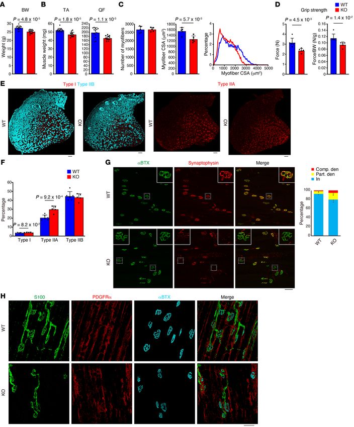
RESEARCH ARTICLE                                                                                      The Journal of Clinical Investigation

                                                                                                   Figure 1. Specific and efficient depletion of
                                                                                                   mesenchymal progenitors. (A) Scheme of mesen-
                                                                                                   chymal progenitor depletion. (B) Immunostaining
                                                                                                   of tibialis anterior (TA) muscle for PDGFRα and
                                                                                                   dystrophin. (C) Quantification of PDGFRα signal
                                                                                                   intensity. n = 5 at 2 days of Tmx, n = 5 for WT/R26-
                                                                                                   DTA and n = 4 for Pα-CE/R26-DTA at 6 weeks of
                                                                                                   Tmx. (D) FACS analysis of hind limb muscles. Red
                                                                                                   polygonal region indicates PDGFRα+ mesenchymal
                                                                                                   progenitors. The percentage of PDGFRα+ cells in
                                                                                                   the total mononucleated cells is shown. n = 4. (E)
                                                                                                   Quantification of cell number per muscle weight.
                                                                                                   n = 4. Data represent the mean ± SD; 2-sided
                                                                                                   unpaired t test (C and E). Scale bar: 20 μm (B).

progenitors in muscle regeneration by conditional depletion of               In this study, through searching for the genes responsible for
PDGFRα+ cells (14). Thus, mesenchymal progenitors play a sup-            mesenchymal progenitor–dependent muscle maintenance, we
portive role by regulating satellite cell behavior. Hence, they are      found that Bmp3b is specifically expressed in mesenchymal pro-
required for efficient and sufficient muscle regeneration.               genitors and its expression is significantly decreased during aging
    Several lines of evidence indicate that regular regeneration         or adipogenic differentiation. We demonstrated the functional
is not required for the maintenance of homeostasis in normal             importance of BMP3B in both in vivo and in vitro experiments.
uninjured myofibers during the process of natural aging (15).            Furthermore, the administration of BMP3B to aged mice resulted
There is little evidence of ongoing myonecrosis and subsequent           in improved energy metabolism and an increase in muscle mass
myogenesis in most people, even those who regularly perform              and strength. Our results demonstrate underlying mechanisms
mild exercise (16). This notion is strongly supported by studies         of the maintenance of muscle health by interstitial mesenchymal
conducted with satellite cell–ablated aged mice, in which most           progenitors and provide a mechanistic insight into how mesenchy-
muscles demonstrate normal phenotype with no exacerbation                mal progenitors contribute to sarcopenia.
of sarcopenia (17, 18). In contrast, a recent study showed that
mice with depleted mesenchymal progenitors exhibit muscle                Results
atrophy under steady-state conditions (14). Therefore, it is             Depletion of mesenchymal progenitors leads to muscle weakness
becoming clearer that mesenchymal progenitors also play an               and atrophy. To elucidate the role of mesenchymal progenitors
essential role in homeostatic muscle maintenance. However,               under steady-state conditions, we utilized Pdgfra-CreER (hereaf-
the mechanism by which mesenchymal progenitors maintain                  ter referred to as Pα-CE) mice (19). Crossing these animals with
muscle integrity is not clear, nor is the precise role of these          R26-EYFP mice followed by tamoxifen (Tmx) administration con-
cells in muscle aging.                                                   firmed the highly specific recombination in PDGFRα+ cells (PDG-

   2                                      J Clin Invest. 2021;131(1):e139617 https://doi.org/10.1172/JCI139617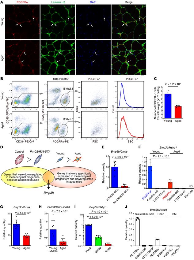
The Journal of Clinical Investigation RESEARCH ARTICLE
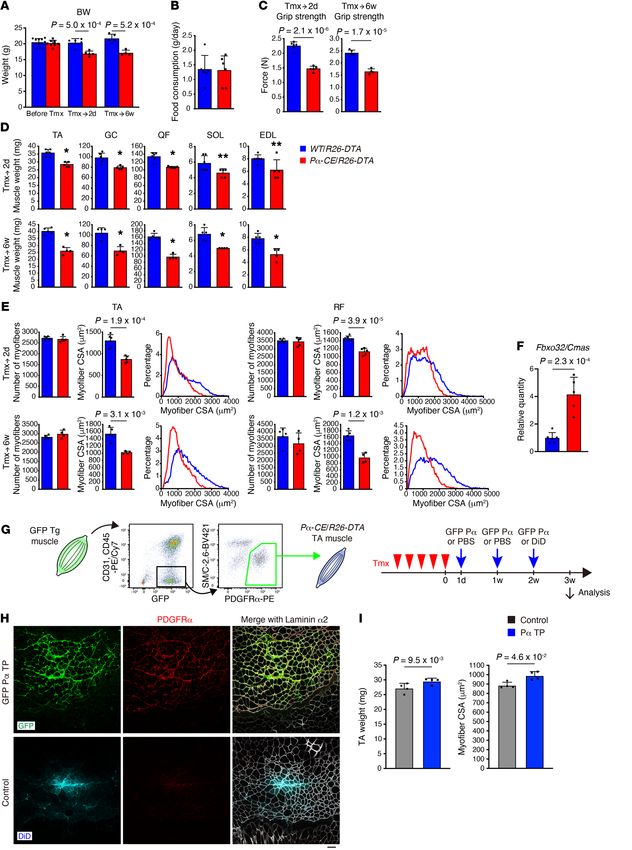
Figure 2. Mesenchymal progenitor depletion leads
to muscle weakness and atrophy. (A–E) Body
weight (A), food intake during the last 3 days of
Tmx injection (B), grip strength (C), muscle weight
(D), and number, CSA, and CSA distribution of
myofibers in TA and RF muscles (E). n = 11, 6, 5 for
WT/R26-DTA and n = 8, 5, 4 for Pα-CE/R26-DTA
prior to Tmx and at 2 days of Tmx and 6 weeks of
Tmx. n = 6 for food intake. TA, tibialis anterior; GC,
gastrocnemius; QF, quadriceps femoris; SOL, soleus;
EDL, extensor digitorum longus; RF, rectus femoris.
(F) Expression of Fbxo32 in the TA muscle. n = 6
for WT/R26-DTA and n = 5 for Pα-CE/R26-DTA.
(G) Scheme of the reconstitution of mesenchymal
progenitors. Contralateral TA was used as a control
with PBS injection. Fluorescent dye (DiD) was used
to visualize the PBS-distributed area in the final
injection. (H) Fluorescence images of GFP, DiD,
PDGFRα, and laminin α2. (I) Muscle weight and CSA
of mesenchymal progenitor–reconstituted mice. n =
4. Data represent the mean ± SD; 2-sided unpaired t
test (A–F), 2-sided paired t test (I). *P < 0.01, **P <
0.05 (D). Scale bar: 50 μm (H).
J Clin Invest. 2021;131(1):e139617 https://doi.org/10.1172/JCI139617 3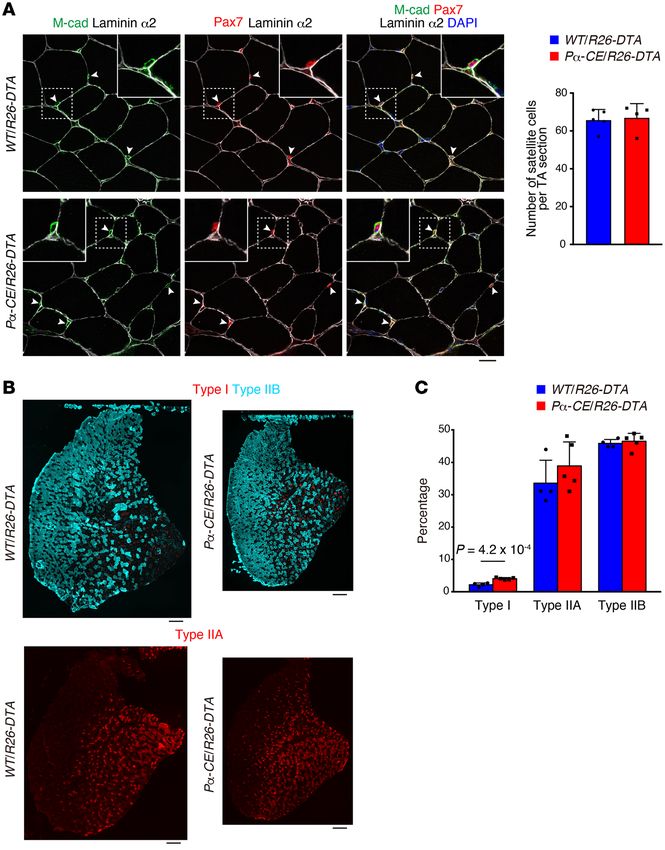
RESEARCH ARTICLE                                                                                       The Journal of Clinical Investigation

FRα+ cell ratio among EYFP+ cells of 99.7% ± 0.98%, n = 3 mice)           cells locally in a specific muscle (14). Our results, together with the
(Supplemental Figure 1, A and B; supplemental material available          results demonstrated by the study conducted by Wosczyna et al.,
online with this article; https://doi.org/10.1172/JCI139617DS1).          strongly suggest that muscle-resident mesenchymal progenitors
PDGFRα+ cells were localized in the interstitial space (Supplemen-        are indispensable for steady-state muscle maintenance.
tal Figure 1C) and were observed more frequently in the perivascu-             Mesenchymal progenitors play a role in maintaining muscle fiber
lar regions (Supplemental Figure 1, D–F), as previously described         type and neuromuscular junctions. To gain further insight into
(5). Next, Pα-CE mice were crossed with R26-DTA mice to deplete           the effect of mesenchymal progenitor depletion, we examined
mesenchymal progenitors (Figure 1A). To exclude the nonspecific           the number of satellite cells. The satellite cell number remained
effects of Tmx, the same amount of Tmx was injected into both             unchanged 2 days after the depletion of mesenchymal progenitors
Pα-CE/R26-DTA mice and WT/R26-DTA control littermate mice.                (Figure 1E). We did not observe any difference in the number of
Following Tmx administration, the majority of PDGFRα+ cells dis-          sublaminar satellite cells even 6 weeks after mesenchymal progen-
appeared (>80% decline in PDGFRα intensity), with this reduc-             itor depletion (Figure 3A). However, a previous study demonstrat-
tion lasting 6 or more weeks (Figure 1, B and C), indicating that the     ed that the number of satellite cells decreased 9 months after the
ablation of PDGFRα+ cells cannot be recovered during this time            depletion of mesenchymal progenitors (14). Hence, the mesenchy-
period. FACS analysis revealed that the number of PDGFRα+ cells           mal progenitors appear to be required for the long-term mainte-
specifically decreased by 73% (Figure 1, D and E). This decline           nance of satellite cells. Muscle wasting is frequently accompanied
rate was underestimated because we normalized cell number by              by fiber type shift; moreover, fast-to-slow shifts also occur during
muscle weight yet Pα-CE/R26-DTA mouse muscle weight signifi-              aging (21). Therefore, we examined the fiber type of mesenchy-
cantly decreased, as described below. Thus, mesenchymal pro-              mal progenitor–depleted mice. Analysis of TA muscle fiber type,
genitors were specifically and efficiently depleted in Pα-CE/R26-         typically consisting of predominantly fast fibers, demonstrated an
DTA mice. The depletion of mesenchymal progenitors resulted               increased ratio of slow fibers in Pα-CE/R26-DTA mice (Figure 3, B
in reduced body weight, which cannot be attributed to decreased           and C), although the change was small.
food intake (Figure 2, A and B), along with a significant reduc-               As proper muscle-nerve interaction constitutes a critical fac-
tion in muscle strength and weight (Figure 2, C and D). Although          tor in the maintenance of muscular health, we examined the rela-
the number of myofibers remained unchanged, Pα-CE/R26-                    tionship between mesenchymal progenitors and motor nerves.
DTA mouse myofibers demonstrated reduced cross-sectional                  Whole-mount immunostaining revealed that mesenchymal pro-
area (CSA) at least in histological assessment (Figure 2E). These         genitors surround the motor nerve axon and also cover the neu-
phenotypes lasted for 6 or more weeks, as depleted mesenchy-              romuscular junction (NMJ) in wild-type (WT) mice (Figure 4A),
mal progenitors did not replenish. In association with the loss of        with the majority of postsynaptic endplates being completely
muscle mass, we observed the upregulation of muscle-specific E3           occupied by presynaptic nerve terminals (Figure 4B). However,
ubiquitin ligase Fbxo32, which is also known as MAFbx/atrogin-1,          Pα-CE/R26-DTA mice exhibited an increased ratio of partially or
in Pα-CE/R26-DTA muscle (Figure 2F). To examine the poten-                completely denervated NMJs, and this ratio tended to increase at
tial toxicity of killed mesenchymal progenitors toward bystander          later time points (Supplemental Figure 3 and Figure 4B). We also
myofibers, myofiber damage was visualized using Evans blue dye            observed that the mesenchymal progenitors were located adja-
(EBD) or IgG staining (20). No EBD- or IgG-positive myofibers             cent to Schwann cells, whereas Pα-CE/R26-DTA mice exhibited
were observed in the Pα-CE/R26-DTA muscle, and we observed                disorganized Schwann cells (Figure 4C). Consistent with these
only weak interstitial IgG staining (Supplemental Figure 2). In           morphological changes, negative regulators of Schwann cell dif-
contrast, the intracellular accumulation of EBD and IgG in dam-           ferentiation, such as Jun and SRY-box 2 (Sox2), were upregulat-
aged myofibers was prominent along with the substantial intersti-         ed in Pα-CE/R26-DTA mice, and the expression of inhibitor of
tial accumulation of IgG in the dystrophic D2-mdx muscle, which           DNA binding 2 (Id2), another negative regulator, was also likely
was used as a positive control (Supplemental Figure 2). Thus, no          to increase, although not to a statistically significant extent (Fig-
apparent myofiber damage or inflammation in Pα-CE/R26-DTA                 ure 4D). Conversely, mature Schwann cell markers (myelin basic
muscle was observed. As PDGFR+ cells also reside in tissues oth-          protein [Mbp] and gap junction protein β-1 [Gjb1]) were downreg-
er than skeletal muscle, systemic deletion of PDGFRα+ cells could         ulated (Figure 4D). Interestingly, similar deterioration of NMJ and
influence the Pα-CE/R26-DTA muscle. To address this issue, we             Schwann cells was also observed in sarcopenic geriatric mice (22).
reconstituted mesenchymal progenitors specifically in the muscles              Bmp3b is a mesenchymal progenitor–specific and sarcopenia-re-
of the mice previously depleted of mesenchymal progenitors. We            lated gene. As Pα-CE/R26-DTA mouse phenotypes were markedly
transplanted freshly isolated mesenchymal progenitors from GFP-           similar to the phenotype observed in sarcopenic conditions, we
Tg mice into the tibialis anterior (TA) muscle of the Pα-CE/R26-          sought to identify factors important for mesenchymal progenitor–
DTA mice (Figure 2G), resulting in successful engraftment (Fig-           dependent muscle maintenance and involved in aging-related sar-
ure 2H). Transplanted cells were exclusively distributed in the           copenia. We first characterized mesenchymal progenitors in aged
interstitial space while maintaining PDGFRα expression, and they          mice. Mesenchymal progenitors were observed to reside in the
did not contribute to myofibers directly (Figure 2H). Notably, the        muscle interstitial space of both young and aged mice in a similar
reconstitution of mesenchymal progenitors led to the recovery of          manner (Figure 5A). However, in the FACS analysis, we observed
muscle weight and fiber CSA specifically in the transplanted mus-         a clear decrease in the PDGFRα+ fraction and a minor decrease in
cle (Figure 2I). Wosczyna et al. also demonstrated the importance         PDGFRα fluorescence intensity in aged mice (Figure 5B). More-
of muscle-resident mesenchymal progenitors by depleting these             over, aged mesenchymal progenitors showed an increase in the

   4                                       J Clin Invest. 2021;131(1):e139617 https://doi.org/10.1172/JCI139617
The Journal of Clinical Investigation                                                                             RESEARCH ARTICLE

                                                                                                         Figure 3. Effect of mesenchymal pro-
                                                                                                         genitor depletion on satellite cells and
                                                                                                         myofiber type. (A) Immunostaining of
                                                                                                         TA muscle for M-cadherin (M-cad), Pax7,
                                                                                                         and laminin α2. The number of satellite
                                                                                                         cells was quantified. Arrowheads: sat-
                                                                                                         ellite cells. Inset: the magnified image
                                                                                                         of the boxed region. n = 5 for WT/R26-
                                                                                                         DTA and n = 4 for Pα-CE/R26-DTA. (B)
                                                                                                         Fluorescence images of type I, type IIB,
                                                                                                         and type IIA myosin heavy chain in TA
                                                                                                         muscle. (C) Percentage of each fiber
                                                                                                         type. n = 4 for WT/R26-DTA and n = 5
                                                                                                         for Pα-CE/R26-DTA. Data represent the
                                                                                                         mean ± SD; 2-sided unpaired t test. Scale
                                                                                                         bars: 20 μm (A) and 250 μm (B).

side scatter (SSC) profile (Figure 5B). Accordingly, mesenchymal          Bmp3b since it is the only factor that exhibits cytokine or growth
progenitor numbers decreased with aging (Figure 5C). These                factor activity based on Gene Ontology and hence assumed to
results indicate the presence of certain differences in the quality       be a candidate molecule responsible for mesenchymal progen-
of cells in addition to the cell number between young and aged            itor–dependent muscle maintenance (Figure 5D and Supple-
mesenchymal progenitors.                                                  mental Table 1). BMP3B, a member of the TGF-β superfamily,
     Subsequently, we searched for genes with decreased expres-           is also known as growth differentiation factor 10 (GDF10). We
sion in Pα-CE/R26-DTA muscle compared with those in the                   first confirmed that Bmp3b is specifically expressed by mes-
control (Figure 5D). As the depletion of mesenchymal progeni-             enchymal progenitors and significantly downregulated in the
tors leads to muscle weakness and atrophy, genes that are func-           Pα-CE/R26-DTA muscle or aged mesenchymal progenitors
tionally important for muscle maintenance and are expressed               (Figure 5, E and F). Bmp3b expression in muscle tissue was also
by mesenchymal progenitors must be selected. However, as                  significantly decreased in aged muscle in both mice (Figure 5G)
genes expressed in other cell types such as satellite, endotheli-         and humans (Figure 5H). Furthermore, freshly isolated mes-
al, and hematopoietic cells might also be included since whole            enchymal progenitors demonstrated a high Bmp3b expression.
muscle tissue was used as a sample, we performed further anal-            However, this expression was decreased after in vitro culture
ysis using FACS-purified cells to identify mesenchymal progen-            and further decreased upon adipogenic differentiation (Figure
itor–specific genes that were also downregulated in aged mice             5I). PDGFRα+ mesenchymal progenitors were also identified in
(Figure 5D). Among genes fulfilling both criteria, we focused on          other tissues including the heart (23, 24) and bone marrow (25),

                                  J Clin Invest. 2021;131(1):e139617 https://doi.org/10.1172/JCI139617                                       5
RESEARCH ARTICLE                                                                                      The Journal of Clinical Investigation

                                                                                         Figure 4. Mesenchymal progenitor depletion results in
                                                                                         defects in neural components. (A) Whole-mount immuno-
                                                                                         fluorescence staining of WT EDL muscle for neurofilament
                                                                                         (NF), acetylcholine receptor (αBTX), and PDGFRα. Arrow-
                                                                                         heads: NMJs. (B) Fluorescence images of acetylcholine
                                                                                         receptor (αBTX) and synaptophysin. Insets show magnified
                                                                                         images of boxed regions. Ratios of completely dener-
                                                                                         vated (Comp. den), partially denervated (Part. den), and
                                                                                         innervated (In) NMJ were calculated at 17 days after Tmx
                                                                                         treatment. n = 6 for WT/R26-DTA, n = 8 for Pα-CE/R26-
                                                                                         DTA. (C) Whole-mount immunofluorescence staining for
                                                                                         PDGFRα, S100, and αBTX. (D) Expression of Schwann cell–
                                                                                         related genes in the mid-belly of the TA muscle. n = 6 for
                                                                                         WT/R26-DTA and n = 5 for Pα-CE/R26-DTA. *P < 0.01, **P
                                                                                         < 0.05. Data represent the mean ± SD; 2-sided unpaired t
                                                                                         test. Scale bars: 20 μm (A: lower panels), 50 μm (A: upper
                                                                                         panels, and C), and 75 μm (B).

although these expressed Bmp3b at a lower level than that by             cant reduction in body weight (Figure 6A) along with decreased
muscle-resident mesenchymal progenitors (Figure 5J).                     muscle weight, which can be attributed to reduced myofiber CSA
    Mesenchymally expressed Bmp3b is functionally important for the      rather than the reduced number of myofibers (Figure 6, B and C).
maintenance of muscle health. To elucidate the function of BMP3B,        A decrease in the grip strength was also observed in Bmp3b-KO
we examined Bmp3b-KO mice (26). Careful examination of the KO            mice, despite normalization by body weight (Figure 6D), suggest-
mouse phenotype demonstrated a minor but statistically signifi-          ing that functional decline is not simply due to decreased body

   6                                      J Clin Invest. 2021;131(1):e139617 https://doi.org/10.1172/JCI139617
The Journal of Clinical Investigation                                                                RESEARCH ARTICLE

                              J Clin Invest. 2021;131(1):e139617 https://doi.org/10.1172/JCI139617                7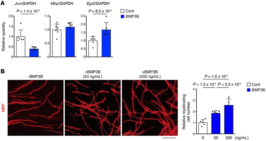
RESEARCH ARTICLE                                                                                           The Journal of Clinical Investigation

Figure 5. Identification of Bmp3b as a mesenchymal progenitor–specific         control cells (Figure 7C), indicating that BMP3B does not affect
and sarcopenia-related gene. (A) PDGFRα and laminin α2 fluorescence            muscle differentiation. This observation was further supported by
images of young and aged muscles. Arrows indicate PDGFRα cells located
                                                                               the significantly decreased expression of Bmp3b in mesenchymal
in the interstitial space. (B) FACS analysis of young and aged muscle. The
percentage of PDGFRα+ cells in the total mononucleated cells is shown. n       progenitors derived from muscle injected with cardiotoxin (CTX),
= 4. (C) Cell number per muscle weight. n = 4. (D) Scheme for the iden-        where active myogenesis was occurring, compared with the cells
tification of genes responsible for mesenchymal progenitor–dependent           derived from intact muscle (Figure 7D). The treatment of fully dif-
muscle maintenance. (E) Expression of Bmp3b in the TA muscle. n = 6 for        ferentiated human myotubes with BMP3B resulted in an induction
WT/R26-DTA and n = 5 for Pα-CE/R26-DTA. (F) Expression of Bmp3b in
                                                                               of myotube hypertrophy without affecting the fusion index (Fig-
FACS-isolated cell populations. As myofibers were difficult to sort, satel-
lite cell–derived myotubes were used. ND, not detected. n = 4 for young, n     ure 7E) along with a moderate activation of Smad1/-5/-8 and the
= 5 for aged. (G) Expression of Bmp3b in the TA muscles of young and aged      Akt pathway, which is the signaling pathway involved in the main-
mice. n = 8 mice. (H) Comparison of Bmp3b expression in human muscle           tenance of muscle mass (28–31), and no effect on Smad2 (Figure
between young-middle-aged and aged groups. n = 6 for young-middle, n =         7F). Conversely, Smad1/-5/-8 and Akt signaling were attenuated
7 for aged. (I) Bmp3b expression in mesenchymal progenitors was analyzed
                                                                               in atrophied Bmp3b-KO muscle (Figure 7G). These results suggest
immediately after sorting (Fresh), after 5 days of culture (GM5d), and after
adipogenic differentiation (Adipo). n = 6 independent samples. (J) Bmp3b       that BMP3B preserves muscle mass by directly acting on differen-
expression in FACS-purified cells from the skeletal muscle, heart, and         tiated myofibers.
bone marrow. n = 3 independent samples. Data represent the mean ± SD;               Next, we investigated the effect of BMP3B on Schwann cells,
2-sided unpaired t test (C and E–H), ANOVA followed by Dunnett’s test (I).     as mesenchymal progenitor depletion and Bmp3b deletion resulted
Scale bar: 20 μm (A).
                                                                               in Schwann cell abnormalities. BMP3B suppressed Jun expression
                                                                               in rat primary Schwann cells (Figure 8A). Although Mbp expres-
                                                                               sion was not affected, BMP3B induced the upregulation of early
weight. In addition, as the weight of other organs such as the liver           growth response 2 (Egr2), an essential regulator of the myelin-
and adipose tissue was comparable to that of WT (Supplemental                  ation program (Figure 8A). In vitro myelination assays performed
Figure 4), the observed muscle phenotype could not be simply                   with murine dorsal root ganglia demonstrated the promotion of
attributed to growth deficit. Bmp3b-KO mice showed a tendency                  myelination by BMP3B (Figure 8B). Hence, BMP3B seems to pos-
of a minor increase in slow fibers and an increase in type IIA fibers          itively regulate the characteristics of Schwann cells by stabilizing
(Figure 6, E and F). We also observed NMJ degeneration (Figure                 the differentiated state.
6G) and disorganized Schwann cells (Figure 6H) in Bmp3b-KO                          BMP3B improves energy metabolism and increases muscle mass
mice, although these phenotypes were less severe than those in                 and strength in aged mice. Since the expression of Bmp3b was
the Pα-CE/R26-DTA mice.                                                        significantly decreased in both purified mesenchymal progeni-
     To gain insight into the function of BMP3B, we used a culture             tors and muscle tissue during aging, we administered BMP3B to
system of FACS-purified cells. Purified WT satellite cells were cul-           aged mice. After 2 weeks of administration, we performed sev-
tured alone or cocultured with PDGFRα+ cells purified from WT,                 eral physiological and metabolic assessments. BMP3B-treated
Bmp3b-KO, or Pα-CE/CAG-CAT-mBmp3b–Tg (hereafter referred                       mice exhibited increasing tendencies of oxygen consumption
to as Pα/Bmp3b-Tg) mice (27), in which Bmp3b overexpression                    and carbon dioxide production, and accordingly demonstrated
was specifically induced in PDGFRα+ cells (Supplemental Figure                 an increased energy expenditure in the light period (Figure 9,
5A). Coculture with WT PDGFRα+ cells resulted in the formation                 A and B). However, the locomotive activity did not differ sig-
of larger myotubes than those in the satellite cell single culture             nificantly (Figure 9C). The administration of BMP3B did not
(Figure 7A). Although not statistically significant, the myotube               affect body weight (Figure 9D) but improved the grip strength
area demonstrated a tendency to increase in coculture with WT                  (Figure 9E) and augmented some of the muscle weight–conse-
PDGFRα+ cells compared with that in coculture with Bmp3b-KO                    quent myofiber hypertrophy (Figure 9, F and G), indicating that
PDGFRα+ cells (Figure 7A). Similar results were observed when                  BMP3B can reverse or inhibit certain phenotypes of sarcope-
conditioned media (CM) harvested from PDGFRα+ cells of each                    nia. These results suggest that the reduction of mesenchymally
genotype was added to mature human myotubes (Supplemental                      expressed BMP3B in the aged muscle represents a contributing
Figure 5B and Figure 7B). Since Bmp3b is downregulated after in                factor in the development of sarcopenia.
vitro culture (Figure 5I), we used Pα/Bmp3b-Tg PDGFRα+ cells to
make the effect of BMP3B clearer. The degree of hypertrophy in                 Discussion
myotubes became more pronounced in coculture with Pα/Bmp3b                     In this study, we describe the essential functions of interstitial
Tg PDGFRα+ cells (Figure 7A), and CM harvested from Pα/B-                      mesenchymal progenitors for the maintenance of muscle integrity
mp3b-Tg PDGFRα+ cells induced myotube hypertrophy more                         under steady-state conditions. Although satellite cells contribute
efficiently compared with that harvested from WT or Bmp3b-KO                   to myofibers with varying extent and timing, satellite cell deple-
PDGFRα+ cells (Figure 7B), with accompanying weak Smad2 and                    tion experiments showed that they are not required globally for the
more obvious Smad1/-5/-8 activation (Supplemental Figure 5C).                  maintenance of myofibers during aging (17, 18). Conversely, mes-
To elucidate whether BMP3B influences muscle differentiation,                  enchymal progenitor–depleted mice exhibited premature aging
we treated mouse satellite cells with recombinant BMP3B during                 phenotypes that were markedly similar to sarcopenia. A recently
the first day of differentiation to examine its effect on early dif-           published paper also demonstrated the importance of mesenchy-
ferentiation response. No difference was observed in the myogen-               mal progenitors in homeostatic muscle maintenance by depleting
in+ cell ratio and fusion index between BMP3B-treated cells and                these cells, but the mechanism by which mesenchymal progeni-

   8                                           J Clin Invest. 2021;131(1):e139617 https://doi.org/10.1172/JCI139617
The Journal of Clinical Investigation                                                                                    RESEARCH ARTICLE

Figure 6. Bmp3b-KO mice show deteriorated muscle phenotypes and defects in neural components. (A–F) Body weight (A), muscle weight (B), myo-
fiber number and CSA (C), grip strength (D), and fluorescence images of each fiber type (E) and fiber type percentage (F). n = 13 (A and B), n = 5 (C), n
= 6 for WT, n = 5 for KO (D and F). (G) Immunostaining for acetylcholine receptor (αBTX) and synaptophysin. Insets show magnified images of boxed
regions. Ratios of completely denervated (Comp. den), partially denervated (Part. den), and innervated (In) NMJ were quantified. n = 5 for WT, n = 4
for KO. (H) Whole-mount immunostaining for S100, PDGFRα, and αBTX. Data represent the mean ± SD; 2-sided unpaired t test (A–D and F). Scale
bars: 200 μm (E), 75 μm (G), and 50 μm (H).

                                     J Clin Invest. 2021;131(1):e139617 https://doi.org/10.1172/JCI139617                                           9
RESEARCH ARTICLE The Journal of Clinical Investigation
tors maintain muscle remains to be elucidated (14). In the present Previous studies have also shown the beneficial function of
study, we comprehensively explored factors critical for mesenchy- BMP3B by demonstrating its antiadipogenic action, metabolically
mal progenitor–dependent muscle maintenance. Consequently, favorable influence on obesity (27), and a neuroprotective role fol-
we concluded that the beneficial effects of mesenchymal progen- lowing stroke (37). Recent single-cell RNA sequencing (RNA-seq)
itors can be explained, at least in part, by the roles of the mesen- analysis of skeletal muscle also confirmed the antiadipogenic role
chymal progenitor–specific gene Bmp3b described in the current of BMP3B (38). Notably, BMP3B expression was decreased during
study. Here, we demonstrated that BMP3B directly ensures mus- aging in both mice and humans, whereas its administration to aged
cle integrity by stimulating signals important for muscle mass mice reversed certain phenotypes of sarcopenia, including the
maintenance. We also uncovered an unexpected relationship improvement of muscle function and mass. BMP3B-treated aged
between mesenchymal progenitors and neural components, along mice showed increased energy expenditure during the light period
with the underlying mechanisms by which BMP3B stabilizes the without the gain of locomotive activity. This may be attributed to
characteristics of Schwann cells. Since Schwann cells are required an increase in basal metabolism owing to increased muscle mass
for the formation and maintenance of the NMJ (32), mesenchy- and the metabolically favorable property of BMP3B (27), although
mally derived BMP3B also presumably establishes muscle health increase in energy expenditure does not necessarily link to health
indirectly via its action on Schwann cells and the NMJ. Mesenchy- improvement. All these results lead to what we believe is a new
mal progenitor–depleted mice and Bmp3b-KO mice showed minor concept, which states that age-related changes in mesenchymal
changes in muscle fiber type. However, it remains to be elucidated progenitors, including reduced BMP3B levels, can be a causal fac-
whether the alterations in fiber type can be attributed to the dimin- tor of sarcopenia. This concept is further supported by the signifi-
ished direct action of BMP3B on myofibers or secondary response cantly decreased BMP3B levels in response to adipogenic differen-
to the deterioration of neural components. Collectively, our study tiation and the strong link between the degree of fat infiltration and
highlights the critical role of stromal components in sustaining decline in motor function in elderly individuals (39).
parenchyma, raising the possibility of the broader importance of Although we revealed the beneficial function of mesenchy-
such mesenchymal-parenchymal interactions in the maintenance mal progenitors in steady-state muscle maintenance, these cells
of homeostasis in diverse tissues. also exert a positive effect on muscle regeneration. Mesenchy-
Although we revealed that BMP3B is specifically expressed mal progenitors stimulate the expansion of muscle stem cells and
in PDGFRα+ cells within the skeletal muscle, we cannot exclude hematopoietic cells and, hence, are required for successful muscle
the effect on muscle maintenance of BMP3B expressed in the regeneration (12, 14). Although the molecular basis for the regen-
cells residing in other tissues. However, previous studies have eration-promoting function of these cells is not yet completely
demonstrated the local paracrine action of the TGF-β super- understood, WNT1-inducible signaling pathway protein 1 (WISP1)
family. One of the well-known functions of the TGF-β super- is a mesenchymal progenitor–derived factor that regulates satel-
family is the regulation of epithelial-mesenchymal transition lite cell–dependent myogenesis and promotes muscle regener-
(EMT). The activation of EMT is mediated via a series of para- ation (40). Recent single-cell RNA-Seq and ATAC-Seq analyses
crine signaling molecules such as TGF-β families, including revealed that mesenchymal progenitors express many kinds of
BMP3B (33, 34). It is also well known that TGF-β superfamily trophic factors and extracellular matrix to orchestrate muscle
members, especially BMPs, act in the restricted region during regeneration (41). In addition to their role in muscle regeneration,
development. Some BMPs have been reported to influence mesenchymal progenitors also exhibit a pathological role as con-
and induce ventral fates only in the regions in which they are tributors to fatty and fibrous degeneration. As mesenchymal pro-
expressed, and the action of BMPs is tightly restricted to the genitors have dual contradictory functions (42, 43), i.e., promotion
regions within and around the cells that produce them (35). of regeneration and the development of pathogenesis, elucidating
Our local reconstitution experiment and previous local deple- mechanisms that contribute to the decision of the fate of mesen-
tion experiment support this paracrine action. We observed the chymal progenitors would be of considerable interest.
recovery of Pα-CE/R26-DTA muscle specifically in mesenchy- In this study, we investigated the role of PDGFRα+ mesen-
mal progenitor–transplanted muscle but not in the contralateral chymal progenitors in the skeletal muscle, but similar PDGFRα+
muscle, while muscle atrophy induced by the local depletion of progenitors also reside in the heart (23, 24) and bone marrow
mesenchymal progenitors cannot be compensated by remain- (25). Our results revealed that muscle-resident mesenchymal
ing cells nearby (14). Hence, it seems likely that mesenchymal progenitors demonstrated the highest expression of BMP3B
progenitors and their secretory products act locally where they among these sources. Similarly, recent single-cell analysis of
reside or are expressed. In addition, we observed that mus- PDGFRα+ cells and mural cells derived from 4 different murine
cle-resident PDGFRα+ cells express significantly higher levels organs identified Bmp3b as one of the skeletal muscle PDGFRα+
of BMP3B than those by PDGFRα+ cells residing in the heart cell–specific genes (36). Thus, there is heterogeneity among
and bone marrow. Recent single-cell analysis of PDGFRα+ cells mesenchymal cell populations, and interorgan heterogeneity
from 4 different organs also revealed preferential expression suggests a tissue-specific role of them. This hypothesis is fur-
of BMP3B in PDGFRα+ cells of the skeletal muscle. (36). Giv- ther supported by the restricted expression of the chemokine
en the paracrine action of the TGF-β superfamily and prefer- CXCL12, which functions as a niche factor for hematopoietic
ential expression of BMP3B in muscle-resident PDGFRα+ cells, stem cells. CXCL12 is highly expressed in bone marrow niche
BMP3B produced in the muscle-resident PDGFRα+ cells may be cells exhibiting mesenchymal progenitor potential (44) but
responsible for muscle maintenance. shows low expression in muscle mesenchymal progenitors (45).
10 J Clin Invest. 2021;131(1):e139617 https://doi.org/10.1172/JCI139617The Journal of Clinical Investigation RESEARCH ARTICLE
J Clin Invest. 2021;131(1):e139617 https://doi.org/10.1172/JCI139617 11RESEARCH ARTICLE The Journal of Clinical Investigation
Figure 7. BMP3B maintains skeletal muscle by stimulating hyper- were provided by Se-Jin Lee (Jackson Laboratory for Genomic
trophic signaling pathway. (A) MyHC staining of satellite cell single Medicine, Farmington, Connecticut, USA). CAG-CAT-mBmp3b–Tg
culture or coculture. As mesenchymal progenitors promote satellite mice have previously been described (27). Genetically engineered
cell proliferation, single culture using 3 times the number of satellite
cells (single ×3) was performed to circumvent the proliferation-stim-
mice were backcrossed to C57BL/6 mice at least 6 times. Tmx (4
ulating effect. n = 12 randomly selected fields. (B) MyHC staining of mg, MilliporeSigma) was injected intraperitoneally for 5 consec-
human myotubes treated with CM. n = 12 randomly selected fields. (C) utive days at 10–11 weeks of age to induce recombination. Thir-
Myogenin and MyHC expression in mouse differentiating myotubes. teen-week-old male Bmp3b/Gdf10-KO and WT littermate mice
Myogenin+ nuclei ratio and fusion index were quantified. n = 5 inde- were used, while female mice were used in other experiments. We
pendent wells. (D) Expression of Bmp3b in mesenchymal progenitors
isolated from intact or CTX-injured muscle. n = 4. (E) MyHC+ area and
used 8- to 12-week-old C57BL/6N females as young mice and 24-
fusion index of human myotubes with or without BMP3B treatment. n to 28-month-old C57BL/6N females as aged mice.
= 11 randomly selected fields. (F) Phosphorylation of Smad2, Smad1/-
5/-8, and Akt in human myotubes was examined. Data of cells derived Human muscle samples
from 2 different subjects are shown. Myostatin (MSTN) and BMP4 were Muscle samples were obtained from 6 subjects with an age range of
used as positive controls. (G) Phosphorylation of Smad2, Smad1/-5/-8,
and Akt in TA muscle was examined. GAPDH was used as an internal
30 to 48 years for the young-middle-aged group, and 7 subjects with
control. Data represent the mean ± SD; 2-sided unpaired t test (C–E), an age range of 73 to 90 years for the aged group. In the young-mid-
ANOVA followed by Tukey’s test (A and B). *P < 0.01, **P < 0.05 (A and dle-aged group, 4 samples were obtained from the gluteus medius
B). Scale bars: 75 μm (A–C). and 2 from the vastus medialis. In the aged group, 4 samples were
obtained from the gluteus medius and another 3 samples were
obtained from the TA.
Thus, mesenchymal progenitor populations in certain tissues
may be specialized to suitably sustain the parenchyma of that Cell isolation
tissue. Although we revealed the indispensability of muscle-res- Skeletal muscle. Cell isolation was performed as described previously (48).
ident mesenchymal progenitors, exploring specific roles for Minced muscles were digested with 0.2% type II collagenase (Worthing-
mesenchymal populations in supporting the parenchyma of oth- ton) for 60 minutes (30 minutes for human muscles) at 37°C. Digested
er tissues will facilitate the understanding of the mechanisms muscles were passed through an 18-G needle several times and further
involved in the homeostasis and aging of diverse organs. digested for 15 minutes at 37°C. Muscle slurries were filtered through a
100 μm cell strainer and then through a 40 μm cell strainer (BD Biosci-
Methods ences). Mouse cells were resuspended in a washing buffer consisting of
2.5% FBS in PBS and stained for cell sorting. Human cells were cultured
Mice on a collagen I–coated dish (Iwaki) in growth medium (GM) consisting of
C57BL/6 mice and GFP-Tg mice were purchased from Japan SLC. DMEM supplemented with 20% FBS, 1% penicillin-streptomycin, and 2.5
Pdgfra-CreER mice (19) (stock 018280), R26-EYFP mice (46) ng/mL bFGF (Katayama Chemical) at 37°C under 5% CO2 and 3% O2 for
(stock 018280), and R26-DTA mice (47) (stock 010527) were pur- several days. When cells reached 80% confluence they were trypsinized
chased from The Jackson Laboratory. Bmp3b/Gdf10-KO mice (26) and stained with primary antibodies followed by secondary staining.
Figure 8. BMP3B stabilizes Schwann cell characteristics. (A) Expression of
Schwann cell–related genes in rat primary Schwann cells with or without
BMP3B stimulation. n = 6 independent cultures. (B) In vitro myelination
assay of mouse dorsal root ganglion. Myelination was visualized by MBP
immunostaining. n = 5 randomly chosen fields from more than 3 explants.
Data represent the mean ± SD; 2-sided unpaired t test (A), ANOVA fol-
lowed by Tukey’s test (B). Scale bar: 50 μm (B).
12 J Clin Invest. 2021;131(1):e139617 https://doi.org/10.1172/JCI139617The Journal of Clinical Investigation RESEARCH ARTICLE
Figure 9. BMP3B administration reverses some of the sarcopenic phenotypes of aged mice. (A) Aged mice (26 months) with or without BMP3B treatment were
monitored in a metabolic chamber, and oxygen consumption (VO2) and carbon dioxide production (VCO2) were measured. (B and C) Energy expenditure (B), and
cumulative home-cage activity (left) and wheel running activity (right) (C) of BMP3B-treated or control mice. (D–F) Body weight (D), grip strength (E), and muscle
weight (F) were measured. (G) The number of myofibers, CSA, and CSA distribution of RF in BMP3B-treated or control mice. n = 5 for control, n = 6 for BMP3B-treat-
ed mice. Data represent the mean ± SEM (A) or the mean ± SD (B–G); 2-sided unpaired t test (B–G). *P < 0.01, **P < 0.05 (F and G).
Heart. The atrium was removed, and each ventricle was finely EBD injection
minced. Minced hearts were digested with 0.2% type II collage- EBD (MillipreSigma) was dissolved in PBS at a concentration of 1%.
nase (Worthington) for 5 minutes at 37°C. Digested hearts were One day prior to the analysis, EBD (10 μL/g) was injected intraperito-
passed through an 18-G needle several times. Digestion and nee- neally to detect damaged myofibers. Damaged myofibers were visual-
dle homogenization were repeated 3 times. Slurries were filtered ized by histological analysis of the intracellular accumulation of EBD
through a 100 μm cell strainer and then through a 40 μm cell strain- and intracellular staining of mouse IgG (20).
er (BD Biosciences). Cells were resuspended in washing buffer and
stained for cell sorting. Immunohistochemistry, cytochemistry, and microscopy
Bone marrow. Bone marrow cells were flushed out from the Fresh muscle samples were rapidly frozen in isopentane cooled with
femurs and tibias and pooled in sterile tubes. The remaining liquid nitrogen. For the detection of EYFP or GFP and the observation
femurs and tibias were cut into small pieces. Bone fragments were of NMJs, muscles were fixed in 4% paraformaldehyde (PFA) for 30
digested with 0.2% type II collagenase (Worthington) with gentle minutes, immersed sequentially in 10% and 20% sucrose, and frozen.
agitation for 2 hours at 37°C. Digested bone fragments were fil- For the observation of NMJ, 40 μm thick longitudinal sections were
tered through a 100 μm cell strainer and then through a 40 μm cell obtained using a cryostat and treated with 1 μg/mL α-bungarotoxin
strainer (BD Biosciences). Filtered cells were mixed with pooled (αBTX) (Invitrogen) for 1 hour. After washing with PBS, the sections
bone marrow cells. Cells were resuspended in washing buffer and were treated with methanol for 5 minutes at −20°C. In other cases, 7
stained for cell sorting. μm thick transverse sections were obtained. Fresh frozen sections were
Reagents used for cell surface staining are listed in Supplemen- fixed with 4% PFA for 5 minutes. For the staining of myosin heavy chain
tal Table 2. Isotype control staining is presented in Supplemental Fig- (MyHC), fresh frozen sections were fixed with acetone for 5 minutes
ure 6. Cell sorting was performed with the MoFlo Astrios (Beckman at −20°C. Cultured cells were fixed with 4% PFA for 5 minutes. Speci-
Coulter) or SORP FACSAria II (BD Biosciences) instrument. mens were blocked with protein-block serum-free reagent (DAKO) for
J Clin Invest. 2021;131(1):e139617 https://doi.org/10.1172/JCI139617 13RESEARCH ARTICLE The Journal of Clinical Investigation
15 minutes and incubated with primary antibodies at 4°C overnight, fol- Assessment of innervation at the NMJs
lowed by secondary staining. Antibodies used are listed in Supplemen- Z-stack images of 40 μm thick longitudinal TA sections were captured
tal Table 2. Specimens were counterstained with DAPI (Invitrogen) and using the TCS SP8 confocal laser scanning microscope system (Lei-
mounted with SlowFade Gold anti-fade reagent (Invitrogen). Images ca), and reconstructed images were displayed by maximum intensity
were captured using an inverted fluorescence microscope DMI6000B projection using LAS X software (Leica). For denervation assessment,
(Leica), BZ-X710 (Keyence), confocal laser scanning microscope sys- we analyzed 50–100 NMJs per TA muscle. The numbers of completely
tem LSM700 (Carl Zeiss), or TCS SP8 (Leica). Negative immunostain- denervated, partially denervated, and innervated NMJs were counted.
ing controls are presented in Supplemental Figure 6.
RNA extraction and quantitative PCR
Quantitative analysis of myofibers Total RNA was extracted from muscle tissues, sorted cells, or cultured
Cross sections were obtained by cutting at the mid-belly of muscle cells by using the miRNeasy Mini Kit or RNeasy Micro Kit (Qiagen),
(approximately 3 mm from the proximal end of the TA muscle or and equal amounts of RNA were reverse transcribed into cDNA using
approximately 4 mm from the distal end of the quadriceps femoris the QuantiTect Reverse Transcription Kit (Qiagen). Real-time quan-
muscle). Immunostaining of laminin or dystrophin was performed, titative PCR was performed with a Thermal Cycler Dice Real-Time
and fluorescence images of entire cross sections were captured with System (TaKaRa) with SYBR Premix Ex Taq II (TaKaRa) under the fol-
a BZ-X710 fluorescence microscope system. Image recognition and lowing cycling conditions: 94°C for 30 seconds followed by 40 cycles
quantification were performed using the Hybrid Cell Count Applica- of amplification (94°C for 5 seconds, 60°C for 20 seconds, 72°C for 10
tion (Keyence). For RF measurement, the region of interest (ROI) was seconds) and dissociation-curve analysis. The following genes were
manually set to RF. Laminin-positive basal lamina or dystrophin-posi- shown to be suitable for each experimental condition as internal con-
tive sarcolemma was first recognized based on the intensity of the flu- trol genes and used for gene expression quantification: cytidine mono-
orescence signal by adjusting the threshold, and myofibers were rec- phospho-N-acetylneuraminic acid synthetase (Cmas) for mouse tissue
ognized using an inversion function. The separation function was used (49, 50), NADH:ubiquinone oxidoreductase subunit A13 (NDUFA13)
to efficiently separate individual myofibers. The small, misrecognized for human samples (51), heat shock factor binding protein 1 (Hsbp1)
areas were excluded by adjusting the lower limit in the histogram for mouse cells (52), and Gapdh for rat cells. Specific primer sequences
function. Finally, errors in the recognition step were corrected man- used for PCR are listed in Supplemental Table 3.
ually. The myofiber number and CSA of individual myofibers were
calculated subsequently. Microarray analysis
Microarray analysis was performed using the 3D-Gene Mouse Oligo
Reconstitution of mesenchymal progenitor cells Chip 24K (Toray) with the following samples: gastrocnemius muscle
Pα-CE/R26-DTA mice were injected with Tmx as described above. derived from Pα-CE/R26-DTA mice or WT/R26-DTA mice, freshly
One day after Tmx injection, CD31−CD45−PDGFRα+ mesenchymal isolated mesenchymal progenitors, satellite cells, and mixed popula-
progenitors were sorted from GFP-Tg mice, and 1 × 10⁵ to 2 × 10⁵ fresh- tions of endothelial and hematopoietic cells obtained from young or
ly sorted cells were transplanted into the TA muscles of Pα-CE/R26- aged mice. We searched for genes that were downregulated (4-fold) in young mesenchymal
injected into the control TA muscles. To assess myofiber CSA, 7 μm progenitors compared with those in young satellite cells or young
thick transverse sections were obtained at 200 μm intervals through- endothelial/hematopoietic cells. Finally, we searched for genes that
out the muscle, and the section showing the maximal distribution of were downregulated (The Journal of Clinical Investigation RESEARCH ARTICLE
gle culture, 2 × 10³ (single) or 6 × 10³ (single ×3) satellite cells were 24-well plates in DMEM containing 10% FBS and 50 ng/mL nerve
cultured per well in a 96-well CellCarrier Ultra plate (PerkinElmer). growth factor (Harlan Bioproducts) for 7 days. Myelination was
For coculture, 2 × 10³ satellite cells and 4 × 10³ mesenchymal progeni- induced by the addition of ascorbic acid. For the analysis of myelin
tors were plated per well. Cells were maintained in GM at 37°C under profiles by immunocytochemistry, the culture was fixed with 4%
5% CO2 and 3% O2 for 4 days and treated with differentiation medium PFA for 5 minutes 10–14 days after initiation of myelination. Myelin-
(DM) consisting of DMEM supplemented with 5% horse serum for 2 ation profiles were visualized by immunofluorescence staining of
days at 37°C under 5% CO2 and 20% O2. The medium was replaced MBP. For quantification, myelination profiles in 5 randomly chosen
with fresh DM, and cells were maintained for an additional 2 days. To fields from more than 3 explants were counted using a 20× objective
analyze the myotube area, cells were fixed with 4% PFA for 5 minutes lens, and the number of myelinated nerve fibers per arbitrary unit
followed by immunofluorescence staining of MyHC. For quantifica- area was calculated.
tion, 6 randomly selected fields from duplicate wells were used. Imag-
es were collected and pooled from 2 independent experiments. The Immunoblotting and capillary-based immunoassays
area of the myotube in 12 randomly selected fields was measured col- Cells were lysed in a lysis buffer consisting of 10 mM Tris-HCl (pH
lectively using the WinROOF software (Mitani Corp). 7.5), 150 mM NaCl, 1% sodium deoxycholate, 1% Triton X-100, pro-
Conditioned culture. The effects of CM or BMP3B on mature tease inhibitor cocktail (Roche), and phosphatase inhibitor cocktail
human myotubes were examined by modifying a previously published (Roche). After sonication for 5 minutes, the lysates were centrifuged at
method (53). Mesenchymal progenitors sorted from Pα/Bmp3b-Tg or 1000g for 15 minutes at 4°C and the supernatants were recovered. The
Bmp3b-KO mice were cultured in GM at 37°C under 5% CO2 and 3% protein concentration was determined using the Pierce 660 nm Pro-
O2 for 5 days. The medium was replaced with a serum-free medium tein Assay (Thermo Fisher Scientific). Aliquots of the lysates contain-
consisting of DMEM with 2% serum replacement 1 reagent (Milli- ing 10 μg of protein were separated by sodium dodecyl sulfate–poly-
poreSigma) for 1 day. The supernatant was collected and used as acrylamide gel electrophoresis and transferred onto polyvinylidene
mesenchymal progenitor CM. CD56+CD82+PDGFRα−CD201− human difluoride membranes. The membranes were probed with primary
myoblasts were prepared as described previously (54). Human myo- antibodies followed by secondary incubation with horseradish peroxi-
blasts were cultured in 96-well CellCarrier Ultra plates (PerkinElmer) dase–conjugated secondary antibodies. After the chemiluminescence
in GM at 37°C under 5% CO2 and 3% O2 and induced to differentiate reaction, images of the developed immunoblots were captured using
into myotubes by culturing in DM at 37°C under 5% CO2 and 20% O2 a light-capture imaging system (ATTO). In some cases, the proteins
for 5 days. Differentiated myotubes were then maintained in GM for were separated and detected using an automated capillary electropho-
2 days. Myotubes were serum-starved by maintenance in serum-free resis system Wes (ProteinSimple) with capillary cartridges of 12–230
medium for 4 hours and stimulation with CM. To assess the phosphor- kDa (ProteinSimple). Signals were analyzed and visualized using
ylation of signaling molecules, myotubes were lysed in lysis buffer for Compass software (ProteinSimple).
30 minutes following stimulation with CM. To analyze the myotube
area, myotubes were maintained in CM for 3 days and the area of the Grip strength test
myotube was measured as described above. The peak grip force was measured using a grip strength meter (Muro-
BMP3B stimulation. Recombinant BMP3B (R&D Systems) was machi Kikai). Measurements were performed 5 times for each mouse,
dissolved in a serum-free medium. The effect of BMP3B on human and the maximum values were recorded.
myotubes was examined similarly to that of the conditioned culture.
Recombinant GDF8/myostatin (R&D Systems) and BMP4 (R&D Sys- BMP3B administration and metabolic experiments
tems) were used as positive controls for Smad2 and Smad1/-5/-8 sig- An osmotic pump (Alzet) with a 200 μL reservoir volume and
naling pathways. a 2-week release duration was filled with 75 μg of recombinant
Culture of primary Schwann cells derived from rats. Primary BMP3B (R&D Systems) dissolved in 4 mM HCl. The control
Schwann cell cultures were prepared as previously described (55) pump was filled with 4 mM HCl. Filled pumps were subcutane-
with minor modifications. Briefly, Schwann cells were obtained ously transplanted into 26-month-old C57BL/6N females. After 2
from the sciatic nerves of postnatal 2-day-old Sprague-Dawley rat weeks, the mice were housed in a metabolic cage equipped with a
pups. Contaminating fibroblasts were removed from the culture running wheel (Shinfactory) for 3 days. Mice were acclimated for
by treating the cells with 10 μM cytosine arabinoside for 48 hours the first 24 hours and then monitored for 48 hours. The oxygen
and complement-mediated cytolysis using anti-Thy1.1 (Serotec) consumption, carbon dioxide production, and energy expendi-
and rabbit complement (Cappel). Schwann cells were propagated ture were measured using an Oxymax open-circuit indirect cal-
on poly-L-lysine–coated plates in DMEM supplemented with 10% orimetry system (Columbus Instruments). Home-cage activity
FBS, 2 μM forskolin, and 20 ng/mL rh-HRG-β1 (MilliporeSigma). and wheel-running activity were measured using the Actimo dual
Schwann cells were stimulated with DMEM containing 10% FBS activity monitoring system (Shinfactory). Mice were subjected to
and 500 ng/mL BMP3B for 4 days and lysed in lysis buffer (Qiagen) the grip strength test prior to euthanizing.
for gene expression analysis.
Statistics
In vitro myelination assay Statistical significance was assessed using Excel (Microsoft) and Prism
In vitro myelination assays were performed as described previous- 8 (GraphPad Software). No statistical methods were used to predeter-
ly (56). Murine DRG explants dissected from the E12.5 embryos of mine the sample size. Body weight, muscle weight, and grip strength
C57BL/6J mice were cultured on poly-L-lysine– and laminin-coated measurements were performed in a blinded manner; other experi-
J Clin Invest. 2021;131(1):e139617 https://doi.org/10.1172/JCI139617 15You can also read