Stem Cell Reports Ar ticle
←
→
Page content transcription
If your browser does not render page correctly, please read the page content below
Please cite this article in press as: Song et al., Treatment of Macular Degeneration Using Embryonic Stem Cell-Derived Retinal Pigment
 Epithelium: Preliminary Results in Asian Patients, Stem Cell Reports (2015), http://dx.doi.org/10.1016/j.stemcr.2015.04.005

Stem Cell Reports
Ar ticle

Treatment of Macular Degeneration Using Embryonic Stem Cell-Derived
Retinal Pigment Epithelium: Preliminary Results in Asian Patients
Won Kyung Song,1,* Kyung-Mi Park,2 Hyun-Ju Kim,2 Jae Ho Lee,3 Jinjung Choi,4 So Young Chong,5
Sung Han Shim,6 Lucian V. Del Priore,7 and Robert Lanza8,*
1Department   of Ophthalmology, CHA Bundang Medical Center, CHA University, Seongnam-si, Gyeonggi-do 463-712, Republic of Korea
2Development    Division, CHA Biotech Co., Ltd., Seoul 135-907, Republic of Korea
3CHA Stem Cell Institute, CHA Biotech Co., Ltd., Seoul 135-907, Republic of Korea
4Division of Rheumatology, Department of Internal Medicine, CHA Bundang Medical Center, CHA University, Seongnam-si, Gyeonggi-do 463-712,

Republic of Korea
5Division of Hematology-Oncology, Department of Internal Medicine, CHA Bundang Medical Center, CHA University, Seongnam-si, Gyeonggi-do 463-712,

Republic of Korea
6Department of Biomedical Science, CHA University, Seoul 135-081, Republic of Korea
7Albert Florens Storm Eye Institute, Medical University of South Carolina, Charleston, SC 29425, USA
8Ocata Therapeutics, Marlborough, MA 01752, USA

*Correspondence: songwkmd@daum.net (W.K.S.), rlanza@ocata.com (R.L.)
 http://dx.doi.org/10.1016/j.stemcr.2015.04.005
This is an open access article under the CC BY license (http://creativecommons.org/licenses/by/4.0/).

SUMMARY

Embryonic stem cells hold great promise for various diseases because of their unlimited capacity for self-renewal and ability to differen-
tiate into any cell type in the body. However, despite over 3 decades of research, there have been no reports on the safety and potential
efficacy of pluripotent stem cell progeny in Asian patients with any disease. Here, we report the safety and tolerability of subretinal trans-
plantation of human embryonic-stem-cell (hESC)-derived retinal pigment epithelium in four Asian patients: two with dry age-related
macular degeneration and two with Stargardt macular dystrophy. They were followed for 1 year. There was no evidence of adverse pro-
liferation, tumorigenicity, ectopic tissue formation, or other serious safety issues related to the transplanted cells. Visual acuity improved
9–19 letters in three patients and remained stable (+1 letter) in one patient. The results confirmed that hESC-derived cells could serve as a
potentially safe new source for regenerative medicine.

INTRODUCTION                                                             and McDonald, 1989; Korte et al., 1984; Leonard et al.,
                                                                         1997). Stargardt macular dystrophy (SMD) is the most com-
Since their discovery and isolation in 1998, human embry-                mon form of juvenile macular degeneration that is due to
onic stem cells (hESCs) have been considered a potentially               the production of defective rim proteins encoded by the
valuable tool for generating replacement cells for therapeu-             ABCA4 gene, leading to the accumulation of di-retinoid-
tic purposes (Lanza et al., 2009). However, despite success              pyridinium ethanolamine (A2E) in the RPE, RPE cell loss,
in numerous animal models, fears over tumorigenicity                     and photoreceptor death (Glazer and Dryja, 2002). There
and immunogenicity, coupled with ethical concerns, and                   are no known effective treatments to prevent or reverse vi-
inefficiencies in differentiation methods have all contrib-              sual loss for either disease. Since RPE loss is implicated in
uted to delays in carrying out human clinical trials. Only               the pathophysiology of both disorders, RPE replacement
one group has reported the results of the safety and possible            has been suggested as a therapeutic intervention for these
biological activity of embryonic stem cell progeny in indi-              conditions.
viduals with any disease (Schwartz et al., 2015), but these                 Proper functioning of the RPE is important for maintain-
investigators only enrolled patients who were mostly                     ing the health and integrity of the outer retina, photorecep-
Caucasian. Here, we confirmed the potential safety and ef-               tors, and choriocapillaris. Healthy RPE cells play many
ficacy of hESC-derived cells in Asian patients.                          crucial roles in the retina, including transportation of nu-
   Loss of the retinal pigment epithelium (RPE) is an impor-             trients such as glucose or vitamin A from blood to the pho-
tant part of the disease process in several retinal disorders,           toreceptors, secretion of growth factors, phagocytosis of
including age-related macular degeneration (AMD) and                     the outer segments of the photoreceptors, formation of
Stargardt disease. AMD is a degenerative disease that is                 the blood-retina barrier by tight junctions, and establish-
the leading cause of visual impairment in developed coun-                ment of immune privilege of the eye (Strauss, 2005; Wim-
tries, with the dry (nonexudative) form of AMD accounting                mers et al., 2007). Based on the central role of RPE in the
for 85% to 90% of cases (Age-Related Eye Disease Study                   pathophysiology of AMD, researchers have attempted allo-
Research Group, 2001). Concurrent RPE and choriocapilla-                 geneic and autologous RPE cell transplantations for cases of
ris atrophy are present in severe, atrophic dry AMD, with                wet AMD (Binder et al., 2002; van Meurs et al., 2004; Alg-
RPE atrophy preceding choriocapillaris atrophy (Schatz                   vere et al., 1994) and dry AMD (Algvere et al., 1997, 1999;

                                                                           Stem Cell Reports j Vol. 4 j 1–13 j May 12, 2015 j ª2015 The Authors 1
Please cite this article in press as: Song et al., Treatment of Macular Degeneration Using Embryonic Stem Cell-Derived Retinal Pigment
Epithelium: Preliminary Results in Asian Patients, Stem Cell Reports (2015), http://dx.doi.org/10.1016/j.stemcr.2015.04.005
Joussen et al., 2007). However, most of these clinical trials derived RPE cells immediately post-thawing, which is
have failed to show functional improvements in macular more relevant to the phenotype of the cells that are
degeneration patients, possibly because of immune rejec- actually transplanted (Figure S1). The percentage of cells
tion and graft failure. phagocytized with fluorescence-labeled bioparticles was
Animal studies have shown that hESC-derived RPE cell measured compared to a negative isotype control and an
transplantation can rescue photoreceptors, resulting in untreated negative control (test group at 37 C: 98.47% ±
the improvement of visual functions in RPE-oriented 0.32%, n = 3; isotype contol at 4 C: 34.47% ± 3.67%,
retinal degeneration models (Lund et al., 2006; Lu et al., n = 3; untreated negative control at 37 C: 5.52% ± 0.72%,
2009). Clinical trials of hESC-derived RPE cell transplanta- n = 3) (Figure S1). 16-STR (short tandem repeat) genetic
tion have begun recently in the United States and Europe, analysis using amplified genomic DNA (gDNA) proved
and Schwartz et al. have reported preliminary safety data that RPE cells originated from MA09. Immunostaining of
on one dry AMD patient and one SMD patient (Schwartz OCT-4 and NANOG was conducted for impurity testing to
et al., 2012), as well as follow-up data with nine dry AMD confirm that no hESCs were present (Figure 1M). We
and nine SMD patients (Schwartz et al., 2015). The patient counted DAPI-stained cells in three different fields and
population studied in this paper was all Caucasian, except calculated the total cell number, and we did not see any cells
for one African American patient with SMD. Our report that stained positive for OCT-4+ or NANOG+ within 21-mm
provides interim results of the first pluripotent stem cell tri- dishes (Figure 1M). Additionally, we performed FACS using
als performed in Asian patients, who may carry different fluorescent labels for OCT-4 and TRA-1-60 and demon-
risk alleles for the development of some retinal disorders strated no contamination by hESCs in the final product
such as AMD. For example, the Y402H and R80G (in the (PRE-0008) when 10,000 cells were analyzed for each
C3 gene) variants have been associated with AMD in Cau- marker: OCT-4, 0.28%; TRA-1-60, 0.02% (positive control:
casians but not in Asians (Chen et al., 2006; Mori et al., OCT-4, 53.26%; TRA-1-60, 40.96% [hES-MA09 cells were
2007; Kim et al., 2008; Ng et al., 2008; Lee et al., 2008; maintained on mouse embryonic fibroblast feeder cells];
Kondo et al., 2009; Goto et al., 2009; Pei et al., 2009). Here- negative control [NPC, neural precursor cells]: OCT-4,
in, we report on four Asian patients with macular degener- 0.47%; TRA-1-60, 0.35%) (Figure 1N). On further safety
ation (two with AMD and two with SMD) who underwent analysis through quality control testing, we confirmed the
subretinal transplantation of hESC-derived RPE and were pathogen- and virus-free status of clinical samples by steril-
followed for 1 year to assess safety and tolerability. ity, mycoplasma, and endotoxin detection following the
Korean Pharmacopoeia and the Ministry of Food and
Drug Safety (MFDS) guidelines for pathogen and virus
RESULTS testing. For the clinical studies, we transplanted >90% of
viable cells after their final formulation in BSS Plus solution.
Derivation of RPE Cells from hESCs
The hESC-derived RPE displayed typical RPE behavior, such Clinical Trial Results
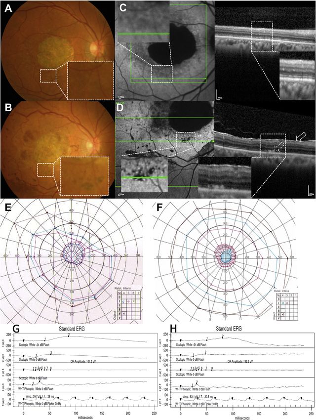
as pigmentation during differentiation and maturation, The first advanced dry-AMD patient was a 79-year-old male
and also exhibited a cuboidal epithelial morphology in tis- with an initial best-corrected visual acuity (BCVA) of the
sue culture. During culture, we observed clusters of pig- study eye of one letter read and of the fellow eye of 20/25
mented RPE cell monolayers that exhibited their unique (80 letters) on a Bailey-Lovie chart. During surgery, retinal
cobblestone morphology at the edges of clusters (Figures detachment was difficult to initiate at the first retinotomy
1A and 1B). Karyotype results using g-banding showed site, and subretinal cells were injected at a second site. A small
46XX, a normal female karyotype (Figure 1C). Thawed cells subretinal hemorrhage was noted at this second site (Fig-
were cultured for 2–3 weeks until fully differentiated to hu- ure 2B). We estimated that 4 3 104 cells were injected subre-
man RPE (hRPE) cells with medium pigmentation (Fig- tinally. The hemorrhage absorbed spontaneously at postop-
ure 1D) and were stained for hRPE markers, including erative 26 weeks (Figure 2C). Immunosuppression was
ZO1, PAX-6, MITF, and Bestrophin (Figures 1E–1I). We stopped 4 weeks postoperatively because of repeated eleva-
observed that >99% of cells expressed hRPE markers. For tion of serum creatinine levels, blood urea nitrogen (BUN)
cell function analysis, we used phagocytosis assay kits using levels, and potassium levels, as well as bone marrow suppres-
fluorescence-labeled bioparticles. Visual imaging of the sion and diarrhea; these adverse events returned to preoper-
differentiated hESC-derived RPE cells with fluorescence mi- ative levels after the cessation of immunosuppression.
croscopy showed that most hRPE cells could phagocytize An epiretinal membrane developed at 2 weeks, with dark
the fluorescently labeled beads (Figures 1J–1L). As for the brown pre-retinal pigmentation from 3 weeks. The epiretinal
quantification of the potency assay, fluorescence-activated membrane enlarged until 8 weeks, causing minimal distor-
cell sorting (FACS) analysis was conducted with hESC- tion of the underlying inner retina, and the pre-retinal
2 Stem Cell Reports j Vol. 4 j 1–13 j May 12, 2015 j ª2015 The Authors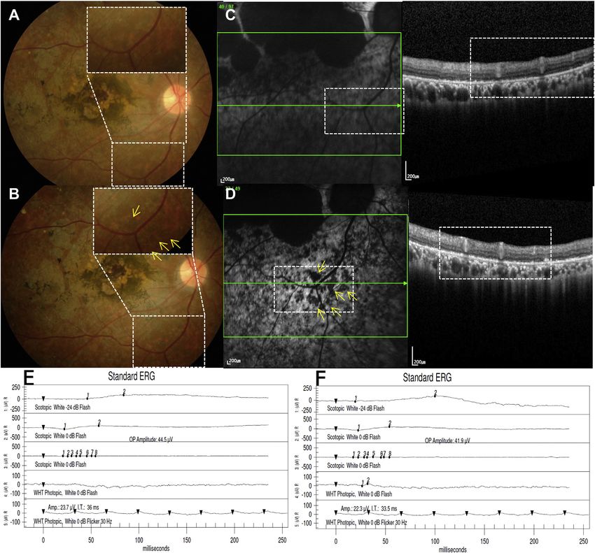
Please cite this article in press as: Song et al., Treatment of Macular Degeneration Using Embryonic Stem Cell-Derived Retinal Pigment
 Epithelium: Preliminary Results in Asian Patients, Stem Cell Reports (2015), http://dx.doi.org/10.1016/j.stemcr.2015.04.005

Figure 1. Characterization of the Clinical Product: Identity, Potency, and Purity
(A) RPE clusters were obtained by culturing an embryoid body attached to a six-well plate for about 8 weeks.
(B) The cells at the edge of the pigmented cluster displayed typical morphology of hRPE with hypo-pigmentation of the leading edge.
(C) A normal female karyotype (46XX) is shown.
(D) A confluent cobblestone monolayer was observed via Hoffman modulation contrast microscopy.
(E and F) Cells were positive for ZO-1 (E) and PAX-6 (F).
(G and H) DAPI staining in (G) was used to identify the location of the nuclei corresponding to ZO-1 (in E) and MITF (H) at the same time.
ZO-1 and MITF were double stained in one sample.
(I) Mature RPE cells were recognized with anti-Bestrophin.
(J–L) Phagocytosis assay results were shown. Fluorescence microscopy image and FACS analyses of the differentiated hESC-derived RPE
cells demonstrate that most of the cells (99.6%) were phagocytized with the fluorescent-labeled particles.
(M and N) Purity was assessed by the absence of hESCs of the final product by immunocytochemical staining for OCT-4 and NANOG (M) and
FACS analysis demonstrating the absence of OCT-4 and TRA-1-60 (N).
Scale bars, 50 mm.

pigmentation area increased and darkened by 13 weeks, with            monthly intravitreal Lucentis (0.5 mg/0.05 ml, Genentech)
no change through 1 year (Figure 2D). Subretinal pigmenta-            injections. At the 1-year visit, BCVA was stable in the
tion started at 3 weeks as an oval-shaped, localized black            study eye (two letters read; Table 1) without subjective symp-
clump. The clump was slightly increased in size until                 toms, and an epiretinal membrane persisted with minimal
13 weeks as the subretinal hemorrhage decreased. Blocked              retinal puckering (Figure 2H). There was minimal enlarge-
autofluorescence was noted at these pre-retinal and subreti-          ment of the central scotoma on Goldmann perimetry and
nal pigmentation areas (Figure 2F). Choroidal neovasculari-           no significant change in electroretinography (ERG) and
zation (CNV) was present on fluorescein angiography tem-              multifocal electroretinography (mfERG) (Figures 2J and 2L;
poral to the area of preoperative geographic atrophy (GA)             Table 2). The BCVA of the fellow eye was 20/32 (75 letters).
at postoperative 33 weeks (Figures 2E and 2F). Leakage on             Coryza, senile purpura, gynecomastia, constipation, and
fluorescein angiography improved (Figure 2G) after three              allergic conjunctivitis were adverse events with no causal

                                                                        Stem Cell Reports j Vol. 4 j 1–13 j May 12, 2015 j ª2015 The Authors 3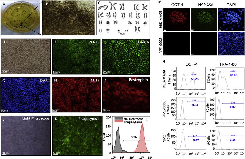
Please cite this article in press as: Song et al., Treatment of Macular Degeneration Using Embryonic Stem Cell-Derived Retinal Pigment
 Epithelium: Preliminary Results in Asian Patients, Stem Cell Reports (2015), http://dx.doi.org/10.1016/j.stemcr.2015.04.005

                                                                                             Figure 2. Ophthalmologic Results of the
                                                                                             First Dry AMD Patient
                                                                                             (A) Baseline fundus photography with
                                                                                             geographic atrophy and drusens.
                                                                                             (B) Small subretinal hemorrhage at post-
                                                                                             operative day 1 at the nasal injection site
                                                                                             (arrow).
                                                                                             (C) Fundus photography at post-operative
                                                                                             26 weeks showing absorption of hemor-
                                                                                             rhage. Subretinal pigment is present at the
                                                                                             nasal injection site (black arrow) and pre-
                                                                                             retinal pigmentation and epiretinal mem-
                                                                                             brane are visible superotemporal to the
                                                                                             fovea (white arrow).
                                                                                             (D and E) Fundus photography (D) and
                                                                                             fluorescence angiography (E) at post-oper-
                                                                                             ative 33 weeks reveal a neovascular mem-
                                                                                             brane temporal to the fovea.
                                                                                             (F) Autofluorescence imaging shows wide-
                                                                                             spread hypo-autofluorescence (left); OCT
                                                                                             demonstrates sub-RPE elevation and sub-
                                                                                             retinal and intraretinal fluid (right).
                                                                                             (G) The CNV is less active on fluorescein
                                                                                             angiography at post-operative 52 weeks
                                                                                             after three monthly intravitreal Lucentis
                                                                                             treatments.
                                                                                             (H) There is no significant change in auto-
                                                                                             fluorescence and OCT.
                                                                                             (I and J) There is minimal enlargement of
                                                                                             central scotoma (J) compared with baseline
                                                                                             (I) based on Goldmann visual field exami-
                                                                                             nation.
                                                                                             (K and L) Electroretinography at baseline
                                                                                             (K) and at the 1-year visit (L) showed no
                                                                                             significant changes.

relationship with the study procedure. The patient com-                    The second dry AMD patient was a 65-year-old male with
pleted a 1-year follow-up with no ocular or systemic serious             an initial BCVA of 20/320 (25 Early Treatment Diabetic
adverse events.                                                          Retinopathy Study [ETDRS] letters) in the study eye and

4 Stem Cell Reports j Vol. 4 j 1–13 j May 12, 2015 j ª2015 The Authors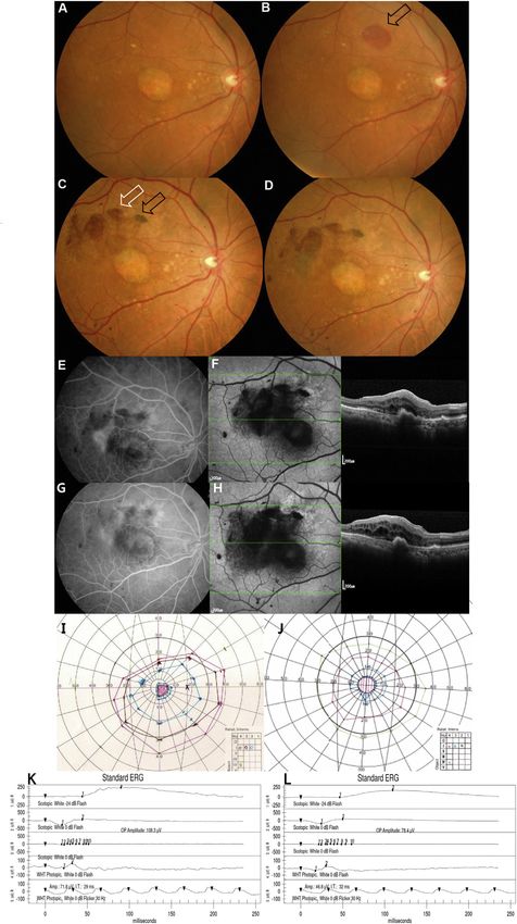
Please cite this article in press as: Song et al., Treatment of Macular Degeneration Using Embryonic Stem Cell-Derived Retinal Pigment
 Epithelium: Preliminary Results in Asian Patients, Stem Cell Reports (2015), http://dx.doi.org/10.1016/j.stemcr.2015.04.005

Table 1. Visual Acuity Changes of the Study Eye of Macular Degeneration Patients after hESC-RPE Cell Injection
               AMD Patient 1                       AMD Patient 2                         SMD Patient 1                         SMD Patient 2
                            ETDRS (Number                        ETDRS (Number                         ETDRS (Number                         ETDRS (Number
Timeline       BCVA         of Letters)            BCVA          of Letters)             BCVA          of Letters)             BCVA          of Letters)
Baseline       CF4ft        1                      20/320        25                      CF2ft           1                     20/640        13

1 week         CF4ft        2                      20/320        27                      CF2ft           0                     20/500        18

2 weeks        CF4ft        4                      20/320        28                      CF2ft           0                     20/500        18

3 weeks        CF4ft        3                      20/250        30                      CF2ft           5                     20/400        22

4 weeks        CF4ft        3                      20/250        33                      CF2ft           5                     20/250        29

6 weeks        CF4ft        2                      20/250        33                      20/800          8                     20/250        31

8 weeks        CF4ft        2                      20/200        35                      20/640        10                      20/200        32

13 weeks       CF4ft        2                      20/250        33                      20/800        10                      20/200        34

26 weeks       CF4ft        3                      20/250        35                      20/640        12                      20/250        35

39 weeks       CF4ft        3                      20/200        34                      20/500        14                      20/200        33

52 weeks       CF4ft        2                      20/200        34                      20/640        13                      20/250        32

CF4ft, counting fingers at 4 ft; CF2ft, counting fingers at 2 ft.

20/80 (55 letters) in the fellow eye. During subretinal injec-                     to the vitrectomy procedure that subsided after the admin-
tion of cells through the first retinotomy at the inferotem-                       istration of topical eye drops. Subretinal pigmentation was
poral macula, the bleb encroached upon the fovea. As per                           observed starting at postoperative 3 weeks as dark brown to
the protocol, the injection was stopped, and another reti-                         black scattered small oval clumps inside the bleb area, and
notomy and subretinal injection of the remaining cells                             the number of clumps increased until 6 weeks and did not
were performed through a distant retinotomy in the super-                          change through 52 weeks (Figures 3A and 3B). After the sur-
otemporal macula. Intraocular pressure elevation, corneal                          gery, stippled hyper-autofluorescence dots were observed
erosions, and corneal abrasion were adverse events related                         at the border of the atrophic zone where the bleb was

Table 2. mfERG Amplitude Changes of Hexagons Including the hESC-RPE-Injected Bleb Site of the Study Eyes Compared with the
Corresponding Area of the Fellow Eye
                                  Study Eyes                                                           Fellow Eyes
Timeline                          Mean ± SDa                 Mediana               pb                  Mean ± SDa                Mediana               pc
Baseline                            9.74 ± 5.22              11.90                 NA                  10.85 ± 4.32              12.41                 0.5309

3 months                          12.27 ± 4.79               13.26                 NA                  14.29 ± 4.67              16.84                 0.4034

6 months                          12.43 ± 5.33               13.61                 NA                  11.61 ± 4.84              13.93                 0.8345

12 months                         13.66 ± 7.68               17.90                 NA                  14.66 ± 7.11              19.23                 0.5309

3 months-baseline   d
                                    2.52 ± 2.28               1.93                 0.1250               3.44 ± 1.48                3.73                0.5309

6 months-baselined                  2.68 ± 3.27               3.67                 0.1875               0.76 ± 1.42                0.63                0.2963

12 months-baselined                 3.92 ± 3.64               1.89                 0.0625               3.81 ± 3.13                4.42                0.8345

Hexagons in which the bleb involved at least half of its area were included. For both study eyes and fellow eyes, n = 5; a total of four eyes and five blebs due to
two blebs in the second AMD patient. NA, not applicable.
a
  Response density (nanovolts per square degree).
b
  Wilcoxon’s signed-rank test of the changes of the study eyes.
c
 Wilcoxon’s rank-sum test between the changes in the study eyes and those in the fellow eyes.
d
  3 months(6 months, 12 months)-baseline indicates the change from baseline to the corresponding month after surgery.

                                                                                     Stem Cell Reports j Vol. 4 j 1–13 j May 12, 2015 j ª2015 The Authors 5
Please cite this article in press as: Song et al., Treatment of Macular Degeneration Using Embryonic Stem Cell-Derived Retinal Pigment
Epithelium: Preliminary Results in Asian Patients, Stem Cell Reports (2015), http://dx.doi.org/10.1016/j.stemcr.2015.04.005
(legend on next page)
6 Stem Cell Reports j Vol. 4 j 1–13 j May 12, 2015 j ª2015 The AuthorsPlease cite this article in press as: Song et al., Treatment of Macular Degeneration Using Embryonic Stem Cell-Derived Retinal Pigment
Epithelium: Preliminary Results in Asian Patients, Stem Cell Reports (2015), http://dx.doi.org/10.1016/j.stemcr.2015.04.005
involved (Figures 3C and 3D). An epiretinal membrane the ERG, and he recovered without sequelae with prophy-
developed at postoperative 2 weeks, with pre-retinal lactic topical antibiotic eye drops. No pigmentation or au-
pigmentation starting at 3 weeks, which increased in size tofluorescence changes were noted after the hESC-RPE
until 15 weeks. Minimal puckering of the underlying retina transplantation in this patient (Figures 4A–4D). At postop-
accompanied this pigmented epiretinal membrane. Stip- erative 52 weeks, his BCVA had gradually improved to
pled hypo-autofluorescence at subretinal pigmented areas 20/640 (13 ETDRS letters; Table 1), and a smaller central
and patchy hypo-autofluorescence at preretinal pigmenta- scotoma was observed via Goldmann visual field examina-
tion areas were noted (Figure 3D). Retinal cysts were noted tion (Figures 4E and 4F). BCVA of the fellow eye was 20/800
at the center of the GA area at 26 weeks on spectral domain (4 ETDRS letters read) before the surgery and 20/500 (13 let-
optical coherence tomography (SD-OCT). There were focal ters) at 52 weeks. There was no change in the anatomic
pinpoint areas of hyperfluorescence on the angiogram at appearance on SD-OCT and no change in either the ERG
26 weeks, with no definite change through 52 weeks. There (Figures 4D and 4H) or mfERG (Table 2) after surgery.
was no sign of intraocular inflammation such as anterior Herpetic vesicles developed on the patient’s right arm
chamber cells or vitreous cells. At the postoperative that were possibly related to immunosuppressive myco-
1-year visit, BCVA had gradually improved to 20/200 (34 phenolate mofetil (MMF); these were treated with topical
ETDRS letters; Table 1) in the study eye. The central sco- acyclovir application without changes in his immunosup-
toma was observed to diminish in intensity via Goldmann pressive regimen. Skin bullae at the left forearm, a contu-
perimetry (Figures 3E and 3F). ERG (Figures 3G and 3H) and sion of the right hand, external otitis, rhinorrhea, sneezing,
mfERG of the injection sites (Table 2) were stable after the fatigue, headache, upper respiratory infection, and chronic
surgery. In the fellow eye, BCVA deteriorated to 20/200 gingivitis were mild adverse events observed that had no
(35 letters). Systemic adverse events considered to be unre- causal relationship with the procedure, as determined by
lated to the study procedures included laryngopharyngeal the rheumatologist and relevant specialists.
reflux that developed at 3 weeks and improved at 13 weeks, The second SMD patient was a 40-year-old male. His
upper respiratory infection with rhinorrhea at 42 weeks, BCVA improved from 20/640 (13 ETDRS letters) to
and potassium level elevation at 52 weeks. These adverse 20/250 (32 letters) at 1 year. BCVA of the fellow eye was
events improved after medical therapy. Diarrhea, indiges- 20/250 (32 letters) at baseline and 20/160 (41 letters) at
tion, and tinnitus were mild events not related to the study 1 year. Subretinal pigmentation started at 4 weeks in the
procedures and subsided spontaneously. The patient expe- bleb area, and the number of pigmentations increased in
rienced intermittent right-hand tremor starting at approx- number until 6 weeks, with hypo-autofluorescence that
imately 15 weeks and received acupuncture several times persisted until 52 weeks (Figures 5A–5D). Multiple
without informing the investigators. At 26 weeks, a neurol- increased autofluorescence spots were also observed inside
ogist evaluated the patient and determined this event as the bleb area where hESC-RPE was injected (Figure 5D).
age-related changes not related to the study procedure. Because of a large initial central scotoma and poor visual
Pneumonia, which may have been related to immunosup- function, visual field examinations were unreliable, and
pression, was diagnosed at 8 weeks at our pulmonary there was no obvious change after surgery. ERG and the
department and then subsided after 3 days of oral antibi- multifocal ERG of the injection site were stable throughout
otics treatment. 52 weeks (Figures 5E and 5F; Table 2). Upper respiratory
Our first SMD patient was a 45-year-old male with an infection, aggravation of reflux esophagitis, and loss of a
initial BCVA of counting fingers (one ETDRS letter). There dental implant were mild adverse events considered unre-
were no signs of anterior chamber or vitreous cell or flare lated to the study procedures.
beyond what would be typically seen in the postoperative The first SMD patient had three missense mutations:
period. The patient had corneal erosions in his left eye after c.983A > T (Glu328Val), c.1933G > A (Asp645Asn), and
Figure 3. Ophthalmologic Results of the Second Dry AMD Patient
(A and B) Baseline (A) and 52-week-postoperative (B) fundus photography of the second dry AMD patient, showing subretinal
pigmentation (B, inset) after surgery.
(C and D) Baseline (C) and 52-week-postoperative (D) autofluorescence imaging and SD-OCT. Note the stippled hypo-autofluorescence
present after surgery at the border of the atrophic zone that may represent blockage from subretinal pigmentation and stippled hyper-
autofluorescence at the same area underlying the bleb (D, left, inset). Subretinal deposits (D, right, dashed arrow on OCT) are seen in the
cell-transplanted areas that were not present prior to surgery. There is an epiretinal membrane present postoperatively (D, right, solid
arrow). Patchy hypo-autofluorescence present at preretinal pigmentation areas (D, left).
(E and F) GVF examinations at baseline (E) and post-operative 52 weeks (F) show a central scotoma of diminished intensity.
(G and H) ERG examinations at baseline (G) and at post-operative 52 weeks (H) show no significant changes.
Stem Cell Reports j Vol. 4 j 1–13 j May 12, 2015 j ª2015 The Authors 7Please cite this article in press as: Song et al., Treatment of Macular Degeneration Using Embryonic Stem Cell-Derived Retinal Pigment
Epithelium: Preliminary Results in Asian Patients, Stem Cell Reports (2015), http://dx.doi.org/10.1016/j.stemcr.2015.04.005
(legend on next page)
8 Stem Cell Reports j Vol. 4 j 1–13 j May 12, 2015 j ª2015 The AuthorsPlease cite this article in press as: Song et al., Treatment of Macular Degeneration Using Embryonic Stem Cell-Derived Retinal Pigment
Epithelium: Preliminary Results in Asian Patients, Stem Cell Reports (2015), http://dx.doi.org/10.1016/j.stemcr.2015.04.005
c.3106G > A (Glu1036Lys). The second SMD patient had two Humphrey visual field examinations were unreliable
missense mutations: c.2894A > G (Asn965Ser) and c.4972A > because of poor fixation and high false-negatives in nearly
C (Ser1658Arg). All of these mutations, except for the all of the participants. Goldmann visual field (GVF) exam-
c.4972A > C mutation in S-004, were previously reported inations were assessed in these patients. There was no
as Stargardt-associated mutations (Jaakson et al., 2003). significant change in the visual field and mfERG examina-
tions in these eyes.
In the two dry AMD patients and one SMD patient,
DISCUSSION increasing subretinal pigmentation developed at the
hESC-RPE cell injection site as has been noted previously
The purpose of this study was to determine the safety and (Schwartz et al., 2012, 2015). In the second dry AMD
tolerability of the subretinal injection of hESC-derived patient and the second SMD patient, multiple spots of
RPE cells as a treatment for dry AMD and SMD. No serious increased autofluorescence were also observed at the injec-
systemic or ocular adverse events occurred in these four tion sites. It is tempting to speculate that the subretinal
Asian patients. Ophthalmological examinations, including pigmentation may represent engrafted RPE, and this
ETDRS visual acuity, visual field examination, color fundus conclusion is strengthened by the observations of small
photography, fundus fluorescein angiography, optical deposits on the inner aspects of Bruch’s membrane after
coherence tomography, fundus autofluorescence photog- surgery on OCT. However, it is known that pigment is not
raphy, ERG, and mfERG did not identify any significant a robust marker of donor cells, since ingestion of donor
safety concerns after surgery. pigment by host macrophages may produce a similar oph-
This present study was an open-label trial of patients who thalmoscopic appearance. Although there is no host RPE
had poor visual function preoperatively with a large central in the regions of atrophy, prior studies have shown that
scotoma, which hindered our ability to perform reliable native adult RPE cells may proliferate and migrate within
measurements of visual functions. In the two dry-AMD 72 hr after retinal detachment in cats (Anderson et al.,
patients, visual acuity in the treated eyes improved by 1981); migration of host RPE and ingestion of pigment by
one letter (stable at counting fingers at 4 ft) and nine letters these cells may also explain the appearance of subretinal
(a two-line improvement from 20/320 to 20/200) at pigment after hESC-RPE injection. It is important to note
52 weeks, respectively. In contrast, the fellow (untreated) that the subretinal pigmentation and stippled hyper-auto-
eyes decreased by 6 and 20 letters, respectively, during fluorescence spots noted after subretinal hESC-RPE injec-
the same time period. In the two SMD patients, visual tion have not been reported in eyes after similar procedures
acuity improved in the treated eyes by 12 letters (counting with Ca- and Mg-free solution in other trials where tran-
fingers at 2 ft to 20/640) and 19 letters (a four-line improve- sient retinal detachment was performed in order to achieve
ment from 20/640 to 20/250), respectively, compared with subretinal viral vector administration (Maguire et al., 2008).
nine letters of improvement in the fellow eyes at 52 weeks One of the largest concerns in the clinical applications of
compared to baseline. The visual acuity improvement hESC-derived cell therapy is potential tumorigenicity. Tera-
noted in the fellow eyes of SMD patients may be due to toma formation is observed within 8 weeks (Klimanskaya
poorer baseline visual acuity than in the fellow eyes of et al., 2005). All of our patients were followed for at least
the dry AMD patients. A 15-letter improvement (a 1 year, and, using color fundus photography, autofluores-
doubling of the visual angle) is generally accepted as a clin- cence imaging, and SD-OCT with 3-mm resolution, we
ically significant change. The sample size in these studies found no unwanted abnormal proliferation suggesting
(SMD phase 1: 3 patients; dry AMD phase 1/2a: total of teratoma formation.
12 patients, with 3 for each dose) was not powered to detect Since the donor hESC-derived cells are allografts, there is
an improvement in visual acuity. Thus, the visual acuity the possibility of graft rejection after transplantation. Pre-
measurement in these preliminary data should be primar- vious allogeneic RPE cell transplantations in dry AMD re-
ily interpreted as a safety parameter to monitor for adverse sulted in graft rejections in some cases, although it started
effects from the transplants. It is important to note that later in the treatment course and was seen less frequently
there was no decline in visual acuity in any of the four eyes. than in patients with wet AMD (Algvere et al., 1997,
Figure 4. Ophthalmologic Results of the First SMD Patient
(A–D) Baseline (A) and 52-week-postoperative (B) fundus photography, baseline (C), and 52-week-postoperative (D) autofluorescence
imaging and SD-OCT showing no signs of immune rejection, tumor formation, or adverse event related to the surgical procedure. No
obvious pigmentation (arrow indicates the cell-injected retinotomy site) was noted after hESC-RPE transplantation.
(E and F) GVF examinations at baseline (E) and post-operative 52 weeks (F) show a central scotoma of diminished size.
(G and H) ERG examinations at baseline (G) and post-operative 52 weeks (H) show no change.
Stem Cell Reports j Vol. 4 j 1–13 j May 12, 2015 j ª2015 The Authors 9Please cite this article in press as: Song et al., Treatment of Macular Degeneration Using Embryonic Stem Cell-Derived Retinal Pigment Epithelium: Preliminary Results in Asian Patients, Stem Cell Reports (2015), http://dx.doi.org/10.1016/j.stemcr.2015.04.005 Figure 5. Ophthalmologic Results of the Second SMD Patient (A and B) Baseline (A) and 52-week postoperative (B) fundus photography show subretinal pigmentation (B, inset, yellow arrows) in the bleb area that may represent engraftment of injected hESC-RPE cells. (C and D) SD-OCT at baseline (C, right) and postoperative 52 weeks (D, right) at corresponding sites showing a monolayer of the RPE layer. Fundus autofluorescence images (left panels of C and D) show stippling of autofluorescence at 52 weeks after surgery (D, inset, yellow arrows). (E and F) There is no change in the electroretinogram from baseline (E) to postoperative 52 weeks (F). 1999). We did not observe any clinically significant intra- able to maintain systemic immunosuppression. The second ocular inflammation and detected no signs of obvious im- dry AMD patient developed intraretinal cysts and dye pool- mune rejection including fluid collection, edema, fibrous ing on fluorescein angiography that did not change for membrane formation, persistent leakage on fluorescein 52 weeks after surgery. No significant changes in visual acu- angiography, or graying or loss of pigmentation of the graft ity, multifocal ERG, symptoms, or other signs of intraocular in patients who underwent graft transplantation and were inflammation accompanied this change. The subretinal 10 Stem Cell Reports j Vol. 4 j 1–13 j May 12, 2015 j ª2015 The Authors
Please cite this article in press as: Song et al., Treatment of Macular Degeneration Using Embryonic Stem Cell-Derived Retinal Pigment
 Epithelium: Preliminary Results in Asian Patients, Stem Cell Reports (2015), http://dx.doi.org/10.1016/j.stemcr.2015.04.005

and pre-retinal pigmentations persisted without change.              Prevention as an imported stem cell line. The comparability of
Cystoid macular edema is present in approximately 20%–               MA09-hRPE cells manufactured from both GMP sites (CHA
40% of patients with epiretinal membrane (Wickham and                Biotech versus Ocata Therapeutics) was confirmed by character-
Gregor, 2013). The retinal cyst may have accompanied                 izing the MA09-hRPE cells in terms of karyotype, genetic analysis,
                                                                     identity, purity, and potency by using real-time PCR, immunocyto-
the epiretinal membrane in this patient. However, this
                                                                     chemistry, FACS analysis, and phagocytosis assays (Supplemental
does not rule out the possibility of graft rejection.
                                                                     Information).
   The moderate adverse events related to immunosuppres-               The MFDS and CHA Bundang Medical Center institutional re-
sion included creatinine level elevation in the first dry            view board (IRB) approval was obtained to carry out two prospec-
AMD patient and pneumonia in the second dry AMD pa-                  tive clinical trials to evaluate the safety and tolerability of the cells
tient. Immunosuppressive medication was stopped in the               in patients with SMD and dry AMD (registered with ClinicalTrials.
first patient because of repeated deterioration of renal func-       gov [https://clinicaltrials.gov]; numbers NCT01625559 and
tion, including creatinine and potassium level elevation.            NCT01674829). The protocols are similar to those of the U.S.
We could continue the dosage of immunosuppression per                studies (NCT01345006 and NCT01344993), with some differences
protocol in three patients. The dose and mandatory period            regarding cancer screening and immunosuppression (see Supple-
of immunosuppression should be determined in the future              mental Information). Cancer screening included complete history
                                                                     recording, physical examination, laboratory tests, and further ul-
trials.
                                                                     trasonography, endoscopy, and biopsy when needed. Immuno-
   The two dry AMD patients developed epiretinal mem-
                                                                     suppression was started at lower dosages of tacrolimus and MMF;
branes with pigmentation. This is relatively high compared           the doses were gradually titrated according to each patient’s serum
with the rate of epiretinal membrane development after the           tacrolimus level (3–7 ng/ml), with tolerability judged by a clinical
vitrectomy procedure. It may arise from inadvertent pre-             rheumatologic specialist, while the ophthalmologists monitored
retinal injection of cells or reflux of transplanted cells from      the patient for changes in the clinical examinations. Lower MMF
the subretinal space. These pre-retinal patches of pigmenta-         dosages of 1.0–1.5 g/day were used according to previous reports
tion did not contract and cause macular wrinkling enough             regarding its safety and efficacy in the Asian organ transplantation
to require additional surgery during the follow-up interval.         patients (Tsang et al., 2000; Kim et al., 2010). Both immunosup-
   The first dry AMD patient developed CNV in the hESC-              pressive drugs were started 1 week prior to the surgical procedure
RPE transplanted eye at 33 weeks. The location of the                and were continued for a period of 7 weeks until post-operative
                                                                     6 weeks. Next, the tacrolimus was discontinued, and the MMF
CNV was not primarily in the bleb area, and only the supe-
                                                                     was continued for an additional 7 weeks before being slowly
rior margin of the CNV adjoined the inferior margin of the
                                                                     tapered thereafter. In the case of SMD, ABCA4 genes were exam-
previous bleb location. This patient had drusenoid subreti-          ined using the exome sequencing method, with a portion of the
nal accumulation with GA in the study eye and drusenoid              blood specimen archived for safety purposes at baseline. Surgeries
pigment epithelial detachment (PED) in the fellow eye. A             were carried out at the CHA Bundang Medical Center by a single
previous report showed a relatively high rate of develop-            surgeon (W.K.S.). Posterior pars plana vitrectomy was performed
ment of CNV (23%) in eyes with drusenoid PED with no                 in the eye with the worse vision, with induction of posterior vitre-
advanced AMD at baseline (Cukras et al., 2010). The CNV              ous detachment (PVD). A volume of 150 ml of RPE reconstituted in
may have developed as a natural course, or it may have               BSS Plus was injected into the subretinal space via a 38-G subretinal
been due to trauma to Bruch’s membrane during the surgi-             cannula, delivering the target dose of 5 3 104 RPE cells. Patients
cal procedure or from the injected hESC-RPE.                         were kept in a supine position for at least 6 hr after the operation.
                                                                     Patients were closely monitored by physical examinations, labora-
   The present report confirmed the feasibility and prelimi-
                                                                     tory examinations, and ophthalmologic examinations, including
nary safety of hESC-RPE cell therapy. Continued follow-up
                                                                     BCVA analysis using a Bailey-Lovie chart, visual field examination
and further study are needed to determine the long-term              using Goldmann visual field testing (Projection perimeter MK-
safety and efficacy of hESC-derived cells as a potential             70ST L-1550, Inami Ophthalmic Instruments) and/or automated
source of replacement cells for the treatment of macular             testing using a validated Humphrey perimeter (Humphrey Field
degeneration.                                                        Analyzer; Carl Zeiss Meditec), fundus photography, fluorescein
                                                                     angiography (KOWA VX-10i; Kowa), SD-OCT, fundus autofluores-
                                                                     cence photography (Spectralis OCT; Heidelberg Engineering), and
EXPERIMENTAL PROCEDURES                                              ERG (UTAS E-3000 system; LKC Technologies) (see Tables S1 and S2
                                                                     for the schedule of assessments).
hESC-derived RPE cells were manufactured in a fully validated
good-manufacturing-practice (GMP) facility under strict environ-     SUPPLEMENTAL INFORMATION
mental control monitoring systems and routine microbial testing
regimens at CHA Biotech. Master and working cell banks were          Supplemental Information includes Supplemental Experimental
established using the hESC line MA09 (Ocata Therapeutics, previ-     Procedures, one figure, and two tables and can be found
ously Advanced Cell Technology) (Schwartz et al., 2012), which       with this article online at http://dx.doi.org/10.1016/j.stemcr.
was registered with the Korea Centers for Disease Control and        2015.04.005.

                                                                     Stem Cell Reports j Vol. 4 j 1–13 j May 12, 2015 j ª2015 The Authors 11
Please cite this article in press as: Song et al., Treatment of Macular Degeneration Using Embryonic Stem Cell-Derived Retinal Pigment
Epithelium: Preliminary Results in Asian Patients, Stem Cell Reports (2015), http://dx.doi.org/10.1016/j.stemcr.2015.04.005
AUTHOR CONTRIBUTIONS Binder, S., Stolba, U., Krebs, I., Kellner, L., Jahn, C., Feichtinger, H.,
Povelka, M., Frohner, U., Kruger, A., Hilgers, R.D., and Krugluger,
Conception and design: R.L., L.V.D.P., and W.K.S.; analysis and
W. (2002). Transplantation of autologous retinal pigment epithe-
interpretation: W.K.S., H.-J.K., J.H.L., J.C., S.Y.C., S.H.S., L.V.D.P.,
lium in eyes with foveal neovascularization resulting from age-
and R.L.; writing the article: W.K.S. and H.-J.K.; critical revision
related macular degeneration: a pilot study. Am. J. Ophthalmol.
of the article: W.K.S., R.L., and L.V.D.P.; final approval of the article:
133, 215–225.
W.K.S., R.L, and L.V.D.P.; data collection: W.K.S., H.-J.K., J.H.L.,
Chen, L.J., Liu, D.T., Tam, P.O., Chan, W.M., Liu, K., Chong, K.K.,
J.C., S.H.S., and K.-M.P.; provision of materials, patients, or re-
Lam, D.S., and Pang, C.P. (2006). Association of complement factor
sources: W.K.S., R.L., and K.-M.P.; literature research: W.K.S.,
H polymorphisms with exudative age-related macular degenera-
H.-J.K., and S.H.S.
tion. Mol. Vis. 12, 1536–1542.
ACKNOWLEDGMENTS Cukras, C., Agrón, E., Klein, M.L., Ferris, F.L., 3rd, Chew, E.Y., Gens-
ler, G., and Wong, W.T.; Age-Related Eye Disease Study Research
We express sincere gratitude to Sung Woo Ha, MD, and Kyung Soon Group (2010). Natural history of drusenoid pigment epithelial
Kim, PhD, for their efforts in the Korean protocol development and detachment in age-related macular degeneration: Age-Related
securing Korean MFDS and IRB approval; the Independent Data Eye Disease Study Report No. 28. Ophthalmology 117, 489–499.
Monitoring Committee, Sung Chul Lee, Oh Woong Kwon, Kwang
Glazer, L.C., and Dryja, T.P. (2002). Understanding the etiology of
Soo Kim, and Hyonggin An; co-investigator Hee Jung Kwon for
Stargardt’s disease. Ophthalmol. Clin. North Am. 15, 93–100, viii.
surgical assistance, co-investigator Sung Woo Choi for mfERG anal-
ysis; clinical research coordinators Myung Hee Choi, Yona Bae, Goto, A., Akahori, M., Okamoto, H., Minami, M., Terauchi, N., Har-
Ju Eun Lee, and Su Youn Kim; and the pioneering patients and their uhata, Y., Obazawa, M., Noda, T., Honda, M., Mizota, A., et al.
families. This research was supported by the CHA Biotech Co., Ltd., (2009). Genetic analysis of typical wet-type age-related macular
by their grant of the Korea Health Technology R&D Project through degeneration and polypoidal choroidal vasculopathy in Japanese
the Korea Health Industry Development Institute (KHIDI), funded population. J. Ocul. Biol. Dis. Infor. 2, 164–175.
by the Ministry of Health & Welfare, Republic of Korea: grant Jaakson, K., Zernant, J., Külm, M., Hutchinson, A., Tonisson, N.,
numbers HI12C1794(A121941) and HI12C0447(A120506). The Glavac, D., Ravnik-Glavac, M., Hawlina, M., Meltzer, M.R., Caruso,
sponsor (CHA Biotech Co., Ltd.) participated in hESC-RPE manu- R.C., et al. (2003). Genotyping microarray (gene chip) for the ABCR
facture and in-use-protocol process, study design, and partial report (ABCA4) gene. Hum. Mutat. 22, 395–403.
preparation. Kyung-Mi Park is an employee of CHA Biotech. Hyun Joussen, A.M., Joeres, S., Fawzy, N., Heussen, F.M., Llacer, H., van
Ju Kim and Jae Ho Lee are employees of CHA Biotech and have Meurs, J.C., and Kirchhof, B. (2007). Autologous translocation of
received stock options. the choroid and retinal pigment epithelium in patients with
geographic atrophy. Ophthalmology 114, 551–560.
Received: October 16, 2014
Kim, K., Lee, S., Hwang, S., Kim, K., Ahn, C., Moon, D., Ha, T., Song,
Revised: April 3, 2015
G., Jung, D., Choi, N., et al. (2010). Does calcineurin inhibitor plus
Accepted: April 6, 2015
mycophenolate mofetil combination therapy decrease the risk of
Published: April 30, 2015
late acute rejection after liver transplantation? J. Korean Soc. Trans-
plant. 24, 93–100.
REFERENCES Kim, N.R., Kang, J.H., Kwon, O.W., Lee, S.J., Oh, J.H., and Chin,
Algvere, P.V., Berglin, L., Gouras, P., and Sheng, Y. (1994). Trans- H.S. (2008). Association between complement factor H gene poly-
plantation of fetal retinal pigment epithelium in age-related mac- morphisms and neovascular age-related macular degeneration in
ular degeneration with subfoveal neovascularization. Graefes Koreans. Invest. Ophthalmol. Vis. Sci. 49, 2071–2076.
Arch. Clin. Exp. Ophthalmol. 232, 707–716. Klimanskaya, I., Chung, Y., Meisner, L., Johnson, J., West, M.D.,
Algvere, P.V., Berglin, L., Gouras, P., Sheng, Y., and Kopp, E.D. and Lanza, R. (2005). Human embryonic stem cells derived
(1997). Transplantation of RPE in age-related macular degenera- without feeder cells. Lancet 365, 1636–1641.
tion: observations in disciform lesions and dry RPE atrophy. Kondo, N., Honda, S., Kuno, S., and Negi, A. (2009). Coding variant
Graefes Arch. Clin. Exp. Ophthalmol. 235, 149–158. I62V in the complement factor H gene is strongly associated
Algvere, P.V., Gouras, P., and Dafgård Kopp, E. (1999). Long-term with polypoidal choroidal vasculopathy. Ophthalmology 116,
outcome of RPE allografts in non-immunosuppressed patients 304–310.
with AMD. Eur. J. Ophthalmol. 9, 217–230. Korte, G.E., Reppucci, V., and Henkind, P. (1984). RPE destruction
Anderson, D.H., Stern, W.H., Fisher, S.K., Erickson, P.A., and Bor- causes choriocapillary atrophy. Invest. Ophthalmol. Vis. Sci. 25,
gula, G.A. (1981). The onset of pigment epithelial proliferation af- 1135–1145.
ter retinal detachment. Invest. Ophthalmol. Vis. Sci. 21, 10–16. Lanza, R., Gearhart, J., Hogan, B., Melton, D., Pedersen, R.,
Age-Related Eye Disease Study Research Group (2001). A random- Thomas, E.D., Thomson, J., and Wilmut, I. (2009). Essentials of
ized, placebo-controlled, clinical trial of high-dose supplementa- Stem Cell Biology, Second Edition (San Diego, CA: Academic
tion with vitamins C and E, beta carotene, and zinc for age-related Press/Elsevier).
macular degeneration and vision loss: AREDS report no. 8. Arch. Lee, K.Y., Vithana, E.N., Mathur, R., Yong, V.H., Yeo, I.Y., Thalamu-
Ophthalmol. 119, 1417–1436. thu, A., Lee, M.W., Koh, A.H., Lim, M.C., How, A.C., et al. (2008).
12 Stem Cell Reports j Vol. 4 j 1–13 j May 12, 2015 j ª2015 The AuthorsPlease cite this article in press as: Song et al., Treatment of Macular Degeneration Using Embryonic Stem Cell-Derived Retinal Pigment
Epithelium: Preliminary Results in Asian Patients, Stem Cell Reports (2015), http://dx.doi.org/10.1016/j.stemcr.2015.04.005
Association analysis of CFH, C2, BF, and HTRA1 gene polymor- neovascular age-related macular degeneration in a chinese popula-
phisms in Chinese patients with polypoidal choroidal vasculop- tion. Curr. Eye Res. 34, 615–622.
athy. Invest. Ophthalmol. Vis. Sci. 49, 2613–2619. Schatz, H., and McDonald, H.R. (1989). Atrophic macular degener-
Leonard, D.S., Zhang, X.G., Panozzo, G., Sugino, I.K., and Zarbin, ation. Rate of spread of geographic atrophy and visual loss.
M.A. (1997). Clinicopathologic correlation of localized retinal Ophthalmology 96, 1541–1551.
pigment epithelium debridement. Invest. Ophthalmol. Vis. Sci.
Schwartz, S.D., Hubschman, J.P., Heilwell, G., Franco-Cardenas, V.,
38, 1094–1109.
Pan, C.K., Ostrick, R.M., Mickunas, E., Gay, R., Klimanskaya, I., and
Lu, B., Malcuit, C., Wang, S., Girman, S., Francis, P., Lemieux, L., Lanza, R. (2012). Embryonic stem cell trials for macular degenera-
Lanza, R., and Lund, R. (2009). Long-term safety and function of tion: a preliminary report. Lancet 379, 713–720.
RPE from human embryonic stem cells in preclinical models of
Schwartz, S.D., Regillo, C.D., Lam, B.L., Eliott, D., Rosenfeld, P.J.,
macular degeneration. Stem Cells 27, 2126–2135.
Gregori, N.Z., Hubschman, J.P., Davis, J.L., Heilwell, G., Spirn,
Lund, R.D., Wang, S., Klimanskaya, I., Holmes, T., Ramos-Kelsey, M., et al. (2015). Human embryonic stem cell-derived retinal
R., Lu, B., Girman, S., Bischoff, N., Sauvé, Y., and Lanza, R. pigment epithelium in patients with age-related macular degener-
(2006). Human embryonic stem cell-derived cells rescue visual ation and Stargardt’s macular dystrophy: follow-up of two open-la-
function in dystrophic RCS rats. Cloning Stem Cells 8, 189–199. bel phase 1/2 studies. Lancet 385, 509–516.
Maguire, A.M., Simonelli, F., Pierce, E.A., Pugh, E.N., Jr., Mingozzi,
Strauss, O. (2005). The retinal pigment epithelium in visual func-
F., Bennicelli, J., Banfi, S., Marshall, K.A., Testa, F., Surace, E.M.,
tion. Physiol. Rev. 85, 845–881.
et al. (2008). Safety and efficacy of gene transfer for Leber’s congen-
ital amaurosis. N. Engl. J. Med. 358, 2240–2248. Tsang, W.K., Tong, K.L., Yeung, S., Lee, W., and Chan, H.W. (2000).
Efficacy and safety of mycophenolate mofetil in different dosages
Mori, K., Gehlbach, P.L., Kabasawa, S., Kawasaki, I., Oosaki, M., Ii-
in Asian renal allograft recipients. Transplant. Proc. 32, 1755–
zuka, H., Katayama, S., Awata, T., and Yoneya, S. (2007). Coding
1756.
and noncoding variants in the CFH gene and cigarette smoking in-
fluence the risk of age-related macular degeneration in a Japanese van Meurs, J.C., ter Averst, E., Hofland, L.J., van Hagen, P.M.,
population. Invest. Ophthalmol. Vis. Sci. 48, 5315–5319. Mooy, C.M., Baarsma, G.S., Kuijpers, R.W., Boks, T., and Stalmans,
P. (2004). Autologous peripheral retinal pigment epithelium trans-
Ng, T.K., Chen, L.J., Liu, D.T., Tam, P.O., Chan, W.M., Liu, K., Hu,
location in patients with subfoveal neovascular membranes. Br. J.
Y.J., Chong, K.K., Lau, C.S., Chiang, S.W., et al. (2008). Multiple
Ophthalmol. 88, 110–113.
gene polymorphisms in the complement factor h gene are associ-
ated with exudative age-related macular degeneration in chinese. Wimmers, S., Karl, M.O., and Strauss, O. (2007). Ion channels in
Invest. Ophthalmol. Vis. Sci. 49, 3312–3317. the RPE. Prog. Retin. Eye Res. 26, 263–301.
Pei, X.T., Li, X.X., Bao, Y.Z., Yu, W.Z., Yan, Z., Qi, H.J., Qian, T., and Wickham, L., and Gregor, Z. (2013). Epiretinal membranes. In
Xiao, H.X. (2009). Association of c3 gene polymorphisms with Retina, Fifth Edition, S.J. Ryan, ed. (Elsevier), pp. 1954–1961.
Stem Cell Reports j Vol. 4 j 1–13 j May 12, 2015 j ª2015 The Authors 13Stem Cell Reports, Volume 4 Supplemental Information Treatment of Macular Degeneration Using Embryonic Stem Cell-Derived Retinal Pigment Epithelium: Preliminary Results in Asian Patients Won Kyung Song, Kyung-Mi Park, Hyun-Ju Kim, Jae Ho Lee, Jinjung Choi, So Young Chong, Sung Han Shim, Lucian V. Del Priore, and Robert Lanza
Figure S1.

Figure S1. Quantification of Phagocytosis Assay Using FACS Analysis, Related to Results

section. Frozen vials of final product were thawed and divided into three experiment groups: No

treatment (A, B); treated with fluorescent E.coli bioparticles and incubated at 4℃ (C, D); and treated

with fluorescent E.coli bioparticles and incubated at 37℃ (E-H). Phagocytized resuts of the hRPE

cells were shown in the image of light microscopy (E), green laser (F) and merged in (G). Insert reveal

magnified area for internalization of bioparticles clearly shown in (H). FACS analysis results of lot#

RPE-0004 shown in (I) as an representative and the quantification of phagocytosis assay for the 3 lots

of hRPE products shown in table (J). Scale bars: 50um.

Table S1. Schedule of Assessments for AMD Study, Related to Experimental Procedures

Section.
Day
          Study Day                  Screeni    Baselin      of
                                                                                                        Post-transplant Assessments
                                       ng          e        Trans
                                                            plant
Assessment                           -30 to -                       Day   Day   Day    Week   Week Week     Week    Week     Week     Week    Week    Week    Week    Week
                                                -7 to -1D   D0
                                       1D                           1     3     7±1D   2±3D   3±3D 4±3D     6±5D    8±5D    13±5D     15±5D   17±5D   26±7D   39±7D   52±7D

Sign informed consent                  X
Assess
                                       X           X
inclusion/exclusion
Blood      collection         for
xenogeneic
                                                   X
transplantation
archiving
Medical history                        X           X         X      X     X      X      X      X    X        X        X       X        X       X       X       X       X
Review of adverse events                           X         X      X     X      X      X      X    X        X        X       X        X       X       X       X       X
Concomitant medications                X           X         X      X     X      X      X      X    X        X        X       X        X       X       X       X       X
Physical          examination,
                                       X                            X     X      X      X      X    X        X        X       X        X       X       X       X       X
vital signs
Serum pregnancy test                   X           X                                                                                                                   X
Clinical laboratory tests              X                            X     X      X      X      X    X        X        X       X        X       X       X       X       X
Cancer screening                       X
Chest    x-ray                         X
ECG                                    X                            X                                                 X       X                        X       X       X
Visual acuity                          X           X                X     X      X      X      X    X        X        X       X        X       X       X       X       X
Refraction                             X           X                X     X      X      X      X    X        X        X       X        X       X       X       X       X
Tonometry                              X           X                X     X      X      X      X    X        X        X       X        X       X       X       X       X
NEI      visual        function
                                       X           X                                           X    X        X        X       X                X                       X
questionnaire VFQ-25
Full    dilated     slit    lamp
                                       X           X                X     X      X      X      X    X        X        X       X        X       X       X       X       X
evaluation
Indirect ophthalmoscope                X           X                X     X      X      X      X    X        X        X       X        X       X       X       X       X
Visual field testing                   X           X                                    X      X    X        X        X       X                X       X       X       X
Spectral Domain OCT                                X                X            X      X      X    X        X        X       X        X       X       X       X       X
Autofluorescence
                                                   X                                                X                         X                        X               X
photography
Fundus photography                                 X                X                          X    X        X        X       X        X       X       X       X       X
Fluorescein angiography                            X                                           X    X                 X       X                X       X       X       X
Electroretinogram                                  X                                                                          X                        X               X
Reading speed                                      X                                                X                         X                        X               X
Axial                      Length
                                       X
Measurement
Tacrolimus administration                          X         X      X     X      X      X      X    X
Tacrolimus blood                                   X         X      X     X      X      X      X    X
Mycophenolate              mofetil
                                                   X         X      X     X      X      X      X    X        X        X      (X)       (X)     (X)     (X)     (X)     (X)
administration
Hospitalization                                    X         X      X
Table S2. Schedule of Assessments for the SMD Study, Related to Experimental Procedures
Section.
Day of
Study Day Screening Baseline Post-transplant Assessments
Transplant
Day Day Day WeekWeek Week Week Week Week Week Week Week
Assessment -30 to -1D -7 to -1D D0
1 3 7±1D 2±3D3±3D 4±3D 6±5D 8±5D 13±5D 26±7D 39±7D 52±7D
Sign informed consent X
Assess inclusion/exclusion X X
Blood collection for xenogeneic
X
transplantation archiving
Medical history X X X X X X X X X X X X X X X
Review of adverse events X X X X X X X X X X X X X X
Concomitant medications X X X X X X X X X X X X X X X
Physical examination, vital signs X X X X X X X X X X X X X
Serum pregnancy test X X X
Clinical laboratory tests X X X X X X X X X X X X X
Cancer screening X
Chest x-ray X
ECG X X X X X X
Visual acuity X X X X X X X X X X X X X X
Refraction X X X X X X X X X X X X X X
Tonometry X X X X X X X X X X X X X X
NEI visual function questionnaire
X X X X X X X X
VFQ-25
Full dilated slit lamp evaluation X X X X X X X X X X X X X X
Indirect ophthalmoscope X X X X X X X X X X X X X X
Visual field testing X X X X X X X X X X
Spectral domain OCT X X X X X X X X X X X X
Autofluorescence photography X X X X
Fundus photography X X X X X X X X X X
Fluorescein angiography X X X X X X X
Electroretinogram X X X
Tacrolimus administration X X X X X X X X
Tacrolimus blood X X X X X X X X
Mycophenolate mofetil
X X X X X X X X X X (X) (X) (X)
administration
Hospitalization X X X
Supplemental Experimental Procedures
Phagocytosis Assay, Related to Results section.
Fluorescent E.coli Bioparticles (Invistrogen Cat #V6694) suspension was prepared prior tophagocytosis assay according to the manufacture’s instruction. Differentiated hRPE cells during the
culture in a 6-well plate were incubated with 600μl/well fluorescent labeled bioparticles overnight at
37 . After washing cells twice with 2ml of phosphate-buffered saline (PBS pH7.4), 2ml of cell culture
medium was added to each well and incubated at 37 for 2 days. For the final product, frozen vials
were thawed, washed twice with culture medium and cells were divided into three 15ml Falcon tubes.
200ul cell culture medium was added for one tube and 200ul fluorescent E.coli bioparticles were
added for the other two tubes. Except isotype negative control tube, the other tubes were incubated at
37 for overnight. As for an isotype negative control tube, which was treated with 200μl fluorescent
labeled bioparticles, was incubated at 4 . After removing the biopartlcles solutions, cells were washed
twice with 10ml cell culture medium, seeded 5x10⁵ cells/well of 6-well plate and incubated at 37 for
2 days. For fluorescence observation, the cells were washed twice with 2ml of phosphate-buffered
saline (PBS pH 7.4) and washed once with 1ml purified water. The cells then were fixed with 400μl of
4% paraformaldehyde solution for 5-10 minutes. The cells were examined under the fluorescence
microscopy (Nikon ECLIPSE Ti-U, S/N: 633088) after removing fixative solution and were washed
twice with 100μl phosphate-buffered saline (PBS pH 7.4). Photo images were taken by using NIS-
Elements BR (version:3.2.3) software. For quantification analysis of the phagocytosis, the rest of the
cells in the 6-well plate were trypsinized with 0.25% Trypsin-EDTA and underwent FACS analysis
using Guaca easyCyteTM Dual Laser System and Guva Software (version 2.2.3).
FACS analysis, Related to Results section.
5
For OCT-4 staining, 1 x 10 cells of each sample were washed with 10% FBS/DMEM:F12 and
resuspended in 1ml 4% paraformaldehyde/PBS and incubated for 10min at room temperature. The
cells were washed twice with 10% FBS/DMEM:F12 and resuspended in 0.1% Triton X-100/PBS and
incubated for 15 minutes on ice. After washing twice with 10% FBS/DMEM:F12, the cells were
resuspended in 95μl phosphate-buffered saline (PBS; pH 7.4) and incubated with 0.1 μg of the
primary antibody (OCT-3/4-Alex 488, BD Biosciences cat# 560217) against OCT-3/4 for 30~60
minutes at 4°C in dark. For TRA-1-60 staining, the cells were washed twice with 1 ml PBS
supplemented with 1% heat inactivated FBS. 1 x 10⁵ cells of each sample were resuspended in 95 μl
phosphate-buffered saline (PBS; pH 7.4) and incubated with 0.1 μg of the primary antibody (TRA-1-You can also read