Targeting hedgehog in hematologic malignancy
←
→
Page content transcription
If your browser does not render page correctly, please read the page content below
From www.bloodjournal.org by guest on September 22, 2015. For personal use only.

Review article

Targeting hedgehog in hematologic malignancy
David A. Irvine1 and Mhairi Copland1
1Paul   O’Gorman Leukaemia Research Centre, College of Medical, Veterinary and Life Sciences, University of Glasgow, Glasgow, United Kingdom

The Hedgehog pathway is a critical media-              controversy over its role in normal hema-            1 clinical trials of hedgehog pathway in-
tor of embryonic patterning and organ                  topoiesis in adult organisms where it may            hibitors are currently underway to test
development, including hematopoiesis. It               be dispensable. Conversely, hedgehog                 this hypothesis in myeloid leukemias. This
influences stem cell fate, differentiation,            signaling appears to be an important sur-            review covers: (1) the hedgehog pathway
proliferation, and apoptosis in respon-                vival and proliferation signal for a spec-           and its role in normal and malignant hema-
sive tissues. In adult organisms, hedge-               trum of hematologic malignancies. Fur-               topoiesis, (2) the recent development of
hog pathway activity is required for                   thermore, hedgehog signaling may be                  clinical grade small molecule inhibitors of
aspects of tissue maintenance and regen-               critical for the maintenance and expan-              the pathway, and (3) the potential utility of
eration; however, there is increasing                  sion of leukemic stem cells and therefore            hedgehog pathway inhibition as a thera-
awareness that abnormal hedgehog sig-                  provides a possible mechanism to selec-              peutic strategy in hemato-oncology.
naling is associated with malignancy.                  tively target these primitive cell subpopu-          (Blood. 2012;119(10):2196-2204)
Hedgehog signaling is critical for early               lations, which are resistant to con-
hematopoietic development, but there is                ventional chemotherapy. Indeed, phase

Introduction
The hedgehog (Hh) signaling pathway was initially discovered by                    (including some hematologic malignancies).8,10-14 Furthermore,
Nüsslein-Volhard and Weischaus in 1980 through a genetic screen                   recent evidence suggests that Hh signaling is critical for the
for factors influencing Drosophila embryonic patterning.1 Charac-                  maintenance and expansion of malignant stem cells, placing Hh
terization of the hh gene revealed that it encodes a secreted protein              signaling at the very heart of the cancer stem cell (CSC) hypothesis
expressed in a repeated pattern across the Drosophila embryo.                      and a key candidate for stem cell-directed anti-leukemia or
Absence of this HH protein prevented normal segmentation and                       anti-tumor therapy.13,15-18
gave the Drosophila embryo a characteristic “prickly” appearance                       In this review, we focus on the nature of the Hh signaling
reminiscent of a curled hedgehog. As studies progressed, it became                 pathway and its relevance to normal and malignant hematopoiesis,
clear that Hh signaling is a conserved process between species and                 with a particular focus on the recent developments in the laboratory
is critically important in vertebrate embryogenesis where it is                    and in early clinical trials, suggesting that Hh signaling blockade
required for development of internal organ, midline, and neuro-                    alone or in combination with other treatments is a new and
logic structures, limb patterning, and development of the hemato-                  potentially effective intervention in hematologic malignancies.
poietic system. It has been well established that absence of
functional Hh signaling causes dysmorphisms, such as holoprosen-
cephaly (cyclopia), limb abnormalities, and improper biologic
system and organ development.2,3                                                   The Hh signaling pathway
    Hh signaling exerts its biologic effect through controlling the
behavior of stem cells within the target tissue. Hh concentration                  Hh is a ligand-dependent signaling pathway. Secretory cells
gradients are generated and, in concert with or in opposition to                   produce 3 possible isoforms of the HH ligand: Sonic (SHH), Desert
other similar signaling pathways, exert a topologic and time-                      (DHH), or Indian (IHH) Hh. These are initially 45-kDa precursor
dependent influence on the apoptosis, proliferation, and differentia-              proteins, which are cleaved and subjected to cholesterol and
tion programs of local stem and progenitor cells. Thus, populations                palmitoyl modification to produce an active N-terminal fragment of
of tissue-specific stem and progenitor cells can be driven to expand               19 kDa.19,20 These proteins are similar in structure and may be
or contract and guided toward a particular specialized function.3-6                partially redundant in function; however, they also have tissue-
    After its critical role in embryogenesis, Hh signaling remains                 specific roles in the developing organism and in the adult stem cell
active in a more limited extent throughout the life of the organism                microenvironment.21 SHH influences the development of many
where it plays a key role in the maintenance and expansion of                      tissues in the embryo, IHH is produced in hematopoietic tissue and
somatic stem cell populations through modulation of self-renewal                   bone and has a role in early hematologic development, and DHH is
activity. It is important in response to injury, tissue stress, healing,           expressed in the testes and nervous system where deficiency leads
and regeneration.7-10                                                              to male sterility and peripheral nerve defects.2,6,22
    Interest in this pathway has been maintained and strengthened                      Hh signaling requires strict spatial limitation and tight modula-
by the recent realization that inappropriate activation of Hh                      tion. Numerous proteins are involved in receiving and transmitting
signaling is associated with many disparate human malignancies                     the Hh signal to the nucleus; positive feedback loops accentuate

Submitted October 4, 2011; accepted December 22, 2011. Prepublished online         © 2012 by The American Society of Hematology
as Blood First Edition paper, January 5, 2012; DOI 10.1182/blood-2011-10-383752.

2196                                                                                                    BLOOD, 8 MARCH 2012 䡠 VOLUME 119, NUMBER 10
From www.bloodjournal.org by guest on September 22, 2015. For personal use only.
BLOOD, 8 MARCH 2012 䡠 VOLUME 119, NUMBER 10 TARGETING HEDGEHOG IN HEMATOLOGIC MALIGNANCY 2197
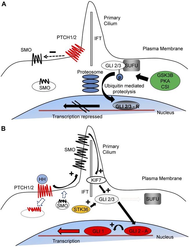
Figure 1. The mechanism of Hh signal transduction.
(A) In the resting state, PTCH 1/2 is expressed on the
plasma membrane and acts to repress SMO activity by
preventing its expression and localization to the primary
cilium. GLI2/3 transcription factors are within a complex,
including SUFU, an inhibitor of Hh signaling. This confor-
mation promotes nonspecific phosphorylation of the
C terminus by GSK3, CSI, and PKA, resulting in
E3 ubiquitin ligase activity and subsequent partial proteo-
somal proteolysis to the C terminal truncated repressor
form. After translocation to the nucleus, the repressive
forms of GLI2 (GLI2-R) and particularly GLI3 (GLI-3R)
potently inhibit the Hh transcriptional program. (B) Interac-
tion of HH ligand with PTCH promotes PTCH internaliza-
tion and degradation and releases the repression of
SMO, causing its accumulation within the primary cilium.
Active SMO in the primary cilium stabilizes the full-length
forms of GLI2 (GLI2-A) and GLI3 (GLI3-A) and accentu-
ates the effect of other positive regulators of Hh signaling,
including serine threonine kinase 36 (STK36) and kinesin
family member 7 (KIF7), which may be involved in
translocation of GLI into the primary cilium. After translo-
cation to the nucleus, GLI2-A potently activates transcrip-
tion of downstream Hh targets, including GLI1 and PTCH1,
and influences chromatin conformation, apoptosis, cell
cycle activity, and differentiation.
and maintain the signal and negative feedback loops switch off the sor, GLI2 exists in both a full-length active form and a truncated
pathway after signal activation. repressor form.29,30 Activated SMO alters the balance between
The central components of the pathway are composed of a pair these forms.10,30 In many vertebrate cell types, this signaling occurs
of 12-span transmembrane receptor proteins, patched (PTCH) 1 and within the primary cilium.31,32 SMO and GLI proteins accumulate
2; a 7-span transmembrane protein, smoothened (SMO); and the there through interaction with the intraflagellar transport (IFT)
family of Glioma (GLI) zinc finger transcription factors GLI1, proteins facilitating activation and onward transduction of the Hh
GLI2, and GLI3 (Figure 1). Unusually, the Hh receptor PTCH is a signal.30,31,33 However, this is dispensable in hematopoietic cells as
negative regulator of pathway activity, whereas the immediately they appear not to possess primary cilia, although, interestingly,
downstream element SMO is the major positive regulator. In the they do express IFT proteins.34
resting state, PTCH represses SMO activity, but after Hh binding When Hh signaling is “off,” GLI2 and GLI3 are retained in the
this repression is removed.23 The mechanism of signal transduction cytoplasm by a protein complex, including the inhibitory molecule
between PTCH and SMO in vertebrates is not known, but the suppressor of fused (SUFU) and are phosphorylated by nonspecific
2 receptors do not appear to interact directly. Patched proteins bear kinase activity mediated by glycogen synthase kinase 3 (GSK3
a striking homology to a transmembrane lipid transporter protein ), casein kinase (CSI), and protein kinase A (PKA).35-38 After
(Neimann-Pick C1 protein), and SMO appears to belong to the phosphorylation, GLI2 and GLI3 undergo C-terminal ubiquitina-
G-protein coupled receptor family.24,25 Current evidence suggests tion and partial proteosome-mediated proteolysis to the truncated
that PTCH influences SMO activity through oxysterol trafficking. inhibitory form before nuclear translocation, resulting in repression
Oxysterols activate Hh signaling through SMO, whereas secretion of the pathway.37 In the presence of Hh ligand, GLI2 and GLI3
of pro-vitamin D3 is inhibitory.26,27 After receptor ligand interac- phosphorylation is prevented and full-length active forms of the
tion, PTCH is internalized, SMO accumulates on the plasma transcription factors translocate to the nucleus.29,38 This activates
membrane and interacts with the GLI family of transcription the transcription of downstream targets that include both positive
factors.28 Whereas GLI3 is predominantly a transcriptional repres- (GLI1) and negative (PTCH1/2) regulatory elements, which driveFrom www.bloodjournal.org by guest on September 22, 2015. For personal use only.
2198 IRVINE and COPLAND BLOOD, 8 MARCH 2012 䡠 VOLUME 119, NUMBER 10
the Hh transcription program and down-regulate ongoing signal- or through 2 different conditional deletion methodologies.13,17,46,47
ing, respectively.30 Dierks et al found no significant difference in the behavior of fetal
HSCs recovered from Smo null mice compared with normal mice.13
Gao et al46 and Hoffman et al47 used the Mx1-Cre system to create a
murine model where Smo deletion could be induced. Neither found
Hh signaling and hematopoiesis any significant difference in the short- or long-term measures of
Hh signaling has multiple complex roles in both adult and hematopoiesis after deletion of Smo and therefore abrogation of Hh
embryonic hematopoiesis, depending on the stage of development signaling. In contrast, Zhao et al demonstrated profound reduction
and whether the hematopoietic system is under regenerative in stem cell activity as indicated by secondary transplant capacity
pressure. IHH, expressed by endodermal cells, specifies hematopoi- after Vav-driven Cre-Lox–mediated conditional Smo deletion.17
etic differentiation at the earliest stages of primitive hematopoiesis Although on the surface these conflicts appear irreconcilable,
and has roles in hematopoietic and vascular development.5,6 the alternative experimental strategies go some way to explaining
However 50% of Ihh null mice survive, suggesting that IHH these differences; the Mx1-Cre system allows deletion in adulthood
functions are partially redundant.39 Definitive hematopoiesis is the and predominantly affects hematopoietic and liver cells, whereas
second wave of hematopoietic activity characterized by the devel- the Vav-Cre-Lox system is active throughout embryogenesis and is
opment of hematopoietic stem cells (HSCs). This process is less selective, affecting both hematopoietic and endothelial tissue,
initiated in the aorta-gonad-mesonephros region but relocates to the raising the possibility that Hh signaling was disrupted in a wider
fetal liver and subsequently the bone marrow.40 Studies in zebrafish range of tissues and at an earlier time point.13,17,46,47
and mice indicate that Hh signaling has a role in establishing Genetic studies have also been performed on the main down-
definitive hematopoiesis.5,41,42 Furthermore, HH ligand has been stream mediators of Hh signaling. Merchant et al characterized the
shown to expand definitive hematopoietic progenitors in mice and effect of Gli1 deletion on hematopoiesis in a Gli1 null murine
humans.5,43,44 model.48 No abnormalities in gross hematopoiesis were found;
Early studies of adult hematopoiesis suggested that Hh signal- however, there was a reduction in proliferation within the stem and
ing caused expansion and increased functional activity of the myeloid progenitor compartments as well as a reduction in myeloid
HSC compartment.9,43 More recently, there has been controversy differentiation and delayed recovery after 5-fluorouracil exposure.
over the role, if any, that Hh signaling plays in adult hematopoiesis Hh acts in concert with other morphogenic signals to influence
because of the differing results of murine studies dissecting the role development of T and B lymphocytes. Thymic stroma and T cells
of Hh mediators in adult hematopoiesis. both express Hh pathway mediators.49,50 Early studies suggested
Dierks et al13 and Trowbridge et al9 demonstrated that Ptch1 that Hh signaling negatively regulated T-cell development51; how-
heterozygosity (hence Hh activation) was associated with increased ever, subsequent work indicates that Hh signaling has a positive
stem cell number, faster hematologic recovery after challenge with regulatory role in early T-cell development.45,49,50,52-54 Both IHH
5-fluorouracil, and increased transplantability in murine models. In and SHH affect T-cell proliferation and maturation.49,52 Conditional
addition, Trowbridge et al found that long-term repopulating cells deletion of Smo at different levels of T-cell development, per-
(the most primitive HSCs) were ultimately exhausted through formed by El Andaloussi et al, suggested that abrogation of Smo
constant Hh activation.9 In contrast to this, Dierks et al found no expression mainly affects primitive thymocytes, leading to thymic
diminution of long-term HSC function.13 Siggins et al examined atrophy and profound defects in early T-cell survival, proliferation,
adult mice subject to conditional deletion of Ptch1 in various and maturation.50 Uhmann et al studied a Ptch1 conditional
tissues, including primitive hematopoietic cells demonstrating that deletion murine model and demonstrated that knockout of Ptch1
conditional deletion of Ptch1 increased circulating primitive hema- led to profound loss of mature T and B cells.53,54 Siggins et al
topoietic cells but did not result in enhanced engraftment in demonstrated that Ptch1 deletion led to significant apoptosis in
transplantation experiments or exhaustion of long-term repopulat- pre-B cells, blocked T-cell specification in bone marrow T cells,
ing capacity.45 Furthermore, their experiments suggested that Ptch1 and caused apoptosis of CD4⫹CD8⫹ T cells because of deletion of
deletion only altered proliferation and mobilization of HSCs when Ptch1 in the supporting microenvironment.45,54
occurring within the supporting bone marrow stroma as, when Overall, it appears that Hh signaling has effects on the
specific deletion limited to HSCs was performed, these differences hematopoietic system dependent on developmental stage and cell
disappeared. This suggests that the effects are through signaling lineage. These effects are the result of a complex interplay between
extrinsic to the HSCs and not a direct effect of Hh on the HSCs the cells and their microenvironment at certain developmental
themselves.45 stages rather than being purely hematopoietic cell-intrinsic mecha-
These discrepancies may be partly explained by the alternative nisms. It is interesting to note that, in early clinical trials of SMO
experimental approaches used. Dierks et al used fetal liver HSCs inhibition in humans, no hematologic toxicities have been noted
from Ptch1⫹/⫺ embryos,13 whereas Trowbridge et al studied adult with up to 6 months of exposure.55 Thus, although Hh signaling
Ptch1⫹/⫺ HSCs.9 In these studies, Ptch1 deletion was present may have a role in normal adult hematopoiesis, it is probably
during embryogenesis. In the germline mutant, Ptch1 heterozygos- dispensable, at least in steady-state conditions, providing a therapeu-
ity would be a feature of all cells and tissues rather than being tic window that may be exploited in hematologic and other
confined to the hematopoietic system. In contrast, Siggins et al used malignancies.
conditional and targeted approaches using the Mx-Cre system.45
They induced deletion of Ptch1 in hematopoietic tissue of mature
mice and also used the SCL enhancer to target Ptch1 deletion to Hh and malignancy
adult murine HSCs.45
Because absence of SMO is lethal, analysis of Smo null mutants Abnormal Hh signaling is associated with diverse human malignan-
is not possible. Four groups have investigated the effect of Smo cies.56 Its oncogenic properties were first identified through the
deletion, either through recovery of Smo null HSCs from fetal liver realization that the genetic abnormality causing Gorlin syndromeFrom www.bloodjournal.org by guest on September 22, 2015. For personal use only.
BLOOD, 8 MARCH 2012 䡠 VOLUME 119, NUMBER 10 TARGETING HEDGEHOG IN HEMATOLOGIC MALIGNANCY 2199
Table 1. Hh pathway inhibitors and clinical trials in hematologic malignancies
Name Action Producer Active hematologic clinical trial Trial number*
Cyclopamine SMO antagonist Generic Nonclinical NA
LDE225 SMO antagonist Novartis Phase 1: CML, in combination with nilotinib NCT1456676
LEQ506 SMO antagonist Novartis Solid tumors only (phase 1 and 2) NA
GDC-0449 SMO antagonist Genentech Phase 1b: myeloma in first remission or first NCT01330173
relapse after ASCT
BMS-833923 SMO antagonist BMS Phase 1: CML, in combination with dasatinib NCT
01218477
Phase 1b: myeloma, in combination with NCT00884546
lenalidomide with dexamethasone or
bortezomib with dexamethasone
IPI926 SMO antagonist Infinity Phase 2: myelofibrosis NCT01371617
PF-04449913 SMO antagonist Pfizer Phase 1: myeloid malignancies, including CML, NCT00953758
in combination with dasatinib or bosutinib
GANT61 Direct GLI inhibition Generic Nonclinical NA
NA indicates not applicable.
*www.clinical trials.gov.
(associated with an excess risk of rhabdomyosarcoma, medulloblas- leukemia (AML), and there is accumulating evidence for their
toma, and basal cell carcinoma) is an inherited inactivating PTCH existence in other hematologic and solid tumors, such as myeloma,
mutation.57 Sporadic cases of the same malignancies are frequently lymphoma, glioblastoma, breast, and prostate cancer.16,18,64-67 In
associated with either inactivating mutations of PTCH or activating these malignancies, CSCs are predominantly quiescent and there-
mutations of SMO, indicating that PTCH and SMO act as tumor fore resistant to conventional therapeutic approaches. Failure to
suppressor and oncogene, respectively.56 Further work confirmed effectively target these cells results in residual low-level disease
that GLI transcription factors were responsible for driving tumor and the likelihood of future relapse. If CSCs are dependent on Hh
formation.58 The association of Hh signaling with oncogenesis is signaling as emerging data suggest, inhibitors of Hh signaling may
not surprising given the critical role that Hh signaling performs in be excellent CSC-targeting therapies.
regulating cell proliferation, cell cycle machinery, apoptosis,
chromatin modeling, self-renewal, and epithelial-to-mesenchymal
transition in responsive cells.59
Recent evidence suggests that Hh signaling may have distinct Targeting the Hh pathway
mechanisms of action in different tumor environments. For ex-
ample, in basal cell carcinoma and medulloblastoma, the most As SMO is the main positive regulator, it has become a logical
common lesions in Hh signaling are gain-of-function SMO muta- pharmaceutical target in modulating ligand-dependent Hh signal-
tions or loss-of-function PTCH mutations.11,60 These are rare in ing. The first Hh inhibitor cyclopamine was identified more than
glioblastoma and lymphoma; however, Hh signaling remains 50 years ago after investigation into a spate of birth defects,
critically important as, in these tumors, malignant cells respond to including cyclopia, in lambs born in sheep farms across Idaho. It
HH ligand secreted from the surrounding microenvironment.12,61
transpired that pregnant ewes were grazing on the corn lily
Furthermore, although Hh signaling is important in pancreatic
Veratrum californicum, a rich source of steroidal alkaloids, includ-
cancer tumorigenesis, this is not through direct signaling to tumor
ing cyclopamine. Cyclopamine was subsequently found to be a
cells but rather because of HH ligand production by tumor cells
potent inhibitor of SMO with anti–tumor activity in in vitro and in
interacting with the local stroma.62 Thus, Hh signaling appears to
be associated with development and maintenance of malignancy vivo systems.68 Although cyclopamine is an effective inhibitor of
through (1) ligand-independent signaling resulting from cell- Hh signaling, it is relatively nonspecific and has significant
intrinsic mutations or (2) autocrine/paracrine signaling between toxicities and therefore has not been effectively used clinically.
tumor cells and stroma or vice versa. Lastly, there is accumulating However, these results provided impetus to synthesize and test
evidence that CSCs are dependent on Hh signaling for population derivatives of cyclopamine or to screen large-scale synthetic
maintenance and expansion. molecule libraries for agents with anti-SMO activity. Several years
on, there are now several clinical grade SMO inhibitors undergoing
preclinical evaluation or early-stage clinical trials (Table 1).
An orally active agent, GDC-0449, showed significant (⬎ 50%
Malignant stem cells response) clinical efficacy against Hh-driven malignancies, such as
The CSC hypothesis suggests that tumors are hierarchical struc- advanced or metastatic basal cell carcinoma in a phase 1 clinical
tures where the capacity for tumor initiation and propagation is trial.55 A subsequent study reported that other solid malignancies
limited to a small number of very primitive cells within the tumor did not respond, although the participants had advanced, multiply
bulk. These cells exhibit similar characteristics to normal stem treated malignancies with relatively short follow-up limiting the
cells, including the ability to undergo symmetric and asymmetric likelihood of a positive result.69 In both cases, SMO inhibition was
self-renewal divisions, thereby maintaining or expanding their well tolerated with little toxicity (predominantly low-grade fatigue,
number in addition to giving rise to more differentiated progeny.63 hyponatremia, and dysgeusia); no maximum tolerated dose was
The existence of such a population has been conclusively demon- reached. Notably, there was a lack of hematologic toxicity in
strated in chronic myeloid leukemia (CML) and acute myeloid treated patients.55,69From www.bloodjournal.org by guest on September 22, 2015. For personal use only.
2200 IRVINE and COPLAND BLOOD, 8 MARCH 2012 䡠 VOLUME 119, NUMBER 10
incidence of leukemia in secondary transplant recipients, and
Hematologic malignancy extended survival.76,77 Therefore, Hh signaling is active in CML, is
critical to the maintenance and expansion of the diseased stem/
There is great interest in using anti-Hh therapies in hematologic progenitor cells, and presents an exciting new target in CML. Phase
malignancies. This extends from accumulating evidence suggest- 1 trials testing the safety of combining TKI and Hh inhibition in
ing that Hh signaling may be critical in the genesis, maintenance, patients with CML of all stages are now open (Table 1;
progression, and relapse of various hematologic conditions through NCT00953758, NCT01218477, NCT1456676).
both direct and indirect mechanisms. Attention has particularly
focused on CML and myeloma where Hh signaling has been
implicated in CSC persistence despite conventional therapy. Multiple myeloma
Myeloma is a malignancy characterized by the malignant clonal
CML expansion of plasma cells and is associated with the production of
monoclonal immunoglobulin. A variety of therapeutic approaches
CML is probably the best understood hematologic malignancy. We have been used but, unfortunately for the majority, myeloma
know a great deal about its clinical behavior and pathophysiology remains incurable because of multiple relapse and progression.
at the cellular and molecular level. It is a paradigm of a stem Evidence is accumulating that myeloma is maintained from a small
cell-driven cancer and is an excellent model for studying stem population of myeloma stem cells, which are resistant to current
cell-directed therapies. CML is a clonal disorder driven by the therapies and may be responsible for the relapsing natural history
constitutively active oncogenic tyrosine kinase BCR-ABL. The of the disease.78 Matsui et al demonstrated that a small population
development of tyrosine kinase inhibitors (TKIs) that block of postgerminal center memory B cells with the surface phenotype
BCR-ABL kinase activity, such as imatinib, nilotinib (Glivec & CD138⫺/CD19⫹/CD27⫹ exist within the tumor, are clonally re-
Tasigna; Novartis Pharma), and dasatinib (Sprycel; Bristol-Myers lated to the circulating tumor bulk, and appear to exhibit stem
Squibb), radically improved management of CML, leading to cell-like characteristics.79,80 These cells are capable of in vitro
unprecedented levels of cytogenetic and molecular responses in colony formation, serial replating, and transplanting the original
chronic-phase patients.70 However, TKI resistance and disease myeloma clone into secondary hosts.79,80 In addition, they express
persistence despite TKI therapy remain ongoing issues. Even constituents of the Hh pathway; and whereas exogenous HH ligand
complete molecular response is not synonymous with cure as caused expansion, inhibition with cyclopamine or a SMO blocking
increasingly sensitive molecular analysis reveals evidence of antibody resulted in contraction of the putative CSC compart-
persistent disease, with the majority of patients having molecular ment.18 Dierks et al demonstrated that HH ligand from supporting
recurrence after TKI withdrawal.71,72 One reason for disease stroma was required for in vitro survival and expansion of murine
persistence despite prolonged TKI therapy is that, while harboring and human primary lymphoma and myeloma cells.12 These obser-
the BCR-ABL fusion protein, primitive CML cells are resistant to vations are particularly relevant as primitive myeloma cells appear
pharmacologically achievable concentrations of TKI and may not to be resistant to standard myeloma therapies, including lenalido-
be dependent on BCR-ABL signaling for survival.70,73 mide, bortezomib, dexamethasone, and cyclophosphamide.80
Hematopoiesis in chronic-phase CML remains hierarchical with
a leukemic stem cell (LSC) giving rise to leukemic progenitor and
effector cells. As CML progresses, the hierarchy is disrupted and Acute leukemia
mature committed progenitors appear to aberrantly reacquire
self-renewal capacity through cooperating events (eg, activation of Less is known about the Hh signaling pathway and its influence on
Wnt signaling).74 Therefore, in CML, self-renewal is a potential AML and acute lymphoid leukemia (ALL). Although there is clear
target both in chronic- and advanced-phase disease. evidence for a LSC population in AML and ALL, recent work has
Hh signaling is increased in BCR-ABL⫹ stem and progenitor suggested that this population is phenotypically variable between
cells, becoming more active with disease progression.13,17,75 Two patients, may not be confined to a single clonal subpopulation, and
groups have explored the effect of Smo deletion in murine models may arise from an HSC or a committed progenitor.81,82 Cells with
of CML. Zhao et al used the Vav-Cre-Lox system to create LSC properties have reduced sensitivity to chemotherapeutic
Smo-deficient mice from which to isolate Smo⫺/⫺ HSCs,17 whereas agents and contribute to treatment failure and disease relapse.83
Dierks et al used fetal liver cells from Smo-deficient mouse Therefore, there is a clear rationale for investigating and targeting
embryos.13 Both groups expressed Bcr-Abl in these cells before mechanisms involved in LSC maintenance and self-renewal.
transplantation. Despite differing approaches, broadly similar re- A variety of cell-extrinsic mechanisms are involved in this process,
sults were obtained. Smo deletion reduced LSC numbers and including oxidative stress, cell-cell contact, and microenviron-
reduced incidence of leukemia with prolonged latency in primary mental signaling in addition to cell intrinsic mechanisms, such as
transplantation and greatly reduced capacity to recrudesce disease constitutive activation of NFB/AKT/PI3K, overexpression of
in secondary hosts. Both groups performed complementary pharma- anti-apoptosis mechanisms, and chromatin modification. Further-
cologic inhibitor studies, demonstrating prolonged survival in more, the leukemogenic translocation is sometimes capable
diseased mice, reduced LSC population, and lower functional of enhancing or conferring self-renewal activity. For example,
activity in vivo and in vitro after cyclopamine treatment.13,17 AML1-ETO translocations interact with the downstream amino-
Interestingly, the combination of TKI therapy with cyclopamine terminal enhancer of split (AES), enhancing self-renewal measure-
resulted in the largest reduction in LSCs in vitro and in vivo.13 ments in vitro, and the MOZ-TIF2 and MLL-ENL translocations
Studies using clinical grade SMO inhibitors alone and com- confer self-renewal properties to progenitor cells.84,85 In this
bined with TKIs both in vitro and in vivo support these conclusions context, blocking one pathway, such as Hh, may not be sufficient to
demonstrating significant reduction in measures of self-renewal, target these LSCs. Hh signaling is completely dispensable for theFrom www.bloodjournal.org by guest on September 22, 2015. For personal use only.
BLOOD, 8 MARCH 2012 䡠 VOLUME 119, NUMBER 10 TARGETING HEDGEHOG IN HEMATOLOGIC MALIGNANCY 2201
development of acute leukemia in a MLL-AF9-mediated murine over normal B cells.97 In addition, in their studies, SMO inhibition
model of myeloid leukemia and a Notch-dependent murine model alone did not influence B-CLL cell survival in vitro. However,
of T-ALL.46,86 In contrast, very recent work in a murine model of GANT61 (a direct Gli1 inhibitor) caused significant apoptosis in
erythroleukemia suggested that development of transplantable B-CLL cells but not normal B cells in vitro, which could be rescued
leukemia depended on proviral activation of Sp1/Pu.1 in cells with by stromal coculture but not recombinant SHH ligand alone. This
an intact Hh signaling pathway.87 In addition, SHH and GLI1 are suggests that other signaling pathways converging on GLI1
expressed in leukemic cell lines and primary leukemic blasts, and expression were responsible.97 Decker et al recently demonstrated
recent evidence suggests that inhibitors of the Hh pathway may that both Hh mediator expression and SMO inhibitor responsive-
have some efficacy.88-90 Lin et al demonstrated Hh signaling ness were variable in different patients. They found that 60% of
activity in precursor B-ALL and that SMO inhibition with cyclo- their CLL cohort responded to SMO inhibition.98 Factors that
pamine or IPI926 reduced in vitro and in vivo measures of predicted likelihood of response included elevated expression of
self-renewal.91 Another preliminary study indicated that cyclo- GLI1 and PTCH1 and carriage of trisomy 12. In addition, trisomy
pamine has some efficacy in AML in vitro, possibly via mitigation 12 CLL cells produced DHH, suggesting that autocrine signaling
of multidrug resistance.92 may be important in this subtype.98
In CSC-driven hematologic malignancies, the rationale for Hh has been implicated in the etiology of T-cell lymphomas.
SMO inhibition is predominantly to target the resistant, diseased Singh et al demonstrated that constituents of the Hh pathway were
stem cell population, attempting to reduce relapse rate and poten- overexpressed in ALK⫹ anaplastic large cell lymphoma.99 They
tially cure these diseases. Although it appears that both CML and demonstrated amplification of the SHH gene locus and conse-
myeloma stem cells rely on Hh signaling for maintenance and quently increased levels of mRNA and protein in ALK⫹ anaplastic
expansion, it is not clear whether this is a cell-intrinsic property, large cell lymphoma cell lines and primary tissue and overexpres-
driven by autocrine/paracrine production of HH ligand, or conse- sion of GLI1 through PI3K/AKT pathway signaling. They also
quent on the effect of Hh signaling on the supportive stromal confirmed that pharmacologic inhibition of SMO led to cell cycle
microenvironment. arrest and apoptosis.99 More recently, it has been reported that
GLI3 is highly expressed in Hodgkin lymphoma cell lines and in
Reed-Sternberg cells from 39 of 39 cases of classic Hodgkin
lymphoma by immunohistochemistry, suggesting a hitherto un-
Lymphoma and CLL known role for Hh signaling in Hodgkin lymphoma.100
Hh pathway inhibition may also be effective in lymphoma and
chronic lymphocytic leukemia (CLL). Hh signaling is critical for
normal B- and T-cell development; however, it is primarily the Conclusion
microenvironmental cells and supportive stroma of the bone In conclusion, despite continued progress in our understanding
marrow and lymphoid organs that produce the HH ligand, which is of the molecular and cellular events that underlie the development
required for lymphoid cell maintenance.45,54 Constituents and and progression of hematologic malignancies, in most instances,
targets of Hh signaling are expressed in various lymphoma cell complete eradication of the malignant clone remains elusive. This
lines and primary tissue.92-94 IHH and SHH have been shown to be has resulted in long-term dependence on drug therapy to maintain
survival factors in B-cell malignancies.94 Dierks et al used the disease control and prevent disease progression and relapse or
E-myc murine model of Burkitt lymphoma to demonstrate that treatment failure.
stromal coculture was necessary for lymphoma cell survival and It has become increasingly clear that the relationship between
expansion, but stroma could be effectively replaced with soluble the tumor cell and its supporting environment plays a critical role in
SHH or IHH.12 Furthermore, lymphoma cells underwent apoptosis treatment resistance through direct pro-survival signaling, control
in the absence or inhibition of Hh signaling both in vitro and in of proliferation, and, at least in some malignancies, the mainte-
vivo. Downstream Hh targets included BCL-2 and BCL-XL; both nance and expansion of CSCs. These relationships are highly
these pro-survival pathways were up-regulated in the presence of complex and are modulated by prevailing chemical conditions,
Hh signaling and down-regulated after inhibition.12,94 such as oxygen tension, and physiologic cues in addition to a
In mantle cell lymphoma, preliminary work has shown that network of signaling pathways, including the conserved embryonic
mantle cell lymphoma cell lines and primary human samples had signaling pathway Hh. Targeting these interactions might provide
increased expression of Hh pathway components compared with an alternative, more effective therapeutic strategy either alone or in
normal B cells and were partially responsive to HH ligand or its combination with conventional treatments. Hh signaling is an
pharmacologic inhibition.95 Direct inhibition of the downstream attractive target as there is a large body of evidence that relates
effectors GLI1 and GLI2 had the most profound effect on mantle abnormal Hh signaling to malignancy; furthermore, from a drug
cell lymphoma cells, indicating that, although canonical Hh development perspective, inhibition of the key positive regulator
signaling may be involved in mantle cell lymphoma, there are SMO is readily achieved. A critical question as to whether SMO
probably other factors driving GLI1 and GLI2 expression. inhibition would be therapeutically useful in hematologic malignan-
Inhibitors of Hh signaling may also have a role in future cies relates to the anticipated level of hematologic toxicity in
treatment of CLL, as initial observations suggest that mediators and humans. Clinical grade SMO inhibitors are currently under trial in
downstream targets of Hh signaling (GLI1, GLI2, and SUFU) are both solid tumors and hematologic malignancies; interestingly,
up-regulated in B-CLL cells and may correlate with clinical there appears to be little hematologic toxicity experienced by trial
outcome.96 Pharmacologic inhibition of Hh signaling abrogated participants.55
stromal microenvironment-mediated survival of B-CLL cells in in The next question relates to the activity and function of Hh
vitro treatment assays with fludarabine.96 In contrast, Desch et al signaling in hematopoietic malignancies. Hh signaling intermedi-
found that key participants of the Hh signaling cascade (including ates are expressed by cells from a wide spectrum of hemato-
SMO and GLI1) were not differentially expressed in B-CLL cells oncologic conditions, and Hh signaling appears to exert its effectFrom www.bloodjournal.org by guest on September 22, 2015. For personal use only.
2202 IRVINE and COPLAND BLOOD, 8 MARCH 2012 䡠 VOLUME 119, NUMBER 10
either through direction of CSC fate or as a survival and prolifera- it remains to be seen whether or how many of these laboratory
tion signal derived from the supporting stromal microenvironment observations translate to real clinical benefits to patients.
as has been shown in models of lymphoma. The degree of evidence
that Hh activity is a legitimate target in different hematologic
malignancies is variable. There is greatest preclinical evidence to Acknowledgments
support the use of Hh antagonists in the context of CML where in D.A.I. was supported by the Chief Scientist Office, Scotland
vitro and in vivo inhibitor studies in mouse and human tissue have (clinical research fellowship) and the Greater Glasgow and Clyde
demonstrated a significant anti-LSC effect. These results are National Health Service Endowment Fund (research funding).
supported by the complementary genetic studies of Zhao et al17 and D.A.I. participated in the Translational Research Training in
Dierks et al13 It must be borne in mind that these were performed Hematology course (run jointly by ASH and EHA). M.C. is a
using models that resulted in quite different data regarding the clinical senior lecturer funded by the Chief Scientist Office,
requirement for Smo in normal HSCs, where Smo deletion was not Scotland, the Scottish Funding Council, and Leukemia and Lym-
specific to HSCs and occurred early in hematopoietic development, phoma Research.
conceivably affecting microenvironmental interactions that are
subsequently reflected in adult HSC function.
In the context of acute leukemias, the published data are Authorship
inconsistent and it is probable that the contribution of Hh signaling, Contribution: D.A.I. wrote and revised the manuscript; and M.C.
and hence the likelihood of benefit from Hh antagonism, is more revised the manuscript and checked the final version.
contextual depending on the transforming mutation. Lastly, in Conflict-of-interest disclosure: The authors declare no compet-
lymphoid malignancies, Hh antagonism could conceivably be used ing financial interests.
to reduce the pro-survival effect of the tumor microenvironment. Correspondence: Mhairi Copland, Paul O’Gorman Leukaemia
Ultimately, the preclinical data suggest that Hh antagonism may Research Centre, College of Medical, Veterinary and Life Sciences,
be an extremely useful therapeutic intervention in several hemato- University of Glasgow, Gartnavel General Hospital, 1053 Great
logic malignancies where it may address the persistence of CSC Western Road, Glasgow G12 0ZD, United Kingdom; e-mail:
and the protective effect of the tumor microenvironment; however, mhairi.copland@glasgow.ac.uk.
References
1. Nüsslein-Volhard C, Wieschaus E. Mutations hedgehog signaling in B-cell malignancies. Nat Niemann-Pick C1 protein. Mol Genet Metab.
affecting segment number and polarity in Dro- Med. 2007;13(8):944-951. 2000;71(1):175-181.
sophila. Nature. 1980;287(5785):795-801. 13. Dierks C, Beigi R, Guo GR, Zirlik K, Stegert MR, 25. Ayers KL, Therond PP. Evaluating Smoothened
2. Chiang C, Litingtung Y, Lee E, et al. Cyclopia and Manley P. Expansion of Bcr-Abl-positive leukemic as a G-protein-coupled receptor for Hedgehog
defective axial patterning in mice lacking Sonic stem cells is dependent on Hedgehog pathway signalling. Trends Cell Biol. 2010;20(5):287-298.
hedgehog gene function. Nature. 1996; activation. Cancer Cell. 2008;14(3):238-249. 26. Bijlsma MF, Spek CA, Zivkovic D, van de Water S,
383(6599):407-413. 14. Jiang J, Hui CC. Hedgehog signaling in develop- Rezaee F, Peppelenbosch MP. Repression of
3. Roessler E, Belloni E, Gaudenz K, et al. Muta- ment and cancer. Dev Cell. 2008;15(6):801-812. smoothened by patched-dependent (Pro-)vitamin
tions in the human Sonic Hedgehog gene cause D3 secretion. Plos Biol. 2006;4(8):e232.
15. Liu S, Dontu G, Mantle ID, et al. Hedgehog sig-
holoprosencephaly. Nat Genet. 1996;14(3):357- 27. Corcoran RB, Scott MP. Oxysterols stimulate
naling and Bmi-1 regulate self-renewal of normal
360. Sonic hedgehog signal transduction and prolif-
and malignant human mammary stem cells. Can-
4. Blank U, Karlsson G, Karlsson S. Signaling path- cer Res. 2006;66(12):6063-6071. eration of medulloblastoma cells. Proc Natl Acad
ways governing stem-cell fate. Blood. 2008; Sci U S A. 2006;103(22):8408-8413.
16. O’Brien CA, Kreso A, Jamieson CHM. Cancer
111(2):492-503. 28. Denef N, Neubüser D, Perez L, Cohen SM.
stem cells and self-renewal. Clin Cancer Res.
5. Cridland SO, Keys JR, Papathanasiou P, 2010;16(12):3113-3120. Hedgehog induces opposite changes in turnover
Perkins AC. Indian hedgehog supports definitive and subcellular localization of patched and
17. Zhao C, Chen A, Jamieson CH, et al. Hedgehog
erythropoiesis. Blood Cells Mol Dis. 2009;43(2): smoothened. Cell. 2000;102(4):521-531.
signalling is essential for maintenance of cancer
149-155. 29. Sasaki H, Nishizaki Y, Hui C, Nakafuku M,
stem cells in myeloid leukaemia. Nature. 2009;
6. Dyer MA, Farrington SM, Mohn D, Munday JR, 458(7239):776-779. Kondoh H. Regulation of Gli2 and Gli3 activities
Baron MH. Indian hedgehog activates hemato- by an amino-terminal repression domain: implica-
poiesis and vasculogenesis and can respecify 18. Peacock CD, Wang Q, Gesell GS, Corcoran- tion of Gli2 and Gli3 as primary mediators of Shh
prospective neurectodermal cell fate in the Schwartz IM, Jones E, Kim J. Hedgehog signal- signaling. Development. 1999;126(17):3915-
mouse embryo. Development. 2001;128(10): ing maintains a tumor stem cell compartment in 3924.
1717-1730. multiple myeloma. Proc Natl Acad Sci U S A.
2007;104(10):4048-4053. 30. Hui CC, Angers S. Gli proteins in development
7. Ingham PW, Placzek M. Orchestrating ontogen- and disease. Annu Rev Cell Dev Biol. 2011;27:
esis: variations on a theme by sonic hedgehog. 19. Lee JJ, Ekker SC, von Kessler DP, Porter JA, 513-537.
Nat Rev Genet. 2006;7(11):841-850. Sun BI, Beachy PA. Autoproteolysis in hedgehog
31. Corbit KC. Vertebrate smoothened functions at
protein biogenesis. Science. 1994;266(5190):
8. Beachy PA, Karhadkar SS, Berman DM. Tissue the primary cilium. Nature. 2005;437(7061):1018-
1528-1537.
repair and stem cell renewal in carcinogenesis. 1021.
Nature. 2004;432(7015):324-331. 20. Beachy PA, Cooper MK, Young KE, et al. Multiple
32. Rohatgi R, Milenkovic L, Scott MP. Patched
roles of cholesterol in hedgehog protein biogen-
9. Trowbridge JJ, Scott MP, Bhatia M. Hedgehog 1 regulates hedgehog signaling at the primary
esis and signaling. Cold Spring Harb Symp Quant
modulates cell cycle regulators in stem cells to cilium. Science. 2007;317(5836):372-376.
Biol. 1997;62:191-204.
control hematopoietic regeneration. Proc Natl 33. Kim J, Kato M, Beachy PA. Gli2 trafficking links
Acad Sci U S A. 2006;103(38):14134-14139. 21. Pathi S, Pagan-Westphal S, Baker DP, et al.
Hedgehog-dependent activation of Smoothened
Comparative biological responses to human
10. Ruiz i Altaba A, Mas C, Stecca B. The Gli code: in the primary cilium to transcriptional activation in
Sonic, Indian, and Desert hedgehog. Mech Dev.
an information nexus regulating cell fate, stem- the nucleus. Proc Natl Acad Sci U S A. 2009;
2001;106(1):107-117.
ness and cancer. Trends Cell Biol. 2007;17(9): 106(51):21666-21671.
438-447. 22. Bitgood MJ, Shen L, McMahon AP. Sertoli cell 34. Finetti F, Paccani SR, Riparbelli MG, et al. Intra-
11. Reifenberger J, Wolter M, Knobbe CB, et al. So- signaling by Desert hedgehog regulates the male flagellar transport is required for polarized recy-
matic mutations in the PTCH, SMOH, SUFUH germline. Curr Biol. 1996;6(3):298-304. cling of the TCR/CD3 complex to the immune
and TP53 genes in sporadic basal cell carcino- 23. Robbins DJ, Hebrok M. Hedgehogs: la dolce vita. synapse. Nat Cell Biol. 2009;11(11):1332-1339.
mas. Br J Dermatol. 2005;152(1):43-51. Workshop on Hedgehog-Gli Signaling in Cancer 35. Humke EW, Dorn KV, Milenkovic L, Scott MP,
12. Dierks C, Grbic J, Zirlik K, Beigi R, Englund NP, and Stem Cells. EMBO Rep. 2007;8(5):451-455. Rohatgi R. The output of Hedgehog signaling is
Guo GR. Essential role of stromally induced 24. Ioannou YA. The structure and function of the controlled by the dynamic association betweenFrom www.bloodjournal.org by guest on September 22, 2015. For personal use only.
BLOOD, 8 MARCH 2012 䡠 VOLUME 119, NUMBER 10 TARGETING HEDGEHOG IN HEMATOLOGIC MALIGNANCY 2203
Suppressor of Fused and the Gli proteins. Genes 54. Uhmann A, van den Brandt J, Dittmann K, et al. Deininger MW, Druker BJ. Human chronic my-
Dev. 2010;24(7):670-682. T-cell development critically depends on prethy- eloid leukemia stem cells are insensitive to ima-
36. Kise Y, Morinaka A, Teglund S, Miki H. Sufu re- mic stromal patched expression. J Immunol. tinib despite inhibition of BCR-ABL activity. J Clin
cruits GSK3beta for efficient processing of Gli3. 2011;186(6):3383-3391. Invest. 2011;121(1):396-409.
Biochem Biophys Res Commun. 2009;387(3): 55. Von Hoff DD, LoRusso PM, Rudin CM, Reddy JC, 74. Jamieson CH, Ailles LE, Dylla SJ, et al.
569-574. Yauch RL, Tibes R. Inhibition of the hedgehog Granulocyte-macrophage progenitors as candi-
37. Tempé D, Casas M, Karaz S, Blanchet-Tournier MF, pathway in advanced basal-cell carcinoma. date leukemic stem cells in blast-crisis CML.
Concordet JP. Multisite protein kinase A and gly- N Engl J Med. 2009;361(12):1164-1172. N Engl J Med. 2004;351(7):657-667.
cogen synthase kinase 3beta phosphorylation 56. Teglund S, Toftgard R. Hedgehog beyond medul- 75. Radich JP, Dai H, Mao M, Oehler V, Schelter J,
leads to Gli3 ubiquitination by SCFbetaTrCP. Mol loblastoma and basal cell carcinoma. Biochim Druker B. Gene expression changes associated
Cell Biol. 2006;26(11):4316-4326. Biophys Acta. 2010;1805(2):181-208. with progression and response in chronic myeloid
57. Gorlin RJ. Nevoid basal cell carcinoma (Gorlin) leukemia. Proc Natl Acad Sci U S A. 2006;103(8):
38. Pan Y, Wang C, Wang B. Phosphorylation of Gli2
syndrome. Genet Med. 2004;6(6):530-539. 2794-2799.
by protein kinase A is required for Gli2 processing
and degradation and the Sonic Hedgehog- 76. Irvine DA, Zhang B, Ho Y, et al. Combination of
58. Grachtchouk M. Basal cell carcinomas in mice
regulated mouse development. Dev Biol. 2009; the hedgehog pathway inhibitor LDE225 and nilo-
overexpressing Gli2 in skin. Nat Genet. 2000;
326(1):177-189. tinib targets the leukemic stem cell population in
24(3):216-217.
chronic myeloid leukemia. Haematologica. 2011;
39. St-Jacques B, Hammerschmidt M, McMahon AP. 59. Katoh Y, Katoh M. Hedgehog target genes: 96(suppl 2):221. Abstract 521.
Indian hedgehog signaling regulates proliferation mechanisms of carcinogenesis induced by aber-
and differentiation of chondrocytes and is essen- 77. Schairer A, Shih A, Geron I, et al. Human blast
rant hedgehog signaling activation. Curr Mol
tial for bone formation. Genes Dev. 1999;13(16): crisis leukemia stem cell inhibition with a novel
Med. 2009;9(7):873-886.
2072-2086. smoothened antagonist. ASH Annual Meeting
60. Wolter M, Reifenberger J, Sommer C, Ruzicka T, Abstracts. 2010;116(21):1223a.
40. Cumano A, Godin I. Ontogeny of the hematopoi- Reifenberger G. Mutations in the human homo-
etic system. Annu Rev Immunol. 2007;25:745- 78. Huff CA, Matsui W. Multiple myeloma cancer
logue of the Drosophila segment polarity gene
785. stem cells. J Clin Oncol. 2008;26(17):2895-2900.
patched (PTCH) in sporadic basal cell carcino-
41. Gering M, Patient R. Hedgehog signaling is mas of the skin and primitive neuroectodermal 79. Matsui W, Huff CA, Wang Q, Malehorn MT,
required for adult blood stem cell formation in tumors of the central nervous system. Cancer Barber J, Tanhehco Y. Characterization of clono-
zebrafish embryos. Dev Cell. 2005;8(3):389-400. Res. 1997;57(13):2581-2585. genic multiple myeloma cells. Blood. 2004;
103(6):2332-2336.
42. Peeters M, Ottersbach K, Bollerot K, Orelio C, 61. Ehtesham M, Sarangi A, Valadez JG, et al.
de Bruijn M, Wijgerde M. Ventral embryonic tis- Ligand-dependent activation of the hedgehog 80. Matsui W, Wang Q, Barber JP, et al. Clonogenic
sues and Hedgehog proteins induce early AGM pathway in glioma progenitor cells. Oncogene. multiple myeloma progenitors, stem cell proper-
hematopoietic stem cell development. Develop- 2007;26(39):5752-5761. ties, and drug resistance. Cancer Res. 2008;
ment. 2009;136(15):2613-2621. 68(1):190-197.
62. Ruiz i Altaba A. Therapeutic inhibition of
Hedgehog-GLI signaling in cancer: epithelial, 81. le Viseur C, Hotfilder M, Bomken S, et al. In child-
43. Bhardwaj G, Murdoch B, Wu D, Baker DP,
stromal, or stem cell targets? Cancer Cell. 2008; hood acute lymphoblastic leukemia, blasts at dif-
Williams KP, Chadwick K. Sonic hedgehog in-
14(4):281-283. ferent stages of immunophenotypic maturation
duces the proliferation of primitive human hema-
have stem cell properties. Cancer Cell. 2008;
topoietic cells via BMP regulation. Nat Immunol. 63. Reya T, Morrison SJ, Clarke MF, Weissman IL. 14(1):47-58.
2001;2(2):172-180. Stem cells, cancer, and cancer stem cells. Na-
ture. 2001;414(6859):105-111. 82. Goardon N, Marchi E, Atzberger A, et al. Coexis-
44. Kobune M, Ito Y, Kawano Y. Indian hedgehog
tence of LMPP-like and GMP-like leukemia stem
gene transfer augments hematopoietic support of 64. Holyoake T, Jiang X, Eaves C, Eaves A. Isolation cells in acute myeloid leukemia. Cancer Cell.
human stromal cells including NOD/SCID- of a highly quiescent subpopulation of primitive 2011;19(1):138-152.
[beta]2m⫺/⫺ repopulating cells. Blood. 2004; leukemic cells in chronic myeloid leukemia.
104(4):1002-1009. Blood. 1999;94(6):2056-2064. 83. de Jonge-Peeters SDPWM, Kuipers F, de Vries
EGE, Vellenga E. ABC transporter expression in
45. Siggins SL, Nguyen N-YN, McCormack MP, et al. 65. Bonnet D, Dick JE. Human acute myeloid leuke- hematopoietic stem cells and the role in AML drug
The Hedgehog receptor Patched1 regulates my- mia is organized as a hierarchy that originates resistance. Crit Rev Oncol Hematol. 2007;62(3):
eloid and lymphoid progenitors by distinct cell- from a primitive hematopoietic cell. Nat Med. 214-226.
extrinsic mechanisms. Blood. 2009;114(5):995- 1997;3(7):730-737.
1004. 84. Steffen B, Knop M, Bergholz U, et al. AML1/ETO
66. Jones RJ, Gocke CD, Kasamon YL, et al. Circu- induces self-renewal in hematopoietic progenitor
46. Gao J, Graves S, Koch U, et al. Hedgehog signal- lating clonotypic B cells in classic Hodgkin lym- cells via the Groucho-related amino-terminal AES
ing is dispensable for adult hematopoietic stem phoma. Blood. 2009;113(23):5920-5926. protein. Blood. 2011;117(16):4328-4337.
cell function. Cell Stem Cell. 2009;4(6):548-558.
67. Brennan SK, Meade B, Wang Q, Merchant AA, 85. Huntly BJP, Shigematsu H, Deguchi K, et al.
47. Hofmann I, Stover EH, Cullen DE, Mao J, Kowalski J, Matsui W. Mantle cell lymphoma acti- MOZ-TIF2, but not BCR-ABL, confers properties
Morgan KJ, Lee BH. Hedgehog signaling is dis- vation enhances bortezomib sensitivity. Blood. of leukemic stem cells to committed murine he-
pensable for adult murine hematopoietic stem cell 2010;116(20):4185-4191. matopoietic progenitors. Cancer Cell. 2004;6(6):
function and hematopoiesis. Cell Stem Cell. 68. Chen JK, Taipale J, Cooper MK, Beachy PA. Inhi- 587-596.
2009;4(6):559-567. bition of Hedgehog signaling by direct binding of 86. Hofmann I, Stover EH, Cullen DE, et al. Hedge-
48. Merchant A, Joseph G, Wang Q, Brennan S, cyclopamine to Smoothened. Genes Dev. 2002; hog signaling is dispensable for adult murine he-
Matsui W. Gli1 regulates the proliferation and dif- 16(21):2743-2748. matopoietic stem cell function and hematopoi-
ferentiation of HSCs and myeloid progenitors. 69. LoRusso PM. Phase I trial of hedgehog pathway esis. Cell Stem Cell. 2009;4(6):559-567.
Blood. 2010;115(12):2391-2396. inhibitor vismodegib (GDC-0449) in patients with 87. Hegde S, Hankey P, Paulson RF. Self renewal of
49. Outram SV, Hager-Theodorides AL, Shah DK, refractory, locally-advanced or metastatic solid leukemia stem cells in friend virus induced eryth-
et al. Indian hedgehog (Ihh) both promotes and tumors. Clin Cancer Res. 2011;17(8):2502-2511. roleukemia requires proviral insertional activation
restricts thymocyte differentiation. Blood. 2009; 70. Irvine DA, Heaney NB, Holyoake TL. Optimising of Spi1 and hedgehog signaling, but not mutation
113(10):2217-2228. chronic myeloid leukaemia therapy in the face of of p53. Stem Cells. 2012;30(2):121-130.
50. El Andaloussi AE, Graves S, Meng F, Mandal M, resistance to tyrosine kinase inhibitors: a synthe- 88. Queiroz KC, Ruela-de-Sousa RR, Fuhler GM,
Mashayekhi M, Aifantis I. Hedgehog signaling sis of clinical and laboratory data. Blood Rev. et al. Hedgehog signaling maintains chemoresis-
controls thymocyte progenitor homeostasis and 2010;24(1):1-9. tance in myeloid leukemic cells. Oncogene. 2010;
differentiation in the thymus. Nat Immunol. 2006; 71. Mahon FX, Rea D, Guilhot J, Guilhot F, Huguet F, 29(48):6314-6322.
7(4):418-426. Nicolini F. Discontinuation of imatinib in patients 89. Ji Z, Mei FC, Johnson BH, Thompson EB, Cheng X.
51. Outram SV, Varas A, Pepicelli CV, Crompton T. with chronic myeloid leukaemia who have main- Protein kinase A, not Epac, suppresses hedge-
Hedgehog signaling regulates differentiation from tained complete molecular remission for at least hog activity and regulates glucocorticoid sensitiv-
double-negative to double-positive thymocyte. 2 years: the prospective, multicentre Stop Ima- ity in acute lymphoblastic leukemia cells. J Biol
Immunity. 2000;13(2):187-197. tinib (STIM) trial. Lancet Oncol. 2010;11(11): Chem. 2007;282(52):37370-37377.
52. Shah DK, Hager-Theodorides AL, Outram SV, 1029-1035. 90. Bai LY, Chiu CF, Lin CW, et al. Differential expres-
Ross SE, Varas A, Crompton T. Reduced thymo- 72. Ross DM, Branford S, Seymour JF, et al. Patients sion of Sonic hedgehog and Gli1 in hematological
cyte development in sonic hedgehog knockout with chronic myeloid leukemia who maintain a malignancies. Leukemia. 2007;22(1):226-228.
embryos. J Immunol. 2004;172(4):2296-2306. complete molecular response after stopping ima- 91. Lin TL, Wang QH, Brown P, Peacock C,
53. Uhmann A, Dittmann K, Nitzki F, et al. The tinib treatment have evidence of persistent leuke- Merchant AA, Brennan S. Self-renewal of acute
Hedgehog receptor Patched controls lymphoid mia by DNA PCR. Leukemia. 2010;24(10):1719- lymphocytic leukemia cells is limited by the
lineage commitment. Blood. 2007;110(6):1814- 1724. hedgehog pathway inhibitors cyclopamine and
1823. 73. Corbin AS, Agarwal A, Loriaux M, Cortes J, IPI-926. Plos One. 2010;5:e15262.From www.bloodjournal.org by guest on September 22, 2015. For personal use only.
2204 IRVINE and COPLAND BLOOD, 8 MARCH 2012 䡠 VOLUME 119, NUMBER 10
92. Kobune M, Takimoto R, Murase K, et al. Drug re- geting of sonic hedgehog-GLI signaling: a poten- els are biomarkers for Hedgehog-inhibitor re-
sistance is dramatically restored by hedgehog tial strategy to improve therapy for mantle cell sponsiveness in CLL [published online ahead of
inhibitors in CD34⫹ leukemic cells. Cancer Sci. lymphoma. Mol Cancer Ther. 2008;7(6):1450- print November 30, 2011]. Blood. 2012. doi:
2009;100(5):948-955. 1460. 10.1182/blood-2011-06-359075.
93. Kim JE, Singh RR, Cho-Vega JH, et al. Sonic 96. Hegde GV, Peterson KJ, Emanuel K, et al.
99. Singh RR, Cho-Vega JH, Davuluri Y, et al. Sonic
hedgehog signaling proteins and ATP-binding Hedgehog-induced survival of B-cell chronic lym-
hedgehog signaling pathway is activated in ALK-
cassette G2 are aberrantly expressed in diffuse phocytic leukemia cells in a stromal cell microen-
positive anaplastic large cell lymphoma. Cancer
large B-cell lymphoma. Mod Pathol. 2009;22(10): vironment: a potential new therapeutic target. Mol
1312-1320. Cancer Res. 2008;6(12):1928-1936. Res. 2009;69(6):2550-2558.
94. Singh RR, Kim JE, Davuluri Y, Drakos E, 97. Desch P, Asslaber D, Kern D, et al. Inhibition of 100. Greaves WO, Kim JE, Singh RR, et al. Glioma-
Cho-Vega JH, Amin HM. Hedgehog signaling GLI, but not Smoothened, induces apoptosis in associated oncogene homologue 3, a hedgehog
pathway is activated in diffuse large B-cell lym- chronic lymphocytic leukemia cells. Oncogene. transcription factor, is highly expressed in Hodg-
phoma and contributes to tumor cell survival and 2010;29(35):4885-4895. kin and Reed-Sternberg cells of classical Hodgkin
proliferation. Leukemia. 2010;24(5):1025-1036. 98. Decker S, Zirlik K, Djebatchie L, et al. Trisomy lymphoma. Hum Pathol. 2011;42(11):1643-
95. Hegde GV, Munger CM, Emanuel K, et al. Tar- 12 and elevated GLI1 and PTCH1 transcript lev- 1652.You can also read