Systematic Identification of Plasmodium Falciparum Sporozoite Membrane Protein Interactions Reveals an Essential Role for the p24 Complex in Host ...
←
→
Page content transcription
If your browser does not render page correctly, please read the page content below
RESEARCH

Systematic Identification of Plasmodium Falciparum
Sporozoite Membrane Protein Interactions Reveals
an Essential Role for the p24 Complex in Host
Infection
Authors
Julia Knöckel, Kirsten Dundas, Annie S. P. Yang, Francis Galaway, Tom Metcalf, Geert-Jan van Gemert,
Robert W. Sauerwein, Julian C. Rayner, Oliver Billker, and Gavin J. Wright

Correspondence                       Graphical Abstract
gw2@sanger.ac.uk

In Brief
Malaria remains one of the
world's most deadly infectious
diseases, and host infection is
initiated when the sporozoite
form of the P. falciparum parasite
is transmitted by the bite of an
infected mosquito. Here, we
compile a library of soluble
recombinant sporozoite cell
surface and secreted proteins
and test them for direct
interactions using an
extracellular protein interaction
assay. We identify three
conserved complexes and show
that one, the p24 complex, is
essential for host liver-stage
infection.

Highlights

•   A library of Plasmodium falciparum sporozoite membrane and secreted proteins.

•   Systematic extracellular interaction screening identifies novel protein complexes.

•   Interactions are conserved across Plasmodium species.

•   Essential role for genes encoding p24 complex proteins in liver-stage infection.

2021, Mol Cell Proteomics 20, 100038
© 2021 THE AUTHORS. Published by Elsevier Inc on behalf of American Society for Biochemistry and
Molecular Biology. This is an open access article under the CC BY license (http://creativecommons.org/
licenses/by/4.0/).
https://doi.org/10.1074/mcp.RA120.002432
RESEARCH

Systematic Identification of Plasmodium
Falciparum Sporozoite Membrane Protein
Interactions Reveals an Essential Role for the
p24 Complex in Host Infection
Julia Knöckel1,2, Kirsten Dundas1,2,‡, Annie S. P. Yang3,‡, Francis Galaway1,2,
Tom Metcalf2, Geert-Jan van Gemert3, Robert W. Sauerwein3, Julian C. Rayner2,
Oliver Billker2,4, and Gavin J. Wright1,2,5,*

Sporozoites are a motile form of malaria-causing Plas-                             invading the mosquito salivary glands and being transmitted
modium falciparum parasites that migrate from the site of                          during a subsequent blood meal. Once deposited in the
transmission in the dermis through the bloodstream to                              dermis, sporozoites disperse in apparently random directions
invade hepatocytes. Sporozoites interact with many cells                           before entering the circulation and eventually enter the liver
within the host, but the molecular identity of these in-
                                                                                   where they invade hepatocytes and continue their develop-
teractions and their role in the pathology of malaria is
                                                                                   ment (2). Targeting the sporozoite is an attractive choice for
poorly understood. Parasite proteins that are secreted and
embedded within membranes are known to be important                                vaccine development because sporozoites are extracellular
for these interactions, but our understanding of how they                          and therefore directly exposed to the host humoral immune
interact with each other to form functional complexes is                           system, this stage of the infection is asymptomatic, and spo-
largely unknown. Here, we compile a library of recombi-                            rozoites are few in number since only tens to hundreds are
nant proteins representing the repertoire of cell surface                          transmitted (3–5). Of importance, malaria vaccines based on
and secreted proteins from the P. falciparum sporozoite                            attenuated whole sporozoites, as well as a subunit vaccine
and use an assay designed to detect extracellular in-                              incorporating components of the major sporozoite surface
teractions to systematically identify complexes. We iden-                          protein CSP, have been shown to be effective in human trials,
tify three protein complexes including an interaction                              although protection was typically short-lived (6).
between two components of the p24 complex that is
                                                                                      Proteins that are displayed on the surface of the parasite or
involved in the trafficking of glycosylphosphatidylinositol-
                                                                                   released from intracellular secretory organelles such as the
anchored proteins through the secretory pathway. Plas-
modium parasites lacking either gene are strongly inhibi-                          rhoptries and micronemes are considered good subunit vac-
ted in the establishment of liver-stage infections. These                          cine targets because they are directly accessible to host an-
findings reveal an important role for the p24 complex in                            tibodies. The main functions of these proteins are likely to be
malaria pathogenesis and show that the library of re-                              the subversion of the immune response or the interaction with
combinant proteins represents a valuable resource to                               host molecules to regulate different stages of infection, for
investigate P. falciparum sporozoite biology.                                      example, to power parasite motility or bind specific receptors
                                                                                   that control the tropism of host cell invasion. Because the
   Malaria is a deadly mosquito-borne infectious disease                           blood stages of Plasmodium falciparum can be cultured
caused by unicellular Plasmodium parasites and despite                             in vitro, we have a comparatively good understanding of which
intensive control efforts is still responsible for almost half a                   parasite proteins are displayed on the surface of the merozoite
million deaths annually (1). Critical to mosquito–human trans-                     and how they interact to form functional complexes. For
mission is the sporozoite: a motile stage that develops within                     example, the major merozoite surface protein MSP1 is found
oocysts located on the midgut of infected mosquitoes before                        in a protein complex with MSP7 and MSP6 (7, 8) and the

From the 1Cell Surface Signalling Laboratory and 2Malaria Programme, Wellcome Sanger Institute, Cambridge, United Kingdom; 3Radboudumc
   Center for Infectious Diseases, Department of Medical Microbiology, Radboud University Medical Center, Nijmegen, The Netherlands; 4The
   Laboratory for Molecular Infection Medicine Sweden (MIMS) and Department of Molecular Biology, Umeå University, Umeå, Sweden; and
   5
    Department of Biology, Hull York Medical School, York Biomedical Research Institute, University of York, York, United Kingdom
This article contains supporting information.
‡
  These authors contributed equally to this work.
* For correspondence: Gavin J. Wright, gw2@sanger.ac.uk.

                                                                                                              Mol Cell Proteomics (2021) 20 100038 1
© 2021 THE AUTHORS. Published by Elsevier Inc on behalf of American Society for Biochemistry and Molecular Biology.
This is an open access article under the CC BY license (http://creativecommons.org/licenses/by/4.0/).                 https://doi.org/10.1074/mcp.RA120.002432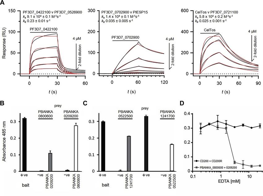
P. Falciparum Sporozoite Membrane Protein Interactions

secreted P41 protein is tethered to the surface of the mero-               Design of the Plasmodium Sporozoite Protein Library
zoite by the glycosylphosphatidylinositol (GPI)-anchored P12            To obtain a catalog of proteins that are expressed by sporozoites,
protein (9). Parasite protein complexes that are known to be         published proteomic data using mass spectrometry–based profiling of
essential for P. falciparum erythrocyte invasion include the         midgut and salivary gland sporozoites (25–27) were combined to
interactions between AMA1 and RON2 (10) and more recently            create a list of unique proteins and protein expression constructs
                                                                     designed essentially as described (23); we additionally included
those proteins that directly bind RH5 including cysteine-rich
                                                                     several proteins expressed by ookinetes. Briefly, cell surface and
protective antigen (11), RH5 interacting protein (12), and           secreted proteins were initially identified by the presence of a signal
P113 (13). Mechanistically, vaccine-elicited antibodies that         peptide and/or transmembrane domains using SignalP v4.0 (28) and
target the components of these protein complexes on the              TMHMM v2.0 (29) software. To define the ectodomain regions, signal
parasite surface may neutralize blood-stage parasites by             sequences and transmembrane domains were removed and all po-
                                                                     tential N-linked glycosylation sites (N-X-S/T, where X is not proline)
inhibiting their function or formation (14). In contrast to the
                                                                     were systematically mutated by substituting the serine/threonine for
blood stages, however, relatively little is known about the          alanine to prevent inappropriate glycosylation when expressed in
biochemical composition and organization of the sporozoite           mammalian cells. Sequences encoding the sporozoite protein ecto-
surface, although genetic targeting has revealed that several        domains were codon optimized for expression in human cells and
parasite surface proteins are required for normal sporozoite         made by gene synthesis (GeneART). Four genes failed synthesis and
                                                                     three could not be subcloned into the mammalian expression plas-
development and infection. For example, circumsporozoite
                                                                     mids, suggesting the insert was toxic in Escherichia coli (Table S1). All
protein (CSP) is required for the development of sporozoites in      sequences were flanked with unique 5′ NotI and 3′ AscI restriction
the oocyst (15, 16), TRAP family proteins (TRAP and TLP) are         enzyme sites to allow inframe cloning in the bait and prey plasmids
important for sporozoite motility and infection (17, 18), and        (30). Both expression plasmids contain a highly efficient mouse vari-
members of the 6-cys family (P36 and P52) are important for          able κ light chain signal peptide and a rat Cd4d3+4 epitope tag fol-
                                                                     lowed by either a peptide sequence allowing biotinylation and 6-his
host receptor usage in hepatocyte invasion (19). Although we
                                                                     tag (baits) or pentamerizing COMP sequences followed by the beta-
have a good cellular description of how the sporozoite in-           lactamase enzyme and a 6-his tag (preys) (31). The same process
teracts with host cells (20), a molecular understanding of these     was followed for the Plasmodium berghei orthologs. All plasmids have
behaviors is incomplete and yet could represent important            been deposited in the Addgene plasmid repository (www.addgene.
new opportunities for pre-erythrocytic malaria vaccines.             org/express).
   Identification of interactions between membrane-embedded
                                                                          Production of the Recombinant Bait and Prey Proteins
proteins is challenging because membrane proteins are both
amphipathic, making them difficult to solubilize in solvents that        All proteins were produced recombinantly as a monobiotinylated
                                                                     bait and a pentamerized prey by transient transfection of HEK293E
retain their native conformation, and because interactions be-
                                                                     (32) or HEK293-6E cells (33) essentially as described (30). All proteins
tween them can be highly transient; together, these features         were purified from the cell culture supernatant up to 6 days after
make it difficult to confidently interpret data from commonly          transfection using Ni2+-NTA resin either using automated purification
used methods such as biochemical purifications using native           systems (GE Healthcare) or manually using Ni-NTA Agarose (Jena
material (21). To address these issues, we have developed an         Biosciences) and dialyzed extensively against Hepes-buffered saline
                                                                     (HBS) or PBS.
approach called AVEXIS (for AVidity-based EXtracellular Inter-
action Screening) (22), which, together with a library of func-                    Normalization of Bait and Prey Proteins
tionally active recombinant blood-stage Plasmodium proteins
                                                                        The expression levels of biotinylated bait and β-lactamase-tagged
(23, 24), has permitted the biochemical identification and            prey proteins were quantified essentially as described (30). Briefly, for
characterization of parasite protein complexes at the surface of     bait proteins, serial dilutions of the purified, dialyzed proteins were
the merozoite including the RH5 complex (13). Here, we use           added to individual wells of a streptavidin-coated microtiter plate
proteomic analyses of Plasmodium sporozoites to compile a            (NUNC). Proteins were captured and washed with HBS-T (HBS/0.1%
recombinant protein library representing the cell surface re-        Tween 20) followed by addition of 1 μg/ml mouse anti-rat Cd4 OX68
                                                                     monoclonal antibody in HBS/0.2% bovine serum albumin (BSA) and
ceptor repertoire and secretome of the P. falciparum sporo-          washed with HBS-T. Proteins were detected by addition of a goat-
zoite. We use this protein library and the AVEXIS approach to        anti-mouse alkaline-phosphatase conjugated antibody (Sigma
identify interactions between sporozoite proteins and charac-        A4656), washing followed by addition of 1 mg/ml alkaline phosphatase
terize their role in the pathogenesis of Plasmodium infections.      substrate 104 dissolved in diethanolamine buffer, and reading the
                                                                     absorbance at 405 nm in an automated plate reader (FluoStar Optima,
                                                                     BMG Labtech). For subsequent assays, the highest protein dilution
                                                                     that saturated the biotin binding capacity of the well was used. The
                  EXPERIMENTAL PROCEDURES                            activity of prey proteins was normalized by quantifying the activity of
                                                                     the β-lactamase enzyme in the prey protein tag. Serial dilutions were
                        Ethics Statement
                                                                     prepared in a 96-well flat bottom microtiter plate, and 20 μl was added
  Experiments involving animals were performed under UK Home         to 60 μl of a 242 μM solution of the colorimetric β-lactamase substrate
Office governmental regulations (project license numbers PD3DA8D1F    nitrocefin (Cayman Chemical Company). The kinetics of substrate
and P98FFE489) and European directive 2010/63/EU. Research was       hydrolysis were followed for 20 min and prey proteins normalized to an
approved by the Sanger Institute Animal Welfare and Ethical Review   activity of ~2 nmol min−1 for use in subsequent assays. To produce
Board.                                                               tetrameric preys to detect the CelTOS-PF3D7_0721100 interaction, a

2 Mol Cell Proteomics (2021) 20 100038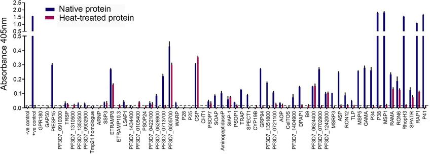
P. Falciparum Sporozoite Membrane Protein Interactions

dilution series of monomeric biotinylated protein (similar to bait          amounts of one of the baits (protein 53–SIAP-1) and therefore this bait
normalization) was incubated with 2 to 5 μg/ml alkaline phosphatase–        was tested against 35 prey proteins. In total, 3437 binding tests were
coupled streptavidin (Sigma) for 45 min to allow the formation of tet-      performed: 46 proteins were present in both prey and bait libraries and
ramers and then used in an ELISA as described above. The lowest             therefore tested in both bait–prey orientations; the other proteins were
dilution that did not give a signal at 405 nm was used for AVEXIS.          consequently tested in one orientation, making a total of 2379 unique
                                                                            interactions tested. The initial screen was performed once, but the
                           Western Blotting                                 identified interactions were repeated in triplicate and further charac-
                                                                            terized using surface plasmon resonance. Control baits and preys
   Purified proteins were resolved by SDS-PAGE using Novex
                                                                            were present in all experiments; typically the rat Cd200–CD200R
NuPAGE 4% to 12% Bis Tris precast gels (Life Technologies). Pro-
                                                                            interaction is used as a positive control. Data are shown as mean
teins were reduced by adding NuPAGE reducing agent to the sample
                                                                            absorbance readings with standard deviations to show reproducibility
and antioxidant (Life Technologies) to the gel running buffer according
                                                                            between replicates.
to the manufacturer's instructions. Proteins were blotted onto nitro-
cellulose membranes (Life Technologies) and protein-binding sites
blocked using a 2% BSA solution. Biotinylated proteins were detected                            Surface Plasmon Resonance
by incubating membranes with High-sensitivity Streptavidin–
                                                                               A Biacore 8K instrument (GE Healthcare) was used for the surface
horseradish peroxidase (HRP) (PIERCE), washing, and addition of
                                                                            plasmon resonance studies essentially as described (35). In brief,
SuperSignal West Pico Chemiluminescent substrate (PIERCE) and
                                                                            biotinylated bait proteins were immobilized on a streptavidin-coated
developed on photographic film.
                                                                            sensor chip (GE Healthcare). Approximately 400 RUs of the control
               Enzyme-Linked Immunosorbant Assay                            bait (biotinylated rat Cd4d3+4) were immobilized in the flow cell used
                                                                            as a reference and the approximate molar equivalents of the query
   ELISAs were performed by capturing biotinylated bait proteins into       protein immobilized in the other flow cells. Purified analyte proteins
individual wells of streptavidin-coated 384-well plates (Nunc). Plates      were resolved by size exclusion chromatography on a Superdex 200
were washed for 30 min with 50 μl PBS-T (0.2% Tween) and blocked            Increase 10/300 column (GE Healthcare) in HBS-P (10 mM Hepes,
with PBS-2% BSA for a minimum of 3 h A volume of 20 μl of a bait            150 mM NaCl, 0.05% v/v P20 surfactant) immediately before use in
protein diluted in PBS-2% BSA at a concentration previously deter-          surface plasmon resonance (SPR) experiments thereby removing any
mined as the amount required to saturate the biotin binding capacity        aggregated protein that might influence kinetic measurements. The
of the well was added in triplicate and incubated for at least 16 h at 4    surface was regenerated with a pulse of 2 M NaCl at the end of each
◦
  C. Where required, a similar set of bait protein dilutions were heat      cycle, and all experiments were performed at 37 ◦ C. Duplicate in-
inactivated for 30 min at 70 ◦ C. Antisera from pooled Malawian adults      jections of the same concentration in each experiment were super-
(23) were centrifuged at 13,000 rpm for a minimum of 1 h, diluted in        imposable demonstrating no loss of activity after regenerating the
PBS-2% BSA and incubated with rocking for at least 16 h at 4 ◦ C            surface. Binding data were analyzed in the manufacturer's Biacore 8K
before adding to the antigen-coated plates for 1 h. Plates were             evaluation software version 1.1 (GE Healthcare).
washed 3× in PBS-T before addition of a 1:10,000 dilution of rabbit-
anti-human polyvalent IgG/A/M (Sigma I8010) in PBS-2% BSA for                        Cloning and Expression of P. berghei Orthologs
1 h, and washed in PBS-T, followed by 1:10,000 dilution of goat anti-
rabbit Poly-HRP IgG/A/M (PIERCE) in PBS-2% BSA for 1 h. Plates                 The P. berghei orthologs of the six identified interacting
were washed in PBS-T, and the HRP substrate ABTS (KPL) was                  P. falciparum proteins were identified using PlasmoDB (36). Se-
added and absorption at 405 nm determined using an automated                quences were modified in the same way as described in Design of the
plate reader (FluoStar Optima, BMG Labtech).                                Plasmodium Sporozoite Protein Library and the genes synthesized by
                                                                            GeneART. The synthetic plasmids were digested with NotI and AscI
 Avidity-Based Extracellular Protein Interaction Screening Assay            restriction enzymes (NEB) and were each subcloned into the bait and
                                                                            prey vectors. Recombinant proteins were produced by transient
   Systematic screening for protein interactions was performed
                                                                            transfection of HEK293-E or 6E cells as described above.
essentially as described (22, 34). Briefly, normalized bait proteins were
immobilized on streptavidin-coated 96-well microtiter plates (Nunc)
and activity-normalized prey proteins added to the arrayed bait pro-                            P. berghei Genetic Targeting
teins and incubated at room temperature for 1 h. The plates were               P. berghei targeting plasmids for PIESP15 (PBANKA_0209200,
washed twice in HBS-T (0.2% Tween 20) and once in HBS before 60 μl          PbGEM-268595) and PBANKA_0800600, PbGEM-331155 were
of 125 μg/ml nitrocefin (Cayman Chemical Company) was added and              obtained from the PlasmoGEM vector resource (plasmogem.sanger.
the absorbance measured at 485 nm in a plate reader. When tetra-            ac.uk) (37). To make targeting plasmids that disrupt the
merized preys were used, 1 mg/ml phosphatase substrate 104 in               PBANKA_0522500 and PBANKA_1241700 loci, oligonucleotides
diethanolamine buffer was added and the absorbance measured at              were designed to amplify 5′ and 3′ homology regions of each gene.
405 nm. The rat Cd200–Cd200R interaction was used as a positive             Each homology arm was approximately 1 kbp long and spans the 5′
control, and a biotinylated Ox68 antibody that recognizes the Cd4-tag       UTR into the open reading frame, and from inside the open reading
in the prey was used as a prey capture control. Some bait and prey          frame into the 3′ UTR of each gene, respectively. Restriction sites were
proteins showed low levels of promiscuous binding in the screen (e.g.,      added to the primer sequence to enable cloning of the PCR products.
bait 56 and prey 27) but because of their lack of specificity and weak       PCRs were performed using Kapa HiFi HotStart Ready Mix (Kapa
binding signals were not investigated further.                              Biosciences), and genomic DNA isolated from P. berghei GFP-LUC
                                                                            parasites (38) was used as a template. PCR conditions were 5 min
           Experimental Design and Statistical Rationale
                                                                            at 95 ◦ C followed by 35 cycles of 95 ◦ C for 30 s, 55 ◦ C for 30 s and 68
                                                                            ◦
   The goal of the protein interaction screening was to identify directly     C for 1 to 3.5 min, and a final extension of 10 min at 68 ◦ C. PCR
interacting proteins within the library of recombinant P. falciparum        products were sequentially cloned into the KpnI and XmaI or the XhoI
sporozoite membrane proteins. Sixty-four bait proteins were tested for      and SacII restriction sites of the pR6K attL1-3xHA-hDHFR-yfcu-attL2
interactions against 54 prey proteins; however, there were insufficient      plasmid backbone (39) and used to transfect P. berghei parasites.

                                                                                                    Mol Cell Proteomics (2021) 20 100038 3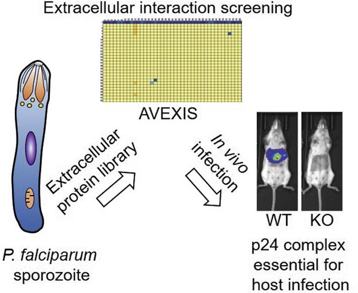
P. Falciparum Sporozoite Membrane Protein Interactions

           Transfection and Cloning by Limiting Dilution                      quantify GFP-positive oocysts. Oocysts were counted manually using
                                                                              the cell counter plugin in ImageJ 1.45s software. To quantify the
   Recombinant parasites were generated by transfecting a parental            number of sporozoites in salivary glands, mosquitoes were anes-
“wildtype” transgenic P. berghei strain expressing green fluorescent           thetized 21 days after infection and salivary glands dissected in PBS.
protein (GFP) and luciferase (38). Transfections were done essentially        Salivary gland sporozoites from 15 to 20 mosquitoes were released by
as described (40). Briefly, 6- to 12-week-old Balb/C or Theiler's orig-        homogenizing the glands with a micropestle, and salivary gland debris
inal mice were used for routine propagation of the parasites. Prior to        were pelleted by centrifugation for 5 min at 100g. Sporozoites were
transfection, 10 μl DNA was digested with NotI restriction enzyme             subsequently counted in a hemocytometer. Groups of four mice were
(NEB) overnight at 37 ◦ C to release the transfection cassette from the       infected by intravenous injection of 5000 salivary gland sporozoites
plasmid backbone. DNA was purified by ethanol precipitation and                per mouse and the development of the parasites was followed in the
resuspended in 10 μl dH2O. Parasites were cultured in vitro for 20 to         whole animal every 24 h for 72 to 96 h by an in vivo imaging system
22 h to obtain synchronized schizonts. Schizonts were purified using a         (IVIS) imaging. For imaging, mice were injected with 200 μl D-luciferin
55% Nycodenz (Sigma)/PBS gradient and washed once in RPMI1640                 (30 mg/ml in Dulbecco's phosphate buffered saline, Source Bio-
medium. Pelleted schizonts were resuspended in P3 Primary cell                sciences) and imaged under isoflurane anesthesia 10 min later using
Nucleofector (Lonza, 18 μl/transfection), and 25 μl of this mix was           an IVIS Spectrum Imaging System (Perkin Elmer).
added to 5 μl of digested DNA. Twenty microliters of the mix was then            Once parasites were observed in the blood by IVIS (usually 72 h
added to an individual well of a 16-well Nucleocuvette Strip and              post sporozoite injection), thin blood smears were taken daily for a
pulsed using the AMAXA 4D-Nucleofector electroporator, program FI-            minimum of 5 days and stained using Giemsa to monitor blood-stage
115. Parasites were immediately resuspended in 100 μl incomplete              parasitemia. To determine parasitemia, at least 500 red blood cells
RPMI1640 medium and injected into the tail vein of Balb/c mice. On            were counted and parasitemia was calculated as a percentage of
the following day in the morning, pyrimethamine (0.07 mg/ml, pH 4.5)          infected red blood cells per total number of red blood cells; percent-
was added to the drinking water for parasite selection. Blood smears          ages were then used for growth curves.
were taken from day 7 after transfection to monitor parasitemia, and
when parasitemia reached 0.5% to 1%, blood was collected for                                   Production of Polyclonal Antisera
stocks and genotyping. Subsequently, a drop of blood was taken from
a mouse infected with the transfected parasites and parasites were               For the production of polyclonal antisera, proteins were expressed
cloned by limiting dilution. Clonality (absence of the WT locus) was          as nonbiotinylated monomers by transient transfection of
confirmed by genotyping, and cloned parasites were then used for               HEK293 cells, purified using the C-terminal his tag, and dialyzed
experiments.                                                                  against PBS. For immunization, 50 μg protein per animal was adju-
                                                                              vanted in Alhydrogel (Invivogen) and delivered intraperitoneally into 6-
    Genotyping of P. berghei Parasites for Validation of Gene                 to 8-week-old female Balb/C mice. Two subsequent boosts were
                          Disruption                                          given with 20 μg per animal at 2 and 4 weeks after the initial injection.
                                                                              Sera were obtained 1 week after the final immunization boost by
   To confirm integration of the transfection cassette into the genome         cardiac bleed and centrifugation of the whole blood after clotting.
of the parasite, we designed specific oligonucleotides for each target
gene, annealing to the chromosome just outside the homology arm of                             P. falciparum Cell Invasion Assays
the plasmid (primer GT). This was paired with a universal primer that
                                                                                 HC-04 cells were routinely cultured in Dulbecco's Modified Eagle
anneals to the yfcu cassette in the targeting plasmid (primer GW2a or
                                                                              Medium: Nutrient Mixture F-12 (DMEM-F12; ThermoFisher
b). To detect absence or presence of the WT locus, a gene-specific
                                                                              Cat#31330038), supplemented with 10% heat-inactivated fetal bovine
primer located inside the gene sequence of the target gene (primer
                                                                              serum and 1× Penicillin/Streptomycin. These cells were incubated at
QCR1) was paired with a reverse primer in the homology arm (QCR2).
                                                                              37 ◦ C under 5% CO2 and split using trypsin when 80% confluency
Primers GW2 and QCR2 were then combined to detect the trans-
                                                                              was reached. Twelve hours before the assay, 50,000 cells per well
fection plasmid, which could be either integrated or not integrated in
                                                                              were seeded in a collagen-coated 96-well flat-bottom plate (Corning
the genome. Because the product size for the primer combination
                                                                              Cat# CLS3603-48EA). On the day of the experiment, salivary glands of
PbPIESP15-GW2+GT was too large, two PCR reactions were done to
                                                                              P. falciparum NF-135-infected mosquitoes were dissected in 1 ml
amplify one half of the fragment each. Therefore, primer GW2b was
                                                                              DMEM-F12 media and crushed using a glass pestle to liberate the
combined with primer PIESP15-rev2 and primer GT was combined
                                                                              sporozoites. Sporozoite numbers were determined using a hemocy-
with primer PbPIESP15-fwd2. Sequences for primers QCR1, QCR2,
                                                                              tometer. Media from each of the hepatocyte-seeded wells were
and GT for PbANKA_0800600 and PbPIESP15 were obtained from the
                                                                              removed and replaced with media containing 50,000 sporozoites per
PlasmoGEM web page. All primer sequences are listed in Table S2.
                                                                              well. The plate was centrifuged at 3000 rpm for 10 min and incubated
PCR reactions were performed using Kapa HiFi HotStart Ready Mix
                                                                              at 37 ◦ C under 5% CO2 for 3 h. After 3 h, the supernatant was
(Kapa Biosciences), and cycling conditions were 5 min at 95 ◦ C fol-
                                                                              removed and each well rinsed three times with PBS and trypsinized for
lowed by 35 cycles of 95 ◦ C for 30 s, 55 ◦ C for 30 s, and 68 ◦ C for 1 to
                                                                              5 min to obtain single-cell suspension. The trypsinized sample was
3.5 min and a final extension of 10 min at 68 ◦ C. Samples were
                                                                              neutralized in equal volumes of PBS supplemented with 10% FCS and
separated on a 0.8% Agarose gel using Hyperladder 1kb (Bioline) as a
                                                                              transferred to a 96-well V-bottom plate (Costar Cat#3897). Samples
size standard.
                                                                              were spun down at 1700 rpm for 4 min. Supernatants were removed,
     In Vivo Phenotyping of Transgenic P. berghei Parasites                   and pellets were resuspended in fixation reagent as per manufac-
                                                                              turer's instructions (eBioscience Foxp3/Transcription factor staining
   Anopheles stephensi mosquitoes were allowed to feed on anaes-              buffer set; ThermoFisher Cat# 00-5523-00) and incubated at 4 ◦ C for
thetized P. berghei-infected Balb/C mice for no more than 15 min and          30 min. Permeabilization buffer was added to each well after the in-
placed in an incubator at 19 ◦ C thereafter. Nine to 11 days after            cubation, and the samples were pelleted at 1700 rpm for 4 min. The
infection, mosquitoes were anesthetized and kept on ice and midguts           supernatant was removed, the pellet was resuspended in per-
were dissected in PBS. Only mosquitoes with developed eggs were               meabilization buffer containing 3SP2 (antibody against PfCSP 1:400
dissected to ensure mosquitoes had taken a blood meal at the time of          dilution) conjugated to FITC and incubated at 4 ◦ C for 30 min. Per-
feeding. Images were taken by fluorescence microscopy and used to              meabilization buffer was added to each well after the incubation, and

4 Mol Cell Proteomics (2021) 20 100038
P. Falciparum Sporozoite Membrane Protein Interactions
the samples were pelleted at 1700 rpm for 4 min. The supernatant was enzymatically monobiotinylated bait proteins by transient
removed, and the pellets were resuspended in 1% paraformaldehyde transfection of suspension-grown HEK293 cells. Using this
in PBS. Each sample was read by fluorescent activated cytometry
approach, 58 of the 69 proteins (84.1%) could be detected
sorting to determine the percentage of FITC-positive (infected) cells.
after his-tag purification from the spent cell culture medium by
Immunostaining of P. berghei Sporozoites ELISA. As is typical for this approach, for those proteins that
To determine the amount and subcellular localization of CSP, sali- were detectably expressed, levels varied widely across several
vary gland sporozoites were dissected from infected A. stephensi orders of magnitude but could be as high as ~50 μg/ml for
mosquitoes 21 to 30 days after blood feeding on P. berghei-infected proteins such as TRAP and CSP. Forty-eight proteins could
mice. Sporozoites were isolated by homogenizing the glands with a be produced in sufficient quantities to be used in the subse-
micropestle and centrifugation for 5 min at 100g to pellet salivary
quent experiments and were resolved by SDS-PAGE and
gland debris. Sporozoites were subsequently counted in a hemocy-
tometer, and 10,000 sporozoites each were placed onto round 13-mm detected by anti-biotin Western blotting to determine their
coverslips and air dried. Coverslips were then stored at −80 ◦ C with integrity (Fig. 1). With the exception of three proteins, PSOP7,
dessicant until used for immunostaining. For immunostaining, the B9, and PF3D7_0624400, whose major species was smaller
coverslips were left to reach room temperature before being removed than expected, the proteins were expressed at the expected
from the bag containing desiccant. Coverslips were then placed into
mass, although some proteins did show evidence of being
individual wells of a 24-well plate (Nunc), and parasites were fixed with
a 4% formalin solution in PBS for 10 min on ice. After washing twice in processed. Those proteins that could not be detected or were
PBS, the coverslips were incubated with 0.1% Triton in PBS on ice for expressed at very low levels included proteins that localize to
15 min to permeabilize the parasites. All slides were washed twice in the rhoptries (RALP1, ARNP, and MAEBL), both the LCCL-
PBS, 1% BSA (Sigma) and incubated with anti-CSP monoclonal domain containing secreted proteins LAP2 and LAP4, two
antibody, 1:1000 in PBS, 1% BSA for 1 h at room temperature. After
members of the TRAP family (CTRP and TREP), and two
washing three times in PBS, 1% BSA parasites were incubated with
Cy3 goat anti-mouse IgG (Life Technologies A10521), 1:500 in PBS, members of the 6-cys family that are also expressed on
1% BSA for 30 min at room temperature, protected from light. Cov- gametes (P47 and P48/45). We have previously observed that
erslips were subsequently washed in PBS, 1% BSA and mounted on proteins localized to the rhoptries of merozoites are also
microscope slides with Vectashield HardSet containing DAPI poorly expressed in this system suggesting that a rhoptry-
(Vectorlabs).
specific chaperone may be required for expression using
this approach (23).
RESULTS
A Library of P. falciparum Sporozoite Surface and Secreted The Recombinant Sporozoite Proteins Are Immunoreactive
Proteins and Contain Conformational Epitopes
To create a recombinant protein library representing the cell To systematically evaluate which of the recombinant pro-
surface receptor repertoire and secretome of the P. falciparum teins within the sporozoite library contained conformational
sporozoite, we analyzed published proteome and tran- epitopes, we quantified the immunoreactivity to purified im-
scriptome datasets from both P. falciparum and Plasmodium munoglobulins pooled from adult donors living in a
yoelii sporozoites (25, 26, 41). To identify proteins that are P. falciparum malaria-endemic region. Over half (35/60, 58%)
likely to be displayed on the surface of the sporozoite or of the proteins were recognized by the pooled immune serum
secreted, we selected those that contain a predicted N-ter- (Fig. 2). We found that the overall immunoreactivity to sporo-
minal signal peptide and/or transmembrane domain using a zoite antigens was lower than for merozoite antigens (23), as
set of bioinformatic tools. The resulting list of 102 proteins expected given that the immune system is exposed to many
included 57 proteins that contained one or more orders of magnitude fewer sporozoites than merozoites during
transmembrane-spanning regions, 11 that encoded a C-ter- an infection. The most immunoreactive proteins were those
minal hydrophobic sequence for the addition of a GPI lipid whose expression is not restricted to the sporozoite stage but
anchor, and 34 that had no obvious way of being tethered to a that are also expressed during the blood stages. Sporozoite
membrane, and so were predicted to be secreted (Table S1). proteins that are not expressed by the blood stages generally
Of these 102 proteins, 25 are also known to be expressed on exhibited a lower immunoreactivity with CSP, PIESP15,
the blood stages (such as P38, RhopH3, and MSP1) and ETRAMP8, and three conserved proteins with unknown
already represented within our library of previously published function: PF3D7_0702900, PF3D7_0713700, and
merozoite proteins (23, 24). For the remaining 77 proteins, we PF3D7_0505700 being the most immunodominant. Except for
designed expression constructs in a similar manner by trun- CSP, the immunoreactivity of the immune serum to heat-
cating the proteins just prior to the predicted transmembrane treated proteins was either completely lost or reduced
region or GPI-anchor and using gene synthesis to codon compared with the untreated proteins, demonstrating that the
optimize the coding regions for expression in human cells (23). immune serum recognized conformational epitopes and sug-
Of these, 69 genes could be successfully synthesized and gesting that most proteins were folded (Fig. 2). The lack of
subcloned into the mammalian expression plasmid (Table S1) heat-labile epitopes in recombinant CSP is consistent with the
and were subsequently expressed as Cd4d3+4-tagged known immunodominance of the repetitive “NANP” repeats,
Mol Cell Proteomics (2021) 20 100038 5P. Falciparum Sporozoite Membrane Protein Interactions
FIG. 1. A library of recombinant P. falciparum sporozoite cell surface and secreted proteins. The library of sporozoite receptor ecto-
domains was expressed as soluble recombinant enzymatically biotinylated proteins in HEK293 cells, and the purified proteins were resolved by
SDS-PAGE under reducing conditions. Proteins were blotted and detected using streptavidin–horseradish peroxidase.
which are predicted to be natively unstructured (42). These HEK293 cells. As expected, the levels of activity varied and 54
data demonstrate that the majority of soluble recombinant prey proteins were expressed at sufficient levels after their
P. falciparum sporozoite proteins expressed in mammalian activities had been normalized to the threshold level required
cells contain conformational epitopes and represent a valu- for the AVEXIS assay (Table S1). In total, 64 bait proteins were
able resource for studying the molecular pathology of systematically tested for interactions against the 54 prey
P. falciparum infections. proteins (Fig. 3A). One of the baits (SIAP-1 - protein 53) was
expressed at low levels and therefore tested against just 35
Identification of P. falciparum Sporozoite Surface Protein
prey proteins so that a total of 3437 binding tests were per-
Complexes by Systematic Protein Interaction Screening
formed. Forty-six of the proteins were present in both bait and
To identify P. falciparum sporozoite extracellular protein prey libraries and consequently tested in both bait–prey ori-
complexes we used the AVEXIS assay, which detects direct entations; the remaining proteins were therefore tested in a
interactions between soluble recombinant ectodomains single orientation only, resulting in a total of 2379 unique in-
expressed as biotinylated bait and highly avid enzyme-tagged teractions tested. We identified three interactions: the first
prey proteins (22). To produce the sporozoite library as prey between a type I cell surface protein called PIESP15 and a
proteins, the ectodomains were subcloned into a plasmid secreted protein: PF3D7_0702900, a second between two
containing tags for pentamer formation and expressed in members of the p24 family of proteins (PF3D7_0422100 and
FIG. 2. The recombinant P. falciparum sporozoite proteins contain heat-labile conformational epitopes. The immunoreactivity of the
recombinant sporozoite proteins to immune serum was quantified by ELISA. Biotinylated proteins were normalized and captured on a
streptavidin-coated microtiter plate and probed with pooled immune sera from Malawian adults (blue). Reduced response of immune serum to
heat-treated (70 ◦ C for 30 min) proteins (pink) demonstrates the presence of heat-labile conformational epitopes. Note that the 11 proteins
between MSP5 and P41 that are grouped on the right of the graph are additionally expressed in blood-stage parasites. Bar charts show means ±
SEM; n = 3. Positive immunoreactivity was defined as mean responses >3 SEM above the averaged control (dashed line).
6 Mol Cell Proteomics (2021) 20 100038P. Falciparum Sporozoite Membrane Protein Interactions
FIG. 3. Identification of three P. falciparum sporozoite complexes by systematic extracellular protein interaction screening. A, the
library of cell surface and secreted sporozoite proteins was expressed as biotinylated bait and enzyme-tagged pentameric prey proteins and
systematically screened for interactions using the AVEXIS assay. The results are presented as a quantitative binding grid. The biotinylated baits
(arranged vertically according to the numbers in Table S1) were immobilized in individual wells of streptavidin-coated microtiter plates and tested
for direct binary interactions with the same proteins expressed as pentameric β-lactamase-tagged preys (arranged horizontally). Binding by
specific baits is quantified by detecting the capture of prey proteins and hydrolysis of the colorimetric β-lactamase substrate nitrocefin. B, the
five named prey proteins that interacted with a bait were retested in triplicate against the arrayed baits and the interactions confirmed. Controls
were: positive (+) Cd200 bait -Cd200R prey; negative (−) Cd200 bait with corresponding sporozoite protein prey. Bars represent mean ± SD;
n = 3.
PF3D7_0526900), and the third between CelTOS and a pre- to probe immobilized baits to test for interactions (43). Using
dicted secreted protein (PF3D7_0721100). The PIESP15- this method, we could show binding of CelTOS and
PF3D7_0702900 and p24 family interactions were identified PF3D7_0721100 in both orientations relative to controls
in both bait–prey orientations in the initial screen; however, (Fig. S1).
because the PF3D7_0721100 protein did not express at suf-
The Interactions Are Conserved Between Orthologous
ficient levels in the prey format, the CelTOS-PF3D7_0721100
Proteins in P. berghei
interaction was initially identified in one orientation. For
those five prey proteins that interacted with a specific bait, we To further characterize these interactions, we first used SPR
repeated the binding assays against all the baits in triplicate to establish whether the proteins could directly interact and
and confirmed the interactions (Fig. 3B). The interaction be- quantify their biophysical binding parameters. For each inter-
tween CelTOS and PF3D7_0721100 was confirmed using a acting pair, one protein was purified and serial dilutions
modified assay where monomeric biotinylated proteins were injected over its binding partner expressed as a biotinylated
clustered around an alkaline phosphatase–streptavidin con- protein and immobilized on a streptavidin-coated sensor chip.
jugate to form avid tetrameric preys, which then could be used Clear binding was observed for each interaction, and the
Mol Cell Proteomics (2021) 20 100038 7P. Falciparum Sporozoite Membrane Protein Interactions FIG. 4. Biophysical characterization of the three sporozoite complex interactions and conservation of interactions with orthologous proteins in P. berghei. A, biophysical characterization of the three P. falciparum sporozoite interactions by surface plasmon resonance. For each interacting pair, one protein was immobilized on the sensor chip and twofold serial dilutions of the indicated corresponding binding partner was injected to quantify binding parameters. The raw sensorgram data (black lines) were fitted to a 1:1 binding model (red lines) to derive the association (ka) and dissociation (kd) rate constants. B, the entire ectodomains of the P. berghei orthologs of PIESP15 (PBANKA_0209200) and PF3D7_0702900 (PBANKA_0800600), as well as PF3D7_0422100 (PBANKA_0522500) and PF3D7_0526900 (PBANKA_1241700) (C), were expressed as both bait and prey proteins and shown to directly interact using the AVEXIS assay in both bait–prey orientations. The rat Cd200– Cd200R interaction was used as a positive control (+ve); negative control bait (−ve) was Cd200. (D) The PIESP15–PBANKA_0800600 interaction but not the control Cd200–Cd200R interaction was inhibited in a dose-dependent manner by addition of EDTA as detected by AVEXIS. Bars and data points indicate means ± SD; n = 3. association and dissociation binding parameters were quan- detect robust interactions between PbPIESP15 and tified by fitting the series of binding traces to a 1:1 binding PbANKA_0800600 and the two P. berghei p24 complex pro- model (Fig. 4A). As expected, the two interactions involving a teins in both bait–prey orientations (Fig. 4, B and C). We were protein predicted to be secreted exhibited slower dissocia- unable to determine if the interaction between CelTOS and tions than the membrane-tethered p24 complex proteins (21). PF3D7_0721100 was conserved between the P. berghei Genome analysis revealed that the six proteins forming orthologs of these proteins because repeated attempts to complexes had identifiable orthologs in the genomes of all produce the P. berghei ortholog of PF3D7_0721100 failed; Plasmodium species sequenced to date (36). To determine if therefore, this interaction was not investigated further. These the protein interactions were conserved across species, we data demonstrate evolutionary conservation of the interaction expressed the orthologs of these proteins from the rodent interfaces, which is striking since the amino acid sequence parasite P. berghei as both baits and preys and tested if they identity of the extracellular regions was relatively modest: 58% could directly interact using the AVEXIS assay. We could and 64% for the two p24 complex proteins and 42% and 75% 8 Mol Cell Proteomics (2021) 20 100038
P. Falciparum Sporozoite Membrane Protein Interactions

for PIESP15 and PF3D7_0702900, respectively. We next                genes (Fig. 5, C and D). This was confirmed by a significant
sought to characterize the biochemical interaction between          delay in the appearance of parasites in the blood of infected
PIESP15 and PF3D7_0702900 in more detail. Both proteins             mice (Fig. 5E).
contain recognizable protein domains in their extracellular
regions: PIESP15 has an L-type lectin domain, so named
                                                                                              DISCUSSION
because it has homologies to lectins found in leguminous
plants (44), and PF3D7_0702900 contains tandem EF-hand                 In this study we have compiled a library of soluble recom-
domains. It seemed unlikely that the PIESP15–                       binant forms of the membrane-embedded and secreted pro-
PF3D7_0702900 interaction involved carbohydrate binding             teins from the P. falciparum sporozoite. We have shown how
because Plasmodium parasites lack the glycosyltransferase           this resource can be used to characterize the host humoral
enzymes necessary for complex carbohydrate modifications             immune response in patients with malaria and identify protein
(45, 46), and because of the design of the recombinant pro-         complexes that are important for sporozoite function. A key
teins the only predicted glycosylation was present within the       feature of this resource is that we have produced the proteins
Cd4 protein tag common to all bait proteins. Both L-type lectin     in a mammalian expression system to increase the chances
and EF hand domains are known to require calcium ion co-            that the proteins contain structurally critical posttranslational
factors in their binding sites (47, 48), and consistent with this   modifications such as disulfide bonds and thereby adopt their
we observed that the PIESP15- PF3D7_0702900 interaction             native conformation, which is essential for retaining extracel-
was inhibited in a dose-dependent manner in the presence of         lular binding functions. A similar library representing the
the divalent ion chelator, EDTA (Fig. 4D).                          secreted and membrane-embedded proteins from the
                                                                    P. falciparum merozoite has proven very valuable to define the
Both Genes Encoding p24 Complex Proteins Are Required
                                                                    serology of the infected host (50), identify both parasite–
          for P. berghei Liver-Stage Infection
                                                                    parasite (9, 13) and host–parasite protein interactions
   The cross-species conservation of both the PIESP15-              (51–54), as well as blood-stage vaccine candidate screening
PF3D7_0702900 and p24 complex protein interactions sug-             (55). An important facet of this protein library is that many
gested that P. berghei was a suitable and experimentally            antigens can be assessed in parallel so that they can be
tractable model and enabled us to further validate these in-        directly compared in a systematic manner rather than the
teractions by asking if parasites lacking genes encoding            more common testing of proteins individually, which in-
interacting proteins had similar infection phenotypes. To make      troduces technical variation that makes comparison very
gene-deficient parasites for all four genes, we used existing        difficult. We therefore envision that this resource will be an
plasmids from the PlasmoGEM resource to target both                 important tool for the malaria community and have an impact
PbANKA_0800600 and PIESP15 (49). Gene targeting plas-               in defining a better molecular understanding of the basic
mids for the P. berghei orthologs of both p24 complex genes,        infection biology of the P. falciparum sporozoite; ultimately,
PBANKA_1241700 and PBANKA_0522500, were not avail-                  these findings will contribute toward providing new opportu-
able, so these were designed and constructed (Fig. S2). Gene-       nities for therapeutic interventions.
deficient parasites for all four genes were made by electro-            Of the three interactions we describe, the interaction be-
porating erythrocytes infected with a GFP-luciferase-               tween the p24 complex proteins PF3D7_0526900 and
transgenic “wildtype” P. berghei parental line and then             PF3D7_0422100 was of particular interest because P. berghei
cloned by limiting dilution. Parasites were genotyped to            parasites lacking the orthologs of either of these genes
confirm recombination at the targeted locus (Figs. S2, B and C       exhibited strong sporozoite in vivo infection phenotypes. p24
and S3). Mosquitoes were infected with the cloned parasite          proteins contain a GOLD (GOLgi Dynamics) domain, and four
lines, and the infection was quantified throughout the life cycle    can be readily identified from their primary protein sequence in
in both the mosquito vector and mammalian host. We                  Plasmodium spp. genomes. Most p24 family proteins are
observed that both the ΔPbPIESP15 and ΔPbANKA_0800600               ubiquitously expressed, although some may show elevated
parasite lines had no overt infection phenotype in either the       expression in different developmental contexts, and at least
mosquito or in mice compared with the wild-type parental line       three are abundant in sporozoites and therefore represented in
(Fig. S4). This lack of infection phenotype was consistent with     our library. We only observed interactions between two of
the inability of polyclonal antibodies to both PIESP15 and          these three proteins suggesting that there is binding speci-
PF3D7_0702900 to prevent invasion of P. falciparum sporo-           ficity within the family, which is most likely mediated by the
zoites in human cells (Fig. S5). By contrast, P. berghei para-      regions of predicted coiled coil in the lumenal domain (56). p24
sites lacking either PBANKA_1241700 or PBANKA_0522500               family proteins are mainly localized to the membranes of the
infected mosquitoes indistinguishably from the wildtype line        early secretory pathway, and experiments in yeast have sug-
(Fig. 5, A and B), whereas intravenous infections of mice with      gested that they play major roles as cargo receptors for GPI-
salivary gland sporozoites resulted in a striking reduction in      linked proteins. Their lumenal (ectodomain) regions function
the number of parasites in the liver stage of infection for both    as a lectin by binding the oligo-mannose glycans in the GPI

                                                                                         Mol Cell Proteomics (2021) 20 100038 9
P. Falciparum Sporozoite Membrane Protein Interactions FIG. 5. Both genes encoding the interacting p24 complex proteins are required for P. berghei mammalian host liver-stage infection. A, the number of oocysts per midgut in A. stephensi mosquitoes infected with P. berghei ΔPBANKA_1241700 or ΔPBANKA_0522500 were counted and found to be not significantly different from the GFP-luciferase transgenic wildtype parental strain. Data points are individual midguts; median is indicated and statistical comparisons were performed using the Mann–Whitney test; p-values > 0.05 were considered nonsignificant (ns). B, average number of salivary gland (SG) sporozoites per mosquito. About 15 to 20 mosquitoes were dissected, and sporozoites isolated from the salivary glands were counted. The columns represent the average number of salivary gland sporozoites across four independent experiments. Columns represent the mean with SD; statistical analysis was performed using an unpaired t test. C, P. berghei ΔPBANKA_1241700 or ΔPBANKA_0522500 parasites do not establish robust liver-stage infections in mice compared with wildtype control. Groups of four mice were infected by intravenous administration of 5000 isolated salivary gland sporozoites, and the resulting parasitemia was quantified in the whole animal using luciferase-based bioluminescence using an in vivo imaging system. Data points are mean ± SD; n = 4. D, normalized bioluminescence images collected on each day after infection; the left-most animal in each cohort is an uninfected control. E, the prepatency period before detection of blood-stage parasitemia is significantly delayed in ΔPBANKA_1241700 and ΔPBANKA_0522500 par- asites relative to the wildtype control. WT animals were culled on day 7 because they showed signs of disease (indicated with a cross). Asexual blood-stage parasitemia was quantified by microscopic analysis of blood smears taken between 4 and 8 days after infection. Shown are the results from one of two independent experiments with similar results. Data points are mean ± SD; n = 4. The prepatency period was less than 4 days for the parental line and 7.0 ± 0.7 (mean ± SD; n = 4) for ΔPBANKA_0522500 and 7 ± 1 for ΔPBANKA_1241700. anchor (57) while their short cytoplasmic tails nucleate the sporozoites lacking the interacting p24 family proteins is that formation of both COPI and COPII vesicle coats through their they are unable to appropriately traffic GPI-anchored proteins dilysine and phenylalanine-containing motifs, respectively that are important for sporozoite invasion of host cells to the (58). One likely molecular interpretation for the phenotypes of sporozoite surface. One obvious candidate is CSP, but we did 10 Mol Cell Proteomics (2021) 20 100038
P. Falciparum Sporozoite Membrane Protein Interactions

not observe any obvious difference in the localization or          P. falciparum sporozoite using a mammalian expression
amount of CSP at the surface of sporozoites that lacked either     system. We believe that these proteins will be particularly
PBANKA_1241700 or PBANKA_0522500 (Fig. S6). It is known            useful to the malaria research community because of the
that p24 family proteins are not essential for GPI-anchored        challenges in obtaining large amounts of sporozoite material
protein trafficking since an engineered yeast strain lacking        and solubilizing membrane-embedded proteins in their
all known p24 family proteins only has a subtle phenotype that     native conformation. Given that targeting the sporozoite
affects the rate and fidelity of protein trafficking (59). This      stage either as attenuated whole sporozoites or as a
suggests that most GPI-anchored proteins in yeast necessary        sporozoite surface protein-based subunit vaccine has
for laboratory growth can be trafficked by “bulk flow” through       shown promise in malaria vaccine development (6), gaining a
the secretory pathway. Perhaps because CSP is so abundant          greater understanding of these proteins and how they
at the sporozoite surface, it mainly relies on bulk flow for        function will not only advance a molecular understanding of
secretion and so is not affected by the loss of p24 family         sporozoite infection biology but is also likely to provide
proteins. The infection phenotype we observed may therefore        important information to improve sporozoite-based malaria
be due to mistrafficking of one or more of the several other        vaccine design.
GPI-anchored proteins that are expressed by sporozoites.
   Of the two other interactions that we identified, we were
                                                                                         DATA AVAILABILITY
able to show that P. berghei parasites genetically targeted for
genes encoding the interacting proteins PIESP15 and                  All the data are available within the article and associated
PbANKA_800600 had no overt infection phenotypes, although          supplementary information. Expression plasmids encoding
because sporozoites were delivered by intravenous injection,       the proteins have been deposited and are available from the
there remains the formal possibility of a phenotype in sporo-      Addgene repository (www.addgene.org).
zoite behavior in the dermis. These findings are consistent
with the lack of in vivo blood-stage growth phenotypes for           Acknowledgments — The authors would like to thank Ellen
knockouts of these genes in P. berghei as part of a large-scale    Bushell and Gareth Girling for plasmids and Youri M. van
genetic screen (49), as well as a previous smaller-scale ge-       Waardenburg for P. falciparum sporozoites.
netic screen focused on identifying genes encoding cell sur-
face and secreted proteins required by the ookinete to infect         Funding and additional information — This work was sup-
mosquitoes (60). There is proteomic evidence from several          ported by the Wellcome Trust grant 206194, Medical
studies that these proteins are not restricted to sporozoites in   Research Council, UK (grant number MR/J002283/1), the
P. falciparum but can be detected in other stages including        Medical Research Council Doctoral Training (MR/J004111/1),
gametocytes (61) and blood stages (62). Of interest, insertional   and EU Horizon 2020 (Agreement 733273), and Dutch
mutagenesis genetic screens have shown that both PIESP15           Research     Council   (NOW)    talent    programme   veni
and PF3D7_0702900 are very important for in vitro blood-           (VI.Veni.192.171).
stage culture in P. falciparum providing evidence that these
genes may play important roles in P. falciparum in other life-       Author contributions — J. K., O. B., and G. J. W. designed
cycle stages (63). It is worth noting that the genes encoding      experiments; J. K., T. M., and F. G. performed experiments
PF3D7_0702900/PbANKA_0800600 are currently annotated               except the P. falciparum sporozoite invasion assays, which
as a “putative centrin” in gene databases, which appears to be     were performed by A. S. P. Y. under the supervision of
a computational prediction based on very low (
P. Falciparum Sporozoite Membrane Protein Interactions
Received November 30, 2020 Published, MCPRO Papers in Press, 18. Moreira, C. K., Templeton, T. J., Lavazec, C., Hayward, R. E., Hobbs, C. V.,
January 27, 2021, https://doi.org/10.1074/mcp.RA120.002432 Kroeze, H., Janse, C. J., Waters, A. P., Sinnis, P., and Coppi, A. (2008)
The Plasmodium TRAP/MIC2 family member, TRAP-Like Protein (TLP), is
involved in tissue traversal by sporozoites. Cell Microbiol. 10, 1505–1516
REFERENCES
19. Manzoni, G., Marinach, C., Topcu, S., Briquet, S., Grand, M., Tolle, M.,
1. WHO. (2019) World malaria report. World Health Organization, Manila, Gransagne, M., Lescar, J., Andolina, C., Franetich, J. F., Zeisel, M. B.,
Philippines, 2019 Huby, T., Rubinstein, E., Snounou, G., Mazier, D., et al. (2017) Plasmo-
2. Dundas, K., Shears, M. J., Sinnis, P., and Wright, G. J. (2019) Important dium P36 determines host cell receptor usage during sporozoite invasion.
extracellular interactions between plasmodium sporozoites and host cells Elife 6, e25903
required for infection. Trends Parasitol. 35, 129–139 20. Tavares, J., Formaglio, P., Thiberge, S., Mordelet, E., Van Rooijen, N.,
3. Aliprandini, E., Tavares, J., Panatieri, R. H., Thiberge, S., Yamamoto, M. M., Medvinsky, A., Menard, R., and Amino, R. (2013) Role of host cell traversal
Silvie, O., Ishino, T., Yuda, M., Dartevelle, S., Traincard, F., Boscardin, S. by the malaria sporozoite during liver infection. J. Exp. Med. 210, 905–915
B., and Amino, R. (2018) Cytotoxic anti-circumsporozoite antibodies 21. Wright, G. J. (2009) Signal initiation in biological systems: The properties
target malaria sporozoites in the host skin. Nat. Microbiol. 3, 1224–1233 and detection of transient extracellular protein interactions. Mol. Biosyst.
4. Daily, J. P. (2018) Shedding light on the role of the skin in vaccine-induced 5, 1405–1412
protection against the malaria sporozoite. mBio 9, e02555-18 22. Bushell, K. M., Sollner, C., Schuster-Boeckler, B., Bateman, A., and Wright,
5. Menard, R., Tavares, J., Cockburn, I., Markus, M., Zavala, F., and Amino, R. G. J. (2008) Large-scale screening for novel low-affinity extracellular
(2013) Looking under the skin: The first steps in malarial infection and protein interactions. Genome Res. 18, 622–630
immunity. Nat. Rev. Microbiol. 11, 701–712 23. Crosnier, C., Wanaguru, M., McDade, B., Osier, F. H., Marsh, K., Rayner, J.
6. Draper, S. J., Sack, B. K., King, C. R., Nielsen, C. M., Rayner, J. C., Higgins, C., and Wright, G. J. (2013) A library of functional recombinant cell-
M. K., Long, C. A., and Seder, R. A. (2018) Malaria vaccines: Recent surface and secreted P. falciparum merozoite proteins. Mol. Cell. Prote-
advances and new horizons. Cell Host Microbe 24, 43–56 omics 12, 3976–3986
7. Pachebat, J. A., Ling, I. T., Grainger, M., Trucco, C., Howell, S., Fernandez- 24. Zenonos, Z. A., Rayner, J. C., and Wright, G. J. (2014) Towards a
Reyes, D., Gunaratne, R., and Holder, A. A. (2001) The 22 kDa component comprehensive Plasmodium falciparum merozoite cell surface and
of the protein complex on the surface of Plasmodium falciparum mero- secreted recombinant protein library. Malar. J. 13, 93
zoites is derived from a larger precursor, merozoite surface protein 7. Mol. 25. Lasonder, E., Janse, C. J., van Gemert, G. J., Mair, G. R., Vermunt, A. M.,
Biochem. Parasitol. 117, 83–89 Douradinha, B. G., van Noort, V., Huynen, M. A., Luty, A. J., Kroeze, H.,
8. Kauth, C. W., Woehlbier, U., Kern, M., Mekonnen, Z., Lutz, R., Mucke, N., Khan, S. M., Sauerwein, R. W., Waters, A. P., Mann, M., and Stunnen-
Langowski, J., and Bujard, H. (2006) Interactions between merozoite berg, H. G. (2008) Proteomic profiling of Plasmodium sporozoite matu-
surface proteins 1, 6, and 7 of the malaria parasite Plasmodium falcipa- ration identifies new proteins essential for parasite development and
rum. J. Biol. Chem. 281, 31517–31527 infectivity. PLoS Pathog. 4, e1000195
9. Taechalertpaisarn, T., Crosnier, C., Bartholdson, S. J., Hodder, A. N., 26. Lindner, S. E., Swearingen, K. E., Harupa, A., Vaughan, A. M., Sinnis, P.,
Thompson, J., Bustamante, L. Y., Wilson, D. W., Sanders, P. R., Wright, Moritz, R. L., and Kappe, S. H. (2013) Total and putative surface prote-
G. J., Rayner, J. C., Cowman, A. F., Gilson, P. R., and Crabb, B. S. (2012) omics of malaria parasite salivary gland sporozoites. Mol. Cell. Prote-
Biochemical and functional analysis of two Plasmodium falciparum omics 12, 1127–1143
blood-stage 6-cys proteins: P12 and P41. PLoS One 7, e41937 27. Swearingen, K. E., Lindner, S. E., Shi, L., Shears, M. J., Harupa, A., Hopp,
10. Lamarque, M., Besteiro, S., Papoin, J., Roques, M., Vulliez-Le Normand, B., C. S., Vaughan, A. M., Springer, T. A., Moritz, R. L., Kappe, S. H., and
Morlon-Guyot, J., Dubremetz, J. F., Fauquenoy, S., Tomavo, S., Faber, B. W., Sinnis, P. (2016) Interrogating the plasmodium sporozoite surface: Iden-
Kocken, C. H., Thomas, A. W., Boulanger, M. J., Bentley, G. A., and Lebrun, tification of surface-exposed proteins and demonstration of glycosylation
M. (2011) The RON2-AMA1 interaction is a critical step in moving junction- on CSP and TRAP by mass spectrometry-based proteomics. PLoS
dependent invasion by apicomplexan parasites. PLoS Pathog. 7, e1001276 Pathog. 12, e1005606
11. Reddy, K. S., Amlabu, E., Pandey, A. K., Mitra, P., Chauhan, V. S., and 28. Petersen, T. N., Brunak, S., von Heijne, G., and Nielsen, H. (2011) SignalP 4.
Gaur, D. (2015) Multiprotein complex between the GPI-anchored CyRPA 0: Discriminating signal peptides from transmembrane regions. Nat.
with PfRH5 and PfRipr is crucial for Plasmodium falciparum erythrocyte Methods 8, 785–786
invasion. Proc. Natl. Acad. Sci. U. S. A. 112, 1179–1184 29. Krogh, A., Larsson, B., von Heijne, G., and Sonnhammer, E. L. (2001)
12. Chen, L., Lopaticki, S., Riglar, D. T., Dekiwadia, C., Uboldi, A. D., Tham, W. Predicting transmembrane protein topology with a hidden Markov model:
H., O'Neill, M. T., Richard, D., Baum, J., Ralph, S. A., and Cowman, A. F. Application to complete genomes. J. Mol. Biol. 305, 567–580
(2011) An EGF-like protein forms a complex with PfRh5 and is required for 30. Kerr, J. S., and Wright, G. J. (2012) Avidity-based extracellular interaction
invasion of human erythrocytes by Plasmodium falciparum. PLoS Pathog. screening (AVEXIS) for the scalable detection of low-affinity extracellular
7, e1002199 receptor-ligand interactions. J. Vis. Exp., e3881
13. Galaway, F., Drought, L. G., Fala, M., Cross, N., Kemp, A. C., Rayner, J. C., 31. Sun, Y., Gallagher-Jones, M., Barker, C., and Wright, G. J. (2012)
and Wright, G. J. (2017) P113 is a merozoite surface protein that binds the A benchmarked protein microarray-based platform for the identification
N terminus of Plasmodium falciparum RH5. Nat. Commun. 8, 14333 of novel low-affinity extracellular protein interactions. Anal. Biochem. 424,
14. Douglas, A. D., Baldeviano, G. C., Jin, J., Miura, K., Diouf, A., Zenonos, Z. 45–53
A., Ventocilla, J. A., Silk, S. E., Marshall, J. M., Alanine, D. G. W., Wang, 32. Durocher, Y., Perret, S., and Kamen, A. (2002) High-level and high-
C., Edwards, N. J., Leiva, K. P., Gomez-Puerta, L. A., Lucas, C. M., et al. throughput recombinant protein production by transient transfection of
(2019) A defined mechanistic correlate of protection against Plasmodium suspension-growing human 293-EBNA1 cells. Nucleic Acids Res. 30, E9
falciparum malaria in non-human primates. Nat. Commun. 10, 1953 33. Loignon, M., Perret, S., Kelly, J., Boulais, D., Cass, B., Bisson, L., Afkha-
15. Coppi, A., Natarajan, R., Pradel, G., Bennett, B. L., James, E. R., Roggero, mizarreh, F., and Durocher, Y. (2008) Stable high volumetric production of
M. A., Corradin, G., Persson, C., Tewari, R., and Sinnis, P. (2011) The glycosylated human recombinant IFNalpha2b in HEK293 cells. BMC
malaria circumsporozoite protein has two functional domains, each with Biotechnol. 8, 65
distinct roles as sporozoites journey from mosquito to mammalian host. 34. Bartholdson, S. J., Bustamante, L. Y., Crosnier, C., Johnson, S., Lea, S.,
J. Exp. Med. 208, 341–356 Rayner, J. C., and Wright, G. J. (2012) Semaphorin-7A is an erythrocyte
16. Menard, R., Sultan, A. A., Cortes, C., Altszuler, R., van Dijk, M. R., Janse, C. receptor for P. falciparum merozoite-specific TRAP homolog, MTRAP.
J., Waters, A. P., Nussenzweig, R. S., and Nussenzweig, V. (1997) Cir- PLoS Pathog. 8, e1003031
cumsporozoite protein is required for development of malaria sporozoites 35. Galaway, F., Yu, R., Constantinou, A., Prugnolle, F., and Wright, G. J. (2019)
in mosquitoes. Nature 385, 336–340 Resurrection of the ancestral RH5 invasion ligand provides a molecular
17. Sultan, A. A., Thathy, V., Frevert, U., Robson, K. J., Crisanti, A., Nussenz- explanation for the origin of P. falciparum malaria in humans. PLoS Biol.
weig, V., Nussenzweig, R. S., and Menard, R. (1997) TRAP is necessary 17, e3000490
for gliding motility and infectivity of plasmodium sporozoites. Cell 90, 36. Aurrecoechea, C., Brestelli, J., Brunk, B. P., Dommer, J., Fischer, S., Gajria,
511–522 B., Gao, X., Gingle, A., Grant, G., Harb, O. S., Heiges, M., Innamorato, F.,
12 Mol Cell Proteomics (2021) 20 100038You can also read