"Science, not Silence" - "Exercise in Schizophrenia: Understanding and Cure" Prof. Dr. Peter Falkai, M.D., Ph.D - MMS ...
←
→
Page content transcription
If your browser does not render page correctly, please read the page content below

5th Munich Medical Student
Science Conference
2021

  „Science,
   not Silence“
                     Keynote Talk
          „Exercise in Schizophrenia:
          Understanding and Cure”
            Prof. Dr. Peter Falkai, M.D., Ph.D.

                              www.mms-sciencecon.med.uni-muenchen.de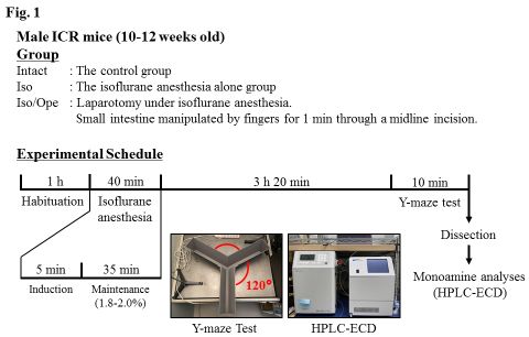
Contents

Welcome message by the Dean of Research                  page 3

Welcome message by the Associate Dean                    page 4

Welcome message by the student coordinator               page 5

Timetable                                                page 6 -7

Keynote talk                                             page 8

Abstracts (in alphabetical order of presenting author)   page 9 - 22

Acknowledgments                                          page 23

Editorial                                                page 24

Welcome message by the Dean of Research

Dear students,

in the past year we have learned to take a closer look at the exchange
between scientists, media and the general public and new habits have
emerged. I am pleased to see the successful virtual continuation of the
Munich Medical Student Science Conference MMS ScienceCon, which
combines students’ research projects from different fields, united by this
year’s conference theme "Science, not silence". These projects show the
broad range of medical research and illustrate curiosity-driven research
activities both during medical training and medical practice.

Research at the University Hospital and the Medical Faculty of LMU Munich
aims to develop new, better therapies for people suffering from diseases and
therefore:

−   to better understand the development of diseases,
−   to find new diagnostic methods,
−   to discover new drugs and non-drug therapies,
−   to develop new strategies for the prevention of diseases.

The Dean's Office supports these principles by funding students’ research
projects as well as Clinician Scientist programs, which make it possible to
follow your curiosity and link healthcare with first-rate science. In this
context, please take a look at the overview of funding and current calls on the
homepage of the Faculty of Medicine: https://forschungsportal.med.uni-
muenchen.de/

I thank Dr Canady as well as the whole team organizing the Munich Medical
Student Science Conference 2021, and I wish you many interesting
contributions and discussions.

Prof. Dr. Stefan Endres
Dean of Research

                                       3
Welcome message by the Associate Dean
Dear students, dear participants at this year’s conference,
What is science? How to distinguish it from non-scientific behaviours?
Sars-CoV-2 pandemic brought those questions to more minds than ever.
So, what is it, what is essential?
Let us see, what others have thought about it.
The astronomer and astrophysicist Carl Sagan writes in his book ´The Demon-haunted world´ (1):
„Again, the reason science works so well is partly that built-in error-correcting machinery. There are
no forbidden questions in science, no matters too sensitive or delicate to be probed, no sacred truths.
That openness to new ideas, combined with most rigorous, sceptical scrutiny of all ideas, sifts the
wheat from the chaff. It makes no difference how smart, august, or beloved you are. You must prove
your case in the face of determined, expert criticism. Diversity and debate are valued. Opinions are
encouraged to contend – substantively and in depth.
The process of science may sound messy and disorderly. In a way, it is. If you examine science in its
everyday aspect, of course you find that scientists run the gamut of human emotion, personality, and
character. But there’s one facet that is really striking to the outsider, and that is the gauntlet of
criticism considered acceptable or even desirable. “
That’s the spirit. Where it does not prevail, there is no science.
He continues with advice for scientists of your age, referring to doctoral thesis oral examinations:
„But they (the students) understand at this critical moment, they have to be able to answer searching
questions posed by experts. So, in preparing to defend their theses, they must practice a very useful
habit of thought. They must anticipate questions; they have to ask: Where in my dissertation is there
a weakness that someone else might find? I’d better identify it before they do.“
All this is not advice for a self-destructive attitude and life. It is the way science develops and
prospers. You feel comfortable with such ideas, even embrace them for your own work, dissertation
or other? Then you are on the best way of becoming a scientist.
Good luck. And let’s hope the virus will be defeated before next year’s conference.
Michael Meyer Martinsried, May 26, 2021
Associate Dean Preclinical Teaching
Professor of Molecular Neurophysiology
(1) Carl Sagan, The Demon-Haunted World – Science as a candle in the dark, Ballantine
Books/Random House, New York 1997, p.31
4Intro by Anne V. Eberhard
Student Coordinator
This was an unprecedented year of unprecedented paradoxes — disappointment and
gratitude, solitude and connection, accomplishments, and stagnation. Questions as “Was my
research good enough? Will I reach my future goals? What do I even want?” may have crossed
your mind. Given that for some of you the undergraduate research opportunity
(Forschungsmodul) has come to an end by now, have faith that you are ready to say yes to all
the adventures outside of your comfort zone that are waiting ahead. Be excited to keep
moving forward even if there are uncertainties. Most of all, be hopeful.
You have shown remarkable flexibility in adapting to new modes of learning and research
since the beginning of the program. You have also found new ways to connect with and
support one another.
Each of you has worked to reimagine our community, and your own role within it, for this
difficult moment in history. Many of you have done so while juggling lectures, additional
family and work responsibilities, or navigating the other unique hardships that this pandemic
has brought. Be assured that your many accomplishments inspire optimism for a brighter
future.
I want to extend my heartfelt thanks to each of you for your tremendous work during the last
year. I am grateful for your dedication in accomplishing your research and advancing our
mission of education and research under extraordinary circumstances.
Personally, I am deeply grateful that I have had this community to walk with through these
challenging times.
While our Online Munich Medical Science Conference is not the in-person event we hoped
for, it still allows us to honor your accomplishments with our warmest wishes as you go out to
make your mark in a changed world – with the promise of coming together again in the future.
Anne was part of the research program at the Department for Neurophysiology in 2018. Then
she pursued novel therapeutic approaches in cardiovascular research for the experimental part
of her doctoral thesis in collaboration with Stanford University at TU Munich, which was
funded by the German Heart Foundation. Since then, she has published in several high impact
journals as co-author, has written a blog-type article for the Nature.Research Community and
assisted peer reviews for the European Heart Journal among others.
Contact for further information:
5Conference Timetable
Time Description
09.00 – 09.30 am Opening Ceremony
Welcome by
Ayse Gertrude Yenicelik
09.30 – 10.30 am Talk Session 1
Searching for new Interactors of the Calbindin-28k
Jara Berneke
Cognitive Function and Hippocampal Volume in Youth Soccer Players
Alexandra Castro Silva
Prevalence and risk factors of atrial fibrillation-
Collective of the SERVE-HF Major Sub-study
Victoria Vaas
Characterization of MIN6 Cells
Florian Fahrenschon & Kevin Bahner
10.30 – 10.45 am Coffee break
10.45 – 11.30 am Talk Session 2
The role of Secretagogin, a calcium-binding protein, regarding insulin
production and secretion
Celine Röder & Maryam Hashemnia
Characterization of the novel TSPO ligand GRT16085N-6 in human
glioblastoma cells
Celine Rohrmus
Tissue specific RNAi knockdown to find a muscle specific function
Marie Dawczynski
11.30 – 11.45 am Coffee break
11.45 – 12.45 pm Talk Session 3
The Nucleus oculomotorius in a Mouse Model of Niemann-Pick Disease
Lara Bucka
Testing the RIST molecular targeted regimen in an in-vitro 3D cell culture
model of neuroblastoma
Carina Kaess
6Phenotypes of m6A related RNA-binding proteins
Mayra Lenz
Indicators for the evaluation of German municipal prevention and health
promotion projects for children and adolescents
Myriam Robert (short talk)
12.45 – 14.00 pm Lunch
14.00 – 14.45 pm Talk Session 4
Establishment of a Novel Postoperative Delirium (POD) Mouse Model
Ran Wang
RIPK3: A novel tumor suppressor in Lung adenocarcinoma
Fabian Allmendinger
Psychological stress among students in health-related fields during the
COVID-19 pandemic: results of a cross-sectional study at selected
Munich universities
Kristina Schröpfer
14.45 – 15.00 pm Coffee break
15.00 – 16.00 pm Keynote talk
Excercise in Schizophrenia: Understanding and Cures
Prof. Peter Falkai, M.D., Ph.D.
16:00 – 16.15 pm Awards and Closing Ceremony
Farewell by
Prof. Dr. Michael Meyer
7Keynote talk
„Excercise in Schizophrenia: Understanding and Cure”
Schizophrenia is a severe brain disorder characterised by positive, negative, affective and
cognitive symptoms and can be regarded as a disorder of impaired neural plasticity. This
lecture focusses on the beneficial role of exercise in schizophrenia and its underlying
mechanisms.
Apart from the established pharmacological treatments in schizophrenia, aerobic exercise
has a profound impact on the plasticity of the brain of both rodents and humans such as
inducing the proliferation and differentiation of neural progenitor cells of the hippocampus
in mice and rats. Aerobic exercise enhances LTP and leads to a better performance in
hippocampus related memory tasks, eventually by increasing metabolic and synaptic
plasticity related proteins in the hippocampus. In healthy humans, regular aerobic exercise
increases hippocampal volume and seems to diminish processes of ageing like brain atrophy
and cognitive decline.
Several meta-analyses demonstrate the beneficial effect of exercise on function, positive as
well as negative symptoms and brain structure in multi-episode schizophrenia.
Prof. Peter Falkai, M.D., Ph.D.
Professor and Chairman at the Department of Psychiatry and Psychotherapy
Ludwig-Maximilians-University Munich, Germany
Professor Peter Falkai has been working in the field of psychiatry for 30 years. His main
research interest is focused on the neurobiology of psychotic disorders, namely
schizophrenia. He has been member of various international and German scientific societies,
such as the Schizophrenia International Research Society (SIRS) and is currently president of
the European Psychiatric Association (EPA).
Prof. Falkai has been leading multidisciplinary teams of researchers, whose clinical and
research expertise focus continuously on the neurobiological origins and
pathomorphological aspects as well as on causal treatment options of psychotic disorders.
8Abstracts in alphabetical order of presenting authors
RIPK3: A novel tumor suppressor in Lung adenocarcinoma.
Fabian Allmendinger1, Deepti Agrawal1, Michelle Dietzen2, 3, 4, Sebastian Vosberg1, 5,
Enkhtsetseg Munkhbaatar1, Caterina Branca1, Nicholas McGranahan2,3,4 and Philipp J.
Jost1, 5.
1. Department of Medicine III, Klinikum rechts der Isar, TUM School of Medicine, Technical University of Munich, Munich,
Germany
2. Cancer Research UK Lung Cancer Center of Excellence, University College London Cancer Institute, Paul O’Gorman
Building, London, UK
3. Cancer Evolution and Genome Instability Laboratory, The Francis Crick Institute, London, UK
4. Cancer Genome Evolution Research Group, University College London Cancer Institute, University College London,
London, UK
5. Division of Clinical Oncology, Department of Medicine, Medical University of Graz, Graz, Austria
Introduction: Receptor-Interacting Serin/Threonin-Protein Kinase 3 (RIPK3) is a key player in a highly
inflammatory programmed cell death pathway, namely necroptosis. Necroptosis induction
elicits the release of intracellular contents and cytokines, which attract and activate anti-tumoral
immune cells. In lung adenocarcinomas (LUAD) RIPK3 is a tumor suppressive aberrational target,
which has been shown to induce immune suppression in the tumor microenvironment.
Methods: To elucidate RIPK3 involvement in LUAD, we took advantage of an in vivo LUAD mouse
model and The Cancer Genome Atlas program (TCGA).
We used genetically modified mice, carrying KrasG12D oncogene (under a floxed stop codon) and
floxed p53 tumor suppressor. Tumor induction was obtained through Cre recombination, which
induced KrasG12D activation as well as p53 deletion.
Results: Analysis at 16 weeks post-induction revealed that advanced-stage lung tumors
(Adenoma, Adenocarcinoma – Ad/AC) have lower RIPK3 expression than early-stage tumors
(bronchial hyperplasia). Furthermore, high RIPK3 expressing tumor lesions showed an
enrichment of anti-tumoral inducible nitric oxide synthase (iNOS)-positive M2-
macrophages and a reduction in Arginase-1 positive anti-inflammatory M1 macrophages. This
difference in macrophage polarization suggests that RIPK3 may initiate an anti-tumoral
immune microenvironment. To evaluate this hypothesis in human settings, we analyzed the
TCGA datasets and found that tumors with higher RIPK3 expression showed higher total
infiltrating leukocytes, specifically T-cells, B-cells, macrophages, and dendritic cells.
Conclusion: Taken together our data clearly point to RIPK3 as a tumor suppressor in LUAD.
This effect may be driven by the inhibition of necroptotic cell death, which in turn alters
immune cell recruitment. Leveraging the crosstalk between inflammatory cell death and immune
micro-environment may be a possible immunogenic therapeutic target.
9Searching for new Interactors of the Calbindin-28k Jara Berneke, Prof. Michael Meyer Molecular Neurophysiology, Meyer Group, Department of Cellular Physiology, Biomedical Center, Ludwig-Maximilians-University of Munich Calbindin-D28k is an EF-hand Ca2+-binding protein and is most known for its calcium buffering function1. It is thought to play a key role in motor coordination, many neurological disorders2, such as Alzheimer and epilepsy and even in stress induced memory loss3. Little is known of its regulatory mechanisms and interactors. Although there are, as mentioned before, many clinical connections to Calbindin, this project focuses on the proteins’ basic molecular characteristics in a cell system. More specifically we want to investigate how Calbindin-D28k interacts with other proteins in cells; confirming known interactors and even detecting new ones. As to known interactors; there are among others4 RanBPM, IMPase, and Caspase 3. Especially Calbindin- D28k’s interaction with the protein Caspase 3 attracted our interest. The protein Caspase 3 is involved in the complicate cascade, regulating apoptosis5. Calbindin-D28k is thought to deactivate Caspase 3 upon binding. For all experiments we used the HN10e cell line6 which we transfected with plasmids expressing Calbindin-D28k-ALFA. Calbindin-D28k-ALFA is a fusion protein of Calbindin-D28k with a peptide called ALFA. The ALFA peptide is a tag for which we have very potent antibodies, called nanobodies7. By linking those nanobodies to agarose beads, we have a good system for co-immunoprecipitation. We additionally transfected our cells with a plasmid for Caspase 3. After co-immunoprecipitation and then separating the proteins by SDS-gel electrophoresis, we did a Western Blot. The results of that blot indicated that Calbindin indeed interacted with Caspase 3. In the future I would like to investigate this interaction further. With the objective of finding new interactors of Calbindin, we conducted a co-immunoprecipitation with cell lysate from the HN10e cells which were transfected only with the plasmid for Calbindin-D28k- ALFA. After this process we separated the proteins by SDS-gel electrophoresis and stained them. The coloration of the gel gave way to the assumption, that we encountered a new interactor of Calbindin- D28k. For further analysis we sent our samples to mass spectrometry which provides accurate mass determination and characterization of unknown proteins. Hoping to identify a new interactor in our samples, we are still waiting for the results. I would like to further investigate the functions of this new interactor und trace those back to the functions of Calbindin-D28k. 1 B.Schwaller, The continuing disappearance of “pure” Ca2+ buffer 2 Jason C You in Nature Medizine, Epigenic suppression of hippocampal calbindin-D28k by ΔFosB drives seizure-related cognitive deficits 3 Ji-Tao Li, Suppressed Calbindin Levels in Hippocampal Excitatory Neurons Mediate Stress-Induced Memory Loss 4 B.Schwaller, The continuing disappearance of “pure” Ca 2+ buffer 5 Benjamin G. Bobay, Structural insights into the calcium-dependent interaction between calbindin-28k and caspase-3 6 Henry J. Lee, Neuronal Properties and Trophic Activities of Immortalized Hippocampal Cells from Embryonic and Young Adult Mice 7 Götzke 2019, The ALFA-tag is a highly versatile tool for nanobody-based bioscience applications
The Nucleus oculomotorius in a Mouse Model of Niemann-Pick Disease

Bucka LA1, Mayadali Ü1, Messoudi A1, Platt F², Horn AKE1

Anatomical Institute, LMU, Munich; Department of Pharmacology, University of Oxford²

Niemann-Pick type C disease (NPC) is a rare lysosomal storage disorder (LSD), which typically manifests as a
neurodegenerative process. In most cases the autosomal recessive disease is caused by a mutation in the NPC1
gene which encodes an integral transmembrane protein of the lysosomal membrane (Platt et al. 2016). Clinical
symptoms include cerebellar ataxia, dysphagia, dementia, destructive neuroinflammation and premature death. A
cardinal sign in NPC patients is a supranuclear gaze palsy for vertical (mainly down) and later horizontal saccades
(Blundell et al. 2017). How these symptoms result from altered lipid metabolism in NPC is still not clear.
Purpose
In an attempt to detect possible neuroanatomical changes in neurons of the oculomotor system a
comparative immunohistochemical study was performed in the oculomotor nucleus (nIII) of a NPC1(-/-) mouse
and wildtype NPC1(+/+) control.
Methods
Selected paraffin sections of both mouse brainstems were stained with different antisera to detect perineural nets
(aggrecan, chondroitin sulfate proteoglycans, hyaluronan binding and link protein), non-phosphorylated
neurofilaments (SMI32), and cholinergic motoneurons (choline acetyltransferase) to distinguish between
different neuron types in nIII.
Results
The experiments revealed a generally stronger immunostaining of the nIII neurons with all markers in the NPC1(-/-)
mouse compared to the wildtype. Additionally, the NPC1(-/-) mouse exhibited ballooned neurons characteristic for
LSD, and SMI32-positive spheroids within cranial nerves most probably representing swollen axons (Bu et al.,
2002). The number of perineuronal net-bearing motoneurons targeting twitch muscle fibers via a single
endplate (SIF) did not differ between the NPC1(-/-) and wildtype mouse. However, the number of perineuronal
net-lacking motoneurons, which correspond to motoneurons targeting non-twitch muscle fibers via multiple
nerve endings (MIF), was elevated in the NPC1(-/-) mouse compared to control (Büttner-Ennever et al., 2001).
Conclusion
Further behavioral studies are required to determine the impact of these findings on the oculomotor
functionality.

References
1. Blundell J., Frisson S., Chakrapani A., Gissen P., Hendriksz C., Vijay S., Olson A. (2018) Oculomotor abnormalities in children
with Niemann-Pick type C. Molecular Genetics and Metabolism. 123, 159-168.
2. Bu B., Li J., Davies P., Vincent I. (2002) Deregulation of cdk5, hyperphosphorylation, and cytoskeletal pathology in the Niemann-
Pick type C murine model. The Journal of Neuroscience. 22, 6515–6525.
3. Büttner-Ennever, J.A., Horn A.K.E., Scherberger H., D'Ascanio P. (2001) Motoneurons of twitch and nontwitch extraocular muscle
fibers in the abducens, trochlear, and oculomotor nuclei of monkeys. The Journal of Comparative Neurology. 438, 318-335.
4. Platt N., Speak A.O., Colaco A., Gray J., Smith D.A., Williams I.M., Wallom K.L., Platt F.M. (2016) Immune dysfunction in
Niemann-Pick disease type C. Journal of Neurochemistry. 136, 74-80.
Cognitive Function and Hippocampal Volume in Youth Soccer Players
Castro-Silva AA, Bonke EM, Koerte IK.

Ms. Castro's research project is in the process of publication and can therefore not be
displayed here.
Tissue specific RNAi knockdown to find a muscle specific function
Dawczynski Marie
Aim/Background:
N6-methyladenosine (m6A) is a methyl modification found on adenine in some portion of
mRNA molecules. Mettl3, Mettl14 and other components of the writer protein complex deposit
the methyl mark on target mRNAs. m6A modification has been suggested to regulate diverse
steps in RNA processing notably alternative splicing. The physiological function of m6A
modification in muscle is poorly understood and we do not know what happens when m6A
levels specifically in muscle are reduced. To test this, we wanted to knockdown the expression
of Mettl3 and Mettl14 specifically in muscle. My aim was to verify which RNAi hairpins work
best for tissue-specific or temporal specific RNAi knock-down.
Methods:
We are using Drosophila melanogaster as our model organism. We crossed different Gal4
lines with different Mettl3/Mettl14 hairpin flies, allowing us to drive RNAi knockdown specifically
in muscle or neural tissues. After sorting flies of the correct genotype, I performed behavioral
assays including a climbing test, a flight test and a wing assay test to determine which RNAi
hairpins give the strongest phenotye.
Results
For RNAi hairpins targeting Mettl3, the strongest hairpin was called NIG. Two other hairpins
gave weak phenotypes, although line BL80431 might be a little stronger than BL80448.
Tubulin-Gal4 and Act5c-Gal4 experiments confirm that RNAi results in only partial knockdown.
We were able to obtain enough flies for behavioural experiments, where this is more
challenging with the mutant. UAS-Dcr2 consistently enhances the Elav-Gal4 RNAi phenotype
with both Mettl3 and Mettl14 hairpins. We tried to compared the RNAi phenotypes of these
lines with Act79B-Gal4 (which is specific to the tubular jump muscle) and Act88F-Gal4 (which
is specific to fibrillar indirect flight muscle), but had inconclusive results as we observed very
little phenotype with Act79B-Gal4 and already observed a phenotype with the Act88F-Gal4
alone.
Conclusion
In agreement with results from other students in the lab, the viability and flight phenotypes
show that the best hairpins for Mettl3 (Ime4) are the NIG and BL80431 lines. The best hairpins
for Mettl14 are 105434 and BL48560. My flight-test results show that Mettl3 is necessary in
muscle as well as in neurons for proper function. Climbing and held-out-wing phenotypes are
much more mild and subtle than flight phenotypes, suggesting that fibrillar flight muscle is
particularly sensitive to loss of m6A modification. Future studies are necessary to identify
sarcomere and neuromuscular junction defects and to identify RNAs that are m6A modified
specifically in muscle.
Characterization of MIN6 Cells

Fahrenschon Florian, Kevin Bahner, Prof. Michael Meyer

Molecular Neurophysiology, Meyer Group, Department of Cellular Physiology, Biomedical Center,
Ludwig-Maximilians-University of Munich

The MIN6 cell-line is among the most commonly used beta-cell-lines in diabetic research. In
comparison to primary beta cells they are more convenient as they do not need to be extracted regularly
from animals, because they have a much longer lifespan and can be cultured more easily. At the same
time they appear to have similar biological characteristics as their primary cell counterpart and thus make
them a favorable proxy. However, the process of cell immortalization e.g. by radiation and thus inducing
an alteration of genes or the extraction from their in-vivo environment and thus the loss of cell-cell
interactions can impose different behavior in some regards that are not fully explored yet.[1]
Specifically, the role of Calbindin in the MIN6 cell line has not been studied sufficiently.

Calbindin-D28k has a calcium buffering function in beta-cells and thus modulates the secretion of insulin.
In 1999 Sooy et al. were the first to postulate this by generating a calbindin knockout.[2] As they didn’t
use MIN6 cells, but primary beta-cells in their experiments, we asked ourselves if we can see the same
effects in the MIN6 cell line.

That is why we further characterized the MIN6 cells after a knock-out (KO) of the calbindin gene locus. At
first we wanted to determine, if calbindin or a protein that interacts with it is altered in its functionality
specifically regarding the glucose stimulated insulin secretion. A dysfunctional insulin secretion is one of
the symptoms of diabetes and thus new insights in these mechanisms are of clinical relevance.
We proved that the KO successfully lowered the expression of calbindin by sequencing the genes and by
performing a SDS-PAGE and a Western Blot. Then we measured the glucose stimulated insulin secretion
with several ELISAs similar as Sooy et al. did. Second, we investigated the role of apoptosis in MIN6 cells
given that apoptosis caused by a nutritional overstimulation of beta cells is claimed to be one reason for
developing type 2 diabetes.[3] Therefore, we tested whether insulin induces calbindin expression in
our cells and if a high sustained level of glucose leads to apoptosis.
However, our results were not as clear as expected. We managed to stimulate insulin secretion, but our
results showed a broad spectrum with almost zero secretion in some cases due to unknown
reasons. We could prove the calbindin KO successfully. However we couldn’t confirm that insulin induces
the calbindin expression. Results for the apoptosis hypothesis are yet to come given that some
experiments are still in progress.

References

1. Skelin, M., M. Rupnik, and A. Cencic, Pancreatic beta cell lines and their applications in diabetes mellitus research. Altex,
2010. 27(2): p. 105-13.
2. Sooy, K., et al., Calbindin-D(28k) controls [Ca(2+)](i) and insulin release. Evidence obtained from calbindin-d(28k) knockout
mice and beta cell lines. J Biol Chem, 1999. 274(48): p. 34343-9.
3. Wali, J.A., S.L. Masters, and H.E. Thomas, Linking metabolic abnormalities to apoptotic pathways in Beta cells in type 2
diabetes. Cells, 2013. 2(2): p. 266-83.
Testing the RIST molecular targeted regimen in an in-vitro 3D cell culture model of
neuroblastoma

Kaess C, Matthes M, Gross J, Heise T, Corbacioglu S, and Sommer G.

Pediatric Hematology, Oncology and Stem Cell Transplantation, Franz-Josef-Strauß-Allee 11, University Hospital
Regensburg, 93053 Regensburg, Germany

Neuroblastoma (NB) is the most common extracranial solid tumor in childhood. Despite
intensive treatment regimens the outcome for patients with high-risk relapsed or treatment
refractory NB remains poor and novel treatment strategies are urgently needed. A new
multimodal treatment design based on metronomically combining molecular targeted
biologicals (Rapaymcin and Dasatinb) with conventional cytostatics (Irinotecan and
Temozolomid), called the RIST therapy, is currently evaluated in a phase II prospective
randomized clinical trial (NCT01467986).
Often promising preclinical results show weak in vivo efficacy due to preclinical drug testing on
conventional two-dimensional (2D) monolayer cell cultures. Aim of this study is to establish a three-
dimensional (3D) cell culture model suitable to further improve the RIST protocol in preclinical
studies. Therefore we used three oncogene MycN-amplified (MNA) (SKNBE(2), Kelly, IMR32), two
MycN-non amplified (MNN) (SKNAS, SKNSH) and one MycN-inducible (MNI) (Tet21N) NB cell
lines. The 3D cell cultures were evaluated by determining the size, number, and viability of
spheroids at various time points after seeding increasing cell numbers, applying distinct growth
supplements and providing different types of ultra-low attachment plates. Furthermore, by
RTqPCR the expression of various cancer stem cell marker was evaluated.
To assess the RIST protocol on spheroid formation, drug treatment was started directly after cell
plating, while spheroid growth was assessed on completely formed spheroids. Our data
demonstrates that the viability of all tested NB spheroids was significantly reduced applying the
same half maximal inhibitory concentration (IC50) as applied in 2D cell cultures.
Interestingly, in contrast to the two MNN NB cell lines two out of three MNA NB cell lines
sustained more viability under RIST therapy. The established 3D cell culture model for
neuroblastoma is suitable for preclinical testing of novel drug analogs even in high-throughput 96-
well formats to improve the efficacy of the RIST therapy on high-risk NB patients.
MUSCLE PHENOTYPES OF m6A RELATED RNA-BINDING PROTEINS
Lenz Mayra, Spletter Maria

Spletter Group, Department of Physiological Chemistry, Ludwig-Maximilians-Universität

PURPOSE/AIM
Methyl modification on adenosine of mRNA is regulated by the N6-methyladenosine (m6A)
pathway. The m6A is a reversible mark that is “written” by the Mettl3/14 complex and can be
removed by an “eraser” enzyme. In addition, m6A is recognized by “readers” such that the m6A
modification regulates mRNA processing including folding, maturation and alternative splicing.
The m6A pathway has been best characterized in neurons, but it is also present in muscle. A
further understanding of the m6A pathway in muscle would add to our knowledge about the impact
of m6A on myogenesis. My aim was to evaluate possible m6A associated RNA binding proteins
(RBPs) and their phenotypes.
METHODS
Drosophila melanogaster was used as a model organism. Rox8, Rpb9 and Fne are RBPs
expressed in muscle that may interact with the Mettl3/14 complex or independently regulate
alternative splicing. YTHDC1 and YTHDF1 are readers that bind m6A modified RNAs to direct RNA
processing. Different genetic crosses and rescues were set. Flies were sorted by intended
genotype and tested in assays of lethality, wing position and flight and climbing ability to identify
their phenotypes.
RESULTS
Rox8 mutants showed a weak phenotype, whereas the phenotype for Rbp9 mutants was
stronger. Different strategies for Rbp9 rescue were shown to be inefficient, producing similar assay
results to Rbp9 mutants. The fne mutant shows no phenotype by itself, but does have a phenotype
when combined with Rbp9 mutants. Low numbers of fne; Rbp9 mutants that were able to be tested
suggest lethality of the cross in earlier development stages. For the reader protein, YTHDC1 mutants
show a strong phenotype. YTHDF1 mutants seem to be lethal at the pupal stage.
CONCLUSION
The different RBPs mutants showed a variety of viability and mobility phenotypes. Rbp9
mutants showed the strongest phenotype of weak mobility out of the writer proteins, and were
enhanced when combined with the fne mutant. There may be a difference between nuclear and
cytoplasmatic readers of m6A, but this needs to be tested further and confirmed.
Indicators for the evaluation of German municipal prevention and health promotion projects
for children and adolescents
Robert M, Jung-Sievers C, Coenen M, Voß S, Hummel J
Purpose
A Delphi is an iterative survey using anonymity and feedback to reach group consensus when
evidence is lacking (1, 2). The attention given to the questionnaire’s development is central for the
survey’s quality. However, how the items are chosen is often a “black box” in prevention and health
promotion Delphi surveys (3). This is even more complex if the items are indicators, as indicators
selection generally is a challenge (4).
This study presents the preparation of a Delphi survey on child health indicators. A final list of
indicators was developed that contains the items that can be assessed by Delphi experts.
The Delphi survey aims to identify relevant indicators that can be used to monitor the health of
children so as to evaluate the prevention chain Freiham and to guide the evaluation of similar
municipal prevention and health promotion projects.
Methods
This study was based on a scoping review on health indices. Health indicators were extracted from
the identified publications following the search and screening strategy of the review. A stepwise
selection was conducted to reduce the indicators’ number through internal consensus building.
A quality appraisal was conducted using following criteria:

i) are the indicators included in major indicators lists?
ii)are the indicators related to the outcomes of the project of the prevention chain Freiham?
Indicators were reviewed independently and discussed within the research team.
Results
720 health indicators were extracted from the review’s publications and categorized into four main
areas.
Taking into account the conducted quality appraisal and team discussions, 512 indicators and indices
were excluded. The final list included 94 health indicators and indices.
Conclusion
The extraction and selection process could produce a comprehensive final list of 94 health indicators
and indices including an appropriate amount to be assessed by experts in a Delphi survey.

References

1.       Hasson F, Keeney S, McKenna H. Research guidelines for the Delphi survey technique. Journal of advanced nursing.
2000;32(4):1008-15.
2.      Linstone HA, Turoff M. The delphi method: Techniques and applications. Newark: New Jersey Institute of
Technology 2002.
3.       Niederberger M, Käfer A-K, König L. Delphi-Verfahren in der Gesundheitsförderung. Ergebnisse eines
systematischen Reviews. Delphi-Verfahren in den Sozial- und Gesundheitswissenschaften: Springer Fachmedien Wiesbaden
GmbH; 2019. p. 301-36.
4.        Nutbeam D. Evaluating health promotion. BMJ. 1999;318(7180):404a.
The role of Secretagogin, a calcium-binding protein, regarding insulin production and secretion

Celine Röder, Maryam Hashemnia, Prof. Dr. Michael Meyer

Molecular Neurophysiology, Meyer Group, Department of Cellular Physiology, Biomedical Center, Ludwig-Maximilians-
University of Munich

Diabetes Mellitus (DM) is one of the leading causes of death worldwide with increasing
incidence, especially in Western countries [1]. The pathological process behind the disease is
malfunctioning of insulin production or secretion, which then leads to rising blood sugar level and
can ultimately cause far-reaching health implications. However, there are several ways this
process can be impaired, all of them leading to diabetes mellitus.

It is known that calcium plays an important role in insulin production as well as insulin secretion in
pancreatic ß-cells [2]. Insulin is being released by an influx of calcium into the cell, thus leading
to depolarisation and exocytosis of membrane vesicles [3]. Interestingly, Calcium-binding
proteins (CBPs) have been found to play a role as calcium-sensors and therefore influence
secretion of insulin [3]. In order to find novel therapeutic targets for customized Diabetes
treatment, the underlying mechanisms of calcium-binding-proteins have to be understood and
explored further.

One of these so-called calcium sensors is Secretagogin (SCGN). This CBP is part of a class of EF-hand
proteins functioning in pancreatic ß-cells, which are depleted in DM and therefore making it an
interesting target[4]. Therefore, knockout cell lines and a wild type cell line were used to investigate
cell functioning in presence or in absence of SCGN. First and foremost, it had to be determined,
whether the knockout to induce a SCGN null mutation was successful. Therefore, we used the
method of Western Blotting. In addition, the genomic makeup was investigated by DNA
characterization by genomic labs. Afterwards, functions of wild type and null mutant cell lines
were compared. Specifically, this included testing all cells under conditions with different
glucose concentrations to investigate how this changed their insulin secretion. One of the most
important methods were enzyme-linked immunosorbent assays (ELISA), which made it possible to
detect different insulin concentrations.

The results so far are inconclusive, making it a subject which definitely should be pursued in the
near future.

References:

1.       World Health Organization. Diabetes.
         13.04.2021 [cited 2021 21.04]; Available from: https://www.who.int/news-room/fact-
         sheets/detail/diabetes.
2.       Wongdee, K., N. Krishnamra, and N. Charoenphandhu, Derangement of calcium metabolism in diabetes mellitus:
         negative outcome from the synergy between impaired bone turnover and intestinal calcium absorption. J Physiol
         Sci, 2017. 67(1): p. 71-81.
3.       Skelin, M., M. Rupnik, and A. Cencic, Pancreatic beta cell lines and their applications in diabetes mellitus research.
         Altex, 2010. 27(2): p. 105-13.
4.       Yang, S.Y., et al., Secretagogin affects insulin secretion in pancreatic β-cells by regulating actin dynamics and focal
         adhesion. Biochem J, 2016. 473(12): p. 1791-803.
Characterization of the novel TSPO ligand GRT16085N-6 in human glioblastoma cells
Rohrmus Celine, Vollmann- Zwerenz Arabel, Hau Peter
Department of Neurology, University Hospital Regensburg
Purpose: The 18-kDa translocator protein (TSPO) is a protein of the outer mitochondrial membrane.
Due to its location, it has been associated with key cellular functions such as proliferation (1)(2),
apoptosis (3)(4), mitochondrial metabolism (5)(6) and oxidative stress (7). It has been shown that
TSPO expression is elevated in a range of brain pathologies (8). This includes malignant cancers such
as Glioblastoma (GBM) which is one of the most frequent and most aggressive primary brain tumors
(9), making TSPO a possible therapeutic target. In this study, we aimed to characterize the effects of
the novel TSPO ligand GRT16085N-6
(GRT16) in a human glioblastoma cell line (BTIC27).
Methods: Proliferation assays were performed to assess cell viability. Furthermore Western blot
technique was used to investigate the role of TSPO on apoptosis. With the aid of the Seahorse
Extracellular Flux Analyzer mitochondrial respiration and glycolysis was evaluated.
Results: At first, we evaluated the IC50 of the novel ligand and the established TSPO ligands XBD173
and Etifoxine in comparison to a DMSO control. Whereby, a cytotoxic effect of the ligand even at
high concentrations could be excluded. We then assessed the effect of GRT16 on apoptosis,
demonstrating that the ligand has no impact on cell death when applied alone. However, in
combination with TNF-related apoptosis inducing ligand (TRAIL) an increase in apoptosis could be
observed when compared to single GRT16 and TRAIL treatment. Analysis of mitochondrial
respiration and glycolysis paralleled these findings, showing no significant changes by ligand
treatment.
Conclusion: Our data imply that GRT16 does not affect cell viability by itself but might promote the
effect of pro-apoptotic agents. Further investigations are needed to fully establish TSPO-mediated
effects and the putative role of GRT16085N-6 in possible drug development for GBM.
References
1. Rechichi M, Salvetti A, Chelli B, Costa B, Da Pozzo E, Spinetti F, et al. TSPO over-expression increases motility, transmigration and
proliferation properties of C6 rat glioma cells. Biochim Biophys Acta - Mol Basis Dis.
2008;1782(2):118–25.
2. Fu Y, Wang D, Wang H, Cai M, Li C, Zhang X, et al. TSPO deficiency induces mitochondrial dysfunction, leading to hypoxia,
angiogenesis, and a growth-promoting metabolic shift toward glycolysis in glioblastoma. Neuro Oncol. 2020;22(2):240–52.
3. Mendonça-Torres MC, Roberts SS. The translocator protein (TSPO) ligand PK11195 induces apoptosis and cell cycle arrest and
sensitizes to chemotherapy treatment in pre- and post-relapse neuroblastoma cell lines. Cancer Biol Ther. 2013;14(4):319–26.
4. Kugler W, Veenman L, Shandalov Y, Leschiner S, Spanier I, Lakomek M, et al. Ligands of the mitochondrial 18 kDa translocator
protein attenuate apoptosis of human glioblastoma cells exposed to erucylphosphohomocholine. Cell Oncol. 2008;30(5):435–
50.
5. Liu GJ, Middleton RJ, Kam WWY, Chin DY, Hatty CR, Chan RHY, et al. Functional gains in energy and cell metabolism after TSPO
gene insertion. Cell Cycle [Internet]. 2017;16(5):436–47. Available from:
http://dx.doi.org/10.1080/15384101.2017.1281477.
6. Betlazar C, Middleton RJ, Banati R, Liu G-J. The Translocator Protein (TSPO) in Mitochondrial Bioenergetics and Immune
Processes. Cells. 2020;9(2):512.
7. Gatliff J, East D, Crosby J, Abeti R, Harvey R, Craigen W, et al. TSPO interacts with VDAC1 and triggers a ROS-mediated inhibition
of mitochondrial quality control. Autophagy. 2014;10(12):2279–96.
8. Batarseh A and VP. Regulation of Translocator Protein 18 kDa (TSPO) Expression in Mol Cell Endocrinol . 2010 October 7;
327(1-2): 1–12. doi:10.1016/j.mce.2010.06.013. Health and Disease States. Mol Cell Endocrinol.
2010;327:1–12.
9. Louis DN, Perry A, Reifenberger G, von Deimling A, Figarella-Branger D, Cavenee WK, et al. The 2016 World Health Organization
Classification of Tumors of the Central Nervous System: a summary. Acta Neuropathol.
2016;131(6):803–20.Psychological stress among students in health-related fields during the COVID-19 pandemic: results of a cross-sectional study at selected Munich universities Schröpfer, Kristina PURPOSE: The COVID-19 pandemic has been a challenging period of upheaval for college students. This study aims to assess the factors associated with psychological stress during the COVID-19 pandemic among a sample of students in health-related fields at Munich universities in Germany. METHODS: Students (N = 623) from KSH Munich and LMU Munich completed an online cross- sectional survey. Information on demographics, academic and everyday difficulties due to the COVID-19 pandemic as well as physical and mental health were collected. Multivariate logistic regression analyses were performed to identify factors associated with the psychological stress. RESULTS: The prevalence for higher psychological stress was 44% among the study population. Identified factors were lower overall satisfaction in life (P
Prevalence and risk factors of atrial fibrillation - Collective of the SERVE-HF Major Sub-study
Victoria Vaas1, Christoph Fisser1, Lara Gall1, Jannis Bureck1, Jörg Priefert1, Dominik Linz2,3,4,5, Holger
Woehrle6, Helmut Teschler7, Martin R Cowie8, Michael Arzt1
1 Department  of Internal Medicine II, University Medical Centre Regensburg, Regensburg, Germany 2Department of
Cardiology, Maastricht University Medical Centre and Cardiovascular Research Institute Maastricht, Maastricht, the
Netherlands
3Department of Cardiology, Radboud University Medical Centre, Nijmegen, the Netherlands
4Department of Biomedical Sciences, Faculty of Health and Medical Sciences, University of Copenhagen, Copenhagen,
Denmark
5Centre for Heart Rhythm Disorders, Royal Adelaide Hospital, University of Adelaide, Adelaide, Australia
6Sleep and Ventilation Center Blaubeuren, Lung Center Ulm, Ulm, Germany
7Department of Pneumology, AFPR, Ruhrlandklinik, West German Lung Center, University Hospital Essen, Essen, Germany
8Faculty of Medicine, National Heart & Lung Institute, Imperial College London, London, United Kingdom

Purpose: Central sleep apnea (CSA) could trigger atrial fibrillation (AF) due to the associated
intermittent hypoxia, recurrent arousals, intrathoracic pressure differences and sympathetic
activation in patients with heart failure and reduced ejection fraction (HFrEF). The aim of the present
work was to analyse the prevalence and risk factors of AF in patients with CSA and HFrEF.

Methods: In the SERVE-HF Major sub-study (NCT01164592) 312 patients were included.
Polysomnography including a nocturnal electrocardiogram that met the appropriate technical
requirements was available at baseline (n=272) and after 3 months (n=180). After exclusion of
patients with pacemakers, 110 patients were analysed for AF.

Results: The prevalence of AF at baseline was 39% (43/110). Patients with AF were older
(72 vs. 67 years, p=0.005), more often assigned to NYHA classes 3 and 4 (84 vs. 64%,
p=0.026) and had a higher mean heart rate (75 vs. 67/min, p=0.006) and blood pressure
(systolic 135 vs. 124mmHg, p=0.004; diastolic 79 vs. 74mmHg, p=0.025). In univariate regression
analysis, established cardiac risk factors such as age, higher systolic (RRsys) and diastolic blood
pressure, NYHA classes 3 and 4, and mean heart rate (HFm), were associated with AF. In contrast to
apnea-hypopnea (odds ratio [OR]: 1.009; 95% confidence interval [95% CI]: 0.984-1.035; p=0.478)
and hypopnea index (OR: 0.969; 95% CI: 0.936-1.003; p=0.077), apnea index (AI; OR: 1.024; 95% CI:
1.001-1.046; p=0.039) was identified as a risk factor. After adjustment for RRsys, HFm and AI, RRsys
and HFm were associated with AF, but AI was not.

Conclusion: The prevalence of AF in patients with CSA and HFrEF is 39%. In addition to known cardiac
risk factors, AI was associated with the occurrence of AF, in contrast to the hypopnea index. The
results suggest that more severe forms of CSA are associated with AF.

Research support:
V.V. received grant support from the German Society of Sleep Medicine; C.F. reports receiving
support from the German Heart Foundation/German Foundation of Heart Research; M.A. has
received consulting fees from ResMed and grant support from ResMed Foundation
Establishment of Novel Postoperative Delirium (POD) Mouse Model

                       Student Name: Ran Wang (王 然)
                                                           Instructor Name: Prof. Shinya Toyokuni (豊國 伸哉 教授)
                               Affiliation Course: Department of Pathology and Biological Responses (Pathology I)

Background and Purpose
       Postoperative delirium (POD) is an acute neuropsychiatric syndrome which is considered to be a complication
occurring frequently among surgical patients of all ages. The symptoms include acute, transient, fluctuating disturbances
in attention, cognition, and/or level of consciousness. Our research group has tried to establish a suitable mouse model for
POD research and found that impairment of both recognition memory and attention could be caused by abdominal surgery
under isoflurane anesthesia (Iso/Ope) at 4 h after surgery in young mice. These behavioral dysfunctions were evaluated by
the novel object recognition test and the water finding test, respectively. In the present study, we measured spatial working
memory using the Y-maze test in the Iso/Ope-treated mice. In addition, we analyzed the changes in monoamine
neurotransmission in the several brain regions.
Methods
      Male ICR mice (10-12 weeks old) were randomly assigned
into 3 groups of the control group (Intact), the isoflurane
anesthesia alone group (Iso), or the Iso/Ope group (Fig. 1).
At 4 h after the anesthesia, the animal was allowed to freely
explore in a Y-shaped maze. The number of arm entries and the
number of triads were recorded in order to calculate the percentage
of alternation. An entry occurred when all four limbs were within
the arm. Each mouse was sacrificed by cervical decapitation under
CO2 euthanasia just after the behavioral testing. Several brain regions were dissected for the measurement of monoamine
and its metabolite contents by a high performance liquid chromatography equipped with an electrochemical detector
(HPLC-ECD). Differences between the groups were evaluated by one-way ANOVA followed by two-tailed Tukey’s test
using a SPSS software. The differences were considered statistically significant for p-values of less than 5%.
Results and Discussion
      In the Y-maze test, no significant difference was observed between the groups, suggesting
that spatial working memory was not impaired in Iso/Ope group. The norepinephrine (NE)
turnover, which was assessed by measuring the ratio of 3-methoxy-4-hydroxyphenylglycol
(MHPG) to NE levels, in both frontal cortex (FC) and hippocampus (HIPPO) of Iso/Ope group
was significantly increased as compared with Intact and Iso groups (Fig. 2). On the other hand,
there were no significant differences in both dopamine and serotonin turnover in the same
regions. These results suggest that behavioral changes in Iso/Ope group that we found in our
previous experiments could be caused by abnormal NE neurotransmission in the FC and HIPPO
regions of the brain. In conclusion, mice in Iso/Ope group at 4 h could be a novel POD model with impairment of
recognition memory and attention, not spatial working memory, and with abnormal NE neurotransmission.
Acknowledgements
      The author thanks Prof. Kimitoshi Nishiwaki and Dr. Atsushi Mori at Department of Anesthesiology for their great
support to complete this research.

                                                          22
Acknowledgements
“Science not silence” was the motto our students chose for this year’s conference. And I am happy
to say that it is indeed very fitting: despite everything, that was going on around you, you couldn’t
be stopped. You still managed to work on your projects and to come here today to be heard.
I am deeply grateful to Anne Eberhard and Ayse Gertrude Yenicelik, who invested lots of their time
and energy to encourage our students to be here today as presenters and jury members – your
enthusiasm and perseverance is inspiring.
With all my heart I want to thank Prof. Michael Meyer, the Associate Dean, for believing in our
students and for keeping the Undergraduate research opportunity (Forschungsmodul) and the MMS
ScienceCon alive.
I want to thank the Dean of Research, Prof. Stefan Endres for supporting the conference format for
five years now.
Furthermore, I am deeply grateful to Prof. Peter Falkai for agreeing to be our keynote speaker on
such short notice. It is a great privilege to have you here.
I also want to thank all the members of the Jury: Prof. Hyunmi Park from Korea University, Prof. Anja
Horn-Bochtler and Prof. Michael Meyer. It makes me especially happy to welcome and thank all the
members of the student Jury: Mr. Michele Rosso, Ms. Daria Reitmeier, Mr. Kyryl Kharytonchuk, Ms.
Lea Merkel, Mr. Tobias Stückler, Ms. Paula Schorlemer, Ms. Luisa Delius and Ms. Elena Nikolova.
In addition, I want to thank the faculty to provide us with enough funding for the student research
projects to continue this year. My sincere hope is that the format can continue in the years to come.
At last, I want to say thank you to all the students who chose to present their projects this year. I am
thrilled to see the MMS ScienceCon growing and to welcome students from all over Bavaria and
even from Korea and Japan.
Stay curious.
Yours sincerely,
Dr. Johanna Canady
Coordinator of research promotion
LMU Medical Faculty
23Editorial Faculty of Medicine Ludwig-Maximilians-Universität München Prof. Dr. Michael Meyer Associate Dean Großhaderner Str. 9 82152 Planegg-Martinsried Tel: 089-2180-71566 Email: michael.meyer@lrz.uni-muenchen.de Dr. Johanna Canady Conference Coordinator Großhaderner Str. 9 82152 Planegg-Martinsried Tel: 089-2180-75335 Email: johanna.canady@med.lmu.de www.mms-sciencecon.med.uni-muenchen.de
You can also read