Biomaterials Science - Royal Society of ...

Page created by Dolores Fisher
 
CONTINUE READING
Biomaterials Science - Royal Society of ...
View Article Online

Biomaterials
                                                                                                                                                                    View Journal

Science
Accepted Manuscript

   This article can be cited before page numbers have been issued, to do this please use: S. Hosseinzadeh,
   S. L. Lindsay, A. G. Gallagher, D. A. Wellings, M. O. Riehle, J. S. Riddell and S. C. Barnett, Biomater. Sci.,
   2020, DOI: 10.1039/D0BM00097C.
                                                                                               Volume 5
                                                                                                               This is an Accepted Manuscript, which has been through the
    Biomaterials
                                                                                               Number 3
                                                                                               March 2017
                                                                                               Pages 343-602
                                                                                                               Royal Society of Chemistry peer review process and has been
                                                                                                               accepted for publication.
    Science                                                                                                    Accepted Manuscripts are published online shortly after acceptance,
    rsc.li/biomaterials-science

                                                                                                               before technical editing, formatting and proof reading. Using this free
                                                                                                               service, authors can make their results available to the community, in
                                                                                                               citable form, before we publish the edited article. We will replace this
                                                                                                               Accepted Manuscript with the edited and formatted Advance Article as
                                                                                                               soon as it is available.

                                                                                                               You can find more information about Accepted Manuscripts in the
                                                                                                               Information for Authors.

                                                                                                               Please note that technical editing may introduce minor changes to the
                                                                                                               text and/or graphics, which may alter content. The journal’s standard
                                  ISSN 2047-4849                                                               Terms & Conditions and the Ethical guidelines still apply. In no event
                                  PAPER
                                  Soo Hyun Kim et al.
                                  Biodegradable vascular stents with high tensile and
                                                                                                               shall the Royal Society of Chemistry be held responsible for any errors
                                  compressive strength, a novel strategy for applying
                                  monofilaments via solid-state drawing and shaped-annealing
                                  processes                                                                    or omissions in this Accepted Manuscript or any consequences arising
                                                                                                               from the use of any information it contains.

                                                                                                                                rsc.li/biomaterials-science
Biomaterials Science - Royal Society of ...
Page 1 of 35                                                                                                                            Biomaterials Science

                                                                                                              A novel poly--lysine based implant, Proliferate®, for promotion of CNS repair
                                                                                                                                                                                          DOI:following
                                                                                                                                                                                                     View Article Online
                                                                                                                                                                                               10.1039/D0BM00097C

                                                                                                              spinal cord injury.

                                                                                                              Sara Hosseinzadeh1, Susan L. Lindsay1, Andrew G. Gallagher2, Donald A. Wellings2, Mathis
          This article is licensed under a Creative Commons Attribution-NonCommercial 3.0 Unported Licence.

                                                                                                              O. Riehle3, John S. Riddell4, Susan C. Barnett1*

                                                                                                                                                                                                                           Biomaterials Science Accepted Manuscript
                                                                                                              1Institute   of Infection, Inflammation and Immunity, Sir Graeme Davies Building, 120 University

                                                                                                              Place, University of Glasgow, Glasgow G12 8TA, United Kingdom.
Open Access Article. Published on 03 June 2020. Downloaded on 6/6/2020 2:17:14 AM.

                                                                                                              2SpheriTech     Ltd, The Heath Business & Technical Park, Runcorn, Cheshire, UK, WA7 4QX

                                                                                                              3Centre   for Cell Engineering, Joseph Black Building, University of Glasgow, Glasgow G12
                                                                                                              8QQ, UK.

                                                                                                              4Institute   of Neuroscience and Psychology, West Medical Building, University of Glasgow,
                                                                                                              Glasgow, G12 8QQ, United Kingdom.

                                                                                                              Running title: Proliferate®, a novel construct for CNS repair.

                                                                                                              *To whom correspondence should be addressed:
                                                                                                              Professor Susan C. Barnett
                                                                                                              University of Glasgow, Institute of Infection, Immunity and Inflammation, College of Medical,
                                                                                                              Veterinary and Life Sciences, GBRC, Room B329, 120 University Place, Glasgow, G12 8TA.
                                                                                                              Telephone: 44 [0]141 330 8409 E-mail: Susan.Barnett@Glasgow.ac.uk

                                                                                                                                                               1
Biomaterials Science - Royal Society of ...
Biomaterials Science                                                 Page 2 of 35

                                                                                                              Abstract                                                                                         View Article Online
                                                                                                                                                                                                      DOI: 10.1039/D0BM00097C

                                                                                                              The limited regenerative capacity of the CNS poses formidable challenges to the repair of
                                                                                                              spinal cord injury (SCI). Two key barriers to repair are i) the physical gap left by the injury,
                                                                                                              and ii) the inhibitory milieu surrounding the injury, the glial scar. Biomaterial implantation into
          This article is licensed under a Creative Commons Attribution-NonCommercial 3.0 Unported Licence.

                                                                                                              the injury site can fill the cavity, provide a substrate for cell migration, and potentially attenuate

                                                                                                                                                                                                                                     Biomaterials Science Accepted Manuscript
                                                                                                              the glial scar. We investigated the biological viability of a biocompatible and biodegradable
                                                                                                              poly-ε-lysine based biomaterial, Proliferate®, in low and high cross-linked forms and when
                                                                                                              coated with IKVAV peptide, for SCI implantation. We demonstrate altered astrocyte
                                                                                                              morphology and nestin expression on Proliferate® compared to conventional glass cell
Open Access Article. Published on 03 June 2020. Downloaded on 6/6/2020 2:17:14 AM.

                                                                                                              coverslips suggesting a less reactive phenotype. Moreover Proliferate® supported myelination
                                                                                                              in vitro, with myelination observed sooner on IKVAV-coated constructs compared with
                                                                                                              uncoated Proliferate®, and delayed overall compared with maintenance on glass coverslips. For
                                                                                                              in vivo implantation, parallel-aligned channels were fabricated into Proliferate® to provide cell
                                                                                                              guidance cues. Extensive vascularisation and cellular infiltration were observed in constructs
                                                                                                              implanted in vivo, along with an astrocyte border and microglial response. Axonal ingrowth
                                                                                                              was observed at the construct border and inside implants in intact channels. We conclude that
                                                                                                              Proliferate® is a promising biomaterial for implantation following SCI.

                                                                                                              Key words: spinal cord injury, Proliferate®, biodegradable, poly-ε-lysine, construct

                                                                                                              Introduction
                                                                                                              Mechanical damage to the spinal cord in traumatic spinal cord injury (SCI) typically induces
                                                                                                              haemorrhage, blood-spinal cord barrier (BSCB) breakdown, oedema, axonal disruption and
                                                                                                              demyelination at the injury site [1]. Over time, the injury progresses to secondary and chronic
                                                                                                              phases commonly comprising cystic cavitation, axonal dieback, demyelination and the
                                                                                                              formation of a perilesional glial scar [1, 2]. Several strategies have been proposed as promising
                                                                                                              treatments for SCI ranging from cell transplantation to biomaterial implantation [2]. Peripheral
                                                                                                              nerve grafts implanted into the injured spinal cord have long been known to promote central
                                                                                                              nervous system (CNS) regeneration by providing a supportive substratum for guided axonal
                                                                                                              growth [3-8]. However, allogenic peripheral nerve transplantation necessitates long term
                                                                                                              immunosuppression and autologous transplantation requires further, often non-recoverable,
                                                                                                              surgery at a secondary site, thereby compounding the existing risk of surgical complications
                                                                                                              [9]. Biocompatible materials can circumvent this unnecessary harm, providing a substrate for

                                                                                                                                                                2
Biomaterials Science - Royal Society of ...
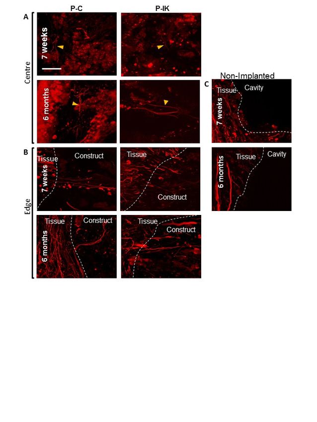
Page 3 of 35                                                                                                                             Biomaterials Science

                                                                                                              regeneration, a delivery vessel for cell and pharmaceutical therapies [10-13], and   a bridge
                                                                                                                                                                                                        View Article Online
                                                                                                                                                                                             DOI: 10.1039/D0BM00097C

                                                                                                              across cystic cavities formed in intermediate stages of SCI [14-16].

                                                                                                              There is evidence for progressive neurodegeneration and spinal cord atrophy years after SCI
          This article is licensed under a Creative Commons Attribution-NonCommercial 3.0 Unported Licence.

                                                                                                              [17], with cavity expansion and continuing die-back of axonal tracts. In pre-clinical studies,

                                                                                                                                                                                                                              Biomaterials Science Accepted Manuscript
                                                                                                              cell transplantation reduces injury area as SCI progresses to chronic stages, a finding primarily
                                                                                                              limited to acute and subacute injury phase transplantations [18, 19]. This demonstrates the
                                                                                                              neuroprotective potential of cell transplantation and consequent physical reconstruction of the
                                                                                                              injury site. However, cell transplantation in human SCI poses a number of challenges,
Open Access Article. Published on 03 June 2020. Downloaded on 6/6/2020 2:17:14 AM.

                                                                                                              including generation of cells at numbers high enough to fill large injury cavities and long-term
                                                                                                              localisation of transplanted cells to the lesion site. By incorporating cells with biomaterials
                                                                                                              prior to transplantation, cells can be more accurately localised to the lesion site than by
                                                                                                              injection alone and can be transplanted at lower densities, with the material acting as a
                                                                                                              substitute ECM to fill the cavity [10]. Several materials, both natural and synthetic, have thus
                                                                                                              far been investigated for this purpose. The mammalian extracellular matrix (ECM) is an
                                                                                                              obvious source of natural biomaterials for regeneration, due to its existing cell-supportive role.
                                                                                                              Indeed, several ECM components have been investigated for SCI implantation, including
                                                                                                              collagen, fibrin, hyaluronic acid and laminin [10, 12, 20-24]. Synthetic polymers, however, are
                                                                                                              often favoured over natural materials for mass reproducibility [10, 12]. These polymers can be
                                                                                                              enriched by incorporation of functional ECM peptides. The most extensively investigated
                                                                                                              examples of these in CNS repair are Arg-Gly-Asp (RGD), the cell adhesive region of ECM
                                                                                                              proteins such as laminin and fibronectin [20-24], and Ile‐Lys‐Val‐Ala‐Val [IKVAV], a
                                                                                                              functional peptide of laminin [23-27]. In particular, IKVAV has been shown to promote
                                                                                                              neuronal plasticity and improved functional outcomes following rodent SCI [28, 29].

                                                                                                              Proliferate®, a polymer based on cross-linked poly--lysine [pK], is a macroporous,
                                                                                                              biocompatible and biodegradable material with tuneable porosity. It is composed of two
                                                                                                              naturally occurring components, pK and a di-carboxylic acid (e.g. decanedioic acid,
                                                                                                              tridecanedioic acid), breaks down upon decomposition into natural, non-toxic components, and
                                                                                                              is reproducible on a mass scale. It is a soft, flexible material interspersed with macropores that
                                                                                                              allow for waste and nutrient exchange, and can bind other bioactive compounds at carboxyl
                                                                                                              and amine moieties. These included amphotericin B and penicillin G to provide a drug eluting
                                                                                                              capacity to advanced corneal bandage lenses and addition of the cell binding peptide H-Gly-

                                                                                                                                                                3
Biomaterials Science - Royal Society of ...
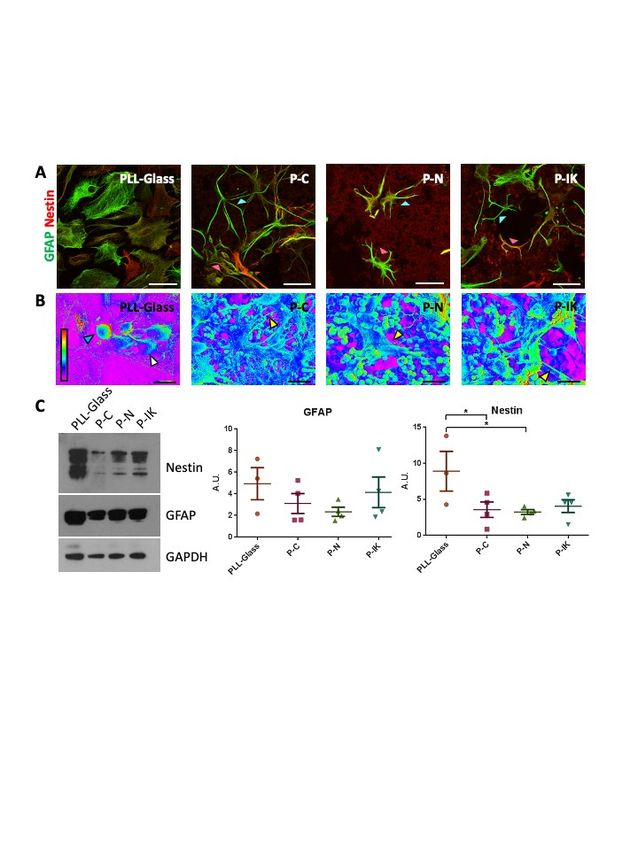
Biomaterials Science                                        Page 4 of 35

                                                                                                              Gly-Arg-Gly-Asp-Gly-Gly-OH (RGD) as well as fragments of larger proteins suchDOI:
                                                                                                                                                                                             as10.1039/D0BM00097C
                                                                                                                                                                                                collagen
                                                                                                                                                                                                     View Article Online

                                                                                                              and fibronectin to promote binding and expansion of corneal endothelial cells in vitro [30-32].

                                                                                                              Here we report that Proliferate®, both in original form and when modified by coating in
          This article is licensed under a Creative Commons Attribution-NonCommercial 3.0 Unported Licence.

                                                                                                              IKVAV, is also a promising candidate for SCI implantation, meeting several criteria for

                                                                                                                                                                                                                           Biomaterials Science Accepted Manuscript
                                                                                                              biomaterials in this field including support of CNS cells, integration with CNS tissue and
                                                                                                              propensity for incorporation of therapeutic compounds.

                                                                                                              Experimental
Open Access Article. Published on 03 June 2020. Downloaded on 6/6/2020 2:17:14 AM.

                                                                                                              Proliferate® synthesis
                                                                                                              Reagents and scaffolds were provided by SpheriTech Ltd (Runcorn, Cheshire, UK).
                                                                                                              Proliferate® was synthesised from pεK (Zhengzhou Bainafo Bioengineering Co. Ltd.,
                                                                                                              Zhengzhou, China) and cross-linked to 95% (excess amine functional groups) or 105% (excess
                                                                                                              carboxyl functional groups) with a mix of decanedioic (Sigma Aldrich, Dorset, UK) and
                                                                                                              tridecanedioic acids (Shanghai Worldyang Chemical Co. Ltd., Shanghai, China) and a polymer
                                                                                                              density of 0.055 g ml-1 using N-hydroxysuccinimide (NHS, CarboSynth ltd., Berkshire, UK)
                                                                                                              and 1-ethyl-3-(3-dimethylaminopropyl) carbodiimide (EDCI, CarboSynth ltd., Berkshire,
                                                                                                              UK).

                                                                                                              A pεK (40.9g, 0.238 mol free amine) monomer stock solution was first prepared
                                                                                                              in dH2O (200 ml) and adjusted to pH 7.1. A separate bis-carboxylic acid cross-linker stock
                                                                                                              solution was also made, composed of tridecanedioic acid (BA; 24.4 g, , 0.2mol of carboxylic
                                                                                                              acid function)), decanedioic acid (SA; 2.24 g, 0.2 mol of carboxylic acid function) and N-
                                                                                                              methylmorpholine (NMM; 25.6 ml, 0.233mol) in dH2O (200 ml). A separate stock solution of
                                                                                                              5% (w/v) tween 80 (VWR International Ltd., Lutterworth, UK) was also prepared. All stock
                                                                                                              solutions were filtered separately to 0.45 µm with a Nylon + GMF syringe filter (Crawford
                                                                                                              Scientific Ltd., Strathaven, Scotland).

                                                                                                              To make carboxyl-functional Proliferate® (P-C, 105% cross-linking, 50 ml), pεK monomer
                                                                                                              solution (10 ml) was combined with bis-carboxylic acid cross-linker solution (11.3 ml) and
                                                                                                              topped up with dH2O (25 ml). Separately, EDCI (6 g, 31.3 mmol) and NHS (0.69 g, 6 mmol)
                                                                                                              were dissolved in dH2O (25 ml) and filter-sterilised before mixing with the pεK/bis-carboxylic

                                                                                                                                                                 4
Biomaterials Science - Royal Society of ...
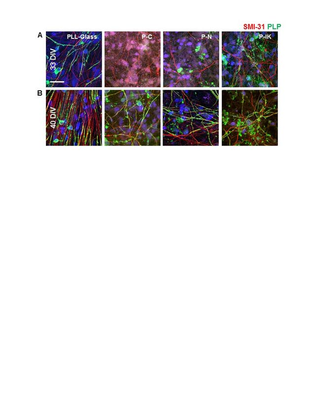
Page 5 of 35                                                                                                                          Biomaterials Science

                                                                                                              acid solution (50 ml total volume). The solution was mixed and immediately poured   into View
                                                                                                                                                                                                        10 Article
                                                                                                                                                                                                            x Online
                                                                                                                                                                                            DOI: 10.1039/D0BM00097C

                                                                                                              10 cm2 Petri dishes (5 ml per dish). The polymer was covered and incubated overnight at 25°C.
                                                                                                              The sheets were then washed 1 x 10 min H2O, 3 x 30 min 0.25 M NaOH, 3x 30 min 0.25 M
                                                                                                              HCl, and 3 x 30 min H2O. Polymer sheets were frozen (approx. -35°C) on an Edwards Super
          This article is licensed under a Creative Commons Attribution-NonCommercial 3.0 Unported Licence.

                                                                                                              Modulyo freeze dryer shelf (Edwards Ltd., West Sussex, UK), the vacuum was then applied

                                                                                                                                                                                                                       Biomaterials Science Accepted Manuscript
                                                                                                              and they were lyophilised overnight with the shelf reaching a final temperature of 27°C
                                                                                                              following an incremental increase. Amine-functional (P-N, 95% cross-linking, 50 ml)
                                                                                                              constructs were made using the same protocol but with different reagent volumes (10.5 ml pεK
                                                                                                              stock solution, 10.75 ml bis-carboxylic acid stock solution, 0.66 g NHS, 5.72 g EDCI).
Open Access Article. Published on 03 June 2020. Downloaded on 6/6/2020 2:17:14 AM.

                                                                                                              The nonapeptide H-Gly-Gly-Ile-Lys-Val-Ala-Val-Gly-Gly-OH was prepared by solid phase
                                                                                                              synthesis and purified by reversed phase HPLC. This peptide containing the laminin binding
                                                                                                              sequence -Ile-Lys-Val-Ala-Val- (IKVAV) was coupled covalently to previously prepared P-C
                                                                                                              constructs prior to freeze-drying with 0.25 M EDCI and NHS, as follows. A 1 cm length of the
                                                                                                              CNS conduit (300 mg) with a carboxylic acid capacity of 0.035 mmol was pre-activated with
                                                                                                              1-Ethyl-3-(3-dimethylaminopropyl)carbodiimide (1.91 g, 10 mmol) and N-hydroxy
                                                                                                              succinimide (1.44 g, 12.5 mmol) in water (50 cm3) for 1h. The pre-activated CNS conduit was
                                                                                                              then washed with water (2 x 50 cm3) and the nonapeptide (72 mg, 0.07 mmol) pre-dissolved in
                                                                                                              water (50 cm3) was added. The coupling reaction was allowed to proceed overnight. The
                                                                                                              nonapeptide coupled conduit was washed with water (2 x 50 cm3), sodium hydroxide solution
                                                                                                              (0.1 mol/dm3, 50 cm3), water (2 x 50 cm3), hydrochloric acid solution (0.1 mol/dm3, 50 cm3)
                                                                                                              and water (2 x 50 cm3). The water was drained off and the sample freeze dried to yield 325
                                                                                                              mg of nonapeptide coupled CNS conduit. The amount of peptide coupled (25 mg) was
                                                                                                              determined gravimetrically.

                                                                                                              Two glycine amino acids either side of the IKVAV have been shown to minimise aggregation
                                                                                                              (Spheritech unpublished observations). The initial loading of IKVAV to the polymer was
                                                                                                              monitored via HPLC. At different time-points in the polymerisation process, the supernatant
                                                                                                              was collected and run on the HPLC to monitor the amount of peptide left in the supernatant.
                                                                                                              The decreasing amount in the supernatant indicated the binding of peptide to the polymer. In
                                                                                                              order to confirm the presence of the IKVAV peptide on the carboxyl scaffold, one dry carboxyl
                                                                                                              scaffold sample and one dry IKVAV carboxyl scaffold sample were sent for amino acid

                                                                                                                                                             5
Biomaterials Science - Royal Society of ...
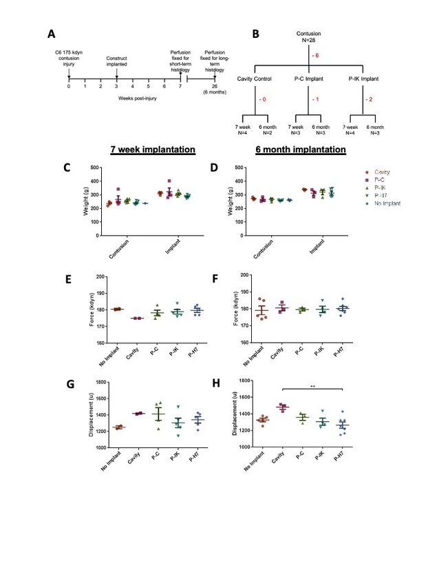
Biomaterials Science                                          Page 6 of 35

                                                                                                              analysis (Severn Biotech Ltd., Kidderminster). The samples were hydrolysed and
                                                                                                                                                                                          DOI:separated,
                                                                                                                                                                                                     View Article Online
                                                                                                                                                                                               10.1039/D0BM00097C

                                                                                                              and the amino acids were detected and quantified compared to their predicted values of 1
                                                                                                              isoleucine, 1 lysine, 2 valine and 1 alanine. The detection of lysine was excluded because of
                                                                                                              the large amounts of lysine found in the poly(ε-lysine) polymer. The IKVAV carboxyl was
          This article is licensed under a Creative Commons Attribution-NonCommercial 3.0 Unported Licence.

                                                                                                              compared to the carboxyl scaffold in relation to their amino acid content. The coupling protocol

                                                                                                                                                                                                                           Biomaterials Science Accepted Manuscript
                                                                                                              is well established and fully validated [33] therefore the only quality control carried out was
                                                                                                              gravimetric analysis of the construct prior and post coupling of the peptide. IKVAV coupling
                                                                                                              to biomaterial scaffolds is a well-established procedure [34-36].
Open Access Article. Published on 03 June 2020. Downloaded on 6/6/2020 2:17:14 AM.

                                                                                                              To make 24-well plate construct inserts, the dry sheets were cut to 0.64 cm2 with a size 5 borer.
                                                                                                              Blank (no membrane attached) 24-well cell culture inserts (SABEU GmbH & Co. KG,
                                                                                                              Northeim, Germany) were adhered to the construct using a dissolution/reprecipitation
                                                                                                              technique. The blank inserts were aligned on top of the 0.64 cm2 constructs before adding
                                                                                                              toluene dropwise (40 µl). The insert was then inverted and left to dry for 1 h. To make tubular
                                                                                                              constructs for implantation, a 1 ml serological pipette (3 mm in diameter) was secured
                                                                                                              vertically using a clamp and 300 soluble polyvinyl alcohol fibres (100 µm diameter, soluble at
                                                                                                              90oC), were inserted vertically through the pipette, and secured at each end with cable ties.
                                                                                                              P-C was prepared as described and poured into a 1 ml syringe upon combination with the cross-
                                                                                                              linker solution. The syringe was inverted and attached to the serological pipette, and the
                                                                                                              polymer solution was syringed upwards to fill the pipette. The construct was left to set for 5 h,
                                                                                                              following which the pipette and polymer within were cut into 40 mm segments using a pipe
                                                                                                              cutter and scalpel respectively. The pipette sections containing the construct were placed in a
                                                                                                              water bath at 100°C for 8 h to dissolve the fibres whilst retaining morphology. The constructs
                                                                                                              were removed from the pipette sections, washed further with water at 100°C (x5) and freeze-
                                                                                                              dried as described above (see schematic in supplementary Fig 1). The constructs were
                                                                                                              examined via SEM for wash efficiency.

                                                                                                              Prior to cell seeding or implantation, constructs were sterilised in 70 % ethanol for >2 h and
                                                                                                              incubated in cell culture medium overnight. For in vivo implantation, constructs were washed
                                                                                                              and stored in endotoxin-free PBS following sterilisation, prior to surgery.

                                                                                                              Mechanical characterisation

                                                                                                                                                               6
Biomaterials Science - Royal Society of ...
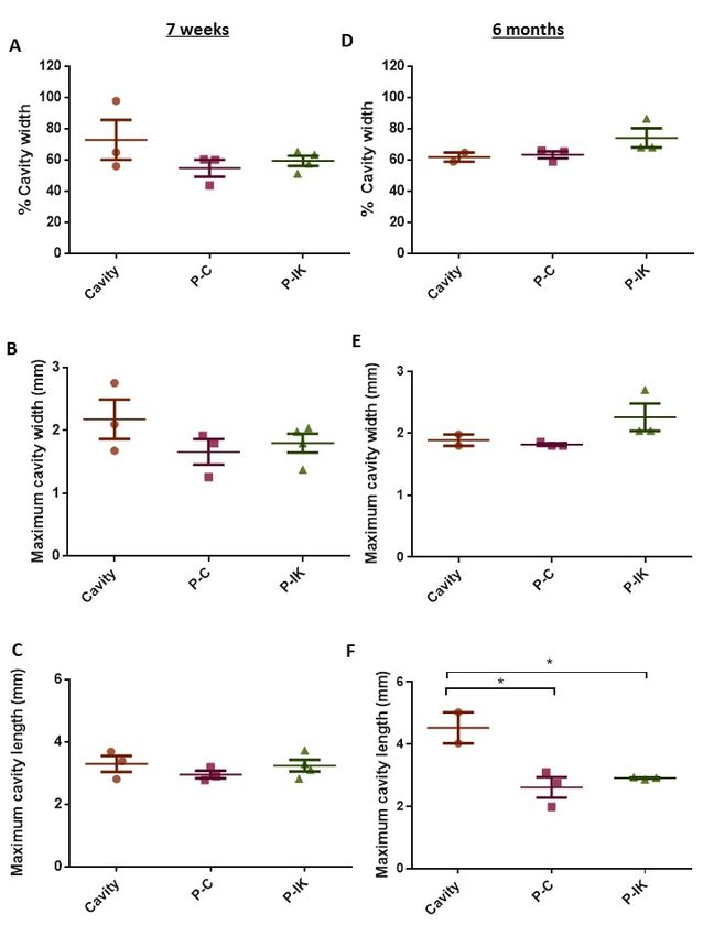
Page 7 of 35                                                                                                                           Biomaterials Science

                                                                                                              Mechanical properties of Proliferate® was measured using a Linkam TST350 tensile   tester
                                                                                                                                                                                                  View Article Online
                                                                                                                                                                                       DOI: 10.1039/D0BM00097C

                                                                                                              (Linkam Scientific Instruments Ltd., Tadworth, England). Stress (σ), strain (ε) and elastic
                                                                                                              modulus (E) were determined for each hydrated sample. Samples were cut to a dog bone shaped
                                                                                                              piece using a bore to provide a 15 mm test length between the clamps of the Linkam stage. The
          This article is licensed under a Creative Commons Attribution-NonCommercial 3.0 Unported Licence.

                                                                                                              width and thickness of each sample was measured with an Absolute Digimatic caliper

                                                                                                                                                                                                                        Biomaterials Science Accepted Manuscript
                                                                                                              (Mitutoyo Ltd., Andover, England) and the readings recorded. The sample was then mounted
                                                                                                              in the Linkam tensile tester and secured in place with the provided clamps. The software was
                                                                                                              run and each sample was elongated until breakage point. A 20 N load cell was used for all
                                                                                                              mechanical analysis with a strain rate of 100 μm s-1. Results were recorded and analysed using
Open Access Article. Published on 03 June 2020. Downloaded on 6/6/2020 2:17:14 AM.

                                                                                                              the supplied Linksys32 software.

                                                                                                              Ethical considerations
                                                                                                              All experimental procedures were approved by the Ethical Review Panel of the University of
                                                                                                              Glasgow and performed in accordance with the UK Animals (Scientific Procedures) Act 1986
                                                                                                              using ARRIVE guidelines. Animals were kept under appropriate light and temperature
                                                                                                              conditions, with food and water available ad libitum.

                                                                                                              Cell culture
                                                                                                              Primary glial and neural cells isolated from Sprague-Dawley (SD) rats (Charles River) were
                                                                                                              maintained in a humidified incubator at 37°C with 7% atmospheric CO2. Astrocytes were
                                                                                                              cultured from postnatal day 1 (P1) rat neurospheres according to standard methods previously
                                                                                                              described [37]. Briefly, neurospheres were obtained from striatum dissections, cultured for 7-
                                                                                                              10 days in vitro (DIV), resuspended and cultured in astrocyte culture media (10% foetal bovine
                                                                                                              serum (FBS) in low glucose Dulbecco’s modified Eagle’s medium (DMEM), both
                                                                                                              ThermoFisher). Astrocyte cultures were fed twice weekly and reached confluency at 5-7 DIV.
                                                                                                              Myelinating cultures were generated from embryonic day 15 (E15) rats and seeded onto
                                                                                                              confluent neurosphere-derived astrocyte monolayers as previously described [37]. E15 spinal
                                                                                                              cord preparations were initially cultured in high glucose DMEM (ThermoFisher) containing
                                                                                                              10 ng/ml biotin (Sigma-Aldrich), 50 nM N1 medium supplement (Sigma-Aldrich), 50 nM
                                                                                                              hydrocortisone (Sigma-Aldrich) and 0.5 mg/ml bovine insulin (ThermoFisher) for 12 days.
                                                                                                              Insulin was removed from culture media for the remaining culture period (up to 40 DIV).
                                                                                                              Myelinating cultures were fed three times weekly. All culture media was supplemented with
                                                                                                              0.5 µg/ml gentamycin.

                                                                                                                                                              7
Biomaterials Science - Royal Society of ...
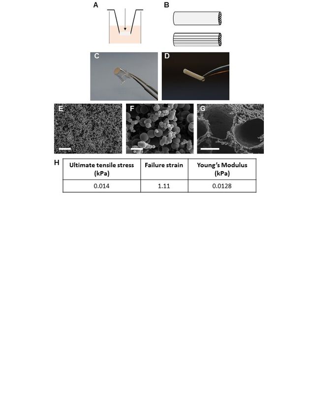
Biomaterials Science                                              Page 8 of 35

                                                                                                              Glia cells or myelinating cultures were plated onto Proliferate® constructs as described    above
                                                                                                                                                                                                           View Article Online
                                                                                                                                                                                                DOI: 10.1039/D0BM00097C

                                                                                                              or onto poly-L-lysine (PLL, Sigma-Aldrich) coated 13 mm glass coverslips (VWR) in 24-well
                                                                                                              plates. 20 ml PLL solution (13.3 μg/ml in dH2O) was used per 96 coverslips. 500 μl culture
                                                                                                              media was added to wells containing suspended construct inserts prior to cell seeding, and a
          This article is licensed under a Creative Commons Attribution-NonCommercial 3.0 Unported Licence.

                                                                                                              50-100 μl cell suspension was added and incubated for 1 h. Wells were then topped up with a

                                                                                                                                                                                                                                 Biomaterials Science Accepted Manuscript
                                                                                                              further 500 µl media. Cultures were fed by removing 500 µl media surrounding the insert and
                                                                                                              replacing with 500 µl media directly onto the insert.

                                                                                                              Implantation in vivo
Open Access Article. Published on 03 June 2020. Downloaded on 6/6/2020 2:17:14 AM.

                                                                                                              Contusion procedures were carried out on adult male Sprague-Dawley rats as previously
                                                                                                              described [19], with modifications to impact force and injury level as follows. Animals were
                                                                                                              anaesthetised with isoflurane and a laminectomy was performed to expose the spinal cord at
                                                                                                              the C6 level. A midline impact with a force of 175 kdyn was administered using an Infinite
                                                                                                              Horizon impactor (Precision Systems Instrumentation). The contusion site was marked by a
                                                                                                              10-0 Ethicon suture in the dura, the wound was closed and animals recovered in heated
                                                                                                              cabinets. Implantation of Proliferate® constructs was performed 3 weeks after contusion,
                                                                                                              allowing sufficient time for cavity formation. The injury site was re-opened and the contusion
                                                                                                              cavity identified by the marking suture. An incision was made in the dura at this site, revealing
                                                                                                              the fluid-filled cavity. Cavity fluid was aspirated with care using a blunted 23 G needle. The
                                                                                                              tubular construct containing parallel-aligned channels was cut to the size of the injury cavity
                                                                                                              and implanted in alignment with the cord. The material is compliant (sponge like) in
                                                                                                              consistency and so could be eased through this slit-like opening, displacing the edges of the
                                                                                                              dorsal columns to fill the cavity. Once this is done, the spared tissue of the dorsal columns
                                                                                                              spring back over the top of the biomaterial so that there is minimal scarring or contact of the
                                                                                                              biomaterial with the dura. Adherence problems and the potential to disrupt the integrity of the
                                                                                                              tissue containing the injury site and implants were therefore minimal due to the injury model
                                                                                                              and implant approach adopted. The dura was closed again over the injury, with a marking
                                                                                                              suture placed at the implantation site. In all procedures, animals received pre- and post-
                                                                                                              operative analgesics (buprenorphine, 0.05 mg/kg and carprofen, 5 mg/kg−1, s.c.). Saline (3–5
                                                                                                              ml) and enrofloxacin (5 mg/kg) were given s.c. for 3 and 7 days respectively following surgery.
                                                                                                              If motor impairment was detected following recovery, animals were excluded from the study.
                                                                                                              Animal numbers and experimental design is summarised in Fig 1. Twenty-eight animals were
                                                                                                              contused, of which 6 were excluded due to motor impairment or inadequate recovery. No
                                                                                                              significant difference was observed in animal weights at contusion and implantation
                                                                                                                                                                 8
Page 9 of 35                                                                                                                          Biomaterials Science

                                                                                                              procedures (Fig 4C, D), or in actual force and displacement achieved DOI:
                                                                                                                                                                                   (Fig10.1039/D0BM00097C
                                                                                                                                                                                           4E-H).
                                                                                                                                                                                             View Article Online

                                                                                                              Subsequently, 7 animals received P-C implants, of which 1 was excluded, and 9 received P-IK
                                                                                                              implants, of which 2 were excluded. Injury histology was assessed at short term (7 weeks) and
                                                                                                              long-term (6 months) time points.
          This article is licensed under a Creative Commons Attribution-NonCommercial 3.0 Unported Licence.

                                                                                                                                                                                                                   Biomaterials Science Accepted Manuscript
                                                                                                              Scanning electron microscopy [SEM]
                                                                                                              Proliferate® constructs were mounted onto specimen stubs (Agar Scientific) using conductive
                                                                                                              carbon tape and silver paint was applied to construct edges. Stubs were coated with 10-20 nm
                                                                                                              gold palladium using a Quorum Q150T high vacuum coater. To visualise cell monolayers
Open Access Article. Published on 03 June 2020. Downloaded on 6/6/2020 2:17:14 AM.

                                                                                                              grown on the construct, samples were prepared prior to coating using the following protocol.
                                                                                                              Cultures were fixed for 1 h at RT using 1.5% glutaraldehyde in 0.1 M sodium cacodylate buffer.
                                                                                                              Samples were then washed in 0.1 M sodium cacodylate buffer, before being incubated in 1%
                                                                                                              osmium tetroxide in sodium cacodylate buffer for 1 h. Samples were rinsed once more in buffer
                                                                                                              before washing 3x 10 min in dH2O, and incubating in 0.5% uranyl acetate in dH2O for 1 h,
                                                                                                              protected from light. Samples were then progressively dehydrated in the following ethanol
                                                                                                              concentrations: 30% (10 min), 50% (10 min), 70% (10 min), 90% (10 min), absolute (4 x 5
                                                                                                              min) and dried absolute (4 x 5 min). The dried absolute ethanol solution contained a 3A
                                                                                                              molecular sieve (Sigma-Aldrich) to remove moisture. A final drying step was then carried out
                                                                                                              in hexamethyldisilazane (HDMS) before mounting onto specimen stubs and coating as
                                                                                                              described. Samples were visualised on a JEOL6400 scanning electron microscope running at
                                                                                                              10 kV, and images were captured using the Olympus Scandium Software.

                                                                                                              Western blot
                                                                                                              Cells were lysed using CellLytic M containing protease inhibitor cocktail (Sigma-Aldrich), and
                                                                                                              protein concentrations were determined using a BCA protein assay (ThermoFisher) according
                                                                                                              to manufacturer’s instructions. Samples were run on tris-acetate gels and transferred using the
                                                                                                              iBlotTM gel transfer device (Invitrogen). Membranes were blocked in 5% milk in 0.2% Triton
                                                                                                              X-100 in TBS (TBS-T) for 1 h before being incubated with anti-GFAP (1:100,000, DAKO)
                                                                                                              for 2 h, and with anti-nestin (1:1000, Merck Millipore) overnight at 4°C. Blots were washed
                                                                                                              in TBS-T and incubated 1 h with secondary antibody (ECL rabbit IgG, ECL mouse IgG, GE
                                                                                                              Healthcare). The protein loading control used was GAPDH (1:1000, Abcam). Band intensities
                                                                                                              were quantified using Image-J and normalised to GAPDH.

                                                                                                              Immunocytochemistry
                                                                                                                                                             9
Biomaterials Science                                        Page 10 of 35

                                                                                                              Cultures were fixed in 4% paraformaldehyde (PFA, in PBS at PH 7.4) and permeabilised    with
                                                                                                                                                                                                     View Article Online
                                                                                                                                                                                          DOI: 10.1039/D0BM00097C

                                                                                                              0.1% triton X-100. Primary antibodies (GFAP anti-rabbit 1:1000, DAKO; nestin anti-mouse
                                                                                                              IgG1 1:500, SMI-31 anti-mouse IgG1 1:1000, both Merck Millipore; β-III-tubulin anti-rabbit
                                                                                                              1:1000, Abcam; PLP 1:00, hybridoma), were diluted in blocking buffer (0.2% gelatin (Sigma-
          This article is licensed under a Creative Commons Attribution-NonCommercial 3.0 Unported Licence.

                                                                                                              Aldrich) in PBS) and incubated for 1 h at RT. Samples were washed three times in PBS before

                                                                                                                                                                                                                           Biomaterials Science Accepted Manuscript
                                                                                                              incubation   with    fluorescent-conjugated      secondary      antibodies   (AlexaFluor   1:1000,
                                                                                                              ThermoFisher) for 45 min at RT. Coverslips were washed and mounted in an aqueous
                                                                                                              mounting medium (25 mg/ml ,4-diazabicyclo[2.2.2]octane (DABCO), Sigma-Aldrich)
                                                                                                              containing Hoechst 33342 nuclear dye (NucBlue™, ThermoFisher). Specimens were imaged
Open Access Article. Published on 03 June 2020. Downloaded on 6/6/2020 2:17:14 AM.

                                                                                                              using an Olympus BX51 fluorescent microscope.

                                                                                                              Histology
                                                                                                              Animals were deeply anaesthetised with intraperitoneal sodium pentobarbital (200 mg/ml
                                                                                                              Euthatal, Vericore) and transcardially perfused with mammalian Ringer solution containing
                                                                                                              0.1% lidocaine. Animals were subsequently perfused with 4% PFA in 0.1 M PBS (1 L PFA
                                                                                                              per animal). Spinal cord segments containing the implant were dissected, leaving
                                                                                                              approximately 10 mm tissue on either side of the injury site, and submerged in cryoprotective
                                                                                                              post-fixation solution (30% sucrose in 4% PFA) overnight. Tissue blocks were then submerged
                                                                                                              in 30% sucrose in PBS for 24 h, or until the tissue was observed to sink. Dura were removed,
                                                                                                              and cords were cut to 6-9 mm blocks containing the injury site, as determined by injury extent.
                                                                                                              Blocks were notched dorso-ventrally before being frozen in OCT. Sixty µm sections were cut
                                                                                                              sagittally using a cryotome at -20°C and incubated free-floating in 0.3M PBS.

                                                                                                              Sections were incubated in 50% ethanol for 30 min, washed 3x in PBS for 10 min, and
                                                                                                              incubated in primary antibodies (GFAP 1:1000, ThermoFisher; nestin anti-mouse IgG1 1:500,
                                                                                                              Merck Millipore; Laminin anti-rabbit 1:500, Sigma-Aldrich; ED-1 anti-mouse IgG1 1:500,
                                                                                                              BioRad; Neurofilament 200 anti-mouse IgG1 1:1000, Sigma-Aldrich) for 72 h at 4°C. Sections
                                                                                                              were washed a further 3x 10 min in PBS before incubation with secondary antibodies
                                                                                                              (AlexaFluor 1:1000, ThermoFisher) for 3 h at 4°C, protected from light. Sections were then
                                                                                                              washed 3x 10 min in PBS, mounted on glass slides in aqueous mounting. Sealed slides were
                                                                                                              stored at -20°C, protected from light.

                                                                                                              All sections were first examined using a Zeiss Axioplan epifluorescence microscope to identify
                                                                                                              sections with the largest cavity size, and to inspect immunolabeling. Representative sections
                                                                                                                                                               10
Page 11 of 35                                                                                                                             Biomaterials Science

                                                                                                              selected were considered the most medial injury sections. Selected illustrative sections    were
                                                                                                                                                                                                          View Article Online
                                                                                                                                                                                               DOI: 10.1039/D0BM00097C

                                                                                                              scanned with a Zeiss LSM 710 confocal system using X20 and X63 objective lenses. Laser
                                                                                                              excitation wavelengths used for scanning were 405, 488, 561 and 633 nm. Tissue sections were
                                                                                                              scanned as tiled composites of multiple fields at low (X20) and high (X63) magnification.
          This article is licensed under a Creative Commons Attribution-NonCommercial 3.0 Unported Licence.

                                                                                                              Sections were scanned throughout the stained tissue to accumulate a series of optical sections

                                                                                                                                                                                                                                Biomaterials Science Accepted Manuscript
                                                                                                              at 2-5 μm z separation. Stacked images were projected into 2D maximum intensity projections
                                                                                                              using Zeiss Zen software (Zeiss, Germany).

                                                                                                              Quantification of cavity extent
Open Access Article. Published on 03 June 2020. Downloaded on 6/6/2020 2:17:14 AM.

                                                                                                              Sagittal sections from each injury block were viewed under an epifluorescence microscope and
                                                                                                              rostral and caudal limits of the injury identified by the construct-tissue border signified by
                                                                                                              construct autofluorescence, or in control sections from cavity rims. Sections containing the
                                                                                                              most extensive sites (at least 3 per block) were selected for confocal imaging and the maximal
                                                                                                              injury length and height were measured using ZEN lite 2010 software (Zeiss). Injury site width
                                                                                                              was quantified by counting the number of sections containing the injury and multiplying by
                                                                                                              section thickness (60 µm).

                                                                                                              Statistical analysis
                                                                                                              All statistical analysis was carried out using GraphPad Prism software. A one-way ANOVA,
                                                                                                              with Dunnett’s or Tukey’s post hoc tests where appropriate, was used for null hypothesis
                                                                                                              statistical testing, where a p
Biomaterials Science                                               Page 12 of 35

                                                                                                                                                                                                              View Article Online
                                                                                                                                                                                                     DOI: 10.1039/D0BM00097C

                                                                                                              Astrocytes alter morphology and reactivity status on Proliferate® in vitro
                                                                                                              Astrocytic interactions with Proliferate® were investigated by culturing neurosphere-derived
                                                                                                              primary neonatal rat astrocytes on suspended Proliferate® cell culture inserts. These types of
          This article is licensed under a Creative Commons Attribution-NonCommercial 3.0 Unported Licence.

                                                                                                              astrocytes were used as they are a feature of the myelinating cultures used to study CNS cell

                                                                                                                                                                                                                                    Biomaterials Science Accepted Manuscript
                                                                                                              differentiation (Section 3.3). Cells were cultured on Proliferate® in carboxyl-functional (P-C,
                                                                                                              high cross-linkage), amine-functional (P-N, low cross-linkage) and IKVAV-coated (P-IK)
                                                                                                              forms. Astrocytes adopted a more fibrous, branched morphology on all forms of Proliferate®
                                                                                                              compared to those grown on PLL-coated coverslips as visualised both by GFAP and nestin
Open Access Article. Published on 03 June 2020. Downloaded on 6/6/2020 2:17:14 AM.

                                                                                                              labelling (Fig 3A) and scanning electron microscopy (SEM, Fig 3B). To investigate astrocyte
                                                                                                              reactivity on Proliferate®, GFAP and nestin expression in these cultures were quantified by
                                                                                                              Western blot as markers of astrocyte reactivity. GFAP upregulation is a well-established
                                                                                                              marker for reactivity in vitro and in vivo, but not a definitive one due to high expression in
                                                                                                              quiescence and heterogeneous expression across CNS tissue and in culture [38]. Nestin was
                                                                                                              therefore used as a supplementary reactivity marker, as GFAP-nestin co-expression is an
                                                                                                              indicator of astrocyte reactivity in vivo [39] and is characteristic of these cultures in vitro [40].
                                                                                                              No significant difference was observed in GFAP expression between astrocyte cultures on
                                                                                                              Proliferate® and controls, however nestin was significantly lower on P-C and P-N than on PLL-
                                                                                                              glass (Fig 3C). Nestin expression also appeared reduced on P-IK, although this difference was
                                                                                                              not significant.

                                                                                                              Neuron survival and neurite extension and myelination on Proliferate® in vitro
                                                                                                              To investigate the potential of Proliferate® for guiding axonal growth in the spinal cord,
                                                                                                              embryonic spinal cord suspensions were cultured on astrocyte monolayers (myelinating
                                                                                                              cultures, MCs) to examine the ability for cells to develop neurites and for myelination. Neurites
                                                                                                              were present at both 33 DIV (Fig 4A) and 40 DIV (Fig 4B). Myelination, as indicated by
                                                                                                              proteolipid protein (PLP, green) immunolabelling, was delayed on constructs compared with
                                                                                                              PLL-glass. Myelination, as seen by the formation of sheaths, is ordinarily observed on MCs at
                                                                                                              28 DIV [37], however this is not clearly observed until 33 DIV on P-IK, and 40 DIV on P-C
                                                                                                              and P-N (Fig 4A, B), showing that constructs can support myelination but that this is delayed
                                                                                                              compared with cultures on PLL-glass. Axonal density appears lower on constructs compared
                                                                                                              with PLL-glass, though this may be due to axonal dispersal through the greater surface area in
                                                                                                              Proliferate® constructs.

                                                                                                                                                               12
Page 13 of 35                                                                                                                          Biomaterials Science

                                                                                                              Proliferate® prevents long-term cavity expansion in vivo                                     View Article Online
                                                                                                                                                                                                  DOI: 10.1039/D0BM00097C

                                                                                                              Proliferate® was implanted into adult rat contusion spinal cord injuries in vivo. As no difference
                                                                                                              was observed in vitro in cell viability, growth and myelination between P-C and P-N, only P-
                                                                                                              C was implanted as the non-coated construct control, alongside the IKVAV-coated P-IK.
          This article is licensed under a Creative Commons Attribution-NonCommercial 3.0 Unported Licence.

                                                                                                              GFAP labelling revealed a distinct astrocytic border surrounding injury cavities and construct

                                                                                                                                                                                                                                 Biomaterials Science Accepted Manuscript
                                                                                                              implants in both implanted and non-implanted injuries (Fig 5) persistent at both 7 weeks (A-
                                                                                                              C) and 6 months post-implantation (D-F). At both time points, however, the GFAP cavity-
                                                                                                              tissue border in non-implanted injuries appears starker than that of implanted injuries, with
                                                                                                              some astrocytes appearing to enter scaffolds at the construct-tissue border in implanted cavities
Open Access Article. Published on 03 June 2020. Downloaded on 6/6/2020 2:17:14 AM.

                                                                                                              (highlighted in Fig 5A-Fii), suggesting that construct-tissue borders are more permissive to
                                                                                                              astrocyte growth and migration than cavity-tissue borders. GFAP delineation coupled with
                                                                                                              non-specific staining of Proliferate® by anti-nestin antibody reveals the full extent of cavities
                                                                                                              and implants (Fig 5B-C, E-F). Quantification of cavity extent (Fig 6) reveals no difference in
                                                                                                              cavity length 7 weeks post-implantation (Fig. 6C) compared with non-implanted control
                                                                                                              cavities, however significantly lower rostral-caudal cavity extremities (cavity length) in P-C
                                                                                                              and P-IK are observed in implanted injuries than in non-implanted injuries 6 months post-
                                                                                                              implantation (p < 0.05. Figure 6F). No significant difference is observed in lateral cavity
                                                                                                              extremities (cavity width) between implanted and non-implanted sections at either time point.
                                                                                                              The longitudinal measurement is more significant in this context as there is evidence that
                                                                                                              contusion cavities continue to expand in this plane for up to 6 months after injury whereas
                                                                                                              changes in width are minimal (J.S. Riddell & M. Hadian, unpublished observations). It is
                                                                                                              notable that channel integrity does not appear to be maintained in implants, with only few open
                                                                                                              channels visible on histological examination (highlighted by dashed lines).

                                                                                                              Proliferate® induces extensive vascularisation and cellular influx in vivo.
                                                                                                              Laminin labelling revealed extensive vascularisation throughout construct implants both 7
                                                                                                              weeks and 6 months post-implantation (Fig 7, Supplementary Fig 2). Clear laminin vessels are
                                                                                                              present throughout P-C and P-IK implanted constructs, abundantly surrounded by nuclei (Fig
                                                                                                              7). Notably, vascularisation and cellular influx occurred throughout constructs both in guidance
                                                                                                              channels and in construct-dense regions. Non-specific construct labelling by Hoechst 33342
                                                                                                              nuclear stain is observed, however nuclei are identifiable by morphology (Supplementary Fig
                                                                                                              3).

                                                                                                              Microglial response to Proliferate® in vivo
                                                                                                                                                             13
Biomaterials Science                                         Page 14 of 35

                                                                                                              To assess the immunological response to construct implants, microglia and macrophages      in
                                                                                                                                                                                                     View Article Online
                                                                                                                                                                                          DOI: 10.1039/D0BM00097C

                                                                                                              injury cavities and construct implants were visualised by anti-ED-1 labelling (green, Fig 8).
                                                                                                              Although microglia were present in constructs both centrally and at the construct-tissue border,
                                                                                                              they accounted for only a small population of Hoechst 33342 stained nuclei within and
          This article is licensed under a Creative Commons Attribution-NonCommercial 3.0 Unported Licence.

                                                                                                              surrounding constructs at both 7 weeks and 6 months post-implantation (Fig 8A,B). No clear

                                                                                                                                                                                                                           Biomaterials Science Accepted Manuscript
                                                                                                              difference was observed in microglial response between 7 week and 6-month time points (see
                                                                                                              Supplementary Fig 2). In non-implanted injuries, a distinct tissue-cavity border was observed
                                                                                                              through which microglia and other cells did not cross due to lack of matrix infilling acting as
                                                                                                              a cell support (Fig 8C). No clear difference was visible in microglial levels surrounding non-
Open Access Article. Published on 03 June 2020. Downloaded on 6/6/2020 2:17:14 AM.

                                                                                                              implanted injury cavities and construct-implanted injuries.

                                                                                                              Neurite growth into Proliferate®-implanted lesions
                                                                                                              Axonal outgrowth from perilesional zones into construct implants remains limited at both
                                                                                                              short- and long-term time points, as visualised by neurofilament labelling (Fig 9). No clear
                                                                                                              difference was observed in axonal growth into constructs between 7 week and 6-month time
                                                                                                              points. Inside constructs, axons remain sparse at both time points, and are limited to segments
                                                                                                              of undisrupted guidance channels (Fig 9A). At the construct tissue border, however, axons
                                                                                                              appear to grow out from perilesional zones into Proliferate® implants with regularity in
                                                                                                              segments with intact guidance channels providing spaces for entry (Fig 9B). Non-implanted
                                                                                                              injuries maintain a binary axonal border between tissue and injury cavity, with no axonal
                                                                                                              outgrowth observed (Fig 8C).

                                                                                                              Discussion
                                                                                                              Previously, we demonstrated in vitro the potential of aligned micropatterned ɛ-
                                                                                                              polycaprolactone (PCL) as an biomaterial for SCI [41, 42], however due to concerns pertaining
                                                                                                              to the stiffness disparity between these PCL scaffolds and the spinal cord, our findings had
                                                                                                              limited potential for in vivo application. We found the stiff PCL was hard to position into a
                                                                                                              lesion and did not appear to integrate well (unpublished observations). Therefore, pK based
                                                                                                              Proliferate® scaffolds were selected as an alternative implantable scaffold with material
                                                                                                              properties more closely comparable to spinal cord tissue. With no preconditioning, the Young’s
                                                                                                              modulus of the adult rat spinal cord has been estimated at 0.15-0.74 kPa [43]. Proliferate® in
                                                                                                              carboxyl-functional form has a Young’s modulus of 0.0128 kPa, comparable closely to these
                                                                                                              recent findings. We therefore concluded that Proliferate® is mechanically compatible with the

                                                                                                                                                              14
Page 15 of 35                                                                                                                            Biomaterials Science

                                                                                                              rat spinal cord and proceeded to test for corresponding CNS biocompatibility using    various
                                                                                                                                                                                                       View Article Online
                                                                                                                                                                                            DOI: 10.1039/D0BM00097C

                                                                                                              neural cell cultures.

                                                                                                              Astrocytes play a large role in the glial scar following SCI, and are highly responsive to
          This article is licensed under a Creative Commons Attribution-NonCommercial 3.0 Unported Licence.

                                                                                                              biomaterial topographies [41, 44]. On the three different Proliferate® substrates tested,

                                                                                                                                                                                                                             Biomaterials Science Accepted Manuscript
                                                                                                              astrocytes adopted a stellate, ramified morphology distinct from the flat, expansive polygonal
                                                                                                              morphology observed on flat PLL-glass coverslips. This morphology is typical of astrocytes in
                                                                                                              vivo [45], therefore suggests that Proliferate® surface topography is comparable to the 3D tissue
                                                                                                              milieu in which these cells inherently reside. The heterogeneously beaded surface topography
Open Access Article. Published on 03 June 2020. Downloaded on 6/6/2020 2:17:14 AM.

                                                                                                              of Proliferate® is more reminiscent of 3D spinal cord tissue than flat glass coverslips typically
                                                                                                              used for cell culture. Consistent with our findings, astrocytes adopt ramified morphologies on
                                                                                                              a range of other 3D biomaterials, including alginate, collagen type 1, collagen-hyaluronic acid
                                                                                                              (HA) and collagen-HA-matrigel hydrogels [46-48], polyurethane nanofibers [49] and
                                                                                                              hydrophobic tripalmitin [50]. It has been shown that when astrocytes are cultured in 3D hyrogel
                                                                                                              their morphology resembles perivascular astrocytes, but they also from round, stellate or
                                                                                                              bipolar shapes within a few days. The latter is induced by aligned microcolumns [47, 51-53],
                                                                                                              however the branched morphology is considered most representative of in vivo phenotypes
                                                                                                              [45]. Moreover, it has been shown that astrocytes form a reparative phenotype as assessed by
                                                                                                              reduced GFAP expression and fewer F‐/G‐actin stress fibres when plated on 3D electrospun
                                                                                                              bioscaffolds relative to 2D astrocytes [54]. The broad consensus in the field is that for better
                                                                                                              understanding of astrocytic behaviour, particularly with respect to the characterisation of the
                                                                                                              spectrum of astrocyte reactivity states, astrocyte culture systems must be optimised to promote
                                                                                                              representative in vivo astrocytic states [40, 55]. To this aim, other modifications of the culture
                                                                                                              environment, separate from substrate characteristics, have also been used to promote a ramified
                                                                                                              morphology. Media modifications, including the removal of serum from culture media, and
                                                                                                              immunopanning are one example of this, being reported to promote astrocyte phenotypes
                                                                                                              closer to those seen in vivo both morphologically and, in protein and gene expression profiles
                                                                                                              [56-58].

                                                                                                              No definitive protein markers of astrocyte reactivity have thus far been identified, though
                                                                                                              increased GFAP expression has long been the accepted hallmark [55, 59]. However, GFAP is
                                                                                                              largely expressed by quiescent astrocytes and astrocytes in vitro. Therefore, alongside
                                                                                                              quantification of GFAP in cultures on Proliferate® and PLL-glass, we also quantified nestin
                                                                                                              expression as an indicator of reactivity. Nestin is an intermediate filament protein
                                                                                                                                                               15
Biomaterials Science                                         Page 16 of 35

                                                                                                              predominantly expressed in neural development [60, 61], and re-expressed DOI:
                                                                                                                                                                                        by10.1039/D0BM00097C
                                                                                                                                                                                            reactive
                                                                                                                                                                                                View Article Online

                                                                                                              astrocytes [62, 63]. Astrocytic nestin expression is common in traditional cell culture
                                                                                                              conditions, however is absent in non-reactive spinal cord in vivo [39]. In traditional astrocyte
                                                                                                              culture, nestin expression is lower on non-coated Proliferate® (P-C, P-N) than on 2D PLL-
          This article is licensed under a Creative Commons Attribution-NonCommercial 3.0 Unported Licence.

                                                                                                              glass, indicating an altered astrocyte reactivity state on the construct reminiscent of in vivo

                                                                                                                                                                                                                        Biomaterials Science Accepted Manuscript
                                                                                                              quiescence. As the astrocyte-mediated glial scar remains one of the greatest barriers to CNS
                                                                                                              repair, the potential induction of a quiescent-like phenotype by Proliferate® could be viewed
                                                                                                              positively, however it is important to note that astrocyte reactivity and the glial scar are also
                                                                                                              implicated in several beneficial functions alongside detrimental ones [38].
Open Access Article. Published on 03 June 2020. Downloaded on 6/6/2020 2:17:14 AM.

                                                                                                              Neuronal morphological differences are also seen between PLL-glass and Proliferate®
                                                                                                              substrates. On flat glass surfaces, axons appear to grow largely in parallel outwards from cell
                                                                                                              body bundles dispersed throughout coverslips [37]. Conversely, on Proliferate® substrates cell
                                                                                                              bodies appear more evenly dispersed throughout the culture without bundling, and axonal
                                                                                                              orientation is no longer organised in parallel fashion. Axonal alignment in the spinal cord is
                                                                                                              important for physiological function, therefore for Proliferate® to be viable as an implantable
                                                                                                              material, the incorporation of guidance cues into the material may be crucial. Myelination was
                                                                                                              delayed in these cultures on Proliferate®, continuous with our previous findings on PCL [41].
                                                                                                              This may due to a lack of neurite organisation or a substrate-specific delay in oligodendrocyte
                                                                                                              maturation, as was postulated in our PCL study. This also may be due to the difference in
                                                                                                              stiffness between glass and proliferate which can also affect aspects of differentiation [64].
                                                                                                              Myelination was, however, observed on P-IK earlier than on P-C and P-N in our study.
                                                                                                              However the important observations is that all CNS glial cells can interact, differentiate and
                                                                                                              form myelin sheaths on Proliferate®.

                                                                                                              For implantation in vivo, Proliferate® was fabricated in tubular form containing parallel
                                                                                                              channels for more organised guidance. However, upon histological examination 7 weeks/6
                                                                                                              months following implantation, channels did not remain intact, with only remnants of the
                                                                                                              channel organisation visible at both time points. We postulate that due to the soft nature of
                                                                                                              Proliferate®, channel walls are prone to adhering and/or collapsing upon compression induced
                                                                                                              by surgical procedures and movement of the spinal column. We believe that by using soluble
                                                                                                              fibres of larger diameter to fabricate channels, channel integrity may be more consistently
                                                                                                              maintained. At 6 months following implantation, non-implanted injuries were more extensive,
                                                                                                              as indicated by higher rostral-caudal cavity lengths, than those implanted with Proliferate®.
                                                                                                                                                             16
Page 17 of 35                                                                                                                            Biomaterials Science

                                                                                                              This indicates stabilisation of the injury by adhesion of constructs to perilesional    tissue,
                                                                                                                                                                                                        View Article Online
                                                                                                                                                                                             DOI: 10.1039/D0BM00097C

                                                                                                              preventing excessive tissue dieback. As rostral-caudal cavity lengths were not significantly
                                                                                                              different in implanted than in non-implanted injuries at 7 weeks post-implantation, it suggests
                                                                                                              that this dieback is a chronic injury response. A distinct astrocytic border was observed
          This article is licensed under a Creative Commons Attribution-NonCommercial 3.0 Unported Licence.

                                                                                                              surrounding constructs in both short and long-term implantations, as is expected of typical non-

                                                                                                                                                                                                                              Biomaterials Science Accepted Manuscript
                                                                                                              implanted contusion injuries, however the reactivity status of these astrocytes is unclear. The
                                                                                                              prevalence of quiescent-like astrocytic phenotypes on Proliferate® substrates in vitro with low
                                                                                                              nestin expression could be indicative of a similar phenotype in vivo, however due to non-
                                                                                                              specific construct labelling by the nestin antibody, co-expression of GFAP and nestin could
Open Access Article. Published on 03 June 2020. Downloaded on 6/6/2020 2:17:14 AM.

                                                                                                              not be accurately quantified by immunohistochemistry. We did note, however, sporadic
                                                                                                              evidence of visible astrocytic movement from the perilesional zones into constructs in areas of
                                                                                                              channel integrity at the border with existing gaps for inward cellular movement. Similar
                                                                                                              astrocytic bordering has been observed surrounding collagen nanofiber implants in C3 rat
                                                                                                              hemisection injury [65] and T10 rat contusion injuries implanted with Poly(lactic-co-glycolic
                                                                                                              acid) (PLGA) and PCL [66]. Astrocytic ingrowth was, however, observed in collagen scaffold
                                                                                                              implanted T8-10 hemisection [67] and transection [68] injuries, and in poly (N-[2-
                                                                                                              hydroxypropyl] methacrylamide) hydrogel (NeuroGel™) implanted into T6-7 complete cat
                                                                                                              transections, where glial scar formation was attenuated [69].

                                                                                                              Evidently, several non-astrocytic cells do move into Proliferate® implants, as nuclei that are
                                                                                                              not associated with either GFAP or nestin are visible extensively throughout constructs at both
                                                                                                              7 weeks and 6 months post-implantation, as is extensive vascularisation. The presence of
                                                                                                              vascular structures across constructs lays the foundations for cell ingrowth. The identity of
                                                                                                              these cells, however, remains elusive. Microglia are one cell type ordinarily prevalent in SCI,
                                                                                                              however we found that microglia represent only a minority of cells in construct implants at
                                                                                                              both time points investigated, appearing in similar quantities both in the centre of constructs
                                                                                                              and at astrocyte-rich construct borders.

                                                                                                              The ultimate goal of construct implantation is to promote neuronal regeneration across the
                                                                                                              injury site. However, as previously discussed, guidance cues are paramount to organised axonal
                                                                                                              growth and alignment. Cell guidance channels are one example of such cues [11], however as
                                                                                                              previously mentioned, guidance channels incorporated into P-C and P-IK implants did not
                                                                                                              retain integrity once implanted. Organised axonal extension across injuries were therefore not
                                                                                                              possible. Congruently, axonal growth into construct implants was observed only in areas where
                                                                                                                                                               17
Biomaterials Science                                          Page 18 of 35

                                                                                                              intact channels existed. It was promising, however, that in such regions axonsDOI:were   able,
                                                                                                                                                                                                       View Article Online
                                                                                                                                                                                                 10.1039/D0BM00097C

                                                                                                              though in small number, to grow into constructs. In these areas, some axons were observed in
                                                                                                              construct centres as well as edges, particularly in implants maintained to 6 months.
          This article is licensed under a Creative Commons Attribution-NonCommercial 3.0 Unported Licence.

                                                                                                              Conclusion

                                                                                                                                                                                                                             Biomaterials Science Accepted Manuscript
                                                                                                              The therapeutic value of biomaterial implantation in SCI is multi-faceted. Our findings have
                                                                                                              shown a novel material, Proliferate®, to possess the necessary material properties for this
                                                                                                              purpose. We have demonstrated the compatibility of CNS cells with Proliferate® in vitro and
                                                                                                              in vivo, showing Proliferate®’s potential both as a solo implant, and as a base upon which other
Open Access Article. Published on 03 June 2020. Downloaded on 6/6/2020 2:17:14 AM.

                                                                                                              therapeutic compounds can be immobilised for delivery of other bioactive molecules to the
                                                                                                              injury site, for example molecules affecting the glial scar or facilitation of axonal regeneration.
                                                                                                              It will then be appropriate to investigate whether these mechanistic actions translate into
                                                                                                              tangible   improvements      in     functional   outcome     by   carrying   out   behavioural     or
                                                                                                              electrophysiological assessments.

                                                                                                              Author Contributions:
                                                                                                              S.H carried out all the in vitro work and in vivo analysis, S.L.L help with in vivo work and
                                                                                                              analysis. J.S.R carried out the animal model of spinal cord injury, S.C.B and M.O.R supervised
                                                                                                              the study and obtained funding. AGG and DAW provided the Proliferate® and supervised the
                                                                                                              biomaterial work. All authors reviewed and commented on the manuscript.

                                                                                                              Acknowledgments: This work was supported by the Medical Research Scotland (SH, grant
                                                                                                              numbers 718-2013-169822-91); the Multiple Sclerosis Society of Great Britain, (SLL grant
                                                                                                              number 56). The authors declare that they have no competing interests.

                                                                                                              Data Availability:
                                                                                                              The raw/processed data required to reproduce these findings cannot be shared at this time due
                                                                                                              to technical or time limitations.
                                                                                                              Conflict of Interest: Proliferate®, based polymer is marketed by SpheriTech but there are no
                                                                                                              financial contribution between the University of Glasgow and SpheriTech.

                                                                                                              References
                                                                                                              1.      Ahuja, C.S., et al., Traumatic spinal cord injury. Nature reviews Disease primers,
                                                                                                                      2017. 3: p. 17018.

                                                                                                                                                                18
Page 19 of 35                                                                                                                            Biomaterials Science

                                                                                                              2.    Tran, A.P., P.M. Warren, and J. Silver, The biology of regeneration failure DOI:
                                                                                                                                                                                                  and10.1039/D0BM00097C
                                                                                                                                                                                                      success
                                                                                                                                                                                                           View Article Online

                                                                                                                    after spinal cord injury. Physiological reviews, 2018. 98(2): p. 881-917.
                                                                                                              3.    Richardson, P.M., U.M. McGuinness, and A.J. Aguayo, Axons from CNS neurones
                                                                                                                    regenerate into PNS grafts. Nature, 1980. 284(5753): p. 264.
                                                                                                              4.    David, S. and A.J. Aguayo, Axonal elongation into peripheral nervous system"
                                                                                                                    bridges" after central nervous system injury in adult rats. Science, 1981. 214(4523):
          This article is licensed under a Creative Commons Attribution-NonCommercial 3.0 Unported Licence.

                                                                                                                    p. 931-933.
                                                                                                              5.    Richardson, P.M., U.M. McGuinness, and A.J. Aguayo, Peripheral nerve autografts to

                                                                                                                                                                                                                                 Biomaterials Science Accepted Manuscript
                                                                                                                    the rat spinal cord: studies with axonal tracing methods. Brain research, 1982.
                                                                                                                    237(1): p. 147-162.
                                                                                                              6.    Richardson, P.M., V.M.K. Issa, and A.J. Aguayo, Regeneration of long spinal axons in
                                                                                                                    the rat. Journal of neurocytology, 1984. 13(1): p. 165-182.
Open Access Article. Published on 03 June 2020. Downloaded on 6/6/2020 2:17:14 AM.

                                                                                                              7.    Bray, G.M., et al., The use of peripheral nerve grafts to enhance neuronal survival,
                                                                                                                    promote growth and permit terminal reconnections in the central nervous system of
                                                                                                                    adult rats. Journal of Experimental Biology, 1987. 132(1): p. 5-19.
                                                                                                              8.    Tabakow, P., et al., Transplantation of autologous olfactory ensheathing cells in
                                                                                                                    complete human spinal cord injury. Cell transplantation, 2013. 22(9): p. 1591-1612.
                                                                                                              9.    Côté, M.-P., et al., Peripheral nerve grafts support regeneration after spinal cord
                                                                                                                    injury. Neurotherapeutics, 2011. 8(2): p. 294-303.
                                                                                                              10.   Liu, S., et al., Biomaterial-supported cell transplantation treatments for spinal cord
                                                                                                                    injury: challenges and perspectives. Frontiers in cellular neuroscience, 2018. 11: p.
                                                                                                                    430.
                                                                                                              11.   Haggerty, A.E. and M. Oudega, Biomaterials for spinal cord repair. Neuroscience
                                                                                                                    bulletin, 2013. 29(4): p. 445-459.
                                                                                                              12.   Straley, K.S., C.W.P. Foo, and S.C. Heilshorn, Biomaterial design strategies for the
                                                                                                                    treatment of spinal cord injuries. Journal of neurotrauma, 2010. 27(1): p. 1-19.
                                                                                                              13.   Ziemba, A.M. and R.J. Gilbert, Biomaterials for local, controlled drug delivery to the
                                                                                                                    injured spinal cord. Frontiers in pharmacology, 2017. 8: p. 245.
                                                                                                              14.   Norenberg, M.D., J. Smith, and A. Marcillo, The pathology of human spinal cord
                                                                                                                    injury: defining the problems. 2004, Mary Ann Liebert, Inc.
                                                                                                              15.   Tator, C.H., Update on the pathophysiology and pathology of acute spinal cord injury.
                                                                                                                    Brain pathology, 1995. 5(4): p. 407-413.
                                                                                                              16.   Milhorat, T.H., et al., Pathological basis of spinal cord cavitation in syringomyelia:
                                                                                                                    analysis of 105 autopsy cases. Journal of neurosurgery, 1995. 82(5): p. 802-812.
                                                                                                              17.   Ziegler, G., et al., Progressive neurodegeneration following spinal cord injury:
                                                                                                                    implications for clinical trials. Neurology, 2018. 90(14): p. e1257-e1266.
                                                                                                              18.   Toft, A., et al., Electrophysiological evidence that olfactory cell transplants improve
                                                                                                                    function after spinal cord injury. Brain, 2007. 130(4): p. 970-984.
                                                                                                              19.   Lindsay, S.L., et al., Human olfactory mesenchymal stromal cell transplants promote
                                                                                                                    remyelination and earlier improvement in gait co-ordination after spinal cord injury.
                                                                                                                    Glia, 2017. 65(4): p. 639-656.
                                                                                                              20.   Amr, S.M., et al., Bridging defects in chronic spinal cord injury using peripheral nerve
                                                                                                                    grafts combined with a chitosan-laminin scaffold and enhancing regeneration
                                                                                                                    through them by co-transplantation with bone-marrow-derived mesenchymal stem
                                                                                                                    cells: Case series of 14 patients. The journal of spinal cord medicine, 2014. 37(1): p.
                                                                                                                    54-71.

                                                                                                                                                               19
Biomaterials Science                                              Page 20 of 35

                                                                                                              21.   Cao, J., et al., The use of laminin modified linear ordered collagen scaffolds  loaded
                                                                                                                                                                                                 DOI:
                                                                                                                                                                                                            View Article Online
                                                                                                                                                                                                      10.1039/D0BM00097C
                                                                                                                    with laminin-binding ciliary neurotrophic factor for sciatic nerve regeneration in rats.
                                                                                                                    Biomaterials, 2011. 32(16): p. 3939-3948.
                                                                                                              22.   Hou, S., et al., The repair of brain lesion by implantation of hyaluronic acid hydrogels
                                                                                                                    modified with laminin. Journal of neuroscience methods, 2005. 148(1): p. 60-70.
                                                                                                              23.   Woerly, S., et al., Spinal cord repair with PHPMA hydrogel containing RGD peptides
          This article is licensed under a Creative Commons Attribution-NonCommercial 3.0 Unported Licence.

                                                                                                                    (NeuroGel™). Biomaterials, 2001. 22(10): p. 1095-1111.
                                                                                                              24.   Hejčl, A., et al., HPMA-RGD hydrogels seeded with mesenchymal stem cells improve

                                                                                                                                                                                                                                  Biomaterials Science Accepted Manuscript
                                                                                                                    functional outcome in chronic spinal cord injury. Stem cells and development, 2010.
                                                                                                                    19(10): p. 1535-1546.
                                                                                                              25.   Tysseling-Mattiace, V.M., et al., Self-assembling nanofibers inhibit glial scar
                                                                                                                    formation and promote axon elongation after spinal cord injury. Journal of
Open Access Article. Published on 03 June 2020. Downloaded on 6/6/2020 2:17:14 AM.

                                                                                                                    Neuroscience, 2008. 28(14): p. 3814-3823.
                                                                                                              26.   Wei, Y.T., et al., Hyaluronic acid hydrogels with IKVAV peptides for tissue repair and
                                                                                                                    axonal regeneration in an injured rat brain. Biomedical Materials, 2007. 2(3): p.
                                                                                                                    S142.
                                                                                                              27.   Park, J., et al., Nerve regeneration following spinal cord injury using matrix
                                                                                                                    metalloproteinase-sensitive, hyaluronic acid-based biomimetic hydrogel scaffold
                                                                                                                    containing brain-derived neurotrophic factor. Journal of Biomedical Materials
                                                                                                                    Research Part A: An Official Journal of The Society for Biomaterials, The Japanese
                                                                                                                    Society for Biomaterials, and The Australian Society for Biomaterials and the Korean
                                                                                                                    Society for Biomaterials, 2010. 93(3): p. 1091-1099.
                                                                                                              28.   Tysseling, V.M., et al., Self-assembling peptide amphiphile promotes plasticity of
                                                                                                                    serotonergic fibers following spinal cord injury. Journal of neuroscience research,
                                                                                                                    2010. 88(14): p. 3161-3170.
                                                                                                              29.   Kazemi, S., et al., IKVAV-linked cell membrane-spanning peptide treatment induces
                                                                                                                    neuronal reactivation following spinal cord injury. Future science OA, 2015. 1(4).
                                                                                                              30.   Gallagher, A.G., et al., A novel peptide hydrogel for an antimicrobial bandage contact
                                                                                                                    lens. Advanced healthcare materials, 2016. 5(16): p. 2013-2018.
                                                                                                              31.   Gallagher, A.G., et al., Development of a Poly-ε-Lysine Contact Lens as a Drug
                                                                                                                    Delivery Device for the Treatment of Fungal Keratitis. Investigative ophthalmology &
                                                                                                                    visual science, 2017. 58(11): p. 4499-4505.
                                                                                                              32.   Kennedy, S., et al., Poly-ε-lysine based hydrogels as synthetic substrates for the
                                                                                                                    expansion of corneal endothelial cells for transplantation. Journal of Materials
                                                                                                                    Science: Materials in Medicine, 2019. 30(9): p. 102.
                                                                                                              33.   Kadler, B., A 3D in vitro co-culture to model peripheral nerve myelination using
                                                                                                                    functionalised poly(ε -lysine) scaffolds., in Faculty of Medical and Human Sciences.
                                                                                                                    2015, The University of Manchester. p. 119.
                                                                                                              34.   Negah, S.S., et al., Laminin-derived Ile-Lys-Val-ala-Val: a promising bioactive peptide
                                                                                                                    in neural tissue engineering in traumatic brain injury. Cell and tissue research, 2018.
                                                                                                                    371(2): p. 223-236.
                                                                                                              35.   Patel, R., et al., Ile-Lys-Val-ala-Val (IKVAV) peptide for neuronal tissue engineering.
                                                                                                                    Polymers for Advanced Technologies, 2019. 30(1): p. 4-12.
                                                                                                              36.   Hosseinkhani, H., et al., Engineering three-dimensional collagen-IKVAV matrix to
                                                                                                                    mimic neural microenvironment. ACS chemical neuroscience, 2013. 4(8): p. 1229-
                                                                                                                    1235.

                                                                                                                                                                20
You can also read