SPIKE1 Activates the GTPase ROP6 to Guide the Polarized Growth of Infection Threads in Lotus japonicus OPEN - Plant Cell
←
→
Page content transcription
If your browser does not render page correctly, please read the page content below
The Plant Cell, Vol. 32: 3774–3791, December 2020, www.plantcell.org ã 2020 ASPB.

SPIKE1 Activates the GTPase ROP6 to Guide the Polarized
Growth of Infection Threads in Lotus japonicus OPEN
                                              [     ]

Jing Liu,a,b Miao Xia Liu,a Li Ping Qiu,a and Fang Xiea,1
a National Key Laboratory of Plant Molecular Genetics, CAS Center for Excellence in Molecular Plant Sciences, Shanghai Institute of

Plant Physiology and Ecology, Chinese Academy of Sciences, Shanghai 200032, China
b University of the Chinese Academy of Sciences, Beijing 100864, China

ORCID IDs: 0000-0002-5092-7627 (J.L.); 0000-0002-5044-4457 (M.X.L.); 0000-0003-4297-9467 (L.P.Q.); 0000-0002-0530-1430 (F.X.)

In legumes, rhizobia attach to root hair tips and secrete nodulation factor to activate rhizobial infection and nodule
organogenesis. Endosymbiotic rhizobia enter nodule primordia via a specialized transcellular compartment known as the
infection thread (IT). The IT elongates by polar tip growth, following the path of the migrating nucleus along and within the root
hair cell. Rho-family ROP GTPases are known to regulate the polarized growth of cells, but their role in regulating polarized IT
growth is poorly understood. Here, we show that LjSPK1, a DOCK family guanine nucleotide exchange factor (GEF), interacts
with three type I ROP GTPases. Genetic analyses showed that these three ROP GTPases are involved in root hair
development, but only LjROP6 is required for IT formation after rhizobia inoculation. Misdirected ITs formed in the root hairs
of Ljspk1 and Ljrop6 mutants. We show that LjSPK1 functions as a GEF that activates LjROP6. LjROP6 enhanced the plasma
membrane localization LjSPK1 in Nicotiana benthamiana leaf cells and Lotus japonicus root hairs, and LjSPK1 and LjROP6
interact at the plasma membrane. Taken together, these results shed light on how the LjROP6-LjSPK1 module mediates the
polarized growth of ITs in L. japonicus.

INTRODUCTION                                                                   primordium via an unwalled droplet (Brewin, 2004). This leads to
                                                                               the formation of a nitrogen-fixing root nodule that provides the
To form symbiotic root nodules, legumes and rhizobia initiate their            proper microenvironment for nitrogen fixation by rhizobia and
symbiotic interaction via a molecular dialog. The host legume                  nutrient exchange between the two partners (Jones et al., 2007;
secretes flavonoid compounds that function as signals sensed by                 Oldroyd, 2013; Roy et al., 2020).
rhizosphere rhizobia, and the expression of nodulation (Nod)                      Genetic studies of L. japonicus and M. truncatula have revealed
genes is induced for the biosynthesis and secretion of nodulation              several genes that are required for IT formation, including LjNAP/
factors (NF)s. NFs are lipochito-oligosaccharide molecular signals             MtRIT1, LjPIR, LjSCARN, and LjARPC1. These genes encode
that are perceived by NF receptors. NFs initiate a series of plant             components of the WAVE/SCAR-ARP2/3 complex, which is re-
signaling and developmental events, including root hair de-                    quired for actin nucleation and cytoskeletal rearrangement.
formation, membrane depolarization, intracellular calcium oscil-               LjNPL/MtNPL encode a legume-specific pectate lyase involved in
lations, and cortex cell division. These events lead to the formation          cell wall remodeling. Mutant analyses revealed some genes that
of the nodule primordium (Ehrhardt et al., 1992; Downie and                    are required for rhizobial infection, but their precise biological/
Walker, 1999; Timmers et al., 1999; Cullimore et al., 2001; Esseling           biochemical functions are unknown. These genes include
et al., 2003; Oldroyd and Downie, 2008).                                       LjRINRK1, encoding an atypical leucine-rich repeat receptor-like
   In temperate legumes such as Lotus japonicus and Medicago                   kinase; LjCERBERUS/MtLIN, encoding a U-box and WD40-
truncatula, rhizobia attach to the host’s root hair tips and redirect          repeat domain protein; MtVAPYRIN, encoding a VAP/MSP and
root hair growth to entrap the rhizobia within an infection chamber            ankyrin-repeats domain protein; MtRPG, encoding a coiled-coil
(Esseling et al., 2003; Murray, 2011; Fournier et al., 2015). Within           protein; and MtCBS1, encoding a DUF21 and cystathionine-
the root hair chamber, an infection pocket filled with bacteria forms           b-synthase domain protein (Arrighi et al., 2008; Kiss et al., 2009;
from a tubular invagination of the cell wall and membrane; this                Yano et al., 2009; Yokota et al., 2009; Miyahara et al., 2010; Murray
structure is known as an infection thread (IT). The IT extends from            et al., 2011; Hossain et al., 2012; Xie et al., 2012; Qiu et al., 2015;
the infection chamber down through the root hairs to the root                  Sinharoy et al., 2016; Li et al., 2019; Liu et al., 2019a).
cortex, where a nodule primordium is produced (Fournier et al.,                   The IT initiates and extends from the root hair tip to the nodule
2008; Fournier et al., 2015). The bacteria grow, divide, and fill the           primordium. This process is directional, indicating that ITs show
IT. The bacteria are then released from the IT into the nodule                 polarized growth. The polarized growth of pollen tubes and root
                                                                               hairs requires a strong calcium gradient at the tip, a polarized actin
1 Address     correspondence to fxie@cemps.ac.cn.                              cytoskeleton, tip-directed vesicle trafficking, exocytosis, and
The author responsible for distribution of materials integral to the findings   signaling by ROP (Rho of plants) GTPases (Samaj et al., 2006;
presented in this article in accordance with the policy described in the       Kost, 2008; Craddock and Yang, 2012; Craddock et al., 2012). The
Instructions for Authors (www.plantcell.org) is: Fang Xie (fxie@cemps.
                                                                               LIN-VAPYRIN-Exo70H4 protein complex in M. truncatula local-
ac.cn).
[OPEN]
       Articles can be viewed without a subscription.                          izes to the tips of elongated pre-ITs, suggesting that an exocytosis
www.plantcell.org/cgi/doi/10.1105/tpc.20.00109                                 process may be involved in the polarized growth of ITs (Liu et al.,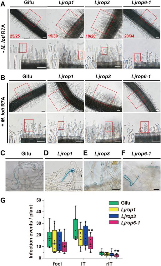
SPK1-ROP6 Mediates Polarized Growth of ITs         3775

2019b). Type I ROP GTPases (including ROP1 to ROP8) play                        Although ITs show polar growth, it is unknown whether a ROP
important roles in root hair and pollen tube tip growth in Arabi-            GTPase regulates this process and, if so, how this ROP GTPase is
dopsis (Arabidopsis thaliana; Kost et al., 1999; Li et al., 1999;            activated. Here, we show that LjSPK1 interacts with LjROP6 and
Jones et al., 2002; Eklund et al., 2010; Craddock et al., 2012). The         activates its ROP GTPase activity. Some ITs were found to be
constitutively active (CA) forms of AtROP4 and AtROP6 do not                 misdirected in the root hairs of the Ljspk1 and Ljrop6 mutants after
function in the polarized growth of root hairs, nor do they form             rhizobial infection. Our results suggest that an LjROP6-LjSPK1
a localized Ca21 gradient at the root hair tip (Molendijk et al., 2001).     signaling module guides the direction of IT growth during the early
Compared to the wild type, plants expressing CA AtROP2 pro-                  stage of rhizobial infection in L. japonicus.
duce additional and misplaced root hairs on the cell surface as well
as longer root hairs, whereas plants expressing dominant-
negative (DN) forms of AtROP2 produce shortened wavy root                    RESULTS
hair (Jones et al., 2002).
   MtROP10 is associated with the NF receptor MtNFP and is                   Expression Pattern of SPK1 in L. japonicus
involved in regulating the polarized growth of root hairs and NF-
                                                                             SPK1 is a DOCK family GEF that activates ROP GTPases (Basu
induced root hair deformation in M. truncatula (Lei et al., 2015). The
                                                                             et al., 2008). Using BLAST tools, we searched for the SPK1 gene in
roles of ROP genes in M. truncatula have been clarified to some
                                                                             the L. japonicus genome. This search identified LjSPK1 (Lotja-
extent by knockdown RNA interference (RNAi) studies. Compared
                                                                             Gi3g1v0299000), which encodes an 1829-amino acid protein
to the wild type, MtROP8 RNAi transgenic plants show larger
                                                                             showing 79.2% identity with AtSPK1. Phylogenetic analysis re-
numbers of infection events and increased nodulation in response
                                                                             vealed that SPK1 is conserved across eukaryotes, usually as
to inoculation, and MtROP9 RNAi plants show enhanced mycor-
                                                                             a single copy gene, with one exception being soybean (Glycine
rhizal and early hyphal root colonization but reduced rhizobial in-
                                                                             max), which has undergone a recent whole-genome duplication
fection (Kiirika et al., 2012; Wang et al., 2014). In L. japonicus, the
                                                                             (Schmutz et al., 2010; Supplemental Figure 1A). Sequence
Nod factor receptor LjNFR5 interacts with LjROP6 to regulate IT
                                                                             analyses revealed three conserved domains in LjSPK1: DHR1
formation and nodulation (Ke et al., 2012), and LjROP6 interacts with
                                                                             (amino acids 446 to 726); DHR2 (amino acids 1299 to 1829), which
LjCHC1, a central component in clathrin-mediated endocytosis, to
                                                                             is the GEF catalytic domain; and DHR3 (amino acids 140 to 287;
regulate rhizobial infection and nodule formation (Wang et al., 2015).
                                                                             Supplemental Figure 1B). To investigate the transcriptional pat-
These findings demonstrate that LjROP6 plays an essential role in
                                                                             tern of LjSPK1 during nodulation, we monitored the transcript
regulating legume-rhizobium symbiosis. However, it is unknown
                                                                             levels of LjSPK1 after NF treatment by RT-qPCR. LjSPK1 tran-
whether ROP GTPase functions in the polarized growth of ITs.
                                                                             script levels slightly increased at 6 and 12 h after NF inoculation
   ROP GTPases act as molecular switches in various signaling
                                                                             versus the 0 h control (Figure 1A).
pathways by cycling between the GTP-bound active and GDP-
                                                                                We analyzed the spatial expression pattern of LjSPK1 by
bound inactive forms (Yang, 2002). The shuttling between the
                                                                             monitoring b-glucuronidase (GUS) expression from the GUS gene
active and inactive forms of ROP GTPases is regulated by three
                                                                             driven by the LjSPK1 promoter (pLjSPK1:GUS) in L. japonicus
types of factors: RhoGEF (guanine nucleotide exchange factor),
                                                                             transgenic hairy roots. In the absence of rhizobial inoculation, we
which controls the transition of these GTPase from the inactive
                                                                             detected GUS signals in root vascular tissues and lateral roots
(GDP-bound) to active (GTP-bound) form; RhoGAP (GTPase-
                                                                             (Figures 1B and 1C). However, after inoculation with Meso-
activating protein), which accelerates GTP hydrolysis; and
                                                                             rhizobium loti R7A/lacZ, GUS signals were present in the dividing
RhoGDI (guanine nucleotide dissociation inhibitor), which inhibits
                                                                             cortex region (Figure 1D), nodule primordia (Figure 1E), bumps
GDP release so that ROP GTPases remain in their inactive state
                                                                             (Figure 1F), young nodules (Figure 1G), and vascular bundles of
(Yang, 2002; Kost, 2008). The M. truncatula genome contains 10
                                                                             mature nodules (Figure 1H). Light microscopy analyses of sec-
genes encoding plant Rop nucleotide exchanger-type RopGEFs.
                                                                             tions of these nodules revealed GUS signals in bump and young
MtRopGEF2 affects the cytosolic Ca21 gradient and the sub-
                                                                             nodule cells (Figures 1I and 1J), but not in the nitrogen-fixation
cellular architecture of root hairs and is required for root hair
                                                                             zone of mature nodules (Figure 1K). These expression patterns
development (Riely et al., 2011).
                                                                             suggest that LjSPK1 might be involved in root nodule symbiosis in
   In addition to plant Rop nucleotide exchanger-type RopGEFs,
                                                                             L. japonicus.
a single DOCK family GEF, SPIKE1 (SPK1), is also present in
plants. In Arabidopsis, spk1 seedlings show a lethal phenotype
and display serious defects in the polarized growth of cotyledons
                                                                             LjSPK1 Is Required for Polarized Growth in Plants and
and leaf epidermal cells (Qiu et al., 2002). The AtSPK1 DOCK ho-
                                                                             Rhizobial Infection
mology region 2 (DHR2) domain binds to ROPs to facilitate nucle-
otide exchange and generates signals to activate two heteromeric             To explore the roles of LjSPK1 in root nodule symbiosis, we
complexes, WAVE/SCAR and ARP2/3, to control actin polymeri-                  obtained two LORE1 insertion lines (30,019,873 and 30,051,253)
zation (Basu et al., 2008). AtSPK1 is involved in activating the             in which LjSPK1 had insertions at amino acid positions 273 and
AtROP6-AtRIC1 pathway, which regulates the auxin-mediated in-                910 from the start codon, respectively. These lines were desig-
ternalization of AtPIN2 (Lin et al., 2012). In rice (Oryza sativa), OsSPK1   nated as Ljspk1-1 (30,019,873) and Ljspk1-2 (30,051,253;
interacts with OsPit, an R protein against rice blast fungus, and            Supplemental Figure 1B). RT-qPCR analysis revealed that en-
regulates the activation of OsRac1 to trigger the immune response            dogenous LjSPK1 transcript levels were significantly reduced in
(Wang et al., 2018).                                                         both Ljspk1 lines (Supplemental Figure 1C). For both Ljspk1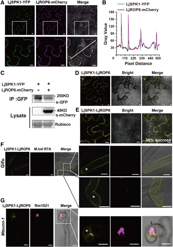
3776       The Plant Cell

Figure 1. LjSPK1 Is Induced by Nod Factors and Is Specifically Expressed in Young Nodules.

(A) RT-qPCR analysis of LjSPK1 transcript levels in roots of wild-type L. japonicus (Gifu) after inoculation with purified Nod factor (0 h, 6 h, 12 h). Expression is
relative to that in mock-treated samples (0 h) and normalized to that of Lotus Ubiquitin. Mean and SD were derived from two biological replicates. Statistical
significance (*P < 0.05) was evaluated by Student’s t test.
(B) and (C) pLjSPK1:GUS expression pattern in wild-type roots without rhizobial inoculation.
(D) to (H) pLjSPK1:GUS expression pattern in wild-type roots after inoculation with M. loti R7A expressing lacZ. GUS activity was detected in nodule
primordia and young nodules (D) to (G) and in vascular bundles of mature nodules (H).
(I) to (K) Section of nodules showing that pLjSPK1:GUS expression in all cell layers in young nodules (I) and (J), but not in mature nodules’ nitrogen-fixation
zone (K).
Transgenic roots were costained with X-Gluc and magenta-Gal to visualize pLjSPK1:GUS expression (in blue) and M. loti (in purple), respectively. Bars 5 100
mm in (B) to (H) and 1 mm in (I) to (K).

alleles, the homozygotes were nearly completely sterile, so we                      number of infection events per centimeter (cm). The number of
could only use seedlings obtained from the segregation of het-                      infection foci and ITs per-cm root did not differ significantly be-
erozygous plants for phenotypic analyses. Soil-grown Ljspk1                         tween the wild type and the Ljspk1 mutants except that Ljspk1-2
mutants survived but showed some growth defects, including                          had fewer rITs (Figure 2F). However, both Ljspk1 mutants had
severe dwarfism of adult plants (Supplemental Figure 1D), few and                    more abnormal infection events than the wild type and Ljspk1
smaller flowers with an abnormal petal shape (Supplemental                           siblings (Figure 2F). These results suggest that the infection events
Figure 1E), and smaller, dark-green leaves (Supplemental Figures                    were not significantly affected, but the polarized growth of ITs was
1F and 1H). Wild-type plants formed elongated, filamentous tri-                      markedly affected in the Ljspk1 mutants. At 2 weeks after in-
chomes on the abaxial midribs of leaves, whereas the Ljspk1                         oculation, the wild type had produced abundant pink mature
mutants formed fewer and smaller trichomes (Supplemental                            nodules, while the Ljspk1 mutants had produced fewer nodules
Figure 1H). Scanning electron microscopy of epidermal cells on                      per plant (Supplemental Figures 2G, 2J, and 2K). Light microscopy
the dorsal leaves of wild-type (Gifu) and Ljspk1 mutants revealed                   of semithin sections of nodules stained with toluidine-blue re-
that wild-type pavement cells had a clear neck and lobe, whereas                    vealed that Ljspk1-1 nodules had fewer infected cells than the wild
those on Ljspk1 leaves were nearly round (Supplemental Figures                      type and Ljspk1-1 siblings’ nodules (Supplemental Figure 2H).
1I and 1J). The phenotypes of these Ljspk1 mutants were similar to                     Next, we explored the role of LjSPK1 by generating LjSPK1-
those reported for the Arabidopsis spk1-1 mutant (Qiu et al., 2002).                overexpressing lines (LjSPK1-OX) and lines with knocked-down
These results suggest that LjSPK1 is involved in the polarized                      LjSPK1 expression (LjSPK1-Ri). The LjSPK1-knockdown lines
growth of cells in L. japonicus.                                                    were generated by introducing an RNAi vector and the LjSPK1-OX
   We analyzed the infection and nodulation phenotypes of the                       lines were generated by introducing the LjSPK1 cDNA driven by
Ljspk1 mutants following inoculation with M. loti R7A constitutively                the L. japonicus Ubiquitin promoter into wild-type L. japonicus
expressing GFP or lacZ. Wild-type (Gifu) and Ljspk1 wild-type sib-                  hairy roots. Compared with empty vector (EV) control, the LjSPK1-
lings (sibling) produced normal elongated ITs in curled root hairs                  Ri lines had markedly fewer infection events, but the LjSPK1-OX
(Figure 2A; Supplemental Figures 2A, 2B, and 2D). In the Ljspk1                     lines had increased numbers of infection events, including in-
mutants, infection initiated normally from a curled root hair, but some             fection foci and ITs (Figures 2G and 2H). The LjSPK1-Ri lines had
ITs showed anomalous growth patterns, including the formation of                    shorter roots than the EV control (Supplemental Figure 2L).
sac-like structures or depolarized loop shapes inside the root hairs                Analysis of the infection events revealed that both the LjSPK1-Ri
(Figures 2B to 2D; Supplemental Figures 2C and 2E). We counted the                  and LjSPK1-OX lines had more abnormal infection events per cm
total number of infection events in the Ljspk1 mutants 1 week after                 of root than the EV control (Figure 2I). The phenotype of LjSPK1-Ri
inoculation. The number of infection events including infection foci                was similar to that of the Ljspk1 LORE1 insertion mutants. RT-
(foci), ITs in root hairs (IT), and ITs extending into cortex cells (rIT) was       qPCR analysis revealed that LjSPK1 transcript levels were sig-
significantly lower in the Ljspk1 mutants than in the wild type. In                  nificantly lower in the LjSPK1-Ri lines and significantly higher in the
addition, there were significantly more abnormal ITs (abnormal in                    LjSPK1-OX lines compared to the control (Supplemental
Figure 2) in the Ljspk1 mutants than in the wild type (Figure 2E).                  Figure 2M). Together, these results demonstrate that LjSPK1 is
   Because the Ljspk1 mutants had shorter primary roots than the                    required for the rhizobial infection process and is involved in the
wild type (Supplemental Figures 2F and 2I), we calculated the                       polarized growth of ITs in L. japonicus.
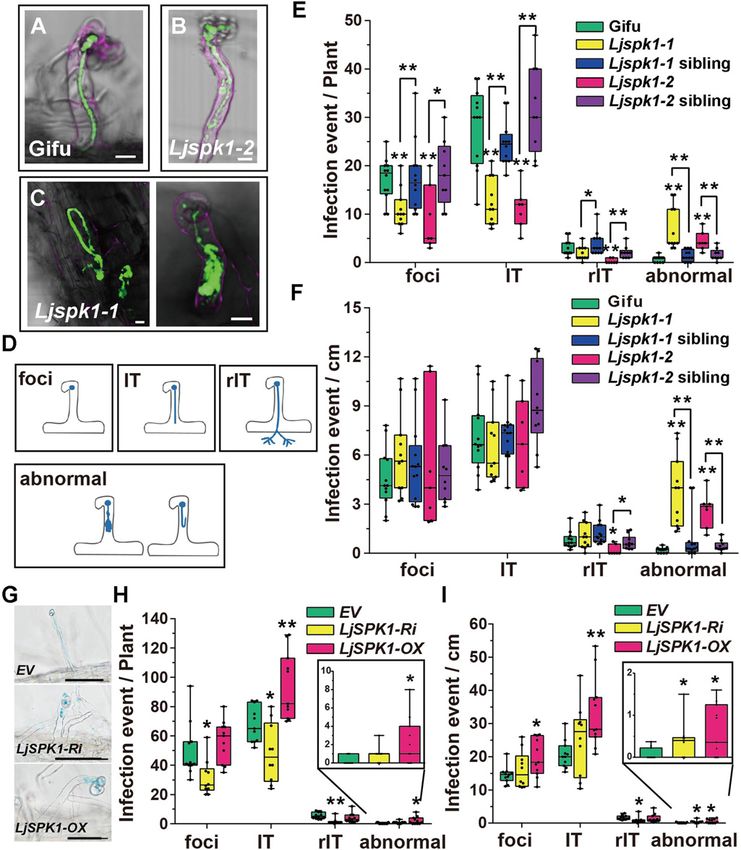
SPK1-ROP6 Mediates Polarized Growth of ITs                   3777

Figure 2. ITs in Ljspk1 Mutants Are Abnormal Compared with Wild-Type L. japonicus.
(A) to (D) Normal elongating ITs in the wild type (Gifu) (A) and typical abnormal infection events in the Ljspk1 mutants (B) and (C) at 1 week after inoculation
with M. loti R7A/GFP. Roots were counterstained with propidium iodide before observation. Green fluorescence shows normal infection foci and a normal IT
in a curled root hair in the wild type, whereas Ljspk1 mutants show abnormal infection processes, such as sac-like or looped ITs (B) and (C). Bars 5 10 mm. (D)
A cartoon diagram of the infection events. Foci, infection foci; IT, IT in an epidermis cell; rIT, IT extending into a cortex cell; abnormal, abnormal IT in root hairs.
(E) and (F) Boxplots representing the number of infection events in wild-type, Ljspk1 siblings, and Ljspk1 mutants. Total number of infection events per plant
(E) and number of infection events per centimeter root (F) were scored at 1 week after inoculation with M. loti R7A/lacZ. Asterisks indicate a significant
difference (*P < 0.05, **P < 0.01, Student’s t test, comparison between the wild type and mutants).
(G) to (I) IT phenotypes (G) and infection events ([H] and [I]) in wild-type L. japonicus hairy roots expressing control plasmid (EV), LjSPK1-Ri, or LjSPK1-OX.
Total number of infection events per plant (H) and number of infection events per centimeter root (I) scored 1 week after inoculation with M. loti R7A/lacZ.
Abnormal infection events are enlarged in the inset in (H) and (I). Asterisks indicate a significant difference (*P < 0.05, **P < 0.01, Student’s t test, comparison
between EV control and experimental group). Bars 5 50 mm.
For each boxplot, the center line in the box shows the median; the box limits are the upper and lower quartiles; the whiskers represent the maximum and
minimum values.
3778 The Plant Cell
SPK1 Interacts with Three L. japonicus ROP GTPases in
Yeast Cells, but Only LjROP6 Is Required for Polarized
Growth of ITs
AtSPK1 is a DOCK-family GEF protein, and DHR2 is the GEF
catalytic domain that facilitates the nucleotide exchange activity
of ROP GTPases (Basu et al., 2008). We were interested in de-
termining which ROP GTPase is activated by LjSPK1 to regulate
rhizobial infection in L. japonicus. BLAST searches of the L. ja-
ponicus genome revealed 10 ROP GTPases. We conducted
phylogenetic analysis of ROP GTPases in Arabidopsis, L. japo-
nicus, and M. truncatula (Supplemental Figure 3A). Because
AtSPK1 interacts and facilitate nucleotide exchange with type I
ROP GTPase, and most ROP GTPases are membrane-localized
proteins, we used the DUAL membrane yeast two-hybrid system
to determine which type I ROP GTPase interacts with the LjSPK1
DHR2 domain (named SPK1-DHR2). Of the four type I and one
type II ROP GTPases examined, three type I ROP GTPases
(LjROP1, LjROP3, and LjROP6) interacted with the LjSPK1 DHR2
domain in yeast (Saccharomyces cerevisiae) cells (Supplemental
Figure 3B). No interaction was detected between LjSPK1 DHR2
and LjROP5 or LjROP10 in yeast cells (Supplemental Figure 3B).
To explore whether these three type I LjROPs function in rhi-
zobial infection, we obtained their LORE1 insertion mutants
(30,000,786 with an insertion at 83 bp in LjROP1; 30,000,537 with
an insertion at 67 bp before the start codon in LjROP3; and
30,031,226 with an insertion at 258 bp in LjROP6). We then ob-
tained homozygotes of these insertion mutants. RT-qPCR anal-
ysis confirmed that endogenous LjROP transcript levels were
significantly reduced in all three Ljrop mutants (Supplemental
Figure 3C). We analyzed the root hair and infection phenotypes of
the mutants. First, we observed the root hairs of the mutants at 2 d
after germination with or without rhizobial inoculation. In the
absence of rhizobial inoculation, the roots of wild-type plants
formed straight root hairs, but more than half of the plants in lines
Ljrop1 (15 out of 30), Ljrop3 (18 out of 26), and Ljrop6-1 (20 out of
34) formed branched root hairs or root hairs with swollen tips
(Figure 3A). At 18 h after M. loti R7A/lacZ inoculation, both the wild Figure 3. Root Hair and IT Phenotypes of Ljrop Mutants.
type and Ljrop mutants displayed rhizobia-induced root hair (A) Light micrographs of root hairs in wild-type (Gifu) and Ljrop mutants at
deformation in the infection zone, with no distinguishable differ- 2 d after germination. The numbers in the lower left corners show the
ences between these lines (Figure 3B). These observations number of plants with the type of root hair shown in the image out of the total
suggest that all three LjROP GTPases play roles in root hair de- number of plants observed. Bars 5 100 mm.
velopment and respond normally to rhizobia. (B) Light micrograph of M. loti R7A-induced root hair deformation in the
We further analyzed IT formation and infection events after infection zone of wild-type and Ljrop mutants at 18 h after M. loti R7A
rhizobial inoculation. There were no differences in IT formation or inoculation. Bars 5 100 mm.
(A) and (B) Insets (bottom) are enlargements of the areas in red boxes (top).
infection events between the wild type and Ljrop1 or Ljrop3, but
(C) to (F) IT phenotypes of wild-type (C) and Ljrop mutants ([D] to [F]) at 5 d
there were fewer infection events in Ljrop6-1 than in the wild type after inoculation with M. loti R7A/LacZ. Bars 5 50 mm.
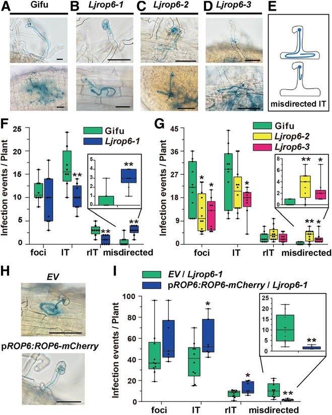
(Figure 3G). Interestingly, Ljrop6-1 contained some misdirected (G) Boxplot representing the number of infection events on roots of wild-
ITs in the root hairs, which were similar to, but more severe than, type and Ljrop mutant roots at 5 d after inoculation with M. loti R7A/lacZ.
those in Ljspk1 (Figure 3F and described in detail below). However, The center line in the box shows the median; the box limits are the upper and
this phenotype was not observed in the wild type, Ljrop1, or Ljrop3 lower quartiles; the whiskers represent the maximum and minimum values.
plants (Figures 3C to 3E). To analyze whether these LjROPs play Asterisks indicate a significant difference (**P < 0.01, Student’s t test,
functionally redundant roles during rhizobial infection, we crossed comparison between wild-type and Ljrop mutants).
Ljrop1 3 Ljrop3 and Ljrop1 3 Ljrop6-1 to obtain the Ljrop1 Ljrop3
and Ljrop1 Ljrop6-1 double mutants. We analyzed the infection produced approximately the same number of misdirected ITs as
events in these mutants after inoculation with M. loti R7A/LacZ. Ljrop6-1 (Supplemental Figure 3D). These results indicate that
Compared with wild-type plants, Ljrop1 Ljrop3 plants had more ROP1, ROP3, and ROP6 affect the polarized growth of root hairs in
infection foci but no misdirected ITs, and Ljrop1 Ljrop6-1 plants L. japonicus, but only LjROP6 is required for the polarized growth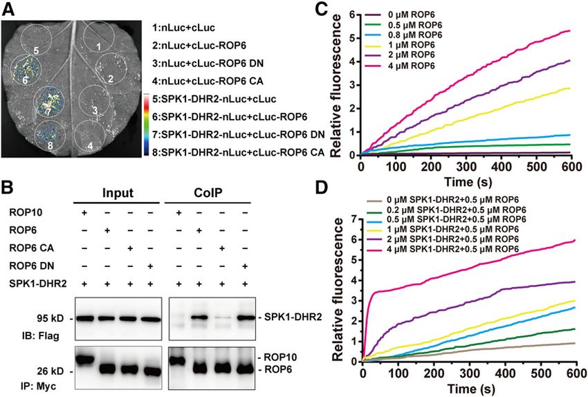
SPK1-ROP6 Mediates Polarized Growth of ITs                  3779

of ITs in root hairs. The results also indicate that the misdirection of
ITs in Ljrop6-1 is not caused by a deficiency in root hair
development.

LjROP6 Is Required for the Polarized Growth of ITs in
L. japonicus

To confirm the notion that ROP6 is required for the polarized
growth of ITs in root hairs, we obtained two more LORE1 insertion
lines: 30,142,846 (Ljrop6-2) and 30,103,232 (Ljrop6-3), with in-
sertions at 505 bp after and 114 bp before the LjROP6 start codon,
respectively (Supplemental Figure 4A). RT-qPCR analysis of
homozygous Ljrop6-2 and Ljrop6-3 plants showed that LjROP6
transcript levels were significantly lower in these plants than in
wild-type plants (Supplemental Figure 4E).
   We observed the root hair phenotypes of the Ljrop6-2 and
Ljrop6-3 mutants at 2 d after germination. Both before and after
rhizobial inoculation, the root hair phenotypes and responses to
rhizobia of the Ljrop6-2 and Ljrop6-3 mutants were similar to those
of Ljrop6-1 (Supplemental Figures 5A and 5B). We analyzed rhi-
zobial symbiosis in these three Ljrop6 mutants after M. loti R7A/
LacZ inoculation. After rhizobial infection, the three Ljrop6 mu-
tants produced normal ITs like those in the wild type (Figure 4A).
However, as noted above, some ITs in the Ljrop6 mutants were
misdirected or tangled in the root epidermal cells (Figures 4B to
4E), suggesting that the ITs became misdirected as they elongated
from the infection chamber down to the cortex nodule primordium.
Compared to the wild type, the Ljrop6 mutants had significantly
                                                                            Figure 4. Disorientated IT Phenotypes of Ljrop6 Mutants and Comple-
fewer infection events and formed more misdirected ITs at 5 d after
                                                                            mentation of IT Phenotype via Hairy Root Transformation of Ljrop6-1.
inoculation with M. loti R7A/lacZ (Figures 4F and 4G). At 2 weeks
after M. loti R7A/lacZ inoculation, the Ljrop6 mutants and wild-            (A) to (E) Normal IT phenotypes of the wild-type (A) and typical misdirected
type plants produced normal mature pink nodules (Supplemental               infection events in all three Ljrop6 mutants in (B) to (D). Bars 5 50 mm. (E) A
                                                                            cartoon diagram of the misdirected ITs. Misdirected indicates misdirected
Figure 4B). The total nodule number was not significantly different
                                                                            or looped IT in root hairs or epidermis cell.
between the Ljrop6 mutants and wild-type plants (Supplemental
                                                                            (F) and (G) Boxplots representing the number of infection events in wild-
Figures 4C and 4D).                                                         type L. japonicus (Gifu) and Ljrop6 mutants at 5 d after inoculation with M.
   To further confirm the notion that LjROP6 is required for the             loti R7A/lacZ. Misdirected infection events are enlarged in the inset. As-
polarized growth of ITs, we generated transgenic hairy roots in the         terisks indicate a significant difference (*P < 0.05, **P < 0.01, Student’s
Ljrop6-1 background using the LjROP6 native promoter to drive               t test, comparison between wild-type and Ljrop6 mutants).
theexpressionofLjROP6cDNAfusedwithmCherry(pLjROP6:LjROP6-                   (H) and (I) IT phenotype (H) and infection events (I) of Ljrop6-1 hairy roots
mCherry). The disorientated ITs in Ljrop6-1 were rescued by comple-         expressing control plasmid (EV) and pLjROP6:LjROP6-mCherry. ITs were
mentation with ROP6-mCherry (Figure 4H). Statistical analyses showed        stained with X-Gal and scored 5 d after inoculation with M. loti R7A/lacZ.
that the complemented group (pLjROP6:LjROP6-mCherry/Ljrop6-1)               Misdirected infection events are enlarged in the inset. Asterisks indicate
                                                                            a significant difference (*P < 0.05, **P < 0.01, Student’s t test, comparison
had more ITs and rITs and fewer misdirected ITs compared with the EV
                                                                            between EV and LjROP6). Bars 5 50 mm.
control at the same time point (Figure 4I). LjROP6 transcript levels were
                                                                            For each boxplot, the center line in the box shows the median; the box limits
significantlyhigherinLjrop6-1hairyrootsthanthecontrol(Supplemental           are the upper and lower quartiles; the whiskers represent the maximum and
Figure 4F). Together, these observations confirm the notion that LjROP6      minimum values.
is required for the polarized growth of ITs in L. japonicus.

                                                                            not detect coprecipitation of LjSPK1 with LjROP10, a type II ROP
LjSPK1 Physically Interacts with LjROP6 In Planta and
                                                                            GTPase, in our co-IP assays (Figure 5B). The activation of ROP
Activates Its GTPase Activity In Vitro
                                                                            GTPases relies on GDP and GTP exchange, and this cycle requires
As described above, LjROP6 interacted with LjSPK1 in yeast cells.           GEFs to facilitate the dissociation of GDP (Kost, 2008). We
We further confirmed their interaction in Nicotiana benthamiana              therefore investigated the ability of LjSPK1 to interact with LjROP6
leaf pavement cells using split-luciferase complementation and              in the DN (D121A) or CA (G15V) form. LjSPK1-DHR2 interacted
coimmunoprecipitation (co-IP) assays. LjROP6 interacted with the            more strongly with the LjROP6 DN form than with the LjROP6 CA
LjSPK1-DHR2 catalytic domain in split-luciferase complemen-                 form in co-IP assays (Figure 5B). These results suggest that
tation (Figure 5A) and co-IP assays (Figure 5B). However, we did            LjSPK1 preferentially interacts with the inactive form, but not the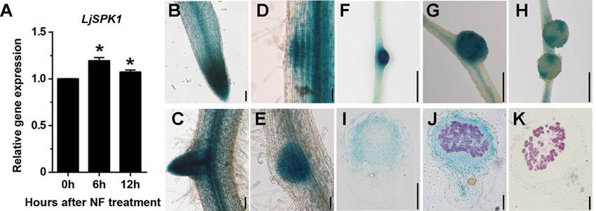
3780      The Plant Cell

Figure 5. LjSPK1 Physically Interacts with LjROP6 and Activates Its GTPase Activity In Vitro.

(A) Luciferase biomolecular complementation assays showing the interaction between LjSPK1-DHR2 and LjROP6, LjROP6 CA, or LjROP6 DN in N.
benthamiana leaf cells. The indicated constructs were transiently coexpressed in N. benthamiana leaves, and luciferase complementation imaging was
captured 2 d after agroinfiltration. nLuc, N-terminal fragment of firefly luciferase; cLuc, C-terminal fragment of firefly luciferase. Fluorescence signal intensity
is indicated.
(B) Co-IP assay showing the interaction between LjSPK1-DHR2 and LjROP6 in N. benthamiana leaves. The indicated constructs were coexpressed in N.
benthamiana leaves. The co-IP assay was performed using anti-Myc antibody, and the proteins were detected by immunoblot analysis with anti-Flag and
anti-Myc antibodies. LjSPK1-DHR2 interacted more strongly with LjROP6 DN than with LjROP6 CA. No interaction was detected between LjSPK1-DHR2
and LjROP10, a type II ROP GTPase.
(C) Time course of intrinsic nucleotide exchange in LjROP6. The intrinsic guanine nucleotide exchange rates of LjROP6 increased with increasing reaction
time and increasing concentration of LjROP6. The assays contained 1 mM mant-GTP and the indicated concentration of GDP-LjROP6.
(D) LjSPK1-DHR2 shows concentration-dependent GEF activity toward LjROP6. The assays contained 1 mM mant-GTP, 0.5 mM GDP-LjROP6, and the
indicated concentrations of LjSPK1-DHR2.
Results are a representative of three independent assays with similar results.

active form, of LjROP6 to activate the nucleotide exchange activity               LjROP6 Colocalizes and Interacts with LjSPK1 in the
of LjROP6.                                                                        Plasma Membrane
   The LjSPK1 DHR2 domain is known to activate small GTPases
(Basu et al. 2008; Wang et al. 2018). To examine whether LjSPK1                   LjROP6 was shown to be localized to the plasma membrane (PM)
                                                                                  and cytoplasm (Ke et al., 2012), but SPK1 is localized to the
facilitates the guanine nucleotide exchange activity of LjROP6, we
tested the GEF activity of the LjSPK1 DHR2 domain in vitro using                  endoplasmic reticulum (ER; Zhang et al., 2010; Wang et al., 2018).
a fluorescence spectroscopy-based procedure (Gu et al., 2006;                      To validate the subcellular localization of LjROP6 and confirm its
Wang et al., 2017). First, we expressed and purified glutathione                   interaction with LjSPK1, we analyzed the subcellular localization
S-transferase-tagged LjSPK1-DHR2 and unlabeled-GDP His-tagged                     of LjROP6 in N. benthamiana leaf cells and L. japonicus roots. We
LjROP6 in Escherichia coli. To start the nucleotide exchange reaction,            coexpressed GFP-LjROP6 or LjROP6-mCherry under the control
we added fluorescently labeled N-methylanthraniloyl (mant)-GTP and                 of the L. japonicus Ubiquitin promoter in N. benthamiana leaf cells.
unlabeled-GDP LjROP6 to the reaction buffer and recorded the fluo-                 In these analyses, LjROP6 localized to the PM, regardless of
rescence values for 600 s. The intrinsic guanine nucleotide exchange              whether it was tagged with a fluorescent marker at its N terminus or
rate of LjROP6 increased with increasing reaction time and with in-               C terminus (Supplemental Figures 6A to 6C). We then expressed
creasing concentrations of LjROP6 (Figure 5C). Next, to investigate the           LjROP6-mCherry in wild-type L. japonicus hairy roots and
effect of LjSPK1 on the guanine nucleotide exchange rate of LjROP6,               monitored its localization before and after inoculation with M. loti
we added LjSPK1-DHR2 protein to reaction buffer containing 0.5 mM                 R7A/mTag. Signals from mCherry were observed at the PM in
unlabeled-GDP LjROP6 protein and 1 mM mant-GTP before recording                   root hairs without rhizobia (Supplemental Figure 6D) and in
the change in fluorescence intensity. The LjSPK1 GEF activity toward               elongating or curled root hairs after inoculation with M. loti R7A
LjROP6 increased with increasing LjSPK1-DHR2 concentrations                       (Supplemental Figure 6E). The LjROP6-mCherry fusion protein
(Figure 5D). These results demonstrate that LjROP6 has intrinsic                  displayed a similar subcellular localization pattern regardless of
guanine nucleotide exchange activity, which is enhanced by SPK1.                  whether it was expressed under the control of the Ubiquitin
SPK1-ROP6 Mediates Polarized Growth of ITs 3781
promoter (Supplemental Figures 6D and 6E) or the LjROP6 native Polarized Growth of Root Hairs and Rhizobial Infection Are
promoter (Supplemental Figure 6F). Affected in LjROP6 Overexpression, CA, and DN Lines
Next, we analyzed the subcellular localization of LjSPK1.
We transformed N. benthamiana leaves with Agrobacterium To explore the role of LjROP6 in root hair development and IT
carrying LjSPK1-yellow fluorescent protein (YFP) and ob- formation, we generated LjROP6 overexpression, LjROP6 CA,
served the localization of LjSPK1 by monitoring the fluores- and LjROP6 DN hairy roots with each construct under the control
cence of YFP by laser scanning confocal microscopy. LjSPK1 of the L. japonicus Ubiquitin promoter. Green fluorescent protein
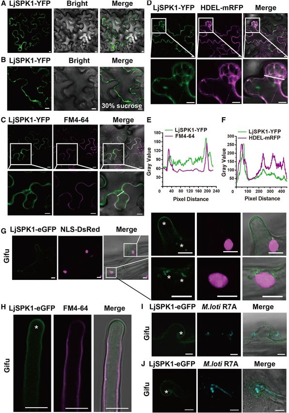
showed a punctate distribution close to the PM or cytoplasm (GFP) signals were visualized in the transformed roots. In the EV
(Figure 6A). Following plasmolysis using 30% (w/v) Suc, control, root hairs showed normal tip growth, and the root hairs
LjSPK1-YFP was not associated with the cell wall (Figure 6B). grew straight and away from the primary root axis (n 5 30)
To examine whether LjSPK1 localizes to the PM, we stained (Supplemental Figures 8A and 8B). However, in hairy roots
the N. benthamiana leaves with the PM localization marker FM4- overexpressing LjROP6, root hairs were morphologically similar to
64. LjSPK1 showed almost no colocalization with FM4-64 (Figures those of the control in that they displayed normal tip growth, but
6C and 6E). AtSPK1 and OsSPK1 are localized to subdomains of the approximately one-third of the hairy roots formed shorter root
ER (Zhang et al., 2010; Wang et al., 2018). To determine whether this hairs (25/82; Supplemental Figures 8A to 8C). Surprisingly, all
is also the case for LjSPK1, we coexpressed LjSPK1-YFP with the LjROP6 CA (42/42) and LjROP6 DN (51/51) lines formed com-
ER marker HDEL-mRFP in N. benthamiana leaves. LjSPK1 colo- pletely depolarized ballooning root hairs on transgenic roots
calized with the ER marker (Figures 6D and 6F). These findings (Supplemental Figures 8A to 8C).
suggest that LjSPK1 is associated with subdomains of the ER. We We analyzed the infection events in the LjROP6 overexpression,
then explored the subcellular distribution of LjSPK1-eGFP in L. LjROP6 CA, and LjROP6 DN transgenic hairy roots after M. loti
japonicus hairy roots. LjSPK1-eGFP displayed PM and punctate R7A/lacZ inoculation. The LjROP6 overexpression line formed
distribution close to the nucleus in L. japonicus root hairs elongated ITs similar to those of the EV control but had a few
(Figure 6G), and LjSPK1-eGFP also colocalized with the PM marker infection foci in uncurled root hairs and formed some disorientated
FM4-64 (Figure 6H). LjSPK1-GFP expressed in Ljrop6-1 hairy roots ITs (Supplemental Figures 8D and 8E). The number of infection
also showed a punctate distribution close to the nucleus events (foci and ITs) in hairy roots appeared to be slightly higher in
(Supplemental Figure 7A), similar to that of LjSPK1-eGFP ex- the LjROP6 overexpression line than in the EV control, but this
pressed in the wild type. After rhizobia inoculation, LjSPK1-eGFP difference was not significant (Supplemental Figure 8H). In the
was detected in the PM of root hairs with infection foci or elongated hairy roots of LjROP6 CA and LjROP6 DN, most ITs were ab-
ITs (Figures 6I and 6J). normally short and formed without root hair curling (Supplemental
We then coexpressed LjSPK1-YFP and LjROP6-mCherry in N. Figures 8F and 8G). In addition, the number of infection events was
benthamiana leaf pavement cells. In contrast to the punctate lo- significantly reduced in LjROP6 CA and LjROP6 DN hairy roots
calization of LjSPK1-YFP when expressed alone, LjSPK1-YFP versus the EV control (Supplemental Figure 8H). LjROP6 transcript
coexpressed with LjROP6-mCherry showed reduced punctate levels in hairy roots were higher in the LjROP6 overexpression,
distribution, and most LjSPK1-YFP signals were localized in LjROP6 CA, and LjROP6 DN lines than in the EV control
patches on the PM (Figures 7A to 7C). This observation suggests (Supplemental Figure 8I). These observations demonstrate that
that LjROP6 promotes the PM localization of LjSPK1. LjROP6 homeostasis is essential for its function in root hair de-
We evaluated the interaction between LjSPK1 and LjROP6 in velopment and IT formation.
planta via bimolecular fluorescence complementation (BiFC) We investigated the functions of LjROP1 and LjROP3 in root hair
assays, with C-terminal and N-terminal split-Venus fragments development by expressing LjROP1 or LjROP3 cDNA under the
fused to LjSPK1 and LjROP6, respectively, driven by the L. ja- control of the LjUb promoter in wild-type L. japonicus hairy roots
ponicus or Arabidopsis Ubiquitin promoter. When this construct and analyzing their CA and DN forms. In all of these transgenic
(pLjUb:LjSPK1-cVenus/pAtUb:LjROP6-nVenus) was agro- lines, the hairy roots formed shorter root hairs than the EV control
infiltrated into N. benthamiana leaf cells, Venus fluores- (Supplemental Figure 9), which is similar to the phenotype of
cence was detected in the PM and in some punctate struc- LjROP6-overexpressing L. japonicus hairy roots.
tures adjacent to the PM (Figure 7D). This distribution was
confirmed by plasmolysis experiments using 30% (w/v) Suc
Actin Filaments Are Disordered in the Root Hairs of Ljrop6-1
(Figure 7E). These results confirm the notion that LjSPK1
Compared with Wild-Type L. japonicus
interacts with LjROP6 at the PM. We then introduced this
construct into wild-type L. japonicus hairy roots and moni- The actin cytoskeleton is important for root hair tip growth. To
tored the localization of Venus fluorescence signals after determine whether LjROP6 regulates the arrangement of actin
inoculation with M. loti R7A/mTag. Venus fluorescence sig- filaments to influence the polarized growth of root hair tips, we
nals were detected in the PM of deformed root hairs (Figure 7F) used Alexa Fluor 488-conjugated phalloidin to stain wild-type and
as well as in infection foci (Supplemental Figure 7B). We also Ljrop6-1 roots. As expected, the root hairs of wild-type plants
expressed these constructs in the M. truncatula sunn-1 predominantly displayed the characteristic arrangement of actin
mutant, which had many more infection events than the filaments in long cables aligned longitudinally (Supplemental
wild type. Venus fluorescence was observed in the infection Figure 10A). However, 70% of Ljrop6-1 root hairs had fewer
chamber of the root hairs at the site of the initiating IT longitudinally aligned actin filaments and significantly more
(Figure 7G; Supplemental Figure 7C). transversely oriented ones compared to the wild type3782 The Plant Cell Figure 6. Subcellular Localization of LjSPK1 in N. benthamiana Leaves and Wild-Type L. japonicus Hairy Roots. (A) to (F) Confocal images of LjSPK1-YFP expressed in N. benthamiana leaf cells. (A) and (B) Subcellular localization of LjSPK1-YFP in N. benthamiana leaves (A) and after plasmolysis via 30% (w/v) Suc treatment (B). (C) LjSPK1-YFP (green) expressed in N. benthamiana leaves and stained with the PM marker FM4-64 dye (magenta), showing that SPK1 does not merge with the PM marker. (D) LjSPK1-YFP (green) and ER marker HDEL-mRFP (magenta) were
SPK1-ROP6 Mediates Polarized Growth of ITs                 3783

(Supplemental Figure 10B). Most of the short swollen or medium-                    2007) and topoisomerase VI (TOPO6A) subunit LjSUNERGOS1 or
length root hairs of Ljrop6-1 showed disordered or web-like ar-                    LjVAG1 (Suzaki et al., 2014; Yoon et al., 2014). These mutants
rangements of actin filaments (Supplemental Figure 10C). These                      exhibit misdirected ITs and highly defective nodule organogen-
results indicate that LjROP6 affects the arrangement of actin fil-                  esis, which in turn affects nodule primordium formation. However,
aments in root hairs to regulate their development.                                in the Ljrop6 mutants observed in this study, the ITs formed loops
                                                                                   and showed disoriented growth in root epidermis cells, but nodule
                                                                                   formation was not affected. This finding suggests that the
DISCUSSION
                                                                                   LjROP6-mediated polar elongation of ITs is not associated with
Rhizobial ITs elongate following the migration of the nucleus along                nodule primordium formation. The Golgi/trans-Golgi-localized
and within the root hairs via polar tip growth. ROP GTPases are key                Rab GTPase PvRabA2 is required for IT progression and the
components required for the polarized growth of cells during                       maintenance of membrane integrity in common bean (Phaseolus
processes such as pollen tube elongation and root hair de-                         vulgaris; Blanco et al., 2009; Dalla et al., 2017). In M. truncatula, the
velopment (Zheng and Yang, 2000; Samaj et al., 2006). LjROP6                       LIN-VPY-Exo70H4 complex shows a polar distribution in pre-ITs,
interacts with the NF receptor LjNFR5 to mediate nodulation, and                   suggesting that this complex mediates the polarized growth of ITs
LjROP6 associates with LjCHC1 (Ke et al., 2012; Wang et al.,                       via exocytosis (Liu et al., 2019b). In the future, it would be in-
2015). The CHC1-hub domain, a dominant effector of clathrin-                       teresting to determine whether LjROP6 interacts with this complex
mediated endocytosis, impairs rhizobial infection (Wang et al.,                    to guide the polarized growth of ITs.
2015). Here, we demonstrated that LjROP6 is required for the                          ROP GTPases regulate cell growth and shape by organizing the
polarized growth of ITs in root hairs. GTPases act as molecular                    actin filaments that drive cytoplasmic streaming and vesicle
switches that fluctuate between an inactive GDP-bound form and                      trafficking in the cytoskeleton. These GTPases also affect the
an active GTP-bound form. We found that the DOCK-family GEF                        arrangement of cortical membrane-associated microtubules,
protein LjSPK1 interacts with, and activates, LjROP6. We also                      which guide cell wall deposition via cellulose synthase complexes
found that LjROP6 promotes the localization of LjSPK1 to the PM                    (Fu et al., 2005; Endler and Persson, 2011; Tominaga and Ito,
and that this may be important for its function. Together, our                     2015). ROP GTPase activity is regulated by GAPs, GEFs, and
results show that the DOCK-family GEF LjSPK1 activates LjROP6                      RhoGDIs (Yang, 2002; Kost, 2008). SPK1 is a DOCK-family GEF
to mediate the polarized growth of ITs, and they provide evidence                  that is conserved in animals and plants (Meller et al., 2005). In
that early NF signaling is connected to morphological changes                      Arabidopsis, AtSPK1 has been implicated in activating ROP
associated with rhizobial infection.                                               signaling to regulate actin polymerization via WAVE and ARP2/3
   Rhizobial invasion into host plants via the IT requires an actively             complexes in leaves (Qiu et al., 2002; Basu et al., 2008). Similarly,
growing root hair. Inside the root hair, an infection chamber forms                in rice, OsPit interacts with OsSPK1 to activate OsRAC1 and
and elongates to form the IT, the tubular structure that guides                    trigger immune responses (Wang et al., 2018). These results in-
bacteria toward cortical cells (Fournier et al., 2008, 2015). ROP                  dicate that LjSPK1 interacts with and activates LjROP6 and that
GTPases play important roles in the polarized growth of cells.                     both LjSPK1 and LjROP6 are involved in the polarized growth of
ROPs are polarized into single, discrete PM domains in tip-                        ITs following rhizobial infection. The formation of ITs requires
growing cells such as pollen tubes and root hairs (Lin et al.,                     several components of the WAVE/SCAR-ARP2/3 complex, in-
1996; Molendijk et al., 2001). The NF receptor LjNFR5 interacts                    cluding NAP, PIR, SCARN, and ARPC1 (Yokota et al., 2009;
with LjROP6 to mediate IT formation and nodulation (Ke et al.,                     Hossain et al., 2012; Qiu et al., 2015). It will be important to in-
2012). In this study, we identified three LORE1 insertion mutants of                vestigate whether the LjSPK1-LjROP6 module interacts with the
Ljrop6. All three mutants were able to form normal ITs after rhi-                  WAVE/SCAR-ARP2/3 complex to regulate actin rearrangement
zobial inoculation. However, compared to the wild type, the                        and mediate IT formation.
mutants had fewer infection events (foci and ITs) and formed many                     Exocytosis and the polar inhibition of clathrin-dependent en-
more disoriented ITs in root hairs. This observation suggests that                 docytosis regulate the polarized growth of cells in both animals
LjROP6 mediates the polarized growth of ITs in root hairs.                         and plants. Studies of ROPs/RACs have revealed that these
   Disoriented ITs have also been observed in other mutants, such                  processes are important targets of Rho-GTPase signaling
as mutants of the cytokine receptor gene LjLhk1 (Murray et al.,                    (Craddock and Yang, 2012; Craddock et al., 2012; Feiguelman

Figure 6. (continued).

coexpressed in N. benthamiana leaves. Image shows merging of LjSPK1-YFP and HDEL-mRFP fluorescence. These genes were driven by the CaMV 35S
promoter. (E) and (F) Intensity profiles of LjSPK1 and FM4-64 or HDEL-mRFP. Plots show fluorescence intensities of LjSPK1-YFP (green) and FM4-64 or
HDEL-mRFP (magenta) in regions of interest (insets in [C] and [D]). (C) and (D) Insets (bottom) are enlargements of the areas in white boxes (top). Bars 5 10
mm.
(G) to (J) Live cell confocal images of LjSPK1-eGFP expressed in root hairs before (G) and (H) and after (I) and (J) M. loti R7A/mTag inoculation in wild-type L.
japonicus hairy roots. (G) LjSPK1-eGFP (green) fluorescence was detected in L. japonicus hairy roots in the PM and puncta in the vicinity of the nucleus. NLS-
DsRed (magenta; nuclear marker) was used as a transgenic marker. (H) LjSPK1-eGFP (green) was detected in L. japonicus hairy root PM and stained with the
PM marker FM4-64 dye (magenta). (I) and (J) LjSPK1-eGFP (green) in the PM of curled root hair (I) or with elongated IT (J) after M. loti R7A/mTag inoculation.
IT is indicated by M. loti R7A/mTag (cyan). Asterisks indicate GFP fluorescence. Expression of LjSPK1 was driven by the L. japonicus Ubiquitin gene
promoter. Insets (right) are enlargements of the areas in white boxes (left). Bars 5 10 mm.
3784 The Plant Cell Figure 7. LjROP6 Colocalizes and Interacts with LjSPK1 at the PM in N. benthamiana Leaves and Hairy Roots in Legumes. (A) to (C) LjSPK1-YFP (green) and LjROP6-mCherry (magenta) were coexpressed in N. benthamiana leaves, showing that LjROP6 promotes LjSPK1 distribution in the PM (A). Insets (bottom) are enlargements of the areas in white boxes (top). (B) Intensity profiles of LjSPK1-YFP and LjROP6-mCherry. Plots show fluorescence intensities of LjSPK1-YFP (green) and LjROP6-mCherry (magenta) in regions of interest (insets in [A]). (C) Immunoblots showing protein levels of LjSPK1 and LjROP6 in N. benthamiana leaves. Total protein was extracted and analyzed by immunoblotting with anti-GFP and anti-mCherry antibodies. Rubisco was used as the loading control. Bars 5 10 mm. (D) to (E) BiFC assay of LjSPK1-cVenus and LjROP6-nVenus expressed in N. benthamiana leaves, before (D) and after (E) plasmolysis with 30% (w/v) Suc treatment. Split Venus fluorescent was detected in the PM. Bars 5 10 mm. (F) to (H) BiFC assay of LjSPK1-cVenus and LjROP6-nVenus in wild-type L. japonicus hairy roots (F) or M. truncatula sunn-1 hairy roots (G) 5 d after rhizobial inoculation. Asterisks represent the Venus fluorescence and indicate that LjROP6 interacts with LjSPK1 in the root hair PM (F) or infection foci (G). Cyan and magenta fluorescence represent M. loti R7A/mTag (cyan, [F]) or Sm1021/mCherry (magenta, G), respectively. Insets (right) are enlargements of the areas in white boxes (left). Bars 5 10 mm. et al., 2018). For example, the yeast exocyst subunit SEC3 is is required for the polar localization of PIN1 (Lavy et al., 2007; a direct effector of the Rho GTPase Cdc42. In Arabidopsis, the Hazak et al., 2010). SPK1 is required for the auxin-activated ROP6- ROP effector ICR1/RIP1 (INTERACTOR OF CONSTITUTIVE RIC1 pathway to inhibit the internalization of PIN2 by stabilizing ACTIVE ROP1) interacts with the Arabidopsis SEC3 homolog and actin microfilaments. These processes affect auxin distribution
SPK1-ROP6 Mediates Polarized Growth of ITs                   3785

and lateral root development (Lin et al., 2012). In L. japonicus,           polar elongation of ITs in root hairs. Perhaps LjROP6 interacts with
LjROP6 interacts with LjCHC1 and is involved in rhizobial infection         LjNFR5 to transduce NF signaling to mediate the progression and
(Wang et al., 2015). Interestingly, LjROP6 alone localizes to the PM        elongation of ITs. Interestingly, in a study of M. truncatula ROP10,
but shows a punctate distribution on the PM when it is coex-                only the MtROP10 CA form showed defects in the polarized
pressed with LjCHC1 (Wang et al., 2015) or LjSPK1, as demon-                growth of root hairs; its infection phenotype was very similar to
strated in the BiFC assay between LjSPK1 and LjROP6 in N.                   those of the LjROP6 CA and LjROP6 DN forms. However, although
benthamiana leaves in this study. Further studies are needed to             MtROP10 was able to interact with the NF receptor MtNFP and it
investigate whether LjSPK1-activated LjROP6 can activate other              was required for root hair deformation, it was not required for the
downstream effectors such as RICs or RIPs. Such interactions                polarized growth of ITs (Lei et al., 2015). Therefore, these and
could control the polarized growth of rhizobial ITs via effects on the      previous findings suggest that different ROP GTPases that are
organization of actin microfilaments, auxin distribution, and                associated with NF signaling mediate different infection processes.
exocytosis and endocytosis pathways.                                           Prenylation is required for the membrane attachment and
   AtSPK1 is localized to subdomains of the ER (Zhang et al.,               function of type I ROP GTPases, and LjROP6 is a type I ROP
2010), and OsSPK1 (Wang et al., 2018) and LjSPK1 (this study)               GTPase. When we examined the subcellular localization of
show a punctate distribution and colocalize with an ER marker.              LjROP6 in N. benthamiana, the placement of the fluorescent
However, coexpressing OsSPK1-OsRAC1 or LjSPK1-LjROP6 in                     protein at the N terminus or C terminus of this protein did not affect
N. benthamiana leaves enhanced the PM localization of SPK1,                 its PM localization. Moreover, in the complementation assay,
suggesting that OsSPK1-OsRAC1 or LjSPK1-LjROP6 interact at                  LjROP6 tagged with mCherry at its C terminus fully rescued the
the PM (Wang et al., 2018). In this study, LjSPK1 alone was dis-            infection phenotype of Ljrop6-1. These results suggest that the
tributed at the PM and in punctate spots around the nucleus in L.           prenylation of LjROP6 has minor effects on its function, which are
japonicus, but it interacted with LjROP6 in the PM and infection            consistent with the results of another study in Arabidopsis (Sorek
pocket in curled root hairs of L. japonicus infected with M. loti. In       et al., 2011).
rice, OsRacGEF1 is transported from the ER to the PM with                      The penetration of bacteria into legume roots is a key step in the
OsCERK1 via a vesicle trafficking pathway (Akamatsu et al., 2013).           specific recognition of compatible rhizobia during the formation
It would be interesting to explore which pathway is involved in the         and progression of ITs. These and previous findings provide
translocation of LjSPK1 from the ER to the PM.                              genetic evidence that the molecular machinery associated with
   GEFs are activated by PM-bound receptor-like kinases                     the LjNFR5-LjROP6-LjSPK1 module is involved in the vesicle
(Akamatsu et al., 2013; Liao et al., 2017). In mammals, the kinase          trafficking and cytoskeleton rearrangements required for the
Akt and the protein phosphatase PP2A interact with the DHR2                 polarized growth of ITs. Thus, we suggest that the LjNFR5-
domain of DOCK6. DOCK6 is phosphorylated by Akt and de-                     LjROP6-LjSPK1 module has been co-opted to participate in some
phosphorylated by PP2A at Ser1194; the phosphorylation status of            events in rhizobial infection, particularly the polarized growth of ITs
DOCK6 determines its ability to promote axon growth (Miyamoto               (Supplemental Figures 11A and 11B). Our results also show that
et al., 2013). A phosphoproteomic study of root nodule symbiosis            LjSPK1 can activate other type I ROP GTPases such as LjROP1
in M. truncatula revealed that two phosphoisoforms of DOCK-                 and LjROP3 and that these ROP GTPases are required for the
family proteins, MtSPK1 and DOCK7, displayed lower phos-                    growth of the root hair tip (Supplemental Figure 11C).
phorylation levels upon NF treatment in nfp and dmi3 than in the
wild type. Those findings suggest that rhizobia infection may
                                                                            METHODS
affect the phosphorylation status of MtSPK1 (Rose et al., 2012).
Thus, it will be important to investigate whether there is a kinase
that affects the phosphorylation status of LjSPK1 to regulate its           Biological Materials and Strains
activity toward LjROP6 during the establishment of symbiosis.               Lotus japonicus accession Gifu B-129 was used in this study. The Ljrop and
   ROP GTPases play an essential role in controlling the polarized          Ljspk1 mutants were obtained from the LORE1 transposon insertion library
growth of pollen tubes and root hairs. In Arabidopsis, AtSPK1               at Lotus Base (https://lotus.au.dk; Fukai et al., 2012; Urbański et al., 2012).
functions as a GEF that interacts with and activates a series of ROP        The mutants were genotyped by PCR and sequenced using the primers
GTPases (Basu et al., 2008). Our results show that LjSPK1 in-               shown in Supplemental Data Set 1.
teracts with three type I ROP GTPases in L. japonicus. All three                The rhizobium strain Mesorhizobium loti R7A carrying pXLGD4 (lacZ),
                                                                            pMP2444 (GFP), or mTag was used for infection and the nodulation assays.
LjROP GTPases are required for the polarized growth of root hairs.
                                                                            The Agrobacterium rhizogenes strain AR1193 was used for hairy root
The Ljrop mutants in this study formed branched and swollen root
                                                                            transformation of L. japonicus and Medicago truncatula roots, and the
hairs, while lines overexpressing the LjROP GTPases and their CA            Agrobacterium tumefaciens strain EHA105 was used for transient ex-
or DN forms produced short, ballooning root hairs. These phe-               pression in N. benthamiana. Plasmids were transformed into Escherichia
nomena are not consistent with the previous finding that plants              coli DH10B or DH5a for cloning or into E. coli Rosetta (DE3) for protein
expressing the DN and CA forms of ROP showed different phe-                 expression. The yeast (Saccharomyces cerevisiae) strain NMY51 was used
notypes (Molendijk et al., 2001; Jones et al., 2002; Lei et al., 2015).     for the DUAL membrane yeast two-hybrid system.
This discrepancy can be explained by the notion that ROP
GTPases function as molecular switches. Because the homeo-
stasis of their activity is strictly regulated, either stronger or weaker   Cloning, DNA Manipulation, and Plasmid Construction
activity will affect their function. Although LjSPK1 can interact with      The coding sequences (CDS) of the LjROPs and LjSPK1 were amplified
three ROP GTPases, only one of them, LjROP6, is required for the            from the Gifu cDNA library by PCR amplification; the CA and DN forms of
3786 The Plant Cell
LjROPs were generated with a Hieff Mut site-directed mutagenesis kit construct LjSPK1-eGFP. The synthesized LjSPK1 CDS in vector pL0V-
(Yeasen Biotechnology) as per the manufacturer’s instructions. SC3 was assembled into EC12850 to generate LjSPK1-cVenus; the
For expression analysis of LjSPK1 in L. japonicus hairy roots, the LjSPK1 LjROP6 PCR product was inserted into pL0V-SC3 following Bbs1 di-
promoter (2 kb upstream of the start codon) was amplified from Gifu ge- gestion, and this construct was assembled into EC12849 to generate
nomic DNA by PCR. The PCR product was inserted into pDONR207 by BP LjROP6-nVenus. Finally, these constructs were assembled into EC50507,
reaction (Invitrogen), and combined into pKGWFS7 to generate the adding p35S:DsRed as a transgenic marker, to generate the BiFC con-
pLjSPK1:GUS construct by LR reaction (Invitrogen). struct pLjUb:LjSPK1-cVerns/pAtUb:LjROP6-nVenus.
For the LjSPK1 RNAi construct, the CDS fragment of LjSPK1 was All PCR amplification was performed using high-fidelity DNA poly-
amplified by PCR. The PCR product was inserted into pDONR207 by BP merase KOD Plus (Toyobo) or MAX (Vazyme), and all constructs were
reaction (Invitrogen) and combined into pUB-GWS-GFP to generate the confirmed by DNA sequencing. The constructs in destination vectors were
LjSPK1-Ri construct by LR reaction (Invitrogen). introduced into A. rhizogenes AR1193 for hairy root transformation in L.
To overexpress LjSPK1 or LjROPs in L. japonicus hairy roots, the japonicus or M. truncatula or into A. tumefaciens strain EHA105 for tran-
LjSPK1, LjROPs, LsROPs CA, and LjROPs DN CDS were transferred from sient expression in N. benthamiana by electroporation. All primers are listed
X-pDONR207 into pUB-GW-GFP by LR reaction to generate the LjSPK1- in Supplemental Data Set 1, and all constructs are listed in the
OX, LjROPs-OX, LjROPs CA, or DN constructs, respectively. Supplemental Table.
For the DUAL membrane yeast two-hybrid system, the PCR products
and pCCW-STE or pDSL-Nx were digested with Sfi1, and the LjROPs and
LjSPK1-DHR2 were inserted into pCCW-STE and pDSL-Nx, respectively, Analysis of Plant Growth, Rhizobial Inoculation, Infection,
using T4 DNA ligase (Takara). Nodulation, Root Hairs, and Actin Rearrangement
To measure guanine nucleotide exchange activity, the LjROP6 and
L. japonicus seeds were scarified, surface sterilized in 10% (v/v) sodium
LjSPK1-DHR2 PCR products were recombined into pDONR207 by BP
hypochlorite for 7 min, and rinsed five times with sterile water. The sterilized
reaction (Invitrogen). LjROP6 pDONR207 and LjSPK1-DHR2 pDONR207
seeds were imbibed in water, transferred to 0.8% (v/v) water agar plates,
were recombined into pHGWA with a His-tag or pGGWA with a glutathione
and grown in 22°C in the dark for 3 to 4 d for germination. The seedlings
S-transferase-tag by LR reaction and transformed into E. coli Rosetta for
were transferred to a mixture of perlite and vermiculite (1:1), cultivated in
protein expression.
a growth chamber under a 16 h/8 h light (250 mmol/m2/s)/dark cycle, and
For the luciferase biomolecular complementation assays in N. ben-
inoculated with rhizobia at 5 to 7 d after transfer. The infection phenotypes
thamiana, the PCR products and luciferase vector pCambia1300-35S-
and events were determined at the indicated time points by laser scanning
nLuc or pCambia1300-35S-cLuc were digested with Kpn1 and Sal1, and
confocal microscopy (FV1000, Olympus) using GFP-marked M. loti R7A or
LjSPK1-DHR2 and LjROP6 were inserted into pCambia1300-X-nLuc or
light microscopy (ECLIPSE Ni, Nikon) after staining the roots with 5-bromo-
pCambia1300-cLuc-X using T4 DNA ligase.
4-chloro-3-indolylbeta-D-galacto-pyranoside (X-Gal).
For co-IP assays in N. benthamiana, the PCR products (LjROP6, LjROP6
To prepare nodule sections, the nodules were fixed in glutaraldehyde
CA, LjROP6 DN, or LjROP10) were inserted into destination vector
(2.5% [v/v]), embedded in Technovit 7100 resin (Kulzer) according to the
pCambia1305-35S-Myc following Kpn1 and Sal1 digestion. The LjSPK1-
manufacturer’s instructions, and cut into 5- to 10-mm transverse sections
DHR2 PCR products were inserted into destination vector pUB-GFP/
with a microtome (RM2265, Leica Microsystems). Root nodule sections
X-FLAG, which was modified from pUB-GFP (Maekawa et al., 2008; Li et al.,
were costained with toluidine blue or magenta-Gal to visualize GUS or
2019), following Kpn1 and Asc1 digestion.
M. loti.
To determine the subcellular localization of LjROP6 in L. japonicus
To analyze the root hair phenotypes of the Ljrop mutants, the seedlings
leaves and L. japonicus hairy roots, LjROP6 pDONR207 was recombined
were transferred to glass slides containing 1 mL liquid Fahraeus medium
into pK7WGF2 to generate GFP-LjROP6 by LR reaction. LjROP6 PCR and incubated overnight. The seedlings were inoculated by adding fresh
products were inserted into pUB-GFP/X-mCherry (this vector was modi- Fahraeus medium with or without M. loti R7A (OD600nm;0.01) and in-
fied from pUB-GFP; Li et al., 2019) following Xba1 and Kpn1 digestion using cubated in the dark for ;18 h before analysis. The root hairs were observed
T4 DNA ligase to generate LjROP6-mCherry. and imaged under a light microscope (Nikon ECLIPSE Ni). For observation
For the complementation assay via hairy root transformation of Ljrop6- root hairs in L. japonicus hairy roots, transformed hairy roots of the LjROPs
1, the LjROP6 promoter (1.8 kb upstream of the start codon) was amplified overexpression, LjROPs CA, and LjROPs DN lines were scored by GFP
from Gifu genomic DNA by PCR amplification. The PCR products and fluorescence using a Nikon SMZ1500 microscope, and then root hairs were
destination vector pUB-GFP were digested with Pst1 and Xba1, and the observed and imaged under a light microscope (Nikon ECLIPSE Ni). The
LjROP6 promoter was inserted into pUB-GFP using T4 DNA ligase to length of root hairs was measured using ImageJ. Five root hair cells were
generate pLjROP6-GFP. pLjROP6-GFP and pUB-GFP/LjROP6-mCherry measured per transformed root, and at least 10 transformed roots were
were digested with Xba1 and Asc1, and LjROP6-mCherry was inserted into scored.
pLjROP6-GFP using T4 DNA ligase to generate pLjROP6:LjROP6- Phalloidin staining was done as previously described by Yokota et al.
mCherry. This vector was also used to determine the subcellular locali- (2009), and actin was observed by laser scanning confocal microscopy
(FV1000, Olympus).
zation of LjROP6 in L. japonicus transformed hairy roots.
All assays were validated in at least three independent experiments.
To determine the subcellular localization of LjSPK1 in N. benthamiana
leaves, the LjSPK1 PCR products were inserted into pCambia1300-35S-
YFP following Sma1 and Spe1 digestion to generate LjSPK1-YFP. For
Analysis of Promoter:GUS, RNAi, Overexpression, and
LjSPK1 localization and BiFC analysis in N. benthamiana leaves, L. ja-
Complementation in L. japonicus Transformed Hairy Roots
ponicus, or M. truncatula hairy roots, the LjSPK1-eGFP and BiFC con-
structs were generated by Golden Gate cloning (Weber et al., 2011). The The indicated constructs in A. rhizogenes AR1193 were introduced into L.
LjSPK1 CDS was synthesized in the level 0 vector pL0V-SC3 (Shanghai japonicus Gifu or Ljrop6-1 roots on 1/2 B5 medium via hairy root trans-
Xitubio Biotechnology). This vector and the EC16570 vector were digested formation. After 2 weeks, the transformed hairy roots were scored by GFP
with Bsa1 to generate LjSPK1-eGFP as the level 1 construct. This level fluorescence, as observed under a Nikon SMZ1500 microscope. The
1 LjSPK1-eGFP was assembled into EC50507 (https://www.ensa.ac.uk/), transformed chimeric plants were transferred to pots filled with a mixture of
adding p35S:NLS-DsRed as a transgenic marker, to generate the level 2 vermiculite and perlite (1:1) and inoculated with M. loti R7A/lacZ at 5 toYou can also read