Setal Field Transects, Evolutionary Transitions and Gecko-Anole Convergence Provide Insights Into the Fundamentals of Form and Function of the ...
←
→
Page content transcription
If your browser does not render page correctly, please read the page content below
ORIGINAL RESEARCH
                                                                                                                                            published: 27 January 2021
                                                                                                                                      doi: 10.3389/fmech.2020.621741

                                           Setal Field Transects, Evolutionary
                                           Transitions and Gecko–Anole
                                           Convergence Provide Insights Into the
                                           Fundamentals of Form and Function of
                                           the Digital Adhesive System of Lizards
                                           Anthony P. Russell 1* and Austin M. Garner 2
                                           1
                                            Department of Biological Sciences, University of Calgary, Calgary, AB, Canada, 2Integrated Bioscience Program, Department of
                                           Biology, The University of Akron, Akron, OH, United States

                                           Recent years have witnessed a multitude of studies focusing on gekkotan adhesion.
                                           Intense interest in this phenomenon was triggered by the discovery of the manner and
                                           magnitude of the forces generated by the hair-like filaments (setae) on the toe pads and
                                           inspired the development of the next generation of smart, reversible synthetic adhesives.
                            Edited by:     Most studies pursuing these goals have concentrated on the generalized form and
                    Stanislav N. Gorb,
                                           properties of gekkotan setae outlined in those key early studies, resulting in the
           University of Kiel, Germany
                                           fabrication of synthetic filaments of uniform dimensions. Although there are over 1,800
                      Reviewed by:
                  Luciano Afferrante,      species of extant geckos, and hundreds of species of anoles (a separate lizard lineage that
             Politecnico di Bari, Italy    has convergently evolved adhesive toe pads), most investigations have used relatively few
               Yoshitaka Nakanishi,
         Kumamoto University, Japan        species as the source of basic information, the Tokay gecko (Gekko gecko) being the most
                  *Correspondence:         prominent among these. Such exemplar taxa generally exhibit structurally intricate setae
                   Anthony P. Russell      and morphologically complex configurations of the adhesive apparatus. Setal structure
                 arussell@ucalgary.ca
                                           taken to be characteristic of these taxa is generally reported by singular statements of
                                           maximal length, diameter, density and branching pattern. Contemporaneous work
                    Specialty section:
         This article was submitted to     focusing on the configuration of setae at locations across the toe pads and upon the
                             Tribology,    evolutionary origin of adhesively competent digits in anoles and specific lineages of
               a section of the journal
  Frontiers in Mechanical Engineering      geckos, however, has revealed extensive variation of setal structure within individuals,
        Received: 26 October 2020          information about how setae may have arisen from non-adhesive filamentous precursors,
      Accepted: 15 December 2020           and how newly adhesively competent digits have been integrated into pre-existing
       Published: 27 January 2021
                                           patterns of locomotor mechanics and kinematics. Such observations provide insights
                             Citation:
                                           into what is minimally necessary for adhesively competent digits to function and reveal the
Russell AP and Garner AM (2021) Setal
          Field Transects, Evolutionary    simplest configuration of components that make this possible. We contend that
         Transitions and Gecko–Anole       information gleaned from such studies will assist those seeking to employ the
Convergence Provide Insights Into the
Fundamentals of Form and Function of       principles of fibrillar-based adhesion, as exemplified by lizards, for bio-inspired
the Digital Adhesive System of Lizards.    applications.
          Front. Mech. Eng 6:621741.
    doi: 10.3389/fmech.2020.621741         Keywords: Anolis, biomimetics, convergent evolution, fibrillar adhesion, Gekkota, synthetic adhesives

Frontiers in Mechanical Engineering | www.frontiersin.org                          1                                          January 2021 | Volume 6 | Article 621741
Russell and Garner                                                                                          Fundamentals of Lizard Adhesive Systems

INTRODUCTION                                                               sufficient for adhesively competent digits to function.
                                                                           Recognition of such attributes should help to simplify the
Recent years have witnessed a deluge of studies focusing on                pathway to the development of effective synthetic setae and
gekkotan adhesion, a remarkable phenomenon whereby geckos                  mechanical mechanisms to operate them (as assessed against
can attach to, and move on, smooth, low friction surfaces using            the performance of the actual structures being mimicked). As
series of expanded scales (scansors or lamellae) that possess              Niewiarowski et al. (2016) note, “no synthetic mimic can yet
arrays of hair-like fibers (setae) carrying multiple flattened,              perform as well as a gecko”.
triangular-shaped tips (spatulae) (Ruibal and Ernst, 1965;
Autumn et al., 2000; Autumn et al., 2002). Intense interest in
this was triggered by the discovery of the manner and magnitude            TRANSECTS OF SETAL FIELDS
of the forces generated by a single seta of the Tokay gecko (Gekko
gecko) (Autumn et al., 2000). These revelations served as                  Large numbers of publications dealing with the attributes of the
inspiration for the development of the next generation of                  adhesive setae of geckos provide information about their
smart, reversible synthetic adhesives. The structure and                   dimensions and morphology (Table 1). Parameters such as
dimensions of the setae examined by Autumn et al. (2000)                   setal length, diameter, density, and spatular width and length
have become the exemplar for gecko filaments. Attempts to                   have been documented. There is, however, considerable
simulate their attributes through fabrication of synthetic fibrils          inconsistency in such reports (Table 1), this perhaps being
have been guided by these findings.                                         attributable to the actual variation expressed between setae,
    It is sobering to realize, however, that there are over 1,800          both within and between species (see below). Generalizations
species of extant geckos (Uetz et al., 2020), as well as hundreds of       about the numbers of terminal branches (hundreds to thousands)
species of anoles (a separate group of lizards that has convergently       that setae bear also abound. Collectively these reports provide
evolved adhesive toe pads–see below; Poe et al., 2017). Relatively         little in the way of insight into which of these features are
few species, however, have been employed as the source of basic            particularly important, or why they should be so. Comparative
information about setal structure, and those that have generally           observations between species (Table 2) intimate that there are
exhibit structurally intricate setae and morphologically complex           species-specific differences in setal structure (Peattie, 2007), with
configurations of the entire adhesive apparatus (Garner et al.,             even closely-related species seemingly exhibiting quite different
2020; Russell et al., 2019; Russell and Gamble, 2019). The setal           dimensional properties (Table 2—see the data for the three
structure taken to be characteristic of these taxa is usually              species of Gekko). Questions arise, therefore, as to what this
represented by a singular statement of seemingly important                 enormous variation might mean and what aspects of it, if any,
dimensions: length, diameter, density and branching pattern.               might be critical for the development of artificial simulacra.
Collectively these oft-repeated properties have led to setal                   The sources cited in Tables 1 and 2 (apart from Ruibal and Ernst,
arrays being conceptualized as organized assemblages of fibrils             1965) do not mention the location on the toe pad from which the
of essentially identical configuration (Garner and Russell, in              exemplar setae were sampled (scansors closer to or farther from the
review).                                                                   toe tip) and give little or no information about whether setal features
    Work focusing on the form of setae at locations across toe             vary predictably along the toe pads. Transects along the entire length
pads, and on the evolutionary origin of adhesively-competent               of toe pads, however, reveal that variation of setal length, basal
digits in specific lineages of geckos has, however, shed light on (1)       diameter and stalk density is extensive within individuals of a given
the variation of setal structure within individuals (Russell et al.,       species (Figures 1–4), and that this variation is regularized and
2007; Johnson and Russell, 2009; Webster et al., 2009; Russell and         predictable both along the length of the entire toe pad, as well as
Johnson, 2014); (2) the manner in which setae may have arisen              locally along the length of each scansor (Russell et al., 2007; Johnson
from non-adhesive filamentous precursors (Russell et al., 2015;             and Russell, 2009; Webster et al., 2009; Russell and Johnson, 2014).
Higham et al., 2017); and (3) the way digits exhibiting the first           There are similarities in the patterns evident in the transect data of
stages of adhesive competence (in that they can support the                the setal fields of Chondrodactylus bibronii (Figure 1) and Gekko
animal during static clinging and locomotion on vertical, low              gecko (Figures 2–4), with the lengths of the setae decreasing from
friction substrata) have been integrated into pre-existing patterns        distal to proximal along the length of each scansor and along the
of locomotor mechanics and kinematics (Russell et al., 2015;               length of the toe pad as a whole, and with the setal stalk diameters
Higham et al., 2017). More recently, similarly-focused                     (and thus the aspect ratio of the setae) changing in a regularized
investigation of the structure of setae and the configuration of            fashion (Figures 1 and 2). There are also notable differences between
the adhesive apparatus of anoline lizards have provided insights           C. bibronii and G. gecko with regard to setal and stalk diameters, with
into which structural and functional attributes are shared with            the setae of the former increasing in diameter from distal to proximal
geckos (Garner et al., 2020), thereby enhancing our                        on each scansor (Figure 1) and those of the latter doing the opposite.
understanding of which features appear to be of fundamental                    The extent of the variance of setal form and stalk density
mechanical importance for the operation of a digit-based                   within a single species becomes more poignant when compared to
adhesive system. We contend that information gleaned from                  the single seta statements for other species. Variation in setal
such studies can be of assistance to those seeking to adapt the            length, basal diameter and stalk density of Chondrodactylus
principles of fibrillar-based adhesion of lizards for technological         bibronii (Figure 1) when compared to those parameters
applications by revealing what is minimally necessary and                  reported by Peattie (2007) for eight other species of gecko

Frontiers in Mechanical Engineering | www.frontiersin.org              2                                     January 2021 | Volume 6 | Article 621741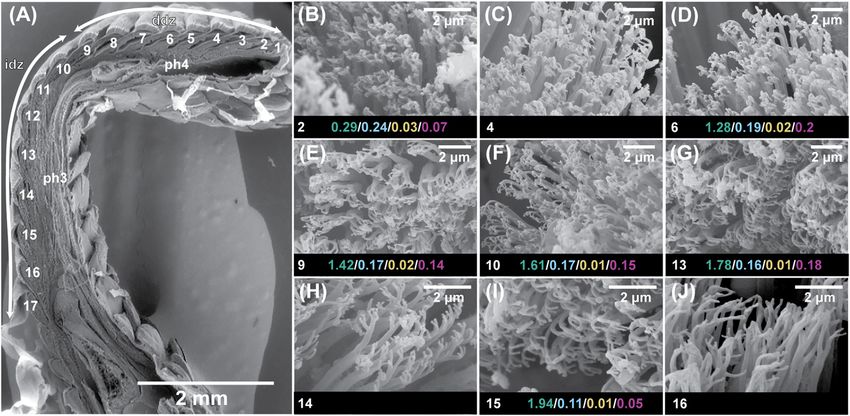
Russell and Garner                                                                                                                                 Fundamentals of Lizard Adhesive Systems

TABLE 1 | Data reported for setal dimensions for geckos in general and for Gekko gecko, indicating the variability in the values. For Gekko gecko, which has been used
extensively in research focusing on gecko adhesion, the values reported span large ranges but generally are not accompanied by information as to where on the toe pad
the measurements were taken from. Apart from a few early investigations, most of the values reported are taken from papers published between 2005 and 2016.

Setal length (µm)                     Setal basal diameter (µm)                         Setal density (mm-2)                       Spatula width (µm)                       Spatula length (µm)

Geckos
  20–10028                                         1–228                                                                                  0.28,24,31                                 0.56,7
  30–12010,30                                      1–523                                                                                0.2–0.36,7,18
  30–1306,7,11,16                                 5–106,7                                                                               0.2–0.511,13
  80–12012                                         2024
  10023
  11029

Gekko gecko
 30–1301,20,25                                      1–22                                         530027                                    0.127                                     0.23,4
 80–10026                                          2.518                                         140006                                  0.1–0.225                                    0.39
 9022                                              3–519                                                                             0.24,5,9,15,17,19,21
 1103,4,15                                        4.23,9,15                                                                             0.2–0.51,20
 1209                                                54
                                                  5–1025
                                                    1022

The sources of the data are indicated by superscript numbers in the table and refer to the following publications: 1Alexander (2006); 2Alibardi and Toni (2005); 3Autumn and Gravish (2008);
4
 Autumn and Hansen (2006); 5Autumn et al. (2006); 6Bhushan (2007); 7Bhushan and Sayer (2007); 8Kellar and Bogue (2008); 9Chen et al. (2008); 10Dalla Valle et al. (2007); 11Del Campo
and Arzt (2007); 12Dellit (1934); 13Filippov and Popov (2006); 14Gao et al. (2005); 15Gravish et al. (2008); 16Hallahan et al. (2008); 17Hansen and Autumn (2005); 18Hiller (1968); 19Huber
et al. (2005); 20Hui et al. (2007); 21Lee et al. (2007); 22Maderson (1964); 23Niewiarowski et al. (2016); 24Northen et al. (2008); 25Pugno and Lepore (2008); 26Rizzo et al. (2006); 27Sun et al.
(2005); 28Toni et al. (2007); 29Yao and Gao (2006); 30Yu et al. (2006); 31Xu et al. (2015).

TABLE 2 | Data ranges furnished by (1) Ruibal and Ernst (1965), (2) Schleich and Kästle (1986) and (3) Bauer (1998) for setal dimensions of individual species of geckos.

Species                                            Family                   Setal length (µm)               Setal basal diameter (µm)                   Setal density (mm2)                   Source

Amalosia lesueurii                          Diplodactylidae                        17+                                   1.5–3+                                 150,000                         (2)
Bavayia cyclura                             Diplodactylidae                        32*+                                   1.3+                                  429,000                         (3)
Bavayia sauvagii                            Diplodactylidae                        31*+                                   1.5+                                  335,000                         (3)
Correlophus sarasinorum                     Diplodactylidae                        36*+                                   1.3+                                  134,000                         (3)
Dactylocnemis pacificus                      Diplodactylidae                     17.5–20+                                  1.5+                                  200,000                         (2)
Dactylocnemis pacificus                      Diplodactylidae                        17+                                    1.5+                                  280,000                         (3)
Eurydactylodes vieillardi                   Diplodactylidae                        19+                                                                          363,000                         (3)
Naultinus elegans                           Diplodactylidae                        15+                                     0.8                                  621,000                         (3)
Naultinus rudis                             Diplodactylidae                        21+                                     0.8                                  490,000                         (3)
Pseudothecadactylus lindneri                Diplodactylidae                        37*+                                   1.2+                                  142,000                         (3)
Rhacodactylus auriculatus                   Diplodactylidae                        38*+                                                                         172,000                         (3)
Toropuku stephensi                          Diplodactylidae                        17+                                   0.8                                    458,000                         (3)
Woodworthia maculata                        Diplodactylidae                        13+                                   0.6                                    342,000                         (3)
Chondrodactylus bibronii                    Gekkonidae                             96*+                                 3.5*+                                   16,000*                         (2)
Hemidactylus bouvieri                       Gekkonidae                           10–50+                               1.5–2.5+                                  110,000                         (2)
Gekko gecko                                 Gekkonidae                      75–108+ 30–130+                        3–4.5*+2,2–4.7*                              14,400*                         (2)
Gekko kuhli                                 Gekkonidae                           24–91+                                                                                                         (2)
Gekko vittatus                              Gekkonidae                          37–78*+                                  3.5*+                                  25,600*                         (2)
Tarentola caboverdiana                      Phyllodactlidae                      27–68+                                  4–4.5*                                 26,000*                         (2)
Thecadactylus rapicauda                     Phyllodactylidae                    50–60*+                                   1.8+                                   6,000                          (2)
Aristelliger praesignis                     Sphaerodactylidae                   46–57*+                                   1.3+                                                                  (1)
Sphaerodactylus cinereus                    Sphaerodactylidae                     10–65                                                                                                         (1)

Values that fall entirely within the ranges of setal dimensions reported for Chondrodactylus bibronii in Figure 1 are denoted by an asterisk (*), and those that fall entirely within the ranges of
setal dimensions reported for Gekko gecko in Figure 2 are denoted by a plus sign (+).

(encompassing representatives of three families, each of which                                       (Figure 2). Further evidence of this overlap of setal
has independently evolved adhesive toe pads–Russell and                                              dimensions between C. bibronii, G. gecko and other species of
Gamble, 2019), reveals that the majority of the variation                                            gecko is evident from the data compiled by Ruibal and Ernst
reportedly encompassed by these eight species (Peattie, 2007)                                        (1965) and Schleich and Kästle (1986), as presented in Table 2.
is subsumed within the range of variation displayed by C.                                               Although the clinging performance of whole animals (Irschick
bibronii. The same is true (even more so) for the setal length                                       et al., 1996) has been correlated with toe pad area, it is evident
and setal stalk diameter variation shown by Gekko gecko                                              from transect surveys of the toe pads that setae in different

Frontiers in Mechanical Engineering | www.frontiersin.org                                        3                                                  January 2021 | Volume 6 | Article 621741
Russell and Garner                                                                                                                      Fundamentals of Lizard Adhesive Systems

 FIGURE 1 | Setal dimensions (length, width, and density) recorded for digit IV, left pes of the gekkonid gecko Chondrodactylus bibronii showing variation in relation
 to location on the toe pad. Data are presented for a proximal (prox), intermediate (int) and distal (dist) scansor (see inset of the ventral view of the digit in the upper right
 corner of the figure). The different symbols on each graph at the left indicate singular values reported for setal length, width and density for eight gecko species by Peattie
 (2007) (see legend). The vertical dashed line to the right of the symbols for each graph represents the range of values expressed by Chondrodactylus bibronii as
 measured by Webster et al. (2009). The lower right inset is a schematic of the setae on the proximal, intermediate and distal scansors. Below this schematic are values of
 particular setal lengths (SL; µm), widths (SW; µm) and densities (SD; mm−2) that correspond to proximal and distal stations along the proximodistal length of each scansor
 (distal to the right).

locations differ in aspect ratio (and therefore bending stiffness)                           setae are idealized as identical structures along the length of the
(Figures 1 and 2) and the number of spatulate tips that the setae                            toe pads, this would suggest that those scansors with the greatest
carry (Figure 4) (Russell et al., 2007). For Gekko gecko, the most                           surface area contribute a proportionally greater amount of the
proximal fibrils recognizable as true setae (with spatulate tips) are                         total adhesive force available to the digit. Average setal tip width
carried on scansor/lamella numbers 14 and 15 (Figure 4H,I).                                  and average setal tip area gradually decrease from distal to
These bear relatively few tips (Figure 4H,I) (certainly not the                              proximal along the length of the toe pad, however, as does
hundreds to thousands typically stated to characterize the setae of                          total setal tip area. Together with changes of setal density and
this species). They are relatively short and range down to less than                         setal stalk diameter along the length of the toe pad (Figures 1 and
25 µm at the proximal ends of these plates (Figure 3E). Bending                              2), the variation of setal structure along the digital transect is
stiffness and the number of adhesive contacts likely directly                                bewilderingly complex. What this means for the potential
influence adhesive force production, thus we find it probable                                  generation of adhesive force at any station within the toe pad
that setae from various regions of the toe pad differentially                                is unknown. It is evident, however, that bringing setae into close
contribute to total adhesive force capacity. In addition to                                  enough contact with the locomotor substratum to enable them to
variability in dimensions, fibrillar outgrowths on the subdigital                             generate van der Waals and frictional adhesive attachment will be
surface of gecko vary considerably in form. The 16th subdigital                              restricted to certain patches on any given footfall (Russell and
plate (Figure 4J) of Gekko gecko bears filaments that are short,                              Johnson, 2007; Russell and Johnson, 2014) rather than the
bifid at their distal tips and lack spatulae. Such forms may very                             entirety of the toe pad creating such engagement. It is possible
well be capable of generating considerable van der Waals                                     that there is compensatory tradeoff along the length of the toe pad
interactions, but whether such interactions are capable of                                   with regard to setal dimensions and configuration such that each
supporting whole animal attachment and locomotion is                                         localized area has the same potential for adhesive force generation
not known.                                                                                   (Russell et al., 2007), but we know nothing about the relative force
    Beyond the setae themselves, the data summarized in Figure 4                             generation attributes of these different setal configurations.
for the Tokay gecko indicate that there are major differences in                                 Such patterns of variation have consequences for the way in
the surface area of the individual scansors/lamellae that make up                            which we conceive and design simulacra of setae. Our
the toe pad, with this increasing from distal to proximal. If all                            understanding of the adhesive capacity of individual setae is

Frontiers in Mechanical Engineering | www.frontiersin.org                                4                                              January 2021 | Volume 6 | Article 621741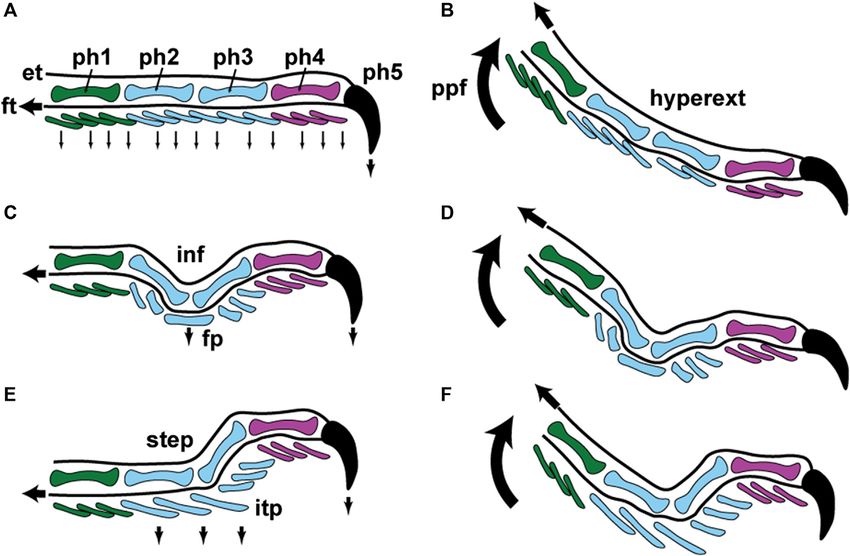
Russell and Garner                                                                                                                     Fundamentals of Lizard Adhesive Systems

 FIGURE 2 | Setal dimensions recorded for digit IV, right manus of the gekkonid gecko Gekko gecko showing variation in relation to location on the toe pad. Data are
 presented for a proximal (prox), intermediate (int) and distal (dist) scansor (see inset of the ventral view of the digit in the upper right corner of the figure). The different
 symbols on each graph at the left indicate singular values reported for setal length and width for eight gecko species by Peattie (2007) (see legend). The vertical dashed
 line to the right of the symbols for each graph represents the range of values expressed by Gekko gecko as measured by Russell et al. (2007). The lower right inset is
 a schematic of the setae on the proximal, intermediate and distal scansors. Below this schematic are values of particular setal lengths (SL; µm) and widths (SW; µm) that
 correspond to proximal and distal stations along the proximodistal length of each scansor (distal to the right).

based upon a single set of observations of filaments taken from an                            the variation must be assessed and appreciated before it can be
undisclosed part of a digit (Autumn et al., 2000). These                                     incorporated into such models. Much of the variation seen (at
observations have neither been repeated for setae with other                                 least within a single species) likely relates to the functioning of the
configurations in the same species, nor for those from other                                  entire setal field and the way in which it is applied to and released
species. There is still much to learn about the mechanical                                   from the substratum. It is the entire setal field upon which the
properties of gecko setae and it is unlikely that the single                                 animal relies to ensure that, upon each footfall, sufficient contact
available set of observations (Autumn et al., 2000) suffices to                               is made to ensure effective attachment. A deeper understanding
explain the properties of all observed configurations. The current                            of the local regional variation in structure of the setae and their
perception of the total clinging ability of a single digit of the                            patterning into fields is required for more effective (and purpose-
Tokay gecko (Gekko gecko), as extrapolated from multiplying the                              specific) fields of artificial biomimetic fibrils to be developed
adhesive force of a single (idealized) seta (Autumn et al., 2000) by                         (Russell et al., 2019). Finding ways of simplifying such surveys of
the stated number of setae carried on such a digit (Sun et al., 2005;                        variation is paramount and we here turn to taxa that exhibit,
Autumn et al., 2006; Tian et al., 2006; Hui et al., 2007) is likely a                        within their ranks, evolutionary transitions from non-adhesively
considerable overestimate. Whole animal observations of clinging                             to adhesively competent digits. Such transitions offer the promise
force per digit result in performance values lower by an order of                            of determining what is minimally necessary and sufficient for a
magnitude or more (Irschick et al., 1996; Autumn et al., 2000,                               functional digital adhesive system to become incorporated into
2002; Autumn and Peattie, 2002). The discrepancy between these                               the pre-existing locomotor mechanics of lizards.
two approaches may reside, to some extent in the variability of the
number of spatulate tips per seta along the length of the toe pad
(Figure 4), differences in setal density at different stations on the                        EVOLUTIONARY TRANSITIONS
toe pad (Figures 1–3), and the number of setae actually making
contact with the surface (which may be reduced based on the                                  The gekkotan adhesive system has been both gained and lost on
surface roughness of the substratum).                                                        multiple occasions (Gamble et al., 2012; Gamble, 2019). Although
    There is still much to learn about how individual setae perform                          geckos are widely known for their possession of adhesive toe pads,
and how that performance relates to their structural properties. It                          somewhere in the region of 40% of the 1800+ living species lack
is likely that much of this can be achieved through modeling, but                            them. Peattie (2008) opined that the discovery of “an extant

Frontiers in Mechanical Engineering | www.frontiersin.org                                5                                             January 2021 | Volume 6 | Article 621741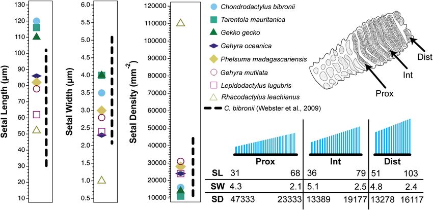
Russell and Garner Fundamentals of Lizard Adhesive Systems
FIGURE 3 | Scanning electron microscopic visualization of the sagittally-sectioned fourth digit of the right manus of the Tokay gecko, Gekko gecko. (A) Visualization
of the entire toe pad showing the location and relative dimensions of the scansors and lamellae (numbered sequentially 1–17 from distal to proximal). The distal region of
the digit (ddz) carries a series of scansors (1–9) that lie beneath the fourth phalanx (ph4). The intermediate region of the digit (idz) carries a series of scansors and lamellae
(10–17) that lie beneath phalanx 3 (ph3), the transition from scansor to lamella occurring between plates 13 and 14. Lamellae are distinguished from scansors by not
being served by branches of the lateral digital tendon system. Panels (B)–(E) depict enlargements of the individual scansors and lamellae, showing the relative lengths of
the plates and the configuration of the fields of setae carried on their outer surfaces. On each plate the lengths of the filaments decrease from a maximum at the distal end
to a minimum at the proximal end, and from distal to proximal along the toe pad the lengths of the filaments decrease, although there is overlap of the span of lengths
between adjacent plates. For plates 1–7, 10–11, and 17 maximum filament length (in µm) is indicated in blue and minimum filament length (in µm) in green. Data from
Russell et al. (2007).
intermediate” would greatly enhance our understanding of how beneath which reside enlarged friction plates (Figure 5A–C).
the gekkotan adhesive system arose. Russell and Gamble (2019) Gonatodes vittatus (Figure 5F–H) has relatively shorter digits
identified a number of candidate taxa for exhibiting such a with a less marked inflection, fewer ventral scales, and a flatter
transition, but only the genus Gonatodes has been studied in proximal portion. Gonatodes humeralis (Figures 5L–N) lacks a
detail in this regard (Russell et al., 2015; Higham et al., 2017). marked digital inflection and bears noticeably enlarged scales at
Examination of the setal fields of Gekko (see above), reveals the base of the digits. Full details of the morphological features of
that the simplest of its spatulated setae are relatively short and the digits of these three species are provided by Russell et al.
subdivided into few terminal branches (Figure 4H). We do not (2015). The salient points of the transition to adhesive
know how effective these are in contributing to the total adhesive competence constitute a correlated suite of small shifts leading
effectiveness of that species. We do know, however, that the to the rather dramatic functional outcome of whole animal
relatively short, sparsely-branched setae of Gontodes humeralis adhesive competency (Higham et al., 2017).
(Russell et al., 2015) are sufficiently developed and numerous to In the shift from strongly inflected digits, in which friction
permit support of its own body mass at rest and during steady plates at the midpoint of the digit (Padian and Olsen, 1984;
vertical locomotion on a low-friction substratum (Higham et al., Russell and Bauer, 1990; Peattie, 2008) enhance traction (Figures
2017). Equivalent locomotor capabilities are not present in any of 5A–C and 6A–D), to the adhesively-competent digits of G.
its close relatives (Higham et al., 2017). Thus, Gonatodes provides humeralis (Figures 5L–N and 6E,F), the digits become
the opportunity, using appropriate phylogenetic comparison and relatively shorter (in relation to overall body size; Figure
the establishment of evolutionary polarity (Russell et al., 2015), to 5A,C,F,H,L,N). This shift is associated with a change in the
establish what might be the minimal set of modifications for the relative proportions of the proximal and distal regions of the
emergence of a functioning digital adhesive system that is digits (Figure 5B,G,M) and a greater discrepancy in size between
effective at the whole organism level. the scales on the underside of the digits in their proximal and
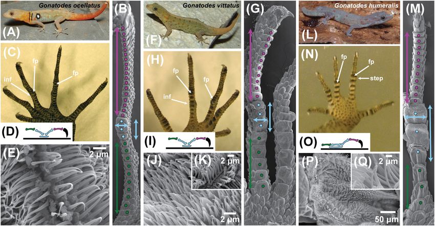
Trends in digit evolution within the genus Gonatodes are distal regions (Figures 5B,G,M and 6A,C,E). Those of the
depicted in Figure 5, which shows the pertinent digital proximal zone become relatively larger (both longer and
features of three exemplar species. Gonatodes ocellatus has wider), markedly fewer in number and exhibit greater overlap
digits that are elongate and slender with a marked inflection, between successive scales (Figure 5B, 5G, and 5M). The relative
Frontiers in Mechanical Engineering | www.frontiersin.org 6 January 2021 | Volume 6 | Article 621741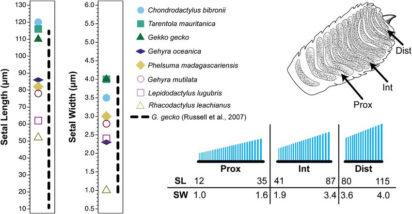
Russell and Garner                                                                                                                  Fundamentals of Lizard Adhesive Systems

 FIGURE 4 | Scanning electron microscopic visualization of the sagittally-sectioned fourth digit of the right manus of the Tokay gecko, Gekko gecko. Panel (A) is
 identical to panel (A) in Figure 3. Panels (B)–(J) depict enlargements of the filaments carried on each plate. For scansors/lamellae 2, 6, 9, 10, 13, and 15 the following
 data are provided: scansor/lamella surface area (mm2) in green/average setal tip width (µm) in blue/average setal tip area (µm2) in yellow/total setal tip area per scansor/
 lamella (mm2) in purple. Data from Russell et al. (2007).

enlargement of the subdigital scales is particularly evident for the                           In all three exemplar species (Figure 5), the epidermal
friction plates (Padian and Olsen, 1984; Russell and Bauer, 1990)                          filaments on the underside of the toes are longest on the
(Figures 5B,G,M and 6C,E) that lie beneath the digital inflection                           friction plates. In G. ocellatus, they are tapered spinules, the
(labeled “inf” on Figures 5C,5H and 6C). In G. humeralis                                   longest being about 3.2 µm in length (Figure 5E). In G. vittatus,
(Figure 5M), this enlargement is accompanied by a flattening                                the longest of these filaments are about 4.0–6.0 µm in length, with
of the proximal part of the digit such that all of the scales on the                       bifid tips, the split occurring at a point about two thirds of their
underside of the proximal region (Figure 5M) are co-planar and                             height from the base (Figure 5K), appearing much like the
contact the substratum as a continuous strip. The inflection of the                         filaments on the proximal lamellae of the digits of Gekko
digit thus shifts from a “v”-like configuration when viewed in                              gecko (Figure 4J). This bifurcation is thought to be associated
profile (Figure 5D,I) to a step (Figure 5N,O and 6E).                                       with friction enhancement (Lange, 1931; Ruibal and Ernst, 1965;
    Within the digits, the changes in digital proportions and                              Schleich and Kästle, 1979; Peattie, 2008; Müller and Hildenhagen,
configuration are accompanied by changes in the arrangement                                 2009; Spinner et al., 2013; Russell et al., 2015). In Gonatodes
of the phalanges. The basalmost phalanx (Figures 5D,I,O and                                humeralis, the lengths, diameters, spacing and form of the
6C,E) increases in relative length whereas the penultimate                                 subdigital spinules are similar to those of G. ocellatus and G.
phalanx (Figures 5D,L,O and 6C,E) becomes relatively                                       vittatus for the scales on the underside of the distal region of the
shorter. The intermediate phalanges (Figures 5D,L,O and                                    digit (Russell et al., 2015). On the incipient toe pads, however, the
6C,E) retain their relative proportions but become reoriented                              distally-situated epidermal outgrowths range from 10.0 to
such that the proximal one becomes linearly aligned with the                               15.0 µm in length, their density is relatively low and the
proximalmost phalanx (Figures 5D,L,O and 6E) and the distal                                spacing relatively great (Russell et al., 2015). These are true
one becomes more vertically-oriented, resulting in the “v”-like                            setae that are divided terminally and carry spatulate tips of
configuration of these two phalanges (Figures 5D and 6C)                                    about 0.12 µm wide (Figure 5P,Q). Such setae are similar in
transforming into a step (Figures 5O and 6E). This transition                              form to those on the most proximal scansors of Gekko gecko
is associated with a proximal extension of the friction plate area                         (Figures 3E and 4H). Unlike the latter, however, the setae on each
(Figures 5D,L,O and 6E) to become more extensive beneath the                               plate are shorter distally than they are more proximally (13.0 vs
proximal part of the digit, resulting in the formation of an                               15.1µm), a pattern also observed in Anolis (see below; Garner
incipient toe pad (Figures 5M,N and 6E).                                                   et al., 2020). The branched, spatula-bearing setae occur only at the

Frontiers in Mechanical Engineering | www.frontiersin.org                              7                                            January 2021 | Volume 6 | Article 621741
Russell and Garner Fundamentals of Lizard Adhesive Systems FIGURE 5 | Trends in digit form within the sphaerodactylid gekkotan genus Gonatodes. This genus is regarded as lacking subdigital toe pads but exhibits digital adhesive competence in one of its species. Shifts in form and proportion in association with the acquisition of whole animal adhesive competence are illustrated using three exemplar species (A) Gonatodes ocellatus, (F) G. vittatus and (L) G. humeralis. For each species a set of illustrations [panels (A)–(E) for G. ocellatus; (G)–(K) for G. vittatus and (M)–(Q) for G. humeralis) is used to illustrate the trends to whole animal adhesive competence in G. humeralis. Panels (B), (G) and (M) depict scanning electron micrographs of the ventral aspect of digit IV of the left pes (hind foot) of the three species, with features of interest indicated by color-coded symbols. Panels (C), (H) and (N) represent the ventral view of the left pes of the three species and panels (D), (I) and (O) depict a schematic of the lateral view of the skeleton of digit IV of the pes of each species, with the phalanges color coded to match the coding in panels (B), (G) and (M). Panels (E), (J) and (K), and (P) and (Q) are scanning electron micrographs of the filamentous outgrowths of the epidermis beneath the mid-digit inflection point of digit IV of the pes. Coding conventions are as follows: purple dots–scales beneath the distal region of the digit; purple double-headed vertical arrows–proximodistal extent of the distal region of the digit; purple phalanx–penultimate phalanx; blue dots–scales comprising the friction plate region of the digit; blue double-headed vertical arrows–length of the friction plate region; blue double-headed horizontal arrows–width of the friction plate region; blue phalanges–intermediate phalanges; blue ellipsoid–extent of the friction plate region below the intermediate phalanges; green dots–scales beneath the basal region of the digit; green double-headed vertical arrows–proximodistal extent of the basal region of the digit; green phalanx–basal phalanx; curved, black claw–ungual phalanx and claw. The blue and green dotted regions combined constitute the proximal region of the digit. Abbreviations: fp–friction plates; inf–digital inflection; step–step-like transition between the proximal and distal digital regions in G. humeralis. distal end of the incipient toe pad plates (Figure 5M) and give (Russell et al., 2007: fig. 5), the latter being multiply branched and way to spinules more proximally. Transition from simple tapered carrying spatulate tips. Thus, Tokay geckos also possess setae that spinules (there is a mixture of unbranched and branched spinules are carried on highly modified scales (lamellae–Figure 3A,E) that far proximally on such scales) to setae is thus evident within the lack the characteristics of scansors. Such scales bear a strong confines of a single scale. structural resemblance to the seta-bearing scales of G. humeralis Although Peattie (2008) suggested that “true adhesive pads and serve to support the idea of a transition from friction- require complex morphological elaborations within the toe” to enhancing to adhesion-promoting structures prior to the enable operation of an adhesive system, Gonatodes humeralis widening of the digits and the acquisition of features that demonstrates that this is not so, there being no modifications of provide the capability for actively controlling the adhesive the digital musculotendinous, circulatory and skeletal systems process via distoproximal digital hyperextension (Russell and that are generally considered to be necessary (Peattie, 2008) for Bels, 2001). the operation of a functional adhesive system in geckos. In terms When climbing vertical, low friction substrata (Higham et al., of their anatomy, the seta-bearing scales of G. humeralis are not 2017), Gonatodes humeralis employs limb and digit kinematics scansors (Russell, 2002), but instead are more akin to the basal that are essentially unchanged from those of lizards in general lamellae found in many geckos. The latter bear elaborate (Brinkman, 1980; Russell and Bauer, 2008) (Figure 6A,B). The epidermal outgrowths, but are not associated with a lateral digits are placed onto the substratum and withdrawn from digital tendon network (see below). The lamellae of the Tokay contact with it such that the distal ends of the digits are the gecko (Gekko gecko) carry epidermal outgrowths that range in last regions to be withdrawn from it. In Gekko gecko, the opposite length from 1.2 µm at their proximal end to over 12.0 µm distally occurs, enabled by the specialized musculature of the digits that Frontiers in Mechanical Engineering | www.frontiersin.org 8 January 2021 | Volume 6 | Article 621741
Russell and Garner                                                                                                                     Fundamentals of Lizard Adhesive Systems

 FIGURE 6 | Schematic representations of lizard digit kinematics during locomotion and inferences about how incipient toe pads became incorporated into this
 pattern. Color conventions for the phalanges (ph1–ph5) and ventral scales of the digit are identical to those in Figure 5, panels (B), (D); (G), (I); and (M), (O). (A) and (B)
 fourth digit of hind foot in lateral view when applied to the substratum (A) and when being raised from contact with it (B). In panel (A) the digit is applied to the substratum
 with the first four phalanges (ph1–ph4) aligned with each other and the fifth (ph5) and its surrounding claw sheath being driven into the substratum via contraction of
 the flexor muscles of the digit through the flexor tendons (ft–proximally-pointing black arrow). The digit thus operates as a directional device along its long axis, the
 phalanges behaving as a series of compression struts with the reaction forces driving the ventral surface of the digit (series of small, ventrally-pointing black arrows) into
 contact with the substratum. In panel (B) the foot is peeled from the substratum by the heel being raised from the surface via pedal plantar flexion (ppf–curved, upwardly-
 pointing arrow). Scales are lifted away from the substratum in a proximal to distal sequence and the claw finally being released from contact through contraction of the
 digital extensor muscles operating through the extensor tendon (et–proximally-pointing black arrow). The digit is bowed as a result of this, becoming hyperextended
 (hyperext). Panels (C) and (D) depict the same situation for a digit with a mid-digital inflection (inf—as encountered in many climbing lizards and as seen in Gonatodes
 ocellatus and G. vittatus (Figure 5, panels C, D; and H, I). Because of the inflection only the claw (ph5) and the friction plates (fp) make contact with the surface. The
 friction plates are driven into contact with the substratum (black, ventrally-directed arrow beneath the friction plates) through tension placed on the flexor tendon (ft) In
 Gonatodes the ventral surface of the friction plates is clad in elongated filaments that may be bifid at their tips to enhance frictional contact (as in G. vittatus–Figure 5,
 panel K). Release of the digit (D) occurs via hyperextension, as depicted in panel B. Panels (E) and (F) show application and release for a gecko with incipient toe pads,
 such as Gonatodes humeralis (Figure 5, panels M, N). The inflection of the digit is modified into a step (step: Figure 5, panel O) and the friction plates are transformed
 into an incipient toe pad (itp) (Figure 5, panels M, N), the plates of which bear a free distal margin on which spatulate-tipped setae (Figure 5, panel Q) are carried. Digital
 mechanics relation to attachment and detachment of the incipient toe pad are essentially unchanged from the patterns depicted in panels A–D.

allows their distal ends to be raised from the substratum while                              removal of the incipient toe pads from adhesive contact with the
their proximal ends remain in contact (Russell, 2002), a process                             substratum (Figure 6E and F). This breaking of adhesive contact
known as hyperextension. When the digits of Gekko gecko make                                 is accommodated within a pre-existing pattern of digit kinematics
contact with the substratum, they do so with their proximal ends                             (Figure 6). Adhesive competence of the digits of G. humeralis has
first. The distal parts, carrying the toe pads, are unfurled after this.                      been subsumed into an essentially unchanged pattern of digit
Gonatodes humeralis, however, like lizards in general (Figure                                mechanics. Geckos with incipient toe pads provide insights into
6A–D), removes its digits from the locomotor surface by raising                              the most basic aspects of digit configuration and kinematics
its heel first and rising onto the tips of the digits, thereby                                compatible with the deployment of an effective adhesive
removing the ventral surface of the digits from contact with                                 system that can assist locomotion (Figure 6).
the substratum in a proximal to distal sequence (Higham et al.,                                  Gonatodes humeralis, unlike other species of Gonatodes, can
2017). This results in hyperextension of the digits (as it does in                           scale smooth, low friction vertically-oriented substrata (Higham
lizards in general), and thus hyperextension is involved with                                et al., 2017). Although the adhesive forces generated are rather

Frontiers in Mechanical Engineering | www.frontiersin.org                                9                                             January 2021 | Volume 6 | Article 621741
Russell and Garner                                                                                                           Fundamentals of Lizard Adhesive Systems

                                                                                    GECKO–ANOLE CONVERGENCE
                                                                                    The evolutionary phenomenon of convergence (the independent
                                                                                    acquisition of form and function–Kuhn et al., 2020) potentially
                                                                                    provides evidence for determining the most fundamental
                                                                                    attributes of complex adaptations (sensu Frazzetta, 1975).
                                                                                    Independent but structurally similar “solutions” to challenges
                                                                                    imposed by the environment provide insights into the
                                                                                    evolutionary responses possible. The adhesive toe pads of
                                                                                    geckos and anoles are regarded as convergent adaptations
                                                                                    (Russell and Garner, in review), their adhesive toe pads having
                                                                                    arisen completely independently. This provides the opportunity
                                                                                    to examine which attributes are repeated in the two lineages and
                                                                                    thus to attempt to establish what is necessary and sufficient for
                                                                                    adhesively competent digits to be incorporated and integrated
                                                                                    into the lacertilian pattern of locomotion.

 FIGURE 7 | Comparison of the maximum adhesive clinging abilities of
 lizards (recorded as Newtons per gram of body mass). Skinks exhibit very
 modest output. Anoles exert, at their greatest output, more adhesive force
 than skinks. Geckos maximally exert adhesive forces well above that of
 the best-performing anoles (although lesser-performing geckos overlap with
 the better-performing anoles). Gekko gecko represents the greatest adhesive
 output yet measured for geckos. In comparison, Gonatodes vittatus
 generates no adhesive attachment forces whereas G. humeralis performs as
 well as many anole species and approaches the lower end of the adhesive
 performance spectrum of geckos with toe pads. Data from Higham et al.
 (2017) and Irschick et al. (1996).

low by gecko standards, they are comparable to those generated
by anoles (Figure 7). Given that the setae of G. humeralis are
similar in form to the simplest setae of Gekko gecko (Figure 4H),
it can be inferred that the latter are also capable of generating
adhesive interactions.
    To this point, we have established the following. (I) That the
setae of geckos with structurally complex toe pads, such as Gekko
gecko and Chondrodactylus bibronii, exhibit clinal variation in
setal dimensions and form along the length of the digit, with the
setae located proximally on the toe pad being relatively short and
only modestly branched. (II) That similarly built setae are present
on the incipient toe pads of geckos such as Gonatodes humeralis,
which exhibits little in the way of the “complex morphological
adaptations” thought to be required for the operation of an
                                                                                     FIGURE 8 | (A) Clinal series of the epidermal outgrowths present on the
adhesive system (Peattie, 2008). The adhesive capabilities of G.                     subdigital pad of anoline lizards (Peterson and Williams, 1981). True setae are
humeralis suggest that the simpler setae occurring more                              outgrowths between 10–30 μm in length that carry a single, expanded
proximally on the digits of Gekko gecko (Figure 4H,I) are also                       spatulate tip. Seta-prong intermediates are outgrowths 5–20 μm in
adhesively effective. These observations demonstrate that an                         length with flattened tips. Prongs terminate in a blunt tip with a slight taper and
                                                                                     are between 5–20 μm in length. Spikes possess straight or recurved, pointed
operational adhesive system is possible in the absence of either
                                                                                     tips and are between 5 and 15 μm in length. Spines are outgrowths up to
complexly branched setae bearing “100–1,000. . .spatulae”                            5 μm in length with pointed recurved tips. (B) Trends of setal morphometrics
(Autumn and Hansen, 2006) and complex anatomical                                     along subdigital pad regions and lamellar zones of Anolis equestris (Garner
modifications promoting attachment and detachment. We now                             et al., 2020). Setal length increases and setal base diameter decreases
ask (III) whether such basic levels of organization are evident in                   proximodistally along pad regions. Setal length is maximal in the intermediate
                                                                                     zones of lamellae. Setal base diameter decreases proximodistally along
toe pads of other taxa, turning our attention to the dactyloid
                                                                                     lamellar zones. Lamella length decreases proximodistally.
iguanian genus Anolis to explore this possibility.

Frontiers in Mechanical Engineering | www.frontiersin.org                      10                                            January 2021 | Volume 6 | Article 621741
Russell and Garner Fundamentals of Lizard Adhesive Systems
Convergence of the Adhesive Fibrils of setae and setal fields into consideration. Anoles appear to
Geckos and Anoles compensate for the lack of structural hierarchy of their
As in geckos, the epidermal outgrowths of the subdigital pads of setae by possessing greater setal density compared to geckos
Anolis (Figure 8A) vary considerably in form along the (Garner et al., 2020), although structural hierarchy of setae
proximodistal axis of the digit (Figures 3–5) and five may be important in other aspects of the gekkotan adhesive
morphotypes have been recognized (Peterson and Williams, system (Persson, 2003; Yao and Gao, 2007). Peterson et al.
1981): (1) true setae, outgrowths 10–30 μm in length with (1982) also note that anoles appear to combat increased
expanded, spatulate tips, (2) seta-prong intermediates, 5–20 μm adhesive demands related to increases in body size by
in length with flattened tips, (3) prongs, 5–20 μm in length possessing greater setal densities. Not all geckos, however,
terminating in blunt tips with a slight taper, (4) spikes, 5–15 μm are characterized by such densities, and the setae of A.
in length with straight or recurved, pointed tips, and (5) spines, up equestris are only 1.75–2.5 times as densely packed as those
to 5 μm in length with recurved, pointed tips. All lamellae (scales of Naultinus elegans, the gecko so far examined to exhibit the
bearing true setae) of Anolis with well-developed subdigital pads highest density and shortest setae, and these are also not
are thought to display a proximodistal clinal gradation of all 5 profusely branched (Bauer, 1998). Table 2 reveals that
morphotypes (from spines to setae) on each lamella, similar to the many diplodactylid geckos have short, slender, densely
clinal variation of epidermal outgrowths on the scales of the packed setae, with values greatly exceeding those for
incipient toe pads of Gonatodes humeralis (Russell et al., 2015). gekkonid geckos (the group to which Chondrodactylus and
As in gekkotans, it is the true setae and their spatulae that are Gekko belong). Very little is known about the adhesive
responsible for the majority of adhesive force capacity in Anolis. In capabilities of diplodactylid geckos, although this is now
Anolis, however, the setae are unbranched (Ruibal and Ernst, 1965; beginning to be explored (Pillai et al., 2020). This again
Williams and Peterson, 1982; Stork, 1983). reveals that there is much more to be learned about the
A recent investigation of the morphometrics of the setal arrays variation in structure and function of gekkotan setae,
of the Cuban knight anole (Anolis equestris), a species similar in cautioning against making sweeping generalizations that
size to Gekko gecko, reveals that its setae increase in length and appear to characterize geckos overall.
decrease in basal diameter proximodistally along regions Theabove-mentioned calculations of adhesive performance
(proximal, intermediate, distal) of the subdigital pad of geckos and anoles, based on setal morphometrics (Garner
(Figure 8B) (Garner et al., 2020). Within a single lamella, et al., 2020), are supported by whole animal observations.
however, setal length is greatest in the intermediate region, Ruibal and Ernst (1965) qualitatively observed gecko and anole
whereas setal basal diameter decreases proximodistally adhesive locomotion on a vertical raceway and noted no
(Figure 8B), a pattern similar to the dimensions observed on obvious differences in performance. More comprehensive
the incipient toe pads of Gonatodes humeralis (see above). Setal work by Irschick et al. (1996) corroborated this finding and
density remains relatively consistent along the entire subdigital found that static clinging performance of geckos and anoles is
pad and along regions (proximal, intermediate, distal) of not markedly different (Figure 7). The general similarity of
individual lamellae. setal form and material properties of anoles and geckos
The profuse branching noted for the archetypal gecko seta indicate the requirement for similar loading conditions
(Autumn et al., 2000) should result in greater adhesive force (normal load followed by shear load) because measurements
production compared to an unbranched seta of similar size, of whole animal adhesive performance indicate that their
such as those of Anolis (Arzt et al., 2003; Peattie and Full, 2007; adhesive systems can be engaged in the same manner
Murphy et al., 2009; Garner et al., 2020). Thus, if geckos and (Irschick et al., 1996). The means by which setal loading
anoles differed only in the nature of the structural hierarchy of and unloading occurs in anoles, however, differs from that
their setae, the setal fields of gekkotans, when scaled against typically attributed to geckos (Russell and Bels, 2001), but is
body mass, should be able to induce greater adhesive forces. similar to that employed by Gonatodes humeralis (Figure
Gekkotan and anoline setae and setal fields, however, differ not 6E,F), with subdigital pad retraction progressing
only in setal size and number of spatulae per seta, but also in proximodistally (as opposed to the distoproximal pattern
the number of fibrils present per unit area (Garner et al., 2020). thought to be typical of geckos). This pattern of release of
Indeed, when compared to setal density patterns in the setae is consistent with setal length increasing proximally
Chondrodactylus bibronii, those of Anolis equestris are along the length of each lamella (Figure 8B), suggesting that
33.3–78 times as densely packed. Application of the clinal variation in setal length is related to the biomechanics of
Johnson-Kendall-Roberts (JKR) theory of elastic contact to subdigital pad peeling (Johnson and Russell, 2009; Garner
the assessment of adhesive performance of gekkotan and et al., 2020), although further empirical work is needed to
anoline setal arrays, in conjunction with the comprehensive validate this.
morphological data obtained for the setal fields of Gekko gecko
(Russell et al., 2007) and Anolis equestris (Garner et al., 2020), Convergence of the Digital Anatomy of
reveal that adhesive performance of gekkotan and anoline setal Geckos and Anoles
arrays should theoretically be similar when taking the The anatomical modifications generally associated with the
differences in the morphology and configuration of their adhesive system in geckos (Peattie, 2008) are only modestly
Frontiers in Mechanical Engineering | www.frontiersin.org 11 January 2021 | Volume 6 | Article 621741Russell and Garner Fundamentals of Lizard Adhesive Systems
FIGURE 9 | Schematic representation of the basic components of the lacertilian digital adhesive system assembled as a model for the operation of this basic
mechanism. Information about structure is derived from a combination of anoles and gekkotan taxa exhibiting incipient toe pads. (A) Diagrammatic lateral view of digit IV.
Phalanges 1–5 (ph1–ph5) and their associated ventral scales (color coded to match the phalanges) that bear micro-ornamentation that is modified into unbranched setae
bearing a single spatulate tip on the free margin of enlarged scales (lamellae/scansors; lam/sca) situated beneath phalanges 2 and 3. The rows of setae on each
lamella/scansor increase in length from distal to proximal. Each lamella/scansor has an expanded proximal zone with an extensive dermal and subdermal core. Its ventral
integument consists of epidermis and a dermal stratum compactum (sc) dominated by linearly-arranged (parallel with the long axis of the digit) collagen fibers that is
continuous with the lateral digital tendons (ldt) lying to either side of the phalanges. This sc/ldt continuum is linked to an aponeurotic network (ap) controlled by muscles in
the flexor compartment of the lower limb and anchored onto skeletal elements (sk) that limit the tensile loading placed upon it. Overlying the setal batteries is a flexible
compliance system (comp) that, when pressurized during application of the lamellae/scansors to the substratum, assists in aligning the setal batteries into a continuous
setal field along the length of the toe pad. Extensor (et) and flexor (ft) tendons, through the actions of their associated muscles, control the claw and flexion and extension
of the entire digit. Lamellae/scansors also possess a thin, flexible free margin (fm). (B) Diagrammatic lateral view of a single lamella/scansor with the setae applied to the
substratum in the adhesion mode. (C) Diagrammatic lateral view of a single lamella/scansor in the process of being released from adhesive contact with the substratum
as a result of digital hyperextension, driven by pedal plantarflexion (ppf).
represented by Anolis, being confined to skeletal (phalangeal) similar location to those of Gonatodes humeralis (Figures
and tendinous features (Russell and Gamble, 2019). The digits 5M,N and 6E,F). Associated with the toe pads of Anolis are
of Anolis exhibit little in the way of muscular modifications lateral digital tendons similar to those of geckos. These course
akin to those exhibited by at least some gecko lineages (Russell along the lateral and medial borders of the phalanges of the
and Gamble, 2019). Anolis does, however, incorporate a digit and branch to serve each lamella. As in geckos, the dense,
compliance-promoting cushioning system (Figure 9A) into collagen rich connective tissue of the lateral digital tendons is
its toe pads, in the form of hypertrophied lacunar cells of the continuous with the stratum compactum of the dermis of the
epidermis (Russell, 2016). This cushioning system is unique to lamellae (the lamellar dermis lacking a stratum laxum). Thus,
Anolis but is analogous and positionally similar to the vascular as in geckos (Russell, 1986), the lateral digital tendon/lamellar
(Russell, 1981) and adipose tissue (Russell and Bauer, 1988) dermis complex furnishes a tensile skeleton that provides
compliance structures found among geckos. connectivity between the setae and skeleton at the
Modifications of the intermediate phalanges enhance both metapodial-phalangeal joint capsules. This chain reinforces
the pressing of the toe pads onto the substratum during the junction between the integument and the underlying
attachment and their hyperextension during release (Figure tissues (Russell, 1986) and permits the tensile load imposed
6E,F). The toe pads of Anolis are located basally on the digits, on the setae (Peterson et al., 1982) to be channeled to points of
ventral to the location of the digital inflection (between resistance deep within the manus and pes, and to be regulated
phalanges 2 and 3 of digit IV) of its close relatives, in a in its magnitude.
Frontiers in Mechanical Engineering | www.frontiersin.org 12 January 2021 | Volume 6 | Article 621741Russell and Garner Fundamentals of Lizard Adhesive Systems
SYNTHESIS AND THE GENERATION OF A morphological modifications that it does show are related to
MODEL OF THE FUNDAMENTALS OF changes of scale size and number on the underside of the digits,
alterations of in phalangeal proportions and orientation, and
FORM AND FUNCTION OF THE DIGITAL transformation of some of the epidermal outgrowths on the
ADHESIVE SYSTEM OF LIZARDS expanded scales of the incipient toe pads into setae with spatulate
Our examination of transects of setal fields constituting the toe tips. There are no evident lateral digital tendons associated with the
pads of geckos, the evolutionary transition to functionally plates of the incipient toe pads, and it appears that tensile loading is
adhesive digits in the naked-toed gecko genus Gonatodes, and placed upon the setae and lamellae by gravitational loading,
the comparison of the structure of gecko and anole adhesive potentially restricting the circumstances in which this
systems has allowed us to deduce what we consider to represent rudimentary adhesive system can be engaged.
the most basic level of organization and mechanical operation of It is interesting to note that all geckos capable of supporting
such a system. Both setal structure, at its most elaborate, and the static and dynamic adhesion on vertical, low friction substrata,
configuration and operation of the entire adhesive system of the including Gekko gecko and Gonatodes humeralis, possess setae
Tokay gecko (Gekko gecko) are complex (Russell, 2002). They that exhibit some level of branching. Comparison of both
permit impressive feats of behavior that are assisted by the Gekko gecko and Gonatodes humeralis with Anolis reveals a
deployment of the adhesive apparatus (Autumn 2006), but are setal structure that is much simpler than that of Gekko gecko or
not typical of all geckos. Gonatodes humeralis. Anolis possesses unbranched setae that
Examination of the variability of setal structure exhibited by are relatively short and present in higher density (compared to
several species of geckos (and anoles) across the entirety of their that of gekkonid geckos–Table 2). Its clinging abilities
setal fields indicates that setae vary considerably in form and (Figure 7) are similar to those of many geckos (Irschick
dimensions depending on the particular location on the digit et al., 1996), despite the absence of structural hierarchy that
(Russell et al., 2007; Johnson and Russell, 2009; Webster et al., purportedly increases the adhesive capacity of a single fiber
2009; Russell and Johnson, 2014; Garner et al., 2020). Such data (Arzt et al., 2003; Peattie and Full, 2007; Murphy et al., 2009;
indicate that there is no single morphology of setae for the toe Garner et al., 2020). The higher setal density of Anolis is
pads of lizards bearing adhesive pads and that these parameters thought to compensate for the lack of branching in anoline
vary regionally, presumably reflecting the functional contribution setae (Garner et al., 2020). A number of species of Anolis also
of particular regions of the toe pads to the totality of the adhesive appear to respond to the increased adhesive demands of
capabilities of the entire setal fields. A common feature, however, increased in body size by increasing setal density (Peterson
regardless of the particular dimensions of setae at different et al., 1982). Like G. humeralis, Anolis exhibits proximal to
stations on the toe pad, is that the setae on individual scansors distal hyperextension of its digits (Figure 6) and its setae
show gradation in length (Figures 1 and 2). In the case of Gekko increase in length from distal to proximal on each lamella
and Chondrodactylus, increase in length of the setae occurs in a (Figure 8B), consistent with the simultaneous release
proximal to distal direction (Figures 1 and 2). This has been hypothesis proposed by Johnson and Russell (2009). In
suggested to be associated with the achievement of simultaneous terms of digital anatomy, Anolis exhibits a simpler
release of all setae on a scansor as the digit is hyperextended manifestation than Gekko, but is more fully elaborated than
(Johnson and Russell, 2009). In the case of Gonatodes humeralis that of Gonatodes humeralis. Anolis exhibits phalangeal
(Russell et al., 2015) and Anolis equestris (Garner et al., 2020), modifications associated with application of the toe pads to
however, setal length decreases proximodistally. Interestingly, the the substratum and their hyperextension upon release. Beyond
locomotor kinematics of Gonatodes humeralis and Anolis are this it also has a lateral digital tendon system and a compliance
essentially unchanged from those typical of lizards in general, the mechanism associated with enhancing contact of the setae with
toes being peeled from the substratum proximodistally as the the substratum.
ankle and wrist extend (a motion called plantarflexion) (Russell The foregoing yields information about what is minimally
and Bels, 2001; Higham et al., 2017) (Figure 6B,D,F). Thus, a necessary and sufficient for the effective operation of a
seta-based adhesive mechanism has been co-opted into a pre- lacertilian digital filamentous adhesive system. Such
existing pattern of foot and digital mechanics in both G. information should potentially be useful for simplifying
humeralis and Anolis. approaches to the development of biomimetic derivatives.
The relatively simple setae, although still branched, at the From the above-mentioned observations we can conclude
proximal end of the setal fields of Gekko gecko somewhat that a functional adhesive system, effective at the whole
resemble those of Gonatodes humeralis (Figure 5M,N). animal level, can exist and operate with relatively short and
Gonatodes humeralis exhibits the early stages of evolutionary unbranched or moderately branched setae, and that
acquisition of adhesively-competent digits (Russell et al., 2015) operational control of such a system requires only minor
and is capable of adhesively-assisted locomotion on vertical, morphological and behavioral changes compared to lizards
smooth, low-friction surfaces (Higham et al., 2017). It lacks any in general. Squamate adhesive systems do not need to be
of the major skeletal, muscular, tendinous and compliance morphologically highly divergent from their ancestral non-
modifications evident in the digits of the Tokay gecko (Russell, adhesive precursors. Basic whole animal adhesive competence
2002), but is still able to attach and detach its adhesively-competent can be attained with relatively simple morphological and
digits in a controlled fashion (Higham et al., 2017). The biomechanical underpinnings.
Frontiers in Mechanical Engineering | www.frontiersin.org 13 January 2021 | Volume 6 | Article 621741You can also read