Etv transcription factors functionally diverge from their upstream FGF signaling in lens development - eLife
←
→
Page content transcription
If your browser does not render page correctly, please read the page content below
RESEARCH ARTICLE

                                    Etv transcription factors functionally
                                    diverge from their upstream FGF
                                    signaling in lens development
                                    Ankur Garg1,2†‡, Abdul Hannan1,2†, Qian Wang1,2†, Neoklis Makrides1,2,
                                    Jian Zhong3, Hongge Li1,2§, Sungtae Yoon1,2, Yingyu Mao1,2, Xin Zhang1,2*
                                    1
                                     Department of Ophthalmology, Columbia University, New York, United States;
                                    2
                                     Department of Pathology and Cell Biology, Columbia University, New York, United
                                    States; 3Burke Neurological Institute and Feil Family Brain and Mind Research
                                    Institute, Weill Cornell Medicine, White Plains, United States

                                    Abstract The signal regulated transcription factors (SRTFs) control the ultimate transcriptional
                                    output of signaling pathways. Here, we examined a family of FGF-induced SRTFs – Etv1, Etv 4, and
                                    Etv 5 – in murine lens development. Contrary to FGF receptor mutants that displayed loss of ERK
*For correspondence:
                                    signaling and defective cell differentiation, Etv deficiency augmented ERK phosphorylation without
xz2369@columbia.edu                 disrupting the normal lens fiber gene expression. Instead, the transitional zone for lens
†
                                    differentiation was shifted anteriorly as a result of reduced Jag1-Notch signaling. We also showed
 These authors contributed
                                    that Etv proteins suppresses mTOR activity by promoting Tsc2 expression, which is necessary for
equally to this work
                                    the nuclei clearance in mature lens. These results revealed the functional divergence between Etv
Present address: ‡Department        and FGF in lens development, demonstrating that these SRTFs can operate outside the confine of
of Physiology, Cardiovascular       their upstream signaling.
research Institute, University of
California, San Francisco, San
Francisco, United States;
§
  Department of Microbiology &
Immunology, Indiana University
School of Medicine, Indianapolis,
                                    Introduction
United States                       The cell signaling networks are commonly depicted in a hierarchical manner, starting with the bind-
                                    ing of extracellular ligands to cell surface receptors, followed by the relay of cytoplasmic mediators,
Competing interests: The
                                    and culminating in the activation of nuclear transcription factors. In this unidirectional view, each sig-
authors declare that no
                                    nal-regulated transcription factor (SRTF) is expected to control a subset of the transcriptional output
competing interests exist.
                                    of upstream signaling (Cvekl and Zhang, 2017). This model has been confirmed in many systems.
Funding: See page 17                For example, the transcriptomic changes induced by BMP, Hedgehog and Wnt signaling can be
Received: 16 September 2019         readily accounted for by their transcription effectors Smad, Gli and b-catenin, respectively. However,
Accepted: 10 February 2020          whether this principle applies to the SRTFs for FGF signaling remains to be determined.
Published: 11 February 2020            The 28 mammalian E26 transformation-specific (ETS) proteins share a highly conserved winged
                                    helix-turn-helix DNA binding domain, which recognizes a core GGA sequence motif (Charlot et al.,
Reviewing editor: Ivan
Topisirovic, Jewish General
                                    2010). Post-translational modifications of these factors, especially serine/threonine phosphorylation
Hospital, Canada                    by ERK, directly affect their subcellular localization, DNA binding and transactivation. In particular,
                                    FGF-ERK signaling induces expression of the Etv (Pea3) subfamily of ETS transcription factors, Etv1
   Copyright Garg et al. This
                                    (Er81), Etv4 (Pea3) and Etv5 (Erm), in addition to enhancing their transcriptional activities during
article is distributed under the
                                    embryonic development (Münchberg and Steinbeisser, 1999; Raible and Brand, 2001; Roehl and
terms of the Creative Commons
Attribution License, which          Nüsslein-Volhard, 2001). Conditional knockouts of these Etv genes disrupted the anterior-posterior
permits unrestricted use and        patterning of the limb bud and branching morphogenesis of the lacrimal gland controlled by FGF
redistribution provided that the    signaling (Garg et al., 2018; Zhang et al., 2009). The significance of Etv factors have also been
original author and source are      demonstrated in studies of human cancer, where aberrant activation of ETV genes has been pro-
credited.                           posed to emulate oncogenic RAS in cellular transformation (Hollenhorst et al., 2011). Thus, the Etv

Garg et al. eLife 2020;9:e51915. DOI: https://doi.org/10.7554/eLife.51915                                                              1 of 21
Research article                                                                                          Developmental Biology

                                   eLife digest Many cells contain proteins known as signal-induced transcription factors, which
                                   are poised to receive messages from the environment and then react by activating genes required
                                   for the cell to respond appropriately. It is commonly thought that these transcription factors
                                   faithfully follow the instructions they receive from the external signal: for instance, if the message
                                   was to encourage the cell to grow, the transcription factors would switch on growth-related genes.
                                       As the eyes of mice and other mammals develop, a signal known as FGF is required for certain
                                   cells to specialize into lens fiber cells: these long, thin, transparent cells form the bulk of the lens,
                                   the structure that allows focused vision. Previous studies suggest that FGF activates three
                                   transcription factors known as Etv1, Etv4 and Etv5, but their precise roles in the development of the
                                   lens has remained unclear.
                                       Here, Garg, Hannan, Wang et al. confirm that FGF signaling does indeed activate all three
                                   proteins. However, mutant mice that lacked Etv1, Etv4 and Etv5 still created lens fiber cells,
                                   suggesting that the transcription factors are largely unnecessary for lens fiber cells formation.
                                       Instead, the Etv proteins participated in a cascade of molecular events involving a protein called
                                   Notch; as a result, if the transcription factors were absent, the lens fiber cells formed prematurely. In
                                   addition, deactivating Etv1, Etv4 and Etv5 also promoted the activity of a protein which interfered
                                   with the removal of internal cell compartments, a process required for lens fiber cells to mature
                                   properly. These findings reveal that the roles of Etv1, Etv4 and Etv5 deviate from and even oppose
                                   FGF signaling in the lenses of mice.
                                       Transcription factors control the ultimate fate of a cell, and there is therefore increased interest in
                                   targeting them for therapy. The work by Garg, Hannan, Wang et al. reveals an unexpected
                                   complexity in how these proteins respond to upstream signals, highlighting the importance of
                                   further dissecting these relationships.

                                   transcription factors Etv1, Etv4 and Etv5 are considered to be SRTFs that are directly downstream of
                                   FGF-Ras-ERK signaling.
                                       FGF signaling is required during several steps of vertebrate lens development, including induc-
                                   tion of the lens vesicle, proliferation of lens epithelial cells, and differentiation of lens fiber cells
                                   (Cvekl and Zhang, 2017; Faber et al., 2001; Robinson, 2006). The mature lens consists of a single
                                   layer of epithelial cells in the anterior and differentiated lens fiber cells in the posterior; the latter
                                   accounts for the bulk of the lens tissue. FGF signaling has been proposed to act in a gradient fash-
                                   ion, promoting proliferation in the lens epithelium at low signaling strength and stimulating differen-
                                   tiation in the lens fiber cells at high strength (Lovicu and McAvoy, 2001; McAvoy and
                                   Chamberlain, 1989; McAvoy et al., 1991). In support of this model, genetic knockouts of FGF
                                   receptors disrupted the expression of lens-specific genes Maf, Prox1, Pax6, Cdh1 and Crystallins,
                                   affecting survival and proliferation of lens epithelial cells and elongation of fiber cells (Chow et al.,
                                   1995; Collins et al., 2018; Garcia et al., 2005; Robinson et al., 1995a; Zhao et al., 2008). Apart
                                   from FGF receptors, their co-receptors heparan sulphates and downstream mediators Frs2, Shp2,
                                   and Crk have also been demonstrated to regulate lens cell differentiation and elongation
                                   (Collins et al., 2018; Li et al., 2019; Li et al., 2014; Madakashira et al., 2012; Pan et al., 2006;
                                   Qu et al., 2011). The knockout phenotypes of these genes were reproduced by overexpression of
                                   negative regulators of FGF-ERK pathway, Sef and Sprouty (Newitt et al., 2010; Shin et al., 2015).
                                   On the other hand, transgenic overexpression of Fgf1 or Fgf3 resulted in premature differentiation
                                   of lens epithelial cells (Collins et al., 2018; Robinson et al., 1998; Robinson et al., 1995b), whereas
                                   over-activation of FGF signaling as a result of Nf1 and Spry1/2 deletion disrupted lens induction and
                                   lens fiber cell differentiation, respectively (Carbe and Zhang, 2011; Kuracha et al., 2011). These
                                   results demonstrated that the FGF signaling cascade is critical for lens development, but the direct
                                   downstream transcriptional effectors of FGF signaling were not well understood.
                                       In this study, we investigated the role of Etv family transcription factors in the lens by genetically
                                   ablating Etv1, Etv4, and Etv5. Instead of the delayed cell differentiation phenotype expected from
                                   FGF signaling deficiency, we observed that the lens epithelial cells differentiated prematurely as a
                                   result of reduced Notch signaling. On the other hand, the expression of FGF targets Maf and

Garg et al. eLife 2020;9:e51915. DOI: https://doi.org/10.7554/eLife.51915                                                              2 of 21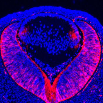
Research article                                                                                        Developmental Biology

                                   Crystallins were largely preserved in Etv1/4/5 mutant lenses. We also showed that mTOR signaling
                                   was aberrantly upregulated, resulting in the failure of nuclei removal in the mature lenses. These
                                   results revealed the critical differences between the function of Etv family transcription factors and
                                   FGF signaling during lens development, demonstrating that these SRTFs can operate outside the
                                   confine of upstream signaling.

                                   Results
                                   MAPK-regulated Etv transcription factors are required for lens
                                   development
                                   Previous studies have shown that Etv transcription factors are controlled by FGF signaling during
                                   embryonic development. To confirm this finding in the lens, we generated conditional knockouts of
                                   Fgfr1 and Fgfr2 using a lens-specific Cre driver Pax6Le-Cre, also known as Le-Cre, which is active in
                                   the lens ectoderm as early as E9.5 (Ashery-Padan et al., 2000). As we and others have previously
                                   reported, genetic ablation of FGF receptors prevented the lens ectoderm from forming the lens ves-
                                   icle in the Pax6Le-Cre; Fgfr1fl/fl; Fgfr2fl/fl (Fgfr CKO) embryo (Figure 1A–H, arrows) (Collins et al.,
                                   2018; Garcia et al., 2005). Importantly, both ERK phosphorylation and expression of Etv1, Etv4 and
                                   Etv5 were also lost in the Fgfr CKO lens ectoderm (Figure 1A–H, outlined), demonstrating that FGF
                                   signaling indeed controls ERK and Etv activities during lens induction. We next ablated Mek and Erk
                                   to investigate whether Etv genes were also regulated by MAPK signaling in the lens. Although the
                                   lenses were formed in Pax6Le-Cre; Mek1(Map2k1)fl/fl; Mek2(Map2k2)-/- (Mek CKO) and Pax6Le-Cre;
                                   Erk1(Mapk3)-/-; Erk2(Mapk1)fl/fl (Erk CKO) embryos, they failed to express any of the Etv genes
                                   (Figure 1I–Q, circled). These results demonstrated that the Etv family transcription factors are con-
                                   trolled by FGF-ERK signaling during lens development.
                                       To investigate the function of Etv family transcription factors in the lens, we used the Pax6Le-Cre
                                   transgenic mouse line to conditionally ablate Etv1 and Etv5 in an Etv4-null background. In Pax6Le-
                                   Cre; Etv1fl/fl; Etv4-/-; Etv5fl/fl embryos (Etv TKO), the lens induction and lens vesicle formation were
                                   unaffected, but the lens size was reduced at E14.5 as shown by Hematoxylin and Eosin staining
                                   (Figure 2A and E). Consistent with this, apoptosis increased in the epithelium and the transitional
                                   zone as indicated by TUNEL staining (Figure 2B,F and I, arrows). On the other hand, proliferation of
                                   the lens epithelial cells was reduced as measured by EdU incorporation assay (Figure 2C,D and I,
                                   arrowheads). In the control lens, the nascent fiber cells exited the cell cycle after the transitional
                                   zone, as shown by the diminution of Cyclin D1 expression and Edu labeling (Figure 2C, arrows). In
                                   contrast, the Etv TKO lens displayed ectopic expression of Cyclin D1 in the posterior lens
                                   (Figure 2G, arrow) and persistent EdU- and pHH3-positive cells in the fiber cell compartment
                                   (Figure 2C,D,G and H, inserts). These phenotypes showed that lens development was disrupted by
                                   loss of Etv transcription factors.

                                   Genetic ablation of Etv causes ectopic activation of ERK signaling
                                   To identify the downstream targets of Etv genes, we performed RNA sequencing analysis to com-
                                   pare the transcriptional landscape of E14.5 control to that of Etv TKO mutant. Since Etv genes are
                                   predominantly expressed in the transitional zone at the lens equator, we isolated these cells by laser
                                   capture microdissection and extracted RNA for cDNA synthesis (Figure 3A). After amplification, the
                                   cDNA library was subjected to high-throughput sequencing to examine the global gene expression
                                   changes. Cluster and PCA analysis of the top 200 differentially expressed genes showed that the
                                   controls and mutants were well segregated across three biological replicates, demonstrating the
                                   consistency of the RNA sequencing data (Figure 3B and Figure 3—figure supplement 1A). Among
                                   the 834 genes showing statistically significant changes in expression (p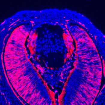
Research article                                                                                                  Developmental Biology

                                                                                   E10.5
                                                         pERK            Etv1                 Etv4                  Etv5
                                                     A            B                    C                     D

                                        Pax6Le-Cre
                                                     E            F                    G                     H
                                         Fgfr CKO

                                                                        E14.5
                                                         Etv1            Etv4                  Etv5
                                                     I            J                    K
                                        Pax6Le-Cre

                                                     L            M                    N
                                         Erk CKO

                                                     O            P                    Q
                                         Mek CKO

                                   Figure 1. Etv transcription factors are controlled by FGF-ERK signaling in the lens. (A–D) At E10.5, the invaginating
                                   lens ectoderm displays ERK phosphorylation and expression of Etv1, 4 and 5 (arrows). (E–H) Genetic ablation of
                                   Fgfr1/2 in Fgfr CKO mutants prevented lens vesicle formation and abrogated phospho-ERK and Etv expression
                                   (arrows). The lens ectoderms are marked by dotted lines. (I–K) At E14.5, Etv1, 4 and 5 are predominantly
                                   expressed in the transitional zone of the lens (arrows). (L–Q) Deletion of either Erk1/2 (Erk CKO) or Mek1/2 (Mek
                                   CKO) abolished expression of Etv genes. The lenses are circled in dotted lines.

                                   supplement 1B). Although this suggests a positive feedback regulation of FGF receptors by Etv
                                   genes, it is unlikely to affect lens development, because previous studies have shown that Fgfr1 and
                                   Fgfr3 double mutant lenses did not exhibit any overt phenotype (Zhao et al., 2008). On the other
                                   hand, the transcript level of Spry2, an inhibitor of receptor tyrosine kinases, was also reduced, which
                                   we confirmed by RNA in situ hybridization in Etv TKO mutant lenses (Figure 3D and F, arrows). This
                                   is consistent with previous ChIP-seq analysis which revealed that SPRY2 is a direct target of PEA3/
                                   ETV family genes (Yan et al., 2013). Since we have shown above that expression of Etv1, Etv4 and
                                   Etv5 in the lens is dependent on ERK signaling (Figure 1I–Q), it is not surprising that Spry2 expres-
                                   sion is also lost in Erk CKO mutants (Figure 3—figure supplement 1C, arrows). Considering that

Garg et al. eLife 2020;9:e51915. DOI: https://doi.org/10.7554/eLife.51915                                                                       4 of 21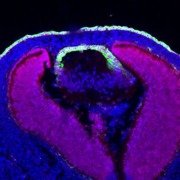
Research article                                                                                                                    Developmental Biology

                                                                                               E14.5
                                                                     H&E               TUNEL           Cyclin D1/EdU                 pHH3/Ecad
                                                               A                   B               C                             D

                                        Pax6Le-Cre
                                                               E                   F               G                             H
                                          Etv TKO

                                           I                               P=0.0283
                                                               80%                                   Pax6Le-Cre                  P=0.0147
                                                                                                     Etv TKO
                                            % positive cells

                                                                                                           100%

                                                                                                            relative area
                                                                                          P=0.0008
                                                               40%

                                                               0%                                                           0%
                                                                             Edu           TUNEL                                 Lens size

                                   Figure 2. Lens development requires Etv transcription factors. (A–H) Hematoxylin and eosin (H and E) staining
                                   reveal reduced lens size in Etv1/4/5 deletion (Etv TKO) mutants (A and E). Etv null lens exhibited increased cell
                                   apoptosis shown by TUNEL staining (B and F, arrows) and reduced cell proliferation as indicated by EdU staining
                                   (C and G, arrowheads). In contrast, ectopic expression of Cyclin D1 (G, arrows) and proliferation markers EdU and
                                   pHH3 (C, D, G and H, inserts) were detected in the posterior lens. (I) Quantification of EdU and TUNEL staining.
                                   The online version of this article includes the following source data for figure 2:
                                   Source data 1. Source data for Figure 2I.

                                   Sprouty proteins are known to inhibit Ras-MAPK signaling (Hanafusa et al., 2002), this suggests
                                   that Etv regulation of Spry constitutes a negative feedback loop to regulate FGF-ERK activity.
                                   Indeed, we notice that ERK phosphorylation was ectopically activated in Etv TKO mutant lenses
                                   (Figure 3E and G, arrows). In support of the upregulation of ERK activity, we also observed increas-
                                   ing expression of Egr1 and Fos (Figure 3C), two early response genes for Ras-MAPK signaling.
                                   Moreover, gene set enrichment analysis (GSEA) showed that Etv deletion enhanced the activity of
                                   the MAP kinase pathway (Figure 3H), which is known to be decreased in FGF receptor mutants.
                                   Therefore, inactivation of FGF receptors or Etv transcription factors in the lens resulted in diametri-
                                   cally different outcomes in MAPK signaling.

                                   Etv deficiency blocks aberrant but not normal lens differentiation
                                   We took three approaches to investigate the role of Etv transcription factors in lens cell differentia-
                                   tion: (1) examining the Etv loss-of-function phenotype, (2) corroborating our results with ERK signal-
                                   ing knockouts and (3) performing genetic epistasis using a FGF gain-of-function model. It was
                                   previously reported that FGF signaling controls the expression of aA-crystallin (Cryaa) in the lens
                                   cells via Etv5 and Maf; the latter also contains Etv-binding sites in its regulatory region (Xie et al.,
                                   2016). Unexpectedly, immunostaining showed that both Maf and a-crystallins proteins were still
                                   present in Etv TKO mutant lenses (Figure 4A,B,F and G). Similarly, we did not detect a significant
                                   reduction in gA-crystallin expression (Figure 4C and H), which is normally induced after aA-crystallin

Garg et al. eLife 2020;9:e51915. DOI: https://doi.org/10.7554/eLife.51915                                                                                      5 of 21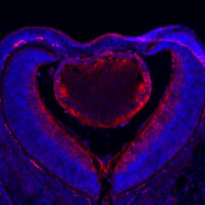
Research article Developmental Biology
in more mature lens fiber cells. To avoid the possibility that protein perdurance may obscure the
dynamic changes in gene expression, we also performed RNA in situ hybridization, which showed
abundant expression of Cryaa and Cryga mRNAs in Etv TKO lenses (Figure 4D,E,I and J). Consistent
with this, there were no statistically significant changes in the transcript levels of Cryaa, Cryga and
Maf in our RNA sequencing data. Based on these results, lens differentiation can apparently proceed
in the absence of Etv transcription factors.
To corroborate the lack of lens differentiation phenotype in Etv loss-of-function mutants, we next
examined ERK signaling knockouts. We reasoned that, since the entire Ets superfamily transcription
factors including Etv are under the control of ERK, inactivation of MAPK may produce a stronger
phenotype than Etv knockouts. Indeed, combined deletion of Erk1(Mapk3) and Erk2(Mapk1)
resulted in a drastic reduction in lens size as previously reported (Xie et al., 2016). However, Maf, a-
and g-crystallins mRNA and protein were still expressed in Erk CKO mutant lenses (Figure 4K–O).
To further confirm this finding, we also examined the lens-specific knockout of Mek1 (Map2k1) and
Mek2 (Map2k2), two kinases upstream of Erk. In the severely diminished Mek CKO mutant lenses,
we again failed to detect significant reduction in the intensity of Maf, a- and g-crystallins staining
(Figure 4P–T). Therefore, genetic ablation of Erk and Etv transcription factors did not abolish lens
differentiation as in FGF receptor mutants.
A B
Etvflox Etv TKO 1
Before LCM
0.5
0
−0.5
After LCM
−1
Etvflox Etv TKO
C H
Etvflox Etv TKO E15.5 E14.5 MAP KINASE
Autoregulation
0.4
Enrichment Score (ES)
Etv4 Nominal p−value: 0.0405
Spry2 pERK FDR: 0.1302
Etv5
0.2
D E ES: 0.3863
Normalized ES: 1.3810
Pax6Le-Cre
Etv1
0.0
Fgfr1
Fgfr3
Feedback
Spry2 Positive Etv vs WT Negative
Egr1 F G
−5 0 5
tTest
Etv TKO
Fos
Targets
Jag1
Tsc2 0 5000 10000 15000
10¹ Rank in ordered gene list
10⁴
Figure 3. Transcriptomic analysis shows ERK signaling dysregulation in Etv mutant lens. (A) The transitional zone
of the lens was isolated by laser capture microscope (LCM) for RNA sequencing analysis. (B) Cluster analysis of the
top differentially expressed genes in the RNA sequencing data. (C) Heat map of the Etv regulated genes. (D and
F) Spry2 is significantly down-regulated in Etv TKO mutants. (H) Gene set enrichment analysis (GSEA) indicates the
MAPK pathway is elevated.
The online version of this article includes the following source data and figure supplement(s) for figure 3:
Source data 1. Source data for Figure 3C.
Figure supplement 1. RNA sequencing analysis identified Etv-regulated genes.
Garg et al. eLife 2020;9:e51915. DOI: https://doi.org/10.7554/eLife.51915 6 of 21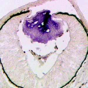
Research article                                                                                                     Developmental Biology

                                                                                              E14.5
                                                      Maf             a-crystallin         g-crystallin           Cryaa                 Cryga

                                                  A               B                    C                     D                     E

                                        Etvflox
                                                  F               G                    H                     I                     J
                                        Etv TKO

                                                  K               L                    M                     N                     O
                                        Erk CKO

                                                  P               Q                    R                     S                     T
                                        Mek CKO

                                   Figure 4. Lens fiber differentiation proceeds in the absence of Etv and ERK. (A–J) Expression of fiber cell markers
                                   Maf, a- and g-crystallin is unaffected in Etv TKO lenses. (K–T) Maf, a- and g-crystallin are still expressed in Erk and
                                   Mek CKO lens despite the severely reduced lens size.

                                      The phenotypic dichotomy between Fgfr and Etv mutants raised the question whether Etv tran-
                                   scription factors play any role in the FGF-induced lens fiber differentiation. To address this issue, we
                                   took a gain-of-function approach to test the genetic epistasis between Etv and FGF signaling. As we
                                   and others have previously reported (Collins et al., 2018; Li et al., 2019; Robinson et al., 1998),
                                   the lens-specific overexpression of Fgf3 in Fgf3OVE391 transgenic animals greatly elevated ERK phos-
                                   phorylation in the lens (Figure 5A and B). Consistent with regulation of Etv and Spry2 by ERK signal-
                                   ing we showed above, this led to increased expression of Etv1, Etv4, Etv5 and Spry2 in the
                                   Fgf3OVE391 lens (Figure 5—figure supplement 1), which was accompanied by ectopic induction of
                                   Maf and g-crystallin but loss of E-cadherin in Pax6-positive anterior lens epithelium (Figure 5F,J and
                                   N, arrows). We hypothesized that if Etv is required for FGF signaling to promote lens differentiation,
                                   then blocking Etv activity should prevent the Fgf3-overexpression phenotype. Since it was cumber-
                                   some to use the triple knockout of Etv1/4/5 to generate compound mutants for the genetic epistasis
                                   experiments, we employed the R26EtvEnR allele, which can be induced to express an Etv4 fusion pro-
                                   tein linked to the Engrailed repressor domain (EtvEnR) after Cre-mediated excision of a transcrip-
                                   tional STOP cassette. This was previously shown to act as a dominant negative protein to suppress
                                   the activities of endogenous Etv transcription factors (Mao et al., 2009). Consistent with this,
                                   Pax6Le-Cre;R R26EtvEnR lenses were reduced in size like those of Etv TKO mutants, while the expres-
                                   sion of E-cadherin, Maf and g-crystallin was maintained (Figure 5G,K,O and Q). After crossing with
                                   Fgf3OVE391, the lens remained smaller in the Pax6Le-Cre; Fgf3OVE391; R26EtvEnR mutant than that of
                                   the control (Figure 5Q). Of note, the expression of Etv1, Etv4, Etv5 and Spry2 in the Pax6Le-Cre;
                                   Fgf3OVE391; R26EtvEnR mutant was diminished but not abrogated compared to that in Fgf3OVE391

Garg et al. eLife 2020;9:e51915. DOI: https://doi.org/10.7554/eLife.51915                                                                           7 of 21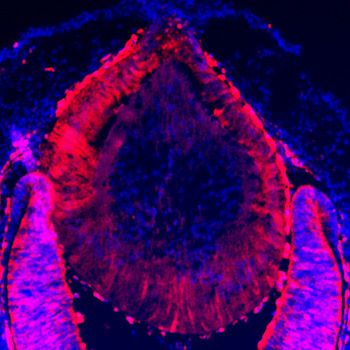
Research article                                                                                                                                 Developmental Biology

                                                                                                                             Pax6Le-Cre;       Pax6Le-Cre;
                                                                                  Pax6Le-Cre            Fgf3OVE391            R26EtvEnR     R26EtvEnR;Fgf3OVE391
                                                                                                                     E14.5

                                                         A                                         B                    C                    D

                                         pERK
                                         Pax6/Ecad       E                                         F                    G                    H

                                                         I                                         J                    K                    L
                                          g-crystallin

                                                         M                                         N                    O                    P
                                         Maf

                                           Q                                                           P=0.0003         P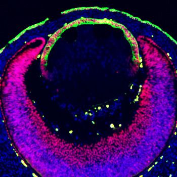
Research article                                                                                        Developmental Biology

                                   (Figure 5—figure supplement 1), suggesting that Etv activities was partially inhibited by EtvEnR.
                                   Nevertheless, in sharp contrast to Fgf3OVE391, Maf and g-crystallins were restricted to the lens fiber
                                   compartment in Pax6Le-Cre; Fgf3OVE391; R26EtvEnR lenses, while E-cadherin was preserved in the
                                   anterior lens epithelium (Figure 5H,L and P). Therefore, inhibition of Etv transcriptional activities
                                   prevented FGF signaling from inducing ectopic expression of the fiber-specific genes in the lens epi-
                                   thelium. Taken together, these results showed that Etv transcription factors were not essential for
                                   normal lens differentiation at the transitional zone, but they were required for FGF signaling to
                                   directly convert lens epithelial cells into lens fibers.

                                   Etv regulates the induction of cell differentiation by promoting Jag1-
                                   Notch signaling
                                   Although the fiber cell differentiation genes were still expressed in Etv TKO mutant lenses, we
                                   noticed that the transitional zone marked by the boundary between the lens epithelial marker Foxe3
                                   and the fiber cell marker Prox1 was moved anteriorly (Figure 6A and E, arrows). As a result, the
                                   length of the lens epithelium marked by Pax6 and E-cadherin staining was shortened compared to
                                   the circumference of the posterior lens (Figure 6B,F and I, arrows). This differentiation pattern is
                                   reminiscent of defective Notch signaling, which is known to cause the lens progenitor cells to
                                   undergo premature differentiation before reaching the lens equator, resulting in an anteriorly shift of
                                   the transitional zone (Jia et al., 2007; Le et al., 2009; Li et al., 2019; Rowan et al., 2008;
                                   Saravanamuthu et al., 2012). Intriguingly, one of the down-regulated genes in Etv TKO mutants
                                   revealed by our transcriptomic analysis was Jag1 (Figure 3C), which encodes the ligand for Notch
                                   signaling. By immunostaining, we confirmed that the expression of Jag1 protein was drastically
                                   reduced in Etv TKO mutant lenses (Figure 6C and G, arrowheads). Jag1 expressed in the lens fiber
                                   cells signals to the Notch receptors in the lens epithelium to maintain the progenitor pool (Jia et al.,
                                   2007; Le et al., 2009). In line with the reduced Notch signaling due to Jag1 deficiency, the anterior
                                   lens epithelium in Etv TKO mutants exhibited severely diminished staining of the Notch1 intracellular
                                   domains (Notch1-ICD), a proteolytic product of Notch1 receptor triggered by Jag1 activation
                                   (Figure 6D and H, arrows). These results support that Etv regulates Jag1-Notch signaling to control
                                   the timing of lens cell differentiation.
                                       To investigate the molecular mechanism of Jag1 regulation by Etv, we turned to cultured lens
                                   cells in vitro. By western blot, we showed that Jag1 expression in the lens epithelial cell culture was
                                   induced by FGF2 after 5 hr treatment and suppressed by Mek inhibitors U0126 and PD0325901 but
                                   not PI3K inhibitor LY294002 (Figure 6J and Figure 6—figure supplement 1). This is consistent with
                                   previous reports that Jag1 expression in the lens is controlled by ERK-mediated FGF signaling
                                   (Li et al., 2019; Saravanamuthu et al., 2009). To determine whether Jag1 is a direct transcriptional
                                   target of Etv, we used the recently published ATAC-seq analysis (Zhao et al., 2019) to search the
                                   open chromatin regions in the developing lens and identified two Etv-binding sites located within
                                   introns 2 and 5 of Jag1 gene (Figure 6K). By chromatin immunoprecipitation (ChIP), we showed that
                                   both introns 2 and 5 sites could be pulled down by Etv5 antibodies but not by IgG control
                                   (Figure 6L), demonstrating that these two sites in Jag1 were occupied by Etv in the lens cells. We
                                   next investigated whether Etv was required for FGF signaling to induce Jag1 expression in vivo.
                                   Jag1 was normally restricted to the nascent lens fibers in wild type control lenses (Figure 5M, arrow-
                                   heads), but it was abolished in Mek and Erk CKO mutants (Figure 6N and O). In Fgf3OVE391 lenses,
                                   however, Jag1 expression was expanded to the entire lens epithelium at the expense of E-cadherin
                                   expression (Figure 6P, arrow), demonstrating that FGF signaling can induce de novo Jag1 expres-
                                   sion in the lens epithelium. In Pax6Le-Cre; R26EtvEnR lenses, Jag1 expression was significantly reduced
                                   compared to those of wild-type control (Figure 6Q, arrowheads), consistent with the role of Etv in
                                   Jag1 regulation. Suppression of Etv activity in Pax6Le-Cre; Fgf3OVE391; R26EtvEnR lenses reversed the
                                   Fgf3 overexpression phenotype, limiting the Jag1 expression domain to the posterior lenses
                                   (Figure 6R, arrowheads). Based on these results, we conclude that Etv mediates the FGF-ERK-Notch
                                   crosstalk to control the induction of lens differentiation.

Garg et al. eLife 2020;9:e51915. DOI: https://doi.org/10.7554/eLife.51915                                                           9 of 21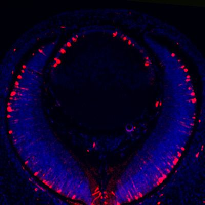
Research article                                                                                                                                      Developmental Biology

                                    Prox1/Foxe3         Pax6 /Ecad            Jag1/Ecad             Notch1-ICD
                               A                   B                      C                     D
                                                                                                                        I
                                                                                                                                                                P=0.0007
             Pax6Le-Cre

                                                                                                                                                  1.0

                                                                                                                       Anterior/posterior ratio
                                                                                                                                                  0.8

                                                                                                                                                  0.6
    E14.5

                               E                   F                      G                     H                                                 0.4
              Etv TKO

                                                                                                                                                  0.2

                                                                                                                                                  0.0
                                                                                                                                                         Pax6Le-Cre Etv TKO

       J                                                                                            K    Jag1
                                                                   FGF2
                                                                                                     E14.5 epi
               Time                    0’    5’    5’      1hr     1hr    5hr     5hr     5hr       E14.5 fiber
            Inhibitor                              LY              LY              U      LY            P0 epi
                                                                                                      P0 fiber
                     Jag1                                                                                    Intron 5 site                                      Intron 2 site

                      pErk                                                                           L                                                  Input          ChIP
                                                                                                                    Marker aEtv5 IgG                              aEtv5    IgG

                              Erk                                                                   Intron 2 site

        pErk/Erk                                                                                    Intron 5 site

                                                                                                                                                  Pax6Le-Cre;         Pax6Le-Cre;
                                      Pax6Le-Cre         Erk CKO                Mek CKO               Fgf3OVE391                                   R26EtvEnR       R26EtvEnR;Fgf3OVE391

                               M                   N                      O                     P                             Q                                    R
                  Jag1/Ecad
     E14.5

Figure 6. Etv induces Jag1 expression to control Notch signaling. (A–H) The transitional zone marked by the boundaries of Prox1, Foxe3, Pax6 and
E-cadherin expressions are shifted anteriorly in Etv TKO mutant lenses (A, B, E and F, arrows). This was caused by reduced expression of Jag1 in the
nascent lens fibers (C and G, arrowheads) and down regulation of Notch signaling as indicated by Notch1-ICD staining in the lens epithelium (D and H,
arrows). (I) Quantification of the anterior and posterior perimeters of the lens shows the relative shortening of the lens epithelium. (J) FGF2 induced
Jag1 expression in the lens culture after 5 hr, which was blocked by Mek inhibitor U1206, but not by PI3K inhibitor LY294002. (K) Bioinformatic analysis
identifies two Etv binding sites within the intron 2 and 5 of the Jag1 locus. The bars indicate the open chromatin regions obtained from the ATAC-seq
analysis of lens epithelium and fibers. (L) Chromatin immunoprecipitation experiment in lens cultures showed that both the introns 2 and 5 sites were
pulled down by Etv5 antibody but not IgG control. (M–R) The endogenous Jag1 expression in lens fiber cells (M, arrowheads) is abolished by deletion
of either Erk or Mek (N and O). Overexpression of Fgf3 induces ectopic Jag1 expression in the lens epithelium (P, arrow), which is suppressed by
dominant negative EtvEnR (Q and R, arrowheads).
The online version of this article includes the following source data and figure supplement(s) for figure 6:
Source data 1. Source data for Figure 6I.
Figure supplement 1. FGF induces Jag1-Notch pathway in an ERK-dependent manner.

                                                                 Deletion of Etv genes led to aberrant activation of mTOR signaling and
                                                                 disruption of nuclei clearance in the lens
                                                                 The final step of lens cell differentiation is the degradation of their cellular organelles, which is crucial
                                                                 for the transparency of the lens (Bassnett et al., 2011). To determine whether Etv transcription fac-
                                                                 tors are required for this process, we examined the clearance of lens cell nuclei by histology. In wild-
                                                                 type control, the maturing fiber cells lost their nuclei as they migrated toward the interior of the
                                                                 lens, eventually forming an organelle free zone (OFZ) (Figure 7A and Figure 7—figure supplement
                                                                 1, arrowheads). In contrast, Etv TKO mutants retained nuclei within the lens core, indicating defec-
                                                                 tive organelle clearance (Figure 7E and Figure 7—figure supplement 1, arrowheads). Previous

Garg et al. eLife 2020;9:e51915. DOI: https://doi.org/10.7554/eLife.51915                                                                                                                              10 of 21
Research article Developmental Biology
p5 E14.5
H&E pmTOR pS6 p4EBP1
A B C D
Pax6Le-Cre
OFZ
E F G H
Etv TKO
I J K L
Tsc1 CKO
Figure 7. Activation of mTOR signaling disrupts nuclei degradation in the Etv mutant. (A–J) The center of the wild
type lens was an organelle free zone (OFZ) at P5 (F), but it still contains nuclei in Etv mutants (F, arrowheads). This
is associated with increased mTOR signaling as indicated by the elevated phosphorylation of mTOR, S6 and
4EBP1 (B-D, F-H, arrowheads). (I–L) The aberrant retention of nuclei and activation of mTOR signaling are
reproduced in Tsc1 knockout lens.
The online version of this article includes the following source data and figure supplement(s) for figure 7:
Figure supplement 1. Persistent nuclei in Etv and Tsc1 mutant lenses.
Figure supplement 1—source data 1. Source data for Figure 7—figure supplement 1D.
studies have shown that inhibition of mTOR signaling accelerated organelle elimination in chick lens
explants (Basu et al., 2014). Interestingly, our RNA sequencing analysis of Etv TKO mutant lenses
showed significant down-regulation of Tsc2, which forms a heterodimer with Tsc1 to inhibit mTOR
activity (Figure 3C). This suggests that the defective nuclei clearance in the Etv TKO mutant may be
caused by elevated mTOR signaling. In support of this hypothesis, we observed that phospho-mTOR
(pmTOR) was localized in the transitional zone of the wild-type control lens, but it expanded into the
fiber cell compartment in Etv TKO mutants (Figure 7B and F, arrowheads). Similarly, Etv TKO
mutant lenses displayed significant increase in pS6 and p4EBP1, two known substrates of mTOR
kinase (Figure 7C,D,G and H). Therefore, inactivation of Etv genes led to increased mTOR signaling
in the lens.
To determine whether mTOR activation indeed interfered with nuclei clearance in the lens, we
generated Pax6Le-Cre; Tsc1flox/flox (Tsc1 CKO) to ablate the Tsc2 binding partner Tsc1. As expected
from the role of Tsc1/2 complex in suppressing mTOR activity, pmTOR was significantly increased in
Tsc1 CKO lenses, which also displayed elevated level of pS6 and p4EBP1 (Figure 7J–L). Importantly,
whereas the wild-type control lens contains a well-defined OFZ in the center, the nuclei were scat-
tered throughout the Tsc1 CKO lenses (Figure 7I and Figure 7—figure supplement 1). The lack of
an OFZ in Tsc1 deficient lenses supports our model that Etv control of mTOR signaling is required
for the terminal differentiation of the lens fiber cells.
Garg et al. eLife 2020;9:e51915. DOI: https://doi.org/10.7554/eLife.51915 11 of 21Research article Developmental Biology
Discussion
FGF signaling plays important roles during vertebrate lens development. As demonstrated by the
profound lens defects in FGF receptor mutants, the primary function of FGF signaling is to promote
differentiation of the lens epithelial cells into the fiber cells. In this study, we have studied the func-
tions of Etv family genes, which are the well-established downstream transcription factors induced
by FGF during embryonic development. Contrary to the defective lens differentiation found in FGF
signaling mutants, deletion of Etv genes did not prevent the expression of lens fiber genes in normal
development. Instead, it caused premature differentiation of the lens progenitors as a result of
reduced Notch signaling. Moreover, Etv mutations led to an increase in the activity of ERK and
mTOR signaling, with the latter responsible for the abnormal retention of the nuclei in the adult lens.
Therefore, unlike BMP, Hedgehog and Wnt signaling that command their downstream signal-regu-
lated transcription factors (SRTFs) as faithful effectors, ETV transcription factors activated by FGF sig-
naling can oppose the intended function of the pathway.
Our study of Etv mutants in lens development has revealed several important features of Etv func-
tion that deviate from the expected role of a faithful executor for FGF signaling. First, deletion of
Etv caused significant derangement of ERK signaling, which is the main intracellular pathway acti-
vated by FGF. Contrary to the loss of ERK activity in mutants lacking either FGF receptors or down-
stream mediators Frs2 and Shp2 (Collins et al., 2018; Li et al., 2014; Pan et al., 2010; Zhao et al.,
2008), Etv-deficient lenses displayed elevated ERK signaling, which was evident in the increased
level of ERK phosphorylation and expression of ERK early response genes Egr1 and Fos. This was
likely due to reduced expression of ERK inhibitor Spry2, which has been shown to be a direct tran-
scriptional target of Etv (Yan et al., 2013). In support of this, deletions of Spry genes have been
found to result in upregulation of ERK signaling in a wide variety of tissues, including the lens
(Kuracha et al., 2011). The aberrant ERK activation in Etv mutants may stimulate downstream tar-
gets normally silenced by loss of FGF signaling, inducing further compensatory changes that rewire
the signaling network. It underscores the intricate role of Etv in stabilizing the intracellular signaling
activated by FGF.
The second distinction of Etv mutants compared to FGF receptor knockouts is the relatively nor-
mal lens differentiation. This was unexpected because a previous study shown that both lens differ-
entiation genes Maf and Cryaa were under direct control of Etv transcription factors in vitro
(Xie et al., 2016). We presented three lines of evidence in support of our conclusion. First, we per-
formed RNA sequencing, RNA in situ hybridization, and immunohistochemistry in Etv mutant lenses,
none of which showed significant reduction in the expression of Maf and Cryaa. Second, we gener-
ated lens-specific knockouts of Erk1/2, which we showed to be required for Etv expression. Even in
these ERK signaling mutants, we still observed expression of Maf and Cryaa. Of note, the previous
study observed that immunostaining for a-crystallin and Maf were severely reduced or even absent
in the Erk1/2 knockout lens (Xie et al., 2016). It is not clear what caused the phenotypic discrepancy
between ours and the previous study, which could be due to a difference in the genetic background
of the animal models or the source of antibodies. It should be noted that g-crystallin expression
appeared to move posteriorly in both Etv and Erk knockouts (Figure 4H,M and R), which may reflect
a slight delay of lens fiber terminal differentiation. Nevertheless, even knockout of ERK kinase Mek
still failed to abolish Maf and Cryaa expression. In the third approach, we explored the role of Etv in
a gain-of-function setting, showing that inhibition of Etv indeed blocked ectopic FGF from stimulat-
ing Maf and g-crystallin expression in the lens epithelium. This result suggests an intrinsic difference
between normal differentiation at the lens equator and ectopic differentiation in the lens epithelium,
which may be shaped by their unique microenvironment and different dosage of FGF signaling.
Taken together, our results demonstrate that Etv genes are dispensable for FGF-induced cell differ-
entiation under the physiological condition during lens development.
Genetic ablation of Etv also differed from inactivation of FGF signaling in its impact on the transi-
tional zone of cell differentiation, which was shifted anteriorly after Etv deletion and posteriorly in
FGF signaling mutants. We recently showed that FGF plays a dual role in specifying the location of
the transitional zone by regulating the timing of lens differentiation; FGF directly promotes differen-
tiation of lens fiber cells, but it also cooperates with PDGF to restrain differentiation of the lens pro-
genitor cells (Li et al., 2019). The latter is mediated by Jag1, which induces Notch signaling in the
anterior lens epithelium to suppress progenitor cell differentiation. In the current study, we
Garg et al. eLife 2020;9:e51915. DOI: https://doi.org/10.7554/eLife.51915 12 of 21Research article Developmental Biology
determined that Jag1 expression is under the direct control of Etv transcription factors and ERK,
delineating a complete molecular cascade that connects FGF to Notch signaling. In the absence of
Etv, loss of Notch signaling leads to premature differentiation of lens progenitor cells indicated by
Foxe3 and Prox1 expression, but the pattern of Crystallin expressions are unchanged. This suggests
the terminal differentiation of lens fibers may be slightly delayed relative to onset of progenitor cells
differentiation. Nevertheless, Etv mutants proceed to express Crystallin genes at the comparable
level as wild-type controls, which is in sharp contrast to the loss of g-crystallin expression in FGF
receptor mutants (Zhao et al., 2008). Therefore, Etv mediates the anti-differentiation function of
FGF to activate Notch signaling but it is largely dispensable for the pro-differentiation aspect of FGF
signaling.
Lastly, our study showed that Etv deficiency augmented mTOR phosphorylation. This is likely
caused by reduced expression of Tsc2, a negative regulator of mTOR. Indeed, we also observed sig-
nificant increase in phosphorylation of S6 and 4EBP1, two downstream targets of mTOR. Impor-
tantly, we observed aberrant retention of the lens fiber cell nuclei in postnatal Etv mutants, which
was mimicked by deletion of Tsc2 partner Tsc1 in the lens. Previous studies have shown that inhibi-
tion of mTOR by rapamycin promotes degradation of the organelle in chick lens explants although
the exact mechanism remained controversial (Basu et al., 2014; Morishita and Mizushima, 2016).
Our study provides the in vivo evidence that mTOR is indeed an important regulator of organelle
removal in lens maturation.
Taken together, our genetic study firmly establishes Etv family transcription factors as SRTFs for
FGF signaling in lens development. SRTFs constitute a special class of transcription factors that relay
intracellular signals to shape the cellular transcriptome, which ultimately determines the identity and
status of the cell. Although many SRTFs have proven to be faithful to their upstream signaling, we
showed that Etv transcription factors deviate or even oppose the function of FGF-ERK signaling in
the lens. This is likely because, unlike other signaling pathways such as BMP, Hedgehog and Wnt
that rely on a single downstream effector, FGF signaling can induce multiple transcription factors.
This generates diversity among the FGF SRTFs that may evolve to acquire their unique or even
opposing functions, which fine tunes the overall transcriptional response. The three PEA3/ETV genes
and ERG are the only ETS genes implicated in tumorigenesis, which has been attributed to their abil-
ity to mimic the oncogenic effect of Ras signaling (Hollenhorst et al., 2011). Our previous work in
lacrimal gland development has indeed found that deletion of Etv transcription factors largely repro-
duced the phenotype of FGF-ERK mutants (Garg et al., 2018). In the lens, however, our current
study paints a more complex picture of Etv activity, suggesting that their functions deviate consider-
ably from their upstream FGF signaling. As transcription factors attract increasing interests as viable
therapeutic targets, it will be important to elucidate the context-dependent function of these SRTFs
in development and diseases.
Materials and methods
Key resources table
Reagent type Additional
(species) or resource Designation Source or reference Identifiers information
Antibody Rabbit monoclonal Cell Signaling Cat.# 55506, IHC-1/200
anti-cyclin D1 RRID:AB_2827374
Antibody Rabbit polyclonal Santa Cruz Cat.#sc-7866, IHC-1/200
anti-Maf Biotechnology RRID:AB_638562
Antibody Mouse monoclonal BD Cat.#610181, IHC-1/500
anti-Ecadherin RRID:AB_397580
Antibody Rabbit monoclonal Cell Signaling Cat.# 4695, WB-1/2000
anti-Erk RRID:AB_390779
Antibody Mouse monoclonal Santa Cruz Cat.#sc-377465 IHC-1/200
anti-Foxe3 Biotechnology
Antibody Rabbit polyclonal Santa Cruz Cat.#sc-8303, IHC-1/200, WB-1/500
anti-Jag1 Biotechnology RRID:AB_649685
Continued on next page
Garg et al. eLife 2020;9:e51915. DOI: https://doi.org/10.7554/eLife.51915 13 of 21Research article Developmental Biology
Continued
Reagent type Additional
(species) or resource Designation Source or reference Identifiers information
Antibody Mouse monoclonal BD Cat.#550609, IHC-1/200
anti-Ki67 RRID:AB_393778
Antibody Rabbit monoclonal Abcam Cat.# ab133256, IHC-1/1000
Lamin A/C RRID:AB_2813767
Antibody Rabbit monoclonal Cell Signalling Cat.#4380, WB-1/1000
Notch1 RRID:AB_10691684
Antibody Rabbit monoclonal Cell Signaling Cat.#4147, IHC-1/200
Notch1-ICD RRID:AB_2153348
Antibody Rabbit polyclonal Covance Cat.#PRB-238C, IHC-1/200
anti-Prox1 RRID:AB_291595
Antibody Rabbit polyclonal Millipore Cat.#06-570, IHC-1/200
anti-pHH3 RRID:AB_310177
Antibody Rabbit anti-pS6 Cell Signaling Cat.#5364, IHC-1/200
RRID:AB_10694233
Antibody Rabbit monoclonal Cell Signaling Cat.#5536, IHC-1/200
anti-pmTOR RRID:AB_10691552
Antibody Rabbit polyclonal Covance Cat.#PRB-278P, IHC-1/200
anti-Pax6 RRID:AB_291612
Antibody Rabbit anti-p4EBP1 Cell Signaling Cat.#2855, IHC-1/200
RRID:AB_560835
Antibody Rabbit monoclonal Cell Signaling Cat.#9101, IHC-1/200
anti-pERK RRID:AB_331646
Antibody Mouse monoclonal Santa Cruz Cat# sc-7383, WB-1/1000
anti-pERK Biotechnology RRID:AB_627545
Antibody Rabbit polyclonal Sam Zigler IHC-1/5000
anti-a-Crystallin (National Eye
Institute)
Antibody Rabbit polyclonal Sam Zigler IHC-1/5000
anti-g-Crystallin (National Eye
Institute)
EdU DNA synthesis Abcam cat. # ab14618
monitoring probe
Commercial Click IT EdU Cell Invitrogen cat. # C10337
assay Kit proliferation kit
Commercial In situ cell death Roche cat.# 1168479510
assay Kit detection kit
Peptide, recombinant ScienCell cat.# 124-02
recombinant protein murine FGF2
Genetic reagent Etv1flox PMID:12741988 MGI:2663693 Dr. Silvia Arber
(M. musculus) (University of Basel,
Basel, Switzerland)
Genetic reagent Etv4-/- PMID:11094084 MGI:2445834 Dr. Xin Sun
(M. musculus) (University of
California
at San Diego,
San Diego, CA)
Genetic reagent Etv5flox PMID:19386269 MGI:3849047 Dr. Xin Sun
(M. musculus) (University of
California
at San Diego, San Diego, CA)
Genetic reagent Tsc1flox PMID:12205640 MGI:2656240 Dr. Stephen Tsang
(M. musculus) (Columbia University,
New York, NY)
Continued on next page
Garg et al. eLife 2020;9:e51915. DOI: https://doi.org/10.7554/eLife.51915 14 of 21Research article Developmental Biology
Continued
Reagent type Additional
(species) or resource Designation Source or reference Identifiers information
Genetic reagent R26EtvEnR PMID:19386268 MGI:3848910 Drs. Andrew McMahon
(M. musculus) (University of
Southern
California,
Los Angeles, CA) and
James Li
(University of
Connecticut
Health Center,
Farmington, CT)
Genetic reagent Map2k1flox PMID:16887817 MGI:3714918
(M. musculus)
Genetic reagent Map2k2KO PMID:12832465 MGI:2668345
(M. musculus)
Genetic reagent Mapk3-/- PMID:11160759 MGI:3042006
(M. musculus)
Genetic reagent Mapk1flox PMID:18596172 MGI:3803954
(M. musculus)
Genetic reagent Fgf3OVE391 PMID:7539358 MGI:6393977 Dr. Michael Robinson,
(M. musculus) Miami University.
Genetic reagent Pax6Le-Cre PMID:11069887 MGI:3045795
(M. musculus)
Genetic reagent Fgfr1flox PMID:16421190 Stock #: 007671
(M. musculus) MGI:3713779
Genetic reagent Fgfr2flox PMID:12756187 MGI:3044679 Dr. David Ornitz,
(M. musculus) Washington
University
Medical School,
St Louis, MO
Mice
Mice carrying Erk1(Mapk3)-/-, Mapk1flox, Mek1(Map2k1)flox and Mek2(Map2k2)-/- alleles were bred
and genotyped as described (Newbern et al., 2008; Newbern et al., 2011). Pax6Le-Cre mice were
from Dr. Ruth Ashery-Padan (Tel Aviv University, Tel Aviv, Israel), Etv1flox mice from Dr. Silvia Arber
(University of Basel, Basel, Switzerland), Etv4+/- and Etv5flox mice from Dr. Xin Sun (University of Cali-
fornia at San Diego, San Diego, CA), R26EtvEnR from Drs. Andrew McMahon (University of Southern
California, Los Angeles, CA) and James Li (University of Connecticut Health Center, Farmington, CT),
Fgf3OVE391 mice were obtained from Dr. Michael Robinson (Miami University, Oxford, OH) and
Fgfr2flox from Dr. David Ornitz (Washington University Medical School, St Louis, MO) (Ashery-
Padan et al., 2000; Mao et al., 2009; Patel et al., 2003; Robinson et al., 1998; Yu et al., 2003;
Zhang et al., 2009). Fgfr1flox mice were from Jackson Laboratory (Stock No: 007671). Tsc1flox mice
were originally obtained from Jackson Laboratory (Stock No:005680) and provided by Dr. Stephen
Tsang (Columbia University, New York, NY). To generate embryos, females in breeding were
checked for vaginal plugs (considered as 0.5 days pc). All the animals were maintained in the mixed
genetic background. Mouse maintenance and experimentation was performed according to proto-
cols approved by Columbia University Institutional Animal Care and Use Committee. Pax6Le-Cre or
Etvflox mice did not display any lens phenotypes and were used as controls. All the experiments
were repeated at least three times.
Histology
Hematoxylin and Eosin staining (H and E) was performed as previously described (Carbe et al.,
2012). Briefly, paraffin blocks were sections at 10 mm and deparaffinized by heating and histosol
washes, followed by rehydration through decreasing percentage of ethanol solutions. The slides
were dipped into hematoxylin for 3 min followed by 10-15 min wash with tap water. The samples
were decolorized with 1% acid alcohol for 15 s, before treatment with Eosin for 1 min. Samples were
Garg et al. eLife 2020;9:e51915. DOI: https://doi.org/10.7554/eLife.51915 15 of 21Research article Developmental Biology
then dehydrated by passing through increasing concentration of ethanol, and transferred to histosol.
The samples were mounted using permount mounting medium.
Immunohistochemistry
For immunohistochemistry, paraffin samples were deparaffinized as described above and cryosec-
tions were briefly washed with PBS to remove OCT. Antigen retrieval was performed with microwave
boiling for 1-2 min followed by heating for 10 min at low-power settings in citrate buffer (10 mM
sodium citrate, pH 6.0). Sections were then washed with PBS and blocked with 5% NGS/0.1% Triton
in PBS. Primary antibody incubation was performed overnight at 4˚C in humid chamber followed by
incubation with florescent-conjugated secondary antibodies for 1 hr at room temperature in dark.
The following primary antibodies were used: Pax6 (PRB-278P), Prox1 (PRB-238C) (both from Cova-
nce, Berkeley, CA), E-cadherin (U3254, Sigma, St Louis, Missouri), Maf (sc-7866), Foxe3 (sc-377465),
Jag1 (sc-8303) (all from Santa Cruz Biotechnology), Lamin A/C (ab133256, Abcam), pHH3 (06-570,
Millpore), phospho-4EBP1 (#2855). phospho-S6 (#5364), phospho-Akt (D9E), phospho-Erk (#4370),
phospho-mTOR (#5536), Cyclin D1 (#55506), Notch1-ICD (# 4147) (all from Cell signaling Technol-
ogy). Antibodies against a- and g-crystallins were kindly provided by Dr. Sam Zigler (National Eye
Institute). For Notch1-ICD antibody staining, samples were paraffin embedded, sectioned and fol-
lowed by antigen retrieval for 20 min in a pressure cooker. To detect phospho-Erk, phospho-mTOR,
phospho-S6, phospho-4EBP1 and NICD, HRP-conjugated secondary antibody and Tyramide signal
amplification kit (Perkin Elemer) were used.
Quantification of lens phenotype
The perimeters of the anterior epithelial layer and posterior fiber cells were measured using ImageJ
and the ratio was calculated for control and mutant lenses. To quantify the nuclei degradation, the
lenses were stained with nuclear markers DAPI and Lamin A/C. The number of nuclei within a con-
centric circle of the lens at the half of the lens radius were counted. For statistical analysis, at least
three embryos of each genotype were taken and two lens sections at the equatorial plane per
embryo were analyzed. The statistical significance was calculated by one-way ANOVA.
TUNEL assay
TUNEL assays were performed on 10-mm paraffin sections following the manufacturer’s instructions
in the Fluoroscein In Situ Cell Death Detection kit (Roche Applied Science, Indianapolis, IN). Apopto-
sis rates were calculated as the ratio of TUNEL-positive cells to DAPI-positive cells in control and
mutant samples, and results were analyzed by t-test.
EdU incorporation assay
Pregnant females were injected with EdU (ab146186, Abcam) dissolved in DMSO at the dosage of
50 mg/kg body weight. After 2 hr, the mice were sacrificed and embryos collected for cryosection.
For EdU detection, the Click-IT EdU Imaging Kit (C10337, Invitrogen) was used according to the
manufacturer’s instructions. The proliferation rates in the lens epithelium were calculated as the
ratios of EdU-positive cells to DAPI-positive cells within the epithelium and the statistical significan-
ces between controls and mutants were evaluated by t-test.
RNA in situ hybridization
Section in situ hybridization was performed as described (Carbe et al., 2013). Briefly, the cryoblocks
were sectioned at 10 mm and hybridized with diluted probes (1:500) at 65˚C overnight. The sections
were washed 3X with wash buffer at 65˚C for 30 min, followed by 2X wash with MABT for 30 min.
The sections were then blocked with blocking buffer for 1 hr at room temperature, followed by incu-
bation with anti-DIG antibody (1:1500) overnight at 4˚C. Next, the slides were washed 4X with MABT
for 20 min and 2X alkaline phosphatase buffer for 10 min, before incubating with BM purple for col-
orimetric reaction for 24 hrs at room temperature. The following probes were used: Pea3, Erm5
(from Dr. Bridget Hogan, Duke University Medical Center, Durham, NC, USA), Er81 (from Dr. Gord
Fishell, New York University Medical Center, New York, NY, USA), Fgfr1 and Fgfr3 (from Suzanne
Mansour, University of Utah, Salt Lake city, UT) and Sprouty2 (from Gail Martin, University of Califor-
nia at San Francisco, San Francisco, CA).
Garg et al. eLife 2020;9:e51915. DOI: https://doi.org/10.7554/eLife.51915 16 of 21Research article Developmental Biology
Laser capture micro-dissection and gene expression profiling
Freshly harvested embryos were frozen in OCT medium (Sakura Finetek), sectioned at 10 mm thick-
ness and transferred to PEN slides (Ziess). To fix and stain the slides, they were dipped in 95% etha-
nol for 2 min to fix the samples, stained with crystal violet stain (3% in ethanol) on ice. The slides
were then rigorously washed in 2 X 70% ethanol for 30-40 s to remove the OCT and dehydrated in
100% ethanol for 2 min. For control and Etv mutant embryos, the lens tissue was micro-dissected
from the transitional zone using Laser capture microscope (Zeiss AxioObserver.Z1 inverted micro-
scope). The RNA was extracted using Qiagen Micro Plus kit. Conversion to cDNA and amplification
were performed using Clontech SMART-seq v4 Ultra low input RNA kit and the cDNA library con-
struction was performed using Nextera XT DNA library preparation kit by the core facility at Colum-
bia university prior to RNA sequencing. The RNAseq data are available from the GEO repository
(GSE137215). Normalization and differential gene expression analysis for RNA-seq data were per-
formed using De-Seq package in R studio. The GSEA analysis was performed using JavaGSEA desk-
top software from Broad Institute.
Cell culture and western blot
Immortalized lens cells were authenticated by immunostaining with lens markers and confirmed to
be free of mycoplasma contamination as previously described (Li et al., 2019). They were cultured
in Dulbecco’s modified Eagle’s medium (DMEM) supplemented with 10% fetal bovine serum (FBS)
and L-glutamine, 1% penicillin-streptomycin. After serum starved for 24 hr, cells were subject to
FGF2 (50 ng/ml) stimulation, with or without Mek inhibitor U0126 (50 mM), PD0325901 (50 mM) or
PI3K inhibitor LY (50 mM) for the indicated time periods, and harvested in CelLytic-M lysis buffer
(Sigma) with proteinase inhibitor cocktail (Thermo fisher). Protein lysates were collected following
centrifugation at 12,000 g for 10 min and resuspended in SDS buffer. Equal amounts of total protein
were loaded for western blot analysis and visualized using an Odyssey SA scanner (LICOR Bioscien-
ces, Lincoln, NE). Antibodies used were Jagged1 (H-114, Santa Cruz Biotechnology), Notch1 (#4380,
Cell signaling Technology), phospho-ERK1/2 (sc-7383, Santa Cruz Biotechnology), ERK (#4695, Cell
signaling Technology).
Chromatin immunoprecipitation
The Chromatin Immunoprecipitation (ChIP) assays were performed in immortalized lens cells as pre-
viously described (Garg et al., 2017; Li et al., 2019). The antibodies used were IgG as isotype con-
trol (#2729, Cell Signaling Technology) and anti-Etv5 (#66657, Proteintech). The primers used for the
intron 2 site are GGTTTCTGCTCCACCTCTGA and GGGAGTGCAAACTTGATGCT, and for the
intron 5 site are AAGAGCCAGCTCAGCTTCAC and AGATCTGTGCCCCAGAGGAT.
Acknowledgements
The authors thank Drs. Silvia Arber, Ruth Ashery-Padan, James Li, Andrew McMahon, Michael Robin-
son, David Ornitz, Xin Sun and Stephen Tsang for mice, Drs. Bridget Hogan, Gord Fishell, Suzanne
Mansour and Gail Martin for in situ probes. We also thank Drs. Carlo Maurer and Kenneth Olive for
help with Laser Capture Microscopy, Drs. Howard Worman and Ji-Yeon Shin for advice on Lamin A/
C antibody, Dr. Mukesh Bansal for bioinformatics analysis, Joseph Ryo for critical reading of the
manuscript. The work was supported by NIH (EY017061 and EY025933 to XZ). The Columbia Oph-
thalmology Core Facility is supported by NIH Core grant 5P30EY019007 and unrestricted funds from
Research to Prevent Blindness (RPB). XZ is supported by Jules and Doris Stein Research to Prevent
Blindness Professorship. AG was a recipient of STARR fellowship. QW is supported by a Postdoctoral
Fellowship from Natural Sciences and Engineering Research Council of Canada.
Additional information
Funding
Funder Grant reference number Author
National Eye Institute EY017061 Xin Zhang
Garg et al. eLife 2020;9:e51915. DOI: https://doi.org/10.7554/eLife.51915 17 of 21You can also read