Regulated Intramembrane Proteolysis of ACE2: A Potential Mechanism Contributing to COVID-19 Pathogenesis?
←
→
Page content transcription
If your browser does not render page correctly, please read the page content below
REVIEW
                                                                                                                                            published: 07 June 2021
                                                                                                                                   doi: 10.3389/fimmu.2021.612807

                                          Regulated Intramembrane
                                          Proteolysis of ACE2: A Potential
                                          Mechanism Contributing to
                                          COVID-19 Pathogenesis?
                                          Sandra M. Gonzalez 1, Abu Bakar Siddik 1,2 and Ruey-Chyi Su 1,2*
                                          1 Department of Medical Microbiology and Infectious Diseases, University of Manitobag, Winnipe, MB, Canada, 2 National

                                          HIV and Retrovirology Laboratories, J.C. Wilt Infectious Diseases Research Centre, National Microbiology Laboratories,
                                          Public Health Agency of Canada, Winnipeg, MB, Canada

                                          Since being identified as a key receptor for SARS-CoV-2, Angiotensin converting enzyme
                                          2 (ACE2) has been studied as one of the potential targets for the development of
                                          preventative and/or treatment options. Tissue expression of ACE2 and the amino acids
                                          interacting with the spike protein of SARS-CoV-2 have been mapped. Furthermore, the
                          Edited by:
                      Jianwei Wang,
                                          recombinant soluble extracellular domain of ACE2 is already in phase 2 trials as a
        Chinese Academy of Medical        treatment for SARS-CoV-2 infection. Most studies have continued to focus on the
  Sciences and Peking Union Medical
                                          ACE2 extracellular domain, which is known to play key roles in the renin angiotensin
                      College, China
                                          system and in amino acid uptake. However, few also found ACE2 to have an immune-
                    Reviewed by:
                   Eric Lazartigues,      modulatory function and its intracellular tail may be one of the signaling molecules in
         Louisiana State University,      regulating cellular activation. The implication of its immune-modulatory role in preventing
                      United States
                 Mark C. Chappell,
                                          the cytokine-storm, observed in severe COVID-19 disease outcomes requires further
    Wake Forest School of Medicine,       investigation. This review focuses on the regulated proteolytic cleavage of ACE2 upon
                      United States
                                          binding to inducer(s), such as the spike protein of SARS-CoV, the potential of cleaved
                 *Correspondence:
                                          ACE2 intracellular subdomain in regulating cellular function, and the ACE2’s immune-
                       Ruey-Chyi Su
                rueyc.su@canada.ca        modulatory function. This knowledge is critical for targeting ACE2 levels for developing
                                          prophylactic treatment or preventative measures in SARS-CoV infections.
                  Specialty section:
        This article was submitted to     Keywords: regulated intramembrane proteolysis, ACE2, SARS-CoV-2, COVID-19, ectodomain shedding,
                     Viral Immunology,    endodomain cleavage
              a section of the journal
             Frontiers in Immunology

      Received: 30 September 2020
                                          INTRODUCTION
           Accepted: 07 May 2021
          Published: 07 June 2021
                                          The recent pandemic caused by the Severe Acute Respiratory Syndrome Coronavirus-2 (SARS-
                           Citation:      CoV-2) has become a catastrophic event threating global health, reaching millions of infected
 Gonzalez SM, Siddik AB and Su R-C        individuals worldwide with a variable temporal estimates of case-fatality rate among the affected
    (2021) Regulated Intramembrane
                                          countries oscillating between 1.6% and 31.4% (1, 2) and requiring the establishment of repeated
     Proteolysis of ACE2: A Potential
          Mechanism Contributing to
                                          lockdowns in many countries to control the spreading and to reduce the impact of the infection.
           COVID-19 Pathogenesis?         Currently, many countries overcame a second wave of infections and there is a potential risk for
         Front. Immunol. 12:612807.       more waves to come as several recent described variants of the virus have been reported in different
   doi: 10.3389/fimmu.2021.612807         countries (3, 4). Nonetheless, the exceptional efficacy exhibited by the available vaccines in reducing

Frontiers in Immunology | www.frontiersin.org                                    1                                         June 2021 | Volume 12 | Article 612807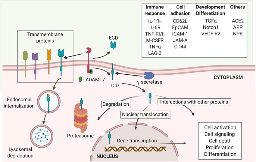
Gonzalez et al.                                                                                     ACE2 Intramembrane Proteolysis by Coronaviruses

severity of coronavirus disease-19 (COVID-19), and deaths has               Sp S1 binding and is followed by the internalization of Sp S1.
brought hope on overcoming this pandemic in a shorter period                SARS-CoV-2 Sp binds more strongly to ACE2 than does SARS-
of time (5–7). Challenges for global distribution and access to             CoV Sp (21, 22); it remains to be sought if internalization of
vaccines remain and it is imperative to vaccinate as many people            SARS-CoV-2 also requires the proteolytic activity of TMPRSS2
as possible to avoid emergence of new variants and surpass                  and/or the cleavage of ACE2. Curiously, the internalization of
the pandemic.                                                               SARS-CoV–1-ACE2 was shown to up-regulate the activity of a
    Our knowledge of SARS-CoV-2 remains limited. However,                   disintegrin and metallopeptidase 17 (ADAM17) (20), resulting in
lessons learned from previous coronavirus (CoV) outbreaks,                  increased proteolysis of the ACE2 ectodomain (19, 23, 24) and
mainly SARS-CoV, which caused the outbreak in 2002-2003 and                 decreased surface ACE2. Reduced cellular ACE2 level is
shares close genotypic and phenotypic similarities with SARS-CoV-           associated with heightened renin-angiotensin system (23) due
2 (8) can inform the immunopathogenic mechanisms triggered                  to increased tissue and serum level of Ang II (25, 26), which is
during SARS-CoV-2 infection and the factors potentially                     associated with inflammation and tissue damage (27–29).
responsible for its rapid transmission and the severe COVID-19                  ACE2 is a potential target of the regulated intramembrane
outcomes. This knowledge is crucial for the development of                  proteolysis (RIP). RIP is a signaling mechanism that follows the
preventive strategies and prophylactic treatment options.                   shedding of the extracellular domain of several trans-membrane
    Like SARS-CoV, SARS-CoV-2 uses the angiotensin-                         proteins, such as the Interleukin-1 receptor type II (IL-1RII), the
converting-enzyme 2 (ACE2) on the target cell as a binding                  IL-6R, TNFa and its receptors TNF-RI and TNF-RII, the
and entry receptor. Both interact with ACE2 via the receptor-               Amyloid precursor protein (APP), the Epithelial adhesion
binding domain (RBD) of the viral spike (S) protein, specifically,           molecule (EpCAM), Notch 1, among many others (30, 31).
the subunit 1 (S1) (9). Following this initial interaction, virions         The cleavage of the extracellular domain of the target proteins
are internalized in the acidic endocytic compartments where                 of RIP to release the ectodomain is mostly mediated by proteases
proteolytic cleavage of the S protein into S1 and S2 subunits by            of ADAM family, like ADAM17, matrix metalloproteases, and
cellular transmembrane protease serine 2 (TMPRSS2) occurs.                  the aspartyl proteases b-site APP-cleaving enzymes (BACE) (24,
Cleavage of S protein allows the exposure of the fusion domain of           30). The second cleavage is the proteolysis of the intracellular
the S2 subunit and the subsequent fusion of the viral envelope              domain, resulting in the release of an intracellular short carboxy-
with the cellular membrane. This process is similar for both                terminal fragment removed from the intra-membrane
SARS-CoV and SARS-CoV-2 (9), and is crucial for the release of              subdomain (30). It is mainly mediated by g-secretase
viral ssRNA genome into cytosol (10). Interestingly, preventing             multiprotein complex (24). While the released extracellular
the S protein cleavage by inhibiting the TMPRSS2 and the                    domains can induce activation and signaling of cells binding to
cysteine proteases CATHEPSIN B/L activity in vitro can block                these fragments, the intracellular subdomains can either be
the SARS-CoV from entering the cells (11) but cannot                        degraded at the proteasome or lysosomes or induce
completely block SARS-CoV-2 (9), suggesting that SARS-CoV-                  cytoplasmic signaling through interaction with other
2 has other cleavage site(s). In support, proteomic analyses                molecules. Some released intracellular subdomains of proteins
identified potential FURIN cleavage sites in SARS-CoV-2 S                    that undergo RIP have been shown to translocate into the nuclei
protein (12) and FURIN protease was shown to cleave MERS-                   to act as messengers and elicit biologic responses, such as
CoV S protein (13). Although inhibitor of FURIN activity could              changes in cellular transcriptional profile and signaling, and
reduce SARS-CoV-2 replication in vitro, it remains to be                    thus, influence the cell fate (24, 30, 31) (Figure 1). The
determined if SARS-CoV-2 infection also utilizes FURIN or                   outcome of the endodomain may depend on the specific
other proteases (14, 15).                                                   cleaved protein and regulatory signals, which remain a gap of
    Furthermore, other molecules such as the transmembrane                  knowledge in the RIP research. Several studies recognize that
receptor CD147 (basigin) expressed by epithelial and immune                 ADAM17, also known as tumor necrosis factor a-converting
cells from lung and skin, and the glycoprotein CD26, highly                 enzyme (TACE) is the main protease, responsible for the
expressed by CD4 and CD8 T cells and innate lymphoid cells                  cleavage of ACE2 (19, 20, 32, 33), even though TMPRSS2, the
(ILC) (16), have been suggested to facilitate SARS-CoV-2 entry              type II membrane serine protease can also cleave ACE2 (19, 33).
via interacting with the viral S protein (17, 18). Indeed, the              The conformational features of ACE2 and its cleavage mediated
antibody against CD147 inhibited in vitro SARS-CoV-2 infection              by ADAM17 (19) suggest that ACE2 may be a target of RIP (30).
(17). Nonetheless, the use of these alternative receptors by SARS-          To support ACE2 being a target of RIP, evidence should be
CoV-2 requires further exploration and confirmation in patients              obtained that following the shedding of ectodomain, the
with COVID-19.                                                              truncated ACE2 is also targeted for intramembrane proteolysis,
    The proteomic activity of TMPRSS2 and the cleavage of                   releasing a soluble ACE2 C-terminal fragment. However,
ACE2 facilitate the internalization of SARS-CoV Sp S1 subunit               whether the intracellular (endo-) domain of ACE2 is cleaved
bound to ACE2 (19). Cleavage-resistant ACE2 mutant failed to                following the binding of SARS-CoV-2 Sp and if the ACE2 endo-
facilitate the internalization of the Sp S1 subunit, but the protease       domain has a regulatory role, like other targets of RIP remains to
that cleaves ACE2 during SARS-CoV-2 infection remains                       be sought.
debatable (19, 20). Together, this in vitro data suggest that                   This review focuses on the proteolysis of ACE2 in
during SARS-CoV infection, ACE2 cleavage occurs following                   physiological and disease contexts and summarizes findings

Frontiers in Immunology | www.frontiersin.org                           2                                   June 2021 | Volume 12 | Article 612807
Gonzalez et al. ACE2 Intramembrane Proteolysis by Coronaviruses
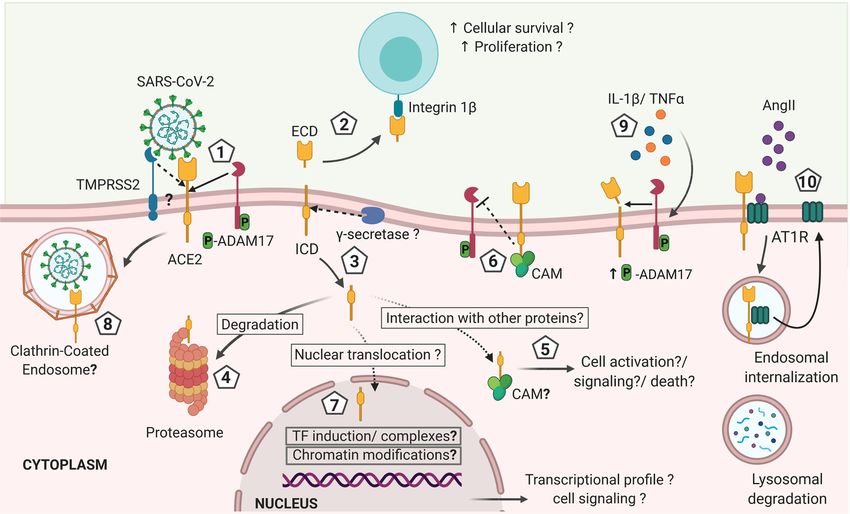
FIGURE 1 | Regulated intramembrane proteolysis process and proteins cleaved by ADAM17. Several proteins are targets of ADAM17 cleavage, including
proteins involved in immune responses, cell adhesion and cellular development and differentiation, among others. The proteolytic activity of ADAM17 can be
triggered through its phosphorylation by PKL2, PKC, and MAPKs. Activated ADAM17 cleaves several transmembrane proteins leading to the release of an
extracellular domain (ECD) and the membrane retention of an intracellular cytoplasmic domain (ICD). The g-secretase multi-protein complex removes this domain
from the membrane allowing it to migrate to the cytoplasm where it can either be degraded at the proteasome or interact with other cytoplasmic proteins.
Moreover, the ICD can trans-locate into the nuclei to induce transcriptional expression of several genes related to cell activation, signaling, death, proliferation and
differentiation (created with BioRender.com).
that support the RIP of ACE2 during SARS-CoV-2 and other encoding a large extracellular domain (also termed ectodomain)
CoVs infections. Moreover, we propose here a hypothesis of the (723aa), a transmembrane domain (21aa) and a short cytoplasmic
potential effects of RIP of ACE2 in cellular fate and COVID-19 domain (also termed endodomain) (44aa) (40). Both ACE2 and its
disease progression and severity. homolog, ACE are fundamental regulators of the renin-angiotensin
system (RAS) that controls blood pressure homeostasis (26, 41) and
the main function of ACE2/angiotensin (1-7) [Ang(1-7)] axis is to
ACE2 CLEAVAGE AND THE counter-balance the effects of ACE/Ang II axis (42). Both enzymes
ENZYMES INVOLVED differ in their substrate specificity: ACE converts Ang I to the potent
vasoconstrictor, Ang II; ACE2 then cleaves Ang II to generate the
ACE2 is a zinc metallopeptidase, type I transmembrane protein vasodilator, Ang(1-7) (27, 28, 41). ACE2 has also been shown to
widely expressed in several tissues and organs, such as vascular cleave Ang I directly, bypassing ACE function to generate Ang(1-9),
endothelia and cardiovascular tissue, brain, the epithelial cells from but at a much-reduced efficiency compared to the cleavage of Ang II
oral and nasopharyngeal mucosa, lungs, kidney, gastrointestinal (43). While inhibition of ACE results in protective cardiovascular
mucosa, Langerhans of the pancreatic tissue, and bone, among effects including reduced blood pressure and lower levels of Ang II,
others (32, 34–38). There is no significant expression of ACE2 in the reduced ACE2 expression is reported in people with hypertension.
lymphoid organs and hematopoietic cells but macrophages (36, 37). However, deletion of Ace2 expression in mouse model did not
Human ACE2 is a 40 kb gene, mapped to chromsomeXp22. ACE2 directly cause hypertension but caused enhanced susceptibility to
transcripts are found in 72 human tissues (39). It encodes 22 introns Ang II-induced hypertension (44). Moreover, reduced ACE2
and 18 exons (40). ACE2 protein consists of ~805 amino acids expression is associated with cardiac dysfunction and heart failure
Frontiers in Immunology | www.frontiersin.org 3 June 2021 | Volume 12 | Article 612807Gonzalez et al. ACE2 Intramembrane Proteolysis by Coronaviruses
that could indirectly result in hypertension (42, 45, 46). These response, cellular development and differentiation, and cell
support the contrasting effects of ACE2/Ang (1-7) and ACE/Ang II adhesion, as well as in several diseases including cancer,
axes (42, 45, 46). Moreover, ACE2 cleaves several other bioactive Alzheimer and others (24, 30, 51, 52). Although it has not
and physiologically important peptides, such as bradykinin, an been completely established if ACE2 shedding corresponds to a
endothelium-dependent vasodilator, the hypotensive peptide RIP event, cleavage of several proteins by ADAM17 is
apelin-13, the opioid peptides dynorphin A and b-casomorphin, accompanied by an intramembrane proteolysis mediated by
the neuropeptide, neurotensin, and the amyloid-b peptide involved the multiprotein g-secretase complex (24) (Figure 1). In a
in Alzheimer disease (43, 47). As mentioned above, ACE2 has been mouse model of neurogenic hypertension, deoxycorticosterone
exploited by coronavirus, such as SARS-CoV, SARS-CoV-2 and acetate-salt treatment caused significant increases in ADAM17
HNL-63-CoV, which causes common cold, for viral entry (9, 48). expression and activity in the hypothalamus and decreased
Hence, a better understanding of ACE2 signaling and function is hypothalamic ACE2 activity and expression, which are
essential for preventative strategies involving the modulation of accompanied by increases in blood pressure, hypothalamic
ACE2 levels, binding and signaling. Ang II levels, and inflammation, and impaired autonomic
An interesting feature shared between ACE2 and ACE is the dysfunction (53). Reduced ACE2 expression and activity in the
shedding of their extracellular domain. ACE is cleaved by the brain also occurred in parallel with an increase of shed ACE2
ACE secretase (43), a sheddase related but different from ectodomains and enzymatic activity in the cerebrospinal fluid
ADAM17, which is the main protease for ACE2 cleavage (25). (53). Chronic knockdown of ADAM17 in the brain was able to
ADAM17 is not shown to be involved in the shedding of ACE blunt the development of hypertension and restored ACE2
(49), partially explained by the minimal sequence homology activity and baroreflex function (53). The caveat with chronic
between ACE and ACE2 at the cleavage sites (43). Both exhibit knockdown of ADAM17 is that there are numerous substrates
only ~42% amino acid homology at their extracellular catalytic for ADAM17 and hence, ADAM17 affects many systems,
domain (40). Whether ACE secretase could induce ACE2 including cytokines that may play a role in hypertension.
shedding is currently unknown but unlikely (43). However, similar effects in blunting the development of
In addition to ADAM17, TMPRSS2, human airway trypsin hypertension and restoring baroreflex function were also
(HAT) and hepsin have also been shown to cleave ACE2. observed when Ace2 was overexpressed in the neurons (53).
TMPRSS2 can interfere with ADAM17-mediated cleavage of Nonetheless, in this model, it is not clear if the pathogenesis was
ACE2 and has been shown to be exploited by CoVs to cleave the caused by reduced ACE2 and Ang (1-7) expression and activity
S protein, required for viral entry into host cells (19, 33). All these in the brain or the increase of Ang II and/or ACE2 ectodomains
three enzymes, TMPRSS2, HAT and hepsin cleave ACE2 at the in the cerebrospinal fluids. The recombinant ACE2 (rhACE2)
same catalytic site, which differs from the proteolytic site for will be a great tool to address this question (54). Moreover, the
ADAM17. Thus, differential outcomes of such cleavages effect of other inflammatory pathways that are also modulated by
compared to ADAM17-mediated shedding may occur (19). It ADAM17 activity on brain inflammation, cannot be ruled out in
is not clear whether these 3 enzymes have redundant roles in the this model (24, 51, 53).
ACE2 signaling and whether HAT and hepsin could also cleave S Spontaneous, constitutive cleavage of ACE2 ectodomain can
protein to facilitate CoV entry. The role of ADAM17 in occur in the absence of disease and may be tissue specific. Soluble
facilitating SARS-CoV-2 infection was examined in a recent ACE2 ectodomain with enzymatic activities are detected in
study using inhibitor (GW280264X) and the siRNA against human bronchoalveolar lavage and urine samples from healthy
ADAM17 to block SARS-CoV-2 infection of HK-2, a human donors (55, 56). In the culture of mouse proximal tubular cells,
kidney cell line, by >90% (50). Data presented in this study the low level, constitutive cleavage of ACE2 ectodomain was
suggests that ADAM17 is the primary sheddase for ACE2. detected (57, 58) but could be further augmented by high levels
The consequential events triggered by ACE2 cleavage remain of D-glucose or Ang II (58). Adding high concentrations of Ang
poorly understood, so do the effects of shedding extracellular and II (10−7 M) or D-glucose (25mM) to the culture of mouse
the intracellular ACE2 domains. Since similar shedding proximal tubular cells resulted in increased ADAM17 activity
processes have been described for other membrane proteins, in the cell lysate and augmented release of ACE2 extracellular
they could potentially provide some insights on the destiny of subdomains and ACE2 enzymatic activity in the culture media in
ACE2 fragments and their functions. a time-dependent manner (58). Hyperactivity of Ang II signaling
pathways have been shown in both clinical trials and animal
models of diabetes to contribute to the development of diabetes
THE PHYSIOLOGICAL ROLE OF ACE2 and diabetic complications (59). In addition, increased soluble
ECTODOMAIN AND THE POTENTIAL ACE2 is found in the urine of patients with diabetes and/or renal
IMPLICATIONS OF ACE2 ENDODOMAIN diseases (59–63). Although these observations imply a role for
ACE2 shedding in diabetes, whether ACE2 cleavage has a
Following the cleavage of the extracellular domains from an ever- compensatory or a pathogenic role in the development of
increasing number of proteins, RIP processing can lead to diabetes remain to be sought.
cellular signaling and modulation of the surrounding In addition to high glucose-D and Ang II, phorbol ester,
microenvironment (30). RIP is a crucial event in immune ionomycin, endotoxin and IL-1b & TNF-a could also acutely
Frontiers in Immunology | www.frontiersin.org 4 June 2021 | Volume 12 | Article 612807Gonzalez et al. ACE2 Intramembrane Proteolysis by Coronaviruses
increase the release of ACE2 ectodomain from primary, proteins have been shown to facilitate nuclear signaling in
polarized human airway epithelial cells and Calu-3 (human altering cellular gene expression under specific cellular
lung adenocarcinoma cell line) (55). Cleaved ACE2 conditions (51). The release of ACE2 endodomain needs to be
ectodomain was detected in the apical secretions of polarized confirmed, and the fate of ACE2 endodomain remains to be
epithelial cells in culture and in the human bronchoalveolar sought. Our current knowledge of how the activity of g-secretase
lavage. Inhibition assay using DPC333 (1.5 nM) and GI254023 complex is triggered to mediate intramembrane proteolysis is
(5mM), inhibitors for ADAM17 and ADAM10, respectively (55), limited; the molecular mechanism(s) leading up to the cleavage
showed that ADAM17 activity is required in both the of ACE2 endodomain are yet to be defined (Figure 1). The
constitutive and acutely induced ACE2 shedding but inhibition intracellular ACE2 endodomains were also detected following
of either ADAM17 or ADAM10 prevented the release of ACE2 the TMPRSS2 mediated cleavage of ACE2 ectodomain in 293 T
ectodomain induced by PMA or ionomycin, respectively (24, cells transfected with full-length ACE2 (19). Furthermore,
52). Evidence supporting ADAM17’s role in regulated ADAM17 and TMPRSS2 do not seem to share the same cleavage
proteolysis of ACE2 is strengthened by the observations that site on the ACE2 ectodomain (19). Interestingly, Heurich et al.
siRNA specific for ADAM17 reduced PMA-induced ACE2 reported the presence of an ACE2 C-terminal fragment (13kDa) in
shedding and overexpression of ADAM17 in HEK293 cells the cellular lysates following TMPRSS2-mediated ACE2 cleavage,
increased ACE2 shedding (24). On the other hand, inhibition and this ACE2 C-terminal fragment was absent in the cell lysate
or siRNA-mediate knockdown of ADAM10 expression had no following ADAM17-mediated ACE2 cleavage (19). It suggests that
impact on PMA-triggered shedding of ACE2 (24, 55, 63). These the trigger for the shedding of the ACE2 ectodomain may affect the
studies are in agreement that while ADAM17 may be the main fate of ACE2 intracellular domain. This is further supported by
sheddase for cleaving ACE2 ectodomain constitutively at low evidence that the endodomains of other proteins that undergo RIP
level or in response to stimulus, such as PMA, ADAM10 is also have different fates depending on the cellular
involved in ACE2 shedding in response to ionomycin (24, 55, microenvironment (30).
63). ADAM10 is also involved in ionomycin-induced shedding There have been limited studies of the fate of ACE2
of CX3CL1 and CXCL16 from leukocytes and EGFR-ligand endodomain following ACE2 shedding. ACE2 seems to exhibit
shedding from fibroblasts (64, 65). The response to PMA and different internalization patterns under different stimulation (19,
ionomycin stimulation of regulated proteolysis of ACE2 24, 55, 72, 73). Heurich et al. (19) observed degradation of ACE2
ectodomain further suggests the involvement of proteinase endodomain following the cleavage by ADAM17. Other studies
kinase C pathway and intracellular calcium signaling in further suggest that during constitutive, low level of ACE2
regulating ACE2 shedding. The shedding of ACE2 ectodomain shedding, mediated by ADAM17, the cytosolic ACE2 fragment
from mice cardiomyocytes could also be induced by Ang II, is degraded via proteasome (19, 24, 55). Moreover, exposure of
which was shown to increase the phosphorylation and activity of ACE2-GFP-transfected Neuro-2A cells, a murine neuroblastoma
ADAM17 (25, 66). In patients with heart failure, increased ACE2 cell line, to high concentrations of Ang II triggered ACE2
ectodomain was reported in plasma, with increased ACE2 internalization and degradation into lysosomes (73). ACE2-
enzymatic activity, suggesting that excessive shedding and internalization triggered by high levels of Ang II is dependent
extracellular release of the ACE2 ectodomain is associated with on the presence of AT1R; this study showed that losartan,
diseases (25, 62). It remains to be shown whether the enzymatic blocker of AT1R or the absence of AT1R in HEK293T cells
activity of circulatory ACE2 ectodomains has any physiological failed to facilitate ACE2 internalization (73). ACE2 degradation
or pathological significance. It is possible that shedding is one of in lysosomes could be blocked by leupeptin, inhibitor of
the mechanisms for regulating ACE2 activity on the cell surface lysosome and was dependent on the ubiquitination of ACE2
(67). Paradoxically, while surface ACE2 facilitates SARS-CoV endodomain, triggered by high levels of Ang II (73).
entry, soluble ACE2 ectodomain has been shown to block SARS- Ubiquitination, a major mechanism known to mediate plasma
CoV infection of cells. In contrast, a recent study showed that membrane protein internalization involves the addition of
soluble ACE2 facilitate SARS-CoV-2 uptake via binding to Ang ubiquitin moiety to lysine residues (72, 74). E3 ligases that
II type 1 receptor (AT1R) (50). Recombinant human ACE2 has ubiquitinate ACE2 includes MDM2 (murine double minutes 2)
been tested in a phase 2-3 trial in acute respiratory distress in vascular endothelial cells and Skp2 in lung epithelial cells (75,
syndrome with interesting results (68), and a pilot trial has been 76). Work by Shen et al. showed that increased levels of MDM2
launched in COVID-19 (NCT04287686) (69, 70). in lung tissues or pulmonary arterial endothelial cells from
Like other proteins that undergo RIP, there is some evidence patients with idiopathic pulmonary arterial hypertension
suggesting that following the release of ACE2 ectodomain, the inversely associated with the levels of ACE2 (75). Furthermore,
ACE2 endodomain is also cleaved off the transmembrane siRNA specific for MDM2 could increase ACE2 expression while
subdomain and released into cytosol (19). The g-secretase overexpression of MDM2 resulted in significant decrease in
complex, a multi-subunit protease complex that catalyzes the ACE2 level in human pulmonary artery endothelial cells (75).
proteolysis of single-pass transmembrane proteins at residues Up-regulating Skp2 by bezo-a-pyrene also resulted in reduced
within the transmembrane domain, catalyzes the RIP of several cellular ACE2 protein level in 16HBE, human bronchial
proteins, such as E-Cadherin, EpCAM, amyloid precursor epithelial cell line, via increased ubiquitination of ACE2 (76).
protein (APP) and CD44 (52, 71). The endodomain of these To summarize, ubiquitination is a major mechanism involved in
Frontiers in Immunology | www.frontiersin.org 5 June 2021 | Volume 12 | Article 612807Gonzalez et al. ACE2 Intramembrane Proteolysis by Coronaviruses regulating cellular ACE2 protein level. In response to increased Furthermore, whether the non-degraded cytosolic ACE2 Ang II, ubiquitination of ACE2 endodomain and interaction of endodomain has a physiological function via interaction with ACE2 with AT1R are critical for the endocytosis and degradation other cytosolic proteins or translocation to the nuclei remains of ACE2 in lysosomes. The interacting AT1R was shown to be unknown (Figure 2). Nonetheless, a candidate nuclear recycled and transported back to the membrane (72, 73). ACE2 localization sequence has been predicted in the ACE2 was shown also to be internalized in response to the binding of endodomain (769RKKKNKA774) using the NLStradamus (http:// the Sp from SAR-CoV-1 and SARS-CoV-2 (19, 24, 55, 77), www.moseslab.csb.utoronto.ca/NLStradamus/) (80) program. It perhaps another mechanism to reduce surface ACE2 expression. remains to be shown whether the ACE2 endodomain has any The route of internalization of the ACE2/SARS-CoV-2 complex regulatory function on gene expression (81). remains under debate, with reports of clathrin- and caveolae- The ACE2 endodomain was shown to interact with Calmodulin independent pathways in HEK293T cells, as well as clathrin- (CAM) that mediates important cellular processes, such as dependent and ACE-2 C-terminus-independent internalization metabolism, cell death and immune responses (82, 83). in COS7, HepG2 and HEK293T-ACE2 cell lines (77–79). Bayati Interestingly, interaction of CAM with the intracellular domain of et al. observed that after engagement with the ACE2 on cell the full-length ACE2 inhibited ACE2 shedding, suggesting a role for surface, the purified spike glycoprotein and the lentivirus the ACE2 intracellular domain in regulating the cleavage of its pseudotyped with the Sp of SARS-CoV-2 underwent rapid, ectodomain (82, 83). In support, ACE2 mutant lacking the clathrin-mediated endocytosis, co-localized with the Rab5- endodomain could not facilitate the proteolysis of ACE2 positive early endosomes in HEK293T-ACE2, Vero-SF-ACF ectodomain induced by SARS-CoV Sp (20). Although this finding and A549 cell lines (79). Inhibitors of clathrin-coated pit suggests a role played by the ACE2 endodomain in regulating formation, such as dynasore and pistop-2 or siRNA against the ADAM17 activity in ACE2 shedding, it could be that deletion of the heavy chain of clathrin blocked the internalization of the SARS- endodomain affected the structural conformation of the ACE2 CoV-2 Sp (79). Inoue et al. made similar observations with the Sp ectodomain, not accessible to ADAM17 (20). ACE2 studies were of SARS-CoV in COS-7 and HepG2 cell lines and further mostly performed in vitro with cell lines. Cell types, culture demonstrated that a low pH condition is required for SARS- condition and stimuli for ACE2 cleavage or internalization may CoV to establish an infection and that the endodomain of ACE2 influence the fate of ACE2 endodomain and should be considered in is not required for the internalization of the SARS-CoV-Sp- study design and interpretation of data. pseudotyped lentivirus (78). This finding contradicts with what Interestingly, CD147, an alternative receptor for SARS-CoV- Haga et al. found that the ACE2 cytoplasmic tail is essential for 2, was also shown to be a target protein of RIP in tumor cell lines the SARS-CoV Sp induced shedding of ACE2 ectodomain which (84). CD147 cleavage is mediated by ADAM10 (84). The released is required for the endocytosis of SARS-CoV Sp (20). CD147 ectodomain was shown to induce the activation of Unfortunately, these two studies did not examine ACE2- fibroblasts (85). Following CD147 shedding, its endodomain is shedding or the fate of ACE2 following its internalization. In a released into cytosol and translocated to lysosomes to be cleaved seemingly contradictory study of the internalization of SARS- again to produce a nuclear localizing subunit, which enhances CoV Sp and Sp-pseudotyped lentivirus, Wang et al. found that autophagy function via the NF-kB–TRAIL–caspase8–ATG3 SARS-CoV bound to ACE2 translocated from cell surface to axis, favoring tumor cell survival (84). It remains to be early endosomes via a pH-dependent, clathrin- and caveolae- explored if SARS-CoV-2 infection triggers RIP of CD147 and independent mechanism (77). In HEK293E-ACE2-GFP cells, ACE2 to modulate cellular activity or function. treatment with ammonium chloride, bafilomycin A1, or chloroquine, resulted in accumulation of ACE2 within perinuclear vacuoles, even after a 14h-incubation. The use of ACE2 AND ITS POTENTIAL IMMUNE- siRNA against clathrin or chlorpromazine, a cationic MODULATORY FUNCTIONS amphiphilic drug that disrupts clathrin-mediated endocytosis had little inhibitory effects (~20%) on the infectivity of the Sp- ACE2 may contribute to maintaining immune regulation (86). pseudotyped lentivirus of VERO cells, suggesting that Sp-ACE2 Overexpression of ACE2 in neurons of a mouse model for induced complex could be internalized via a clathrin-independent neurogenic hypertension resulted in reduced arterial pressure, and mechanism. The study further concluded that the 14h- inflammation, indicated by decreased expression of pro- incubation allowed ACE2 to be ‘recycled’ to the cell surface inflammatory cytokine genes such as TNFA, IL-1b and IL-6 in (77). However, the study did not provide strong data to support hypothalamus (53). All of these cytokines (TNFa, IL-1b, IL-6) were this conclusion. The ACE2 observed at 14h-post-exposure to Sp found in high levels in COVID-19 patients and associated with or Sp-pseudotyped lentivirus could be newly synthesized ACE2. severe disease outcomes (87–91). In agreement, bone marrow Blockers of protein-synthesis or protein trafficking should be derived macrophages from an ACE2-deficient, atherosclerosis used to investigate whether ACE2 was recycled back out to the mouse model exhibited a higher baseline level of pro- cell surface. Although these studies provide evidence that inflammatory cytokines (e.g., TNFa and IL-6) and hyper- ACE2 can be internalized via different mechanisms, the fate responsiveness to LPS and TNF-a stimulation (92). Moreover, in of internalized ACE2 or ACE2 endodomain remains to a murine model of experimentally induced lung injury, be sought. administration of a high dose of recombinant ACE2 protected the Frontiers in Immunology | www.frontiersin.org 6 June 2021 | Volume 12 | Article 612807
Gonzalez et al.                                                                                                        ACE2 Intramembrane Proteolysis by Coronaviruses

 FIGURE 2 | Hypothetical model of ACE2 shedding by ADAM17 and g-secretase after ACE2 interaction with SARS-CoV-2 and its effects on the immune response
 against the infection. 1). Initial interaction between SARS-CoV-2 spike protein and the transmembrane cellular receptor ACE2 may trigger proteolytic cleavage of
 ACE2 mediated by ADAM17, releasing the extracellular domain of this receptor (ectodomain). Other proteases such as TMPRSS2, HAT and Hepsin may also cleave
 ACE2 at a different catalytic site than ADAM17, thus potentially exhibiting differential outcomes. 2) Soluble ACE2 can interact with other cells through integrins acting
 like a cell adhesion molecule and triggering signals associated with cell proliferation and survival. 3) The cytoplasmic domain is likely released from the cellular
 membrane by g-secretase, as described for other RIP proteins and it is either: 4) degraded at the proteasome, or 5) interacts with cytoplasmic proteins as it has
 been described for calmodulin (CAM) that can have effects on cellular activation, inflammation and cells death. 6) If CAM interacts with the transmembrane full-length
 ACE2, the shedding of ACE2 may be abrogated. 7) The intracellularly released ACE2 domain may also be translocated into the nuclei. At the nuclei, the endodomain
 may induce transcriptional expression of different genes by a still unknown mechanism that can include induction of transcription factors or formation of transcription
 factors-like complexes or chromatin modifications, influencing the cellular response and fate. In addition, the increased levels of IL-1b and TNFa induced during
 COVID-19 may increase ADAM17 phosphorylation and activity favoring a sustained shedding of ACE2. Thus, the shedding of ACE2 induced by SARS-CoV-2
 infection is likely influencing the outcome of COVID-19 by mechanisms that are still unknown and that remain to be explored in order to design preventive and
 therapeutic approaches (created with BioRender.com).

mice from developing severe acute lung failure associated with                           signaling, including decreased phosphorylation of the focal
reduced pro-inflammatory responses. On the contrary, loss of                              adhesion kinase (FAK) (95). Moreover, high concentrations of
pulmonary ACE2 expression led to pulmonary malfunction with                              ACE2 ectodomain (0.1 mg/ml or 1mg/ml) could promote Akt
enhanced vascular permeability and lung edema (93). Similarly, in a                      expression and its phosphorylation that have implications in
mouse model, experimental SARS-CoV infection decreased ACE2                              promoting cell activation, proliferation and survival. The ACE2
expression in the lung that correlated with lung injury. The observed                    ectodomain concentration tested is approximately 200-fold higher
ACE2 reduction was induced by SARS-CoV Sp (94). In summary, a                            than the baseline circulating soluble ACE2 concentration (1-2ng/ml
decrease in cellular ACE2 may reduce the susceptibility of cells to                      serum) (41, 96–98). Although high soluble ACE2 level is probably
SARS-CoV-2 but leads to greater, undesirable immune activation                           required to offset the effects of endogenous ACE, which is ~100-200
and more severe tissue damage. In contrast, high abundance of                            fold higher than the baseline ACE2 level, these findings are
ACE2 on the cell membrane is associated with increased                                   encouraging and should be pursued further to test whether
susceptibility to viral particles, but with less damage, due to less                     soluble ACE2 could affect immune activation directly at
inflammation. On the note of inflammation, exogenous soluble                               physiological or pathogenic concentrations (95).
ACE2 ectodomain was shown to promote inflammation. ACE2                                       Most literature showed that ACE2 modulate immune response
ectodomain was found to bind to the adhesion molecule, integrin                          ‘indirectly,’ via its enzymatic function in cleaving Ang II to generate
b1, enhancing cell-cell interactions and regulating cell induced                         Ang(1-7), which have anti-inflammatory property (54, 86, 99, 100).

Frontiers in Immunology | www.frontiersin.org                                        7                                           June 2021 | Volume 12 | Article 612807
Gonzalez et al.                                                                                      ACE2 Intramembrane Proteolysis by Coronaviruses

As Ang II has pro-inflammatory property, via reducing Ang II,                against non-RBD regions are found in COVID-19 patients, a
ACE2 can promote immune quiescence. In support, treatment of                hypothesis that requires further exploration (108).
rats with Ang(1-7) reduced inflammatory damage of cardiac tissue,                Binding of the S protein to ACE2 triggers the shedding of
which was associated with decreased NF-kB activity and expression           ACE2 ectodomain (20), which explains the reduction in cellular
of NF-kB-regulated genes (101). Moreover, treating LPS-activated            membrane ACE2 observed during SARS-CoV infection (94).
macrophages with Ang(1-7) resulted in reduced expression of pro-            Such reduction has also been observed in vitro in VERO cells,
inflammatory cytokine genes, such as TNFA and IL-6, most likely              Caco-2 cells and ACE2-transfected HEK293T cells exposed to
via modulating Src kinase activities (29). However, contrasting             recombinant S protein (94). Nonetheless, it is yet to be explored
findings showed that Ang(1-7) treatment of mice with an                      if SARS-CoV-2 S protein also triggers ACE2 shedding (109). A
established inflammatory condition further aggravated the                    recent study supports this hypothesis. Convalescent COVID-19
inflammatory response, resulting in pronounced apoptosis and                 patients exhibited elevated ACE2 activity in plasma, compared to
increased NF-kB activity (102). Some in vitro studies showed no             matched-healthy donors. The plasma ACE2 activity remains
effect of Ang (1-9) on NF-kB or Akt expression and                          high for a long period of time after recovering and the level
phosphorylation (95). In discerning the differences between the             and length are positively associated with the disease severity
two inflammatory animal models (cardiac tissue verses renal tissue)          (110). It is not clear how ACE2 shedding is regulated in
and the macrophage study (29, 95, 101, 102), understanding how              convalescent COVID-19 patients and how plasma ACE2 level
Ang(1-7) affects NF-kB activity in these two different tissues may          affects the recovery process.
help in resolving this discrepancy.                                             Further supports for the potential impacts of ACE2 shedding
    Moreover, the accumulated Ang II level in the absence or                in immune activation and disease severity came from HNL-63-
reduction of ACE2 was shown to favor pro-inflammatory states.                CoV- (common cold virus) infected VERO cells (20). HNL-63-
Exposing alveolar epithelial cells to pro-fibrotic apoptotic inducers        CoV infection seems to be acquired during childhood and is
while inhibiting ACE2 activity resulted in increased Ang II and             usually not associated with severe disease (111).
decreased Ang(1-7) in cell culture which were associated with                   HNL-63-CoV also engages target cells through binding of its
increased caspase activation, nuclear fragmentation and JNK                 S protein to ACE2. However, SARS-CoV Sp binds to ACE2 with
phosphorylation (103). In fact, Ang II has been shown to have               higher efficiency than HNL-63-CoV Sp (111). HNL-63-CoV Sp
important pro-inflammatory properties, including inducing the                binding to ACE2 did not trigger ACE2 shedding nor ADAM17-
expression of adhesion molecules, recruiting inflammatory cells,             activation (20). Shedding most likely accounted for the observed
and increasing the production of pro-inflammatory cytokines, such            ACE2 down regulation. Glowacka et al. showed that HNL-63-
as IL-1b, IL-18, IFNg, TNFa and IL-6 (104). In summary, changes             CoV infection had no effects on the surface level of ACE2 on
in ACE2 expression or enzymatic activity can modulate Ang II level          VERO cells and that recombinant SARS-CoV Sp bound to ACE2
to affect immune regulation. Although these in vitro and murine             and induced ACE2 shedding with higher efficiency than did the
studies suggest a critical role for ACE2 activities in regulating the       HNL-63-CoV Sp. While ACE2 shedding might be associated
pro-inflammatory state, the inherent limitations in murine and               with disease severity follow coronavirus infection, it was found to
in vitro models however, prevent the extrapolation of these findings         be dispensable for the spread of SARS-CoV and HNL-63-CoV
to defining the physiological roles of human ACE2.                           (111). The efficiency of S protein binding to ACE2 may be
                                                                            explained in part by the differences in the amino acid
                                                                            sequences between the S proteins from both viruses (112). The
                                                                            affinity of S protein binding to ACE2 may have influence over the
BINDING OF CoV Sp TO ACE2 INDUCES                                           activation of ACE2 cleavage and the outcome of infection.
THE PROTEOLYTIC CLEAVAGE OF ACE2                                                The differential proteolysis of ACE2 could also be attributed to
POTENTIALLY INFLUENCING THE                                                 i) a differential requirement of ADAM17 during infection, since
SEVERITY OF INFECTION                                                       silencing of ADAM17 significantly reduced SARS-CoV infection
                                                                            but not HNL-63-CoV (20), and ii) the differential cellular
As described above, the initial binding of the SARS-CoV Sp or               microenvironment requirements for processing S protein. SARS-
SARS-CoV-2 Sp with ACE2 is the first step for viral entry. In this           CoV infection and the cleavage of its S protein depend on the
regard, current studies are evaluating strategies to reduce                 availability of cathepsin L and low endosomal pH, which is not the
transmission or severity of COVID-19 through direct blockade                case for the HNL-63-CoV infection (113, 114). Other additional
of this interaction (105, 106). Interestingly, although both                mechanisms that were not discussed here can also be involved.
coronaviruses share only ~79% genetic similarity (8), and 76%               Thus, ACE2 cleavage induced by SARS-CoV S protein is most likely
homology in their amino acids sequences, both SARS-CoV and                  a unique mechanism exploited by SARS-CoV that may likely
SARS-CoV-2 harbor crucial amino acid residues at the receptor               influence the viral pathogenesis of the disease. Whether ACE2
binding domain (RBD) of S protein required for binding to                   shedding mediated by ADAM17 or TMPRSS2 occurs during SARS-
ACE2 (9). However, the SARS-CoV-2 RBD binds to ACE2 with a                  CoV-2 infection and if ACE2 proteolysis is a mechanism
10-20 fold higher affinity than does the SARS-CoV RBD (22,                   contributing to the differential pathogenesis and disease outcome
107). It is highly likely that additional mechanism(s) are involved         caused by SARS-CoV, SARS-CoV-2 and HNL-63-CoV remains to
in SARS-CoV-2 binding to ACE2, since neutralizing antibodies                be explored.

Frontiers in Immunology | www.frontiersin.org                           8                                    June 2021 | Volume 12 | Article 612807
Gonzalez et al. ACE2 Intramembrane Proteolysis by Coronaviruses
ACE2 cleavage was shown to be essential for efficient SARS- virus-induced generation of ACE2 ectodomain and endodomain
CoV infection (20). However, it may not be the case for SARS- remain to be defined. Understanding the functional properties of
CoV-2 infection or spread. Whether ACE2 proteolysis is these domains induced by coronavirus infection will enhance our
required for SARS-CoV-2 infection and/or replication remains knowledge in the pathogenesis, potentially mediated by ACE2
debatable. Reduced ACE2 surface expression with increased shedding and hence, facilitate the design of preventative tools
ACE2-shedding and their relationship with COVID-19 disease against a severe disease outcome.
severity are yet to be evaluated in SARS-CoV-2 infection. Collectively, these supporting evidence suggest that ACE2 is
Although ACE2 cleavage by TMPRSS2 enhances the uptake of an important regulator of the immune activation, ACE2
SARS-CoV (19), the mechanism involved is still unclear. It was shedding, loss of surface ACE2 expression or loss of ACE2
proposed that ACE2 fragments harbors internalization signals that enzymatic activities can result in enhanced immune activation,
may favor a more efficient uptake of SARS-CoV in a cathepsin L- and pro-inflammatory milieu. The mechanisms underlying these
dependent pathway (19). Even though TMPRSS2 has been shown correlative observations are yet to be defined. As ACE2 shares
to compete with ADAM17 in cleaving ACE2, ADAM17-mediated certain features with other RIP proteins, it is highly plausible that
ACE2 cleavage had no effect on the efficiency of viral uptake (19, 33). the ACE2 intracellular domain has a key regulatory function,
Work is ongoing in determining the differences in the ACE2 responsible for the trans-activation of pro-inflammatory genes in
fragments released by TMPRSS2 versus ADAM17. epithelial cells and alveolar macrophages. The contribution of
Furthermore, the establishment of a pro-inflammatory ACE2 shedding to the cytokine storm observed during SARS-
environment as it is observed during SARS-CoV-2 infection may CoV-2 requires urgent investigation. It remains to be confirmed
also play a role in triggering ACE2 shedding via up regulation of whether there is a functional or consequential difference in the
ADAM17 activity. In vitro activation of human airway epithelial ACE2 shedding by ADAM17 versus TMPRSS2. While there are
cells by PMA/Ionomycin or IL-1b and TNFa resulted in increased studies on the cleavage of ACE2 by ADAM17, little is known of
ADAM17 proteolytic activity and ACE2 shedding (55). The TMPRSS2-mediated ACE2 shedding, including whether the
increased plasma levels of both IL-1b and TNFa in COVID-19 TMPRSS2-cleaved ACE2 ectodomain enhances the virulence of
patients (115, 116) suggest their potential contribution to increased coronavirus. This knowledge is key to employing inhibition of
ADAM17-mediated ACE2 shedding and consequently increased ACE2 shedding as a therapeutic strategy during early COVID-19
plasma Ang II and heightened renin-angiotensin system, leading to to avoid excessive inflammation and disease severity, caused by
enhanced inflammation and triggering lung failure in COVID-19 lung damages.
patients. However, this hypothesis remains to be tested. An
approximated scenario derived from a mouse model of induced
acute lung injury showed that SARS-CoV Sp could reduce ACE2
expression and enhance the severity of lung damage by inducing a
AUTHOR CONTRIBUTIONS
pro-inflammatory state (94). SG participated in the searching and reviewing of research
Several questions remain unsolved regarding ACE2 proteolysis articles, defining the hypothesis, writing and revising the
during CoV infection, especially in SARS-CoV-2, including: 1) Is manuscript, and designing the figures. AS participated in the
ACE2 cleavage triggered by SARS-CoV-2 infection? If so, 2) is searching and reviewing of research articles, writing and
ACE2 cleavage a RIP event? 3) Are the extracellular and revising the manuscript. R-CS participated in the searching
intracellular ACE2 subunits involved in regulation of cellular and reviewing of research articles, defining the hypothesis and
transcription, signaling and fate? 4) Is the exacerbated immune writing, reviewing and revising the manuscript. All authors
response and disease severity influenced by ACE2 proteolysis by contributed to the article and approved the submitted version.
ADAM17 or TMPRSS2? The answers to these questions could be
key for understanding the immunopathogenesis of COVID-19 and
for directing therapeutic approaches.
FUNDING
CONCLUSIONS The work is funded primarily by the Public Health Agency of
Canada. SMG is supported by the operating grant from
Although ACE2 shedding is not a novel concept, its regulation Canadian Institute of Health Research to R-CS (CIHR/
during coronavirus infection and the functional roles of the NIH-154043).
Canada and the United States. CMAJ (2020) 192(25):E666–70. doi:
REFERENCES 10.1503/cmaj.200711
1. Mizumoto K, Dahal S, Chowell G. Spatial Variability in the Risk of Death 3. Emerging SARS-Cov-2 Variants. CDC (2021). https://www.cdc.gov/
From COVID-19 in Italy. Int J Tuberc Lung Dis (2020) 24(8):829–37. doi: coronavirus/2019-ncov/more/science-and-research/scientific-brief-
10.5588/ijtld.20.0262 emerging-variants.html.
2. Abdollahi E, Champredon D, Langley JM, Galvani AP, Moghadas SM. 4. Burki T. Understanding Variants of SARS-Cov-2. Lancet (Lond Engl) (2021)
Temporal Estimates of Case-Fatality Rate for COVID-19 Outbreaks in 397(10273):462. doi: 10.1016/S0140-6736(21)00298-1
Frontiers in Immunology | www.frontiersin.org 9 June 2021 | Volume 12 | Article 612807Gonzalez et al. ACE2 Intramembrane Proteolysis by Coronaviruses
5. Polack FP, Thomas SJ, Kitchin N, Absalon J, Gurtman A, Lockhart S, et al. 23. Chung MK, Karnik S, Saef J, Bergmann C, Barnard J, Lederman MM, et al.
Safety and Efficacy of the BNT162b2 Mrna Covid-19 Vaccine. N Engl J Med SARS-Cov-2 and ACE2: The Biology and Clinical Data Settling the ARB and
(2020) 383(27):2603–15. doi: 10.1056/NEJMoa2034577 ACEI Controversy. EBioMedicine (2020) 58:102907. doi: 10.1016/
6. Voysey M, Clemens SAC, Madhi SA, Weckx LY, Folegatti PM, Aley PK, j.ebiom.2020.102907. Elsevier B.V.
et al. Safety and Efficacy of the Chadox1 Ncov-19 Vaccine (AZD1222) 24. Lambert DW, Yarski M, Warner FJ, Thornhill P, Parkin ET, Smith AI, et al.
Against SARS-Cov-2: An Interim Analysis of Four Randomised Controlled Tumor Necrosis Factor-A Convertase (ADAM17) Mediates Regulated
Trials in Brazil, South Africa, and the UK. Lancet (2021) 397(10269):99–111. Ectodomain Shedding of the Severe-Acute Respiratory Syndrome-
doi: 10.1016/ S0140-6736(20)32661-1 Coronavirus (SARS-Cov) Receptor, Angiotensin-Converting Enzyme-2
7. Baden LR, El Sahly HM, Essink B, Kotloff K, Frey S, Novak R, et al. Efficacy (ACE2). J Biol Chem (2005) 280(34):30113–9. doi: 10.1074/jbc.M505111200
and Safety of the Mrna-1273 SARS-Cov-2 Vaccine. N Engl J Med (2021) 384 25. Patel VB, Zhong JC, Grant MB, Oudit GY. Role of the ACE2/Angiotensin 1-
(5):403–16. doi: 10.1056/NEJMoa2035389 7 Axis of the Renin-Angiotensin System in Heart Failure. Circ Res (2016)
8. Mousavizadeh L, Ghasemi S. Genotype and Phenotype of COVID-19: Their 118:1313–26. Lippincott Williams and Wilkins. doi: 10.1161/
Roles in Pathogenesis. J Microbiol Immunol Infect (2020) 54(2):159–63. doi: CIRCRESAHA.116.307708
10.1016/j.jmii.2020.03.022 26. Zisman LS, Keller RS, Weaver B, Lin Q, Speth R, Bristow MR, et al. Increased
9. Hoffmann M, Kleine-Weber H, Schroeder S, Krüger N, Herrler T, Erichsen Angiotensin-(1-7)-Forming Activity in Failing Human Heart Ventricles:
S, et al. SARS-Cov-2 Cell Entry Depends on ACE2 and TMPRSS2 and Is Evidence for Upregulation of the Angiotensin-Converting Enzyme
Blocked by a Clinically Proven Protease Inhibitor. Cell (2020) 181(2):271– Homologue ACE2. Circulation (2003) 108(14):1707–12. doi: 10.1161/
80.e8. doi: 10.1016/j.cell.2020.02.052 01.CIR.0000094734.67990.99
10. Gallagher TM, Buchmeier MJ. Coronavirus Spike Proteins in Viral Entry and 27. Samavati L, Uhal BD. ACE2, Much More Than Just a Receptor for SARS-
Pathogenesis. Virology (2001) 279(2):371–4. doi: 10.1006/viro.2000.0757 COV-2. Front Cell Infect Microbiol (2020) 10:317. doi: 10.3389/
11. Kawase M, Shirato K, van der Hoek L, Taguchi F, Matsuyama S. fcimb.2020.00317
Simultaneous Treatment of Human Bronchial Epithelial Cells With Serine 28. Tikellis C, Thomas MC. Angiotensin-Converting Enzyme 2 (ACE2) is a Key
and Cysteine Protease Inhibitors Prevents Severe Acute Respiratory Modulator of the Renin Angiotensin System in Health and Disease. Int J Pept
Syndrome Coronavirus Entry. J Virol (2012) 86(12):6537–45. doi: (2012) 2012:256294. doi: 10.1155/2012/256294
10.1128/JVI.00094-12 29. Souza LL, Costa-Neto CM. Angiotensin-(1-7) Decreases LPS-Induced
12. Coutard B, Valle C, de Lamballerie X, Canard B, Seidah NG, Decroly E. The Inflammatory Response in Macrophages. J Cell Physiol (2012) 227
Spike Glycoprotein of the New Coronavirus 2019-nCoV Contains a Furin- (5):2117–22. doi: 10.1002/jcp.22940
Like Cleavage Site Absent in CoV of the Same Clade. Antiviral Res (2020) 30. Lal M, Caplan M. Regulated Intramembrane Proteolysis: Signaling Pathways
176:104742. doi: 10.1016/j.antiviral.2020.104742 and Biological Functions. Physiology (2011) 26(1):34–44. doi: 10.1152/
13. Burkard C, Verheije MH, Wicht O, van Kasteren SI, van Kuppeveld FJ, physiol.00028.2010
Haagmans BL, et al. Coronavirus Cell Entry Occurs Through the Endo-/ 31. Lichtenthaler SF, Haass C, Steiner H. Regulated Intramembrane Proteolysis -
Lysosomal Pathway in a Proteolysis-Dependent Manner. PLoS Pathog Lessons From Amyloid Precursor Protein Processing. J Neurochem (2011)
(2014) 10(11):e1004502. doi: 10.1371/journal.ppat.1004502 117(5):779–96. doi: 10.1111/j.1471-4159.2011.07248.x
14. Xia S, Lan Q, Su S, Wang X, Xu W, Liu Z, et al. The Role of Furin Cleavage 32. Queiroz-Junior CM, Santos ACPM, Galvão I, Souto GR, Mesquita RA, Sá
Site in SARS-Cov-2 Spike Protein-Mediated Membrane Fusion in the MA, et al. The Angiotensin Converting Enzyme 2/Angiotensin-(1-7)/Mas
Presence or Absence of Trypsin. Signal Transduct Targeted Ther (2020) Receptor Axis as a Key Player in Alveolar Bone Remodeling. Bone (2019)
5:1–3. doi: 10.1038/s41392-020-0184-0. Springer Nature. 128:115041. doi: 10.1016/j.bone.2019.115041
15. Bestle D, Heindl MR, Limburg H, van Lam van T, Pilgram O, Moulton H, 33. Shulla A, Heald-Sargent T, Subramanya G, Zhao J, Perlman S, Gallagher TA.
et al. TMPRSS2 and Furin are Both Essential for Proteolytic Activation of Transmembrane Serine Protease Is Linked to the Severe Acute Respiratory
SARS-Cov-2 in Human Airway Cells. Life Sci Alliance (2020) 3(9): Syndrome Coronavirus Receptor and Activates Virus Entry. J Virol (2011)
e202000786. doi: 10.26508/lsa.202000786 85(2):873–82. doi: 10.1128/JVI.02062-10
16. Radzikowska U, Ding M, Tan G, Zhakparov D, Peng Y, Wawrzyniak P, et al. 34. Xia H, Lazartigues E. Angiotensin-Converting Enzyme 2 in the Brain:
Distribution of ACE2, CD147, CD26, and Other SARS-Cov-2 Associated Properties and Future Directions. J Neurochem (2008) 107(6):1482–94.
Molecules in Tissues and Immune Cells in Health and in Asthma, COPD, doi: 10.1111/j.1471-4159.2008.05723.x
Obesity, Hypertension, and COVID-19 Risk Factors. Allergy Eur J Allergy 35. Xu H, Zhong L, Deng J, Peng J, Dan H, Zeng X, et al. High Expression of
Clin Immunol (2020) 75(11):2829–45. doi: 10.1111/all.14429 ACE2 Receptor of 2019-Ncov on the Epithelial Cells of Oral Mucosa. Int J
17. Wang K, Chen W, Zhou Y-S, Lian J-Q, Zhang Z, Du P, et al. CD147-Spike Oral Sci (2020) 12(1):1–5. doi: 10.1038/s41368-020-0074-x
Protein is a Novel Route for SARS-CoV-2 Infection to Host Cells. Signal 36. Hamming I, Timens W, Bulthuis MLC, Lely AT, Navis GJ, van Goor H.
Transduct Target Ther (2020) 5(1):283. doi: 10.1038/s41392-020-00426-x Tissue Distribution of ACE2 Protein, the Functional Receptor for SARS
18. Vankadari N, Wilce JA. Emerging WuHan (COVID-19) Coronavirus: Coronavirus. a First Step in Understanding SARS Pathogenesis. J Pathol
Glycan Shield and Structure Prediction of Spike Glycoprotein and Its (2004) 203(2):631–7. doi: 10.1002/path.1570
Interaction With Human CD26. Emerg Microbes Infect (2020) 9(1):601– 37. Jing Li L, Jie G, Ya-Ping X, Tong-Liang Z, Yao-Ying J, Lou J. Expression of
604. doi: 10.1080/22221751.2020.1739565 Severe Acute Respiratory Syndrome Coronavirus Receptors, ACE2 and
19. Heurich A, Hofmann-Winkler H, Gierer S, Liepold T, Jahn O, Pohlmann S. CD209L in Different Organ Derived Microvascular Endothelial Cells.
TMPRSS2 and ADAM17 Cleave ACE2 Differentially and Only Proteolysis Zhonghua Yi Xue Za Zhi (2007) 87(12):833–7.
by TMPRSS2 Augments Entry Driven by the Severe Acute Respiratory 38. Niu MJ, Yang JK, Lin SS, Ji XJ, Guo LM. Loss of Angiotensin-Converting
Syndrome Coronavirus Spike Protein. J Virol (2014) 88(2):1293–307. doi: Enzyme 2 Leads to Impaired Glucose Homeostasis in Mice. Endocrine
10.1128/JVI.02202-13 (2008) 34(1–3):56–61. doi: 10.1007/s12020-008-9110-x
20. Haga S, Yamamoto N, Nakai-Murakami C, Osawa Y, Tokunaga K, Sata T, et al. 39. Harmer D, Gilbert M, Borman R, Clark KL. Quantitative Mrna
Modulation of TNF-A-Converting Enzyme by the Spike Protein of SARS-Cov Expression Profiling of ACE 2, a Novel Homologue of Angiotensin
and ACE2 Induces TNF-A Production and Facilitates Viral Entry. Proc Natl Converting Enzyme. FEBS Lett (2002) 532(1–2):107–10. doi: 10.1016/
Acad Sci U S A (2008) 105(22):7809–14. doi: 10.1073/pnas.0711241105 S0014-5793(02)03640-2
21. Nguyen HL, Lan PD, Thai NQ, Nissley DA, O’Brien EP, Li MS. Does SARS- 40. Tipnis SR, Hooper NM, Hyde R, Karran E, Christie G, Turner AJ. A Human
Cov-2 Bind to Human ACE2 More Strongly Than Does SARS-Cov? J Phys Homolog of Angiotensin-Converting Enzyme: Cloning and Functional
Chem B (2020) 124(34):7336–47. doi: 10.1021/acs.jpcb.0c04511 Expression as a Captopril-Insensitive Carboxypeptidase. J Biol Chem
22. Wrapp D, Wang N, Corbett KS, Goldsmith JA, Hsieh C-L, Abiona O, et al. (2000) 275(43):33238–43. doi: 10.1074/jbc.M002615200
Cryo-EM Structure of the 2019-Ncov Spike in the Prefusion Conformation. 41. Chappell MC. Biochemical Evaluation of the Renin-Angiotensin System:
Science (80- ) (2020) 367(6483):1260–3. doi: 10.1126/science.abb2507 The Good, Bad, and Absolute? Am J Physiol - Heart Circulatory Physiol
Frontiers in Immunology | www.frontiersin.org 10 June 2021 | Volume 12 | Article 612807You can also read