Gut Microbiota Modulation as a Potential Target for the Treatment of Lung Infections - Frontiers
←
→
Page content transcription
If your browser does not render page correctly, please read the page content below
REVIEW
                                                                                                                                      published: 07 September 2021
                                                                                                                                     doi: 10.3389/fphar.2021.724033

                                           Gut Microbiota Modulation as a
                                           Potential Target for the Treatment of
                                           Lung Infections
                                           Clênio Silva Cruz, Mayra Fernanda Ricci *† and Angélica Thomaz Vieira *†
                                           Laboratory of Microbiota and Immunomodulation (LMI), Department of Biochemistry and Immunology, Institute of Biological
                                           Sciences, Federal University of Minas Gerais, Belo Horizonte, Brazil

                                           The gastrointestinal and respiratory systems are colonized by a complex ecosystem of
                                           microorganisms called the microbiota. These microorganisms co-evolved over millions of
                                           years with the host, creating a symbiotic relationship that is fundamental for promoting host
                          Edited by:       homeostasis by producing bioactive metabolites and antimicrobial molecules, and regulating the
         Helioswilton Sales-Campos,
Universidade Federal de Goiás, Brazil
                                           immune and inflammatory responses. Imbalance in the abundance, diversity, and function of the
                        Reviewed by:
                                           gut microbiota (known as dysbiosis) have been shown to increase host susceptibility to infections
                    Paulo José Basso,      in the lungs, suggesting crosstalk between these organs. This crosstalk is now referred to as the
       University of São Paulo, Brazil
                                           gut-lung axis. Hence, the use of probiotics, prebiotics, and synbiotics for modulation of gut
       Gislane Lelis Vilela de Oliveira,
   São Paulo State University, Brazil      microbiota has been studied based on their effectiveness in reducing the duration and severity of
                   Abhishek Mohanty,       respiratory tract infections, mainly owing to their effects on preventing pathogen colonization and
  University of Ottawa Heart Institute,
                               Canada
                                           modulating the immune system. This review discusses the role and responses of probiotics,
                                           prebiotics, and synbiotics in the gut-lung axis in the face of lung infections.
                   *Correspondence:
                Mayra Fernanda Ricci       Keywords: symbiotics, mucosal immmunity, gut-lung axis, prebiotcs, probiotics, immunobiotics, inflammation,
                riccimayra@gmail.com       microbiota
               Angélica Thomaz Vieira
        angelicathomazvieira@ufmg.br
                              †
                                           INTRODUCTION
                            ORCID:
                Mayra Fernanda Ricci
                                           Microorganisms and humans have co-evolved for thousands of years, and many survival functions
     orcid.org/0000-0003-2074-1204
               Angélica Thomaz Vieira
                                           have been defined throughout this time for both. All body surfaces are colonized by complex and
     orcid.org/0000-0002-4556-7671         dynamic communities of symbiotic microorganisms, including bacteria, viruses, fungi, helminths,
                                           and protists, called microbiota (Grice and Segre, 2012; Sender et al., 2016). As demonstrated by next-
                                           generation sequencing, the lungs and gut possess unique microbiota that differ mainly in
                    Specialty section:     composition and structure, with bacteria being the most predominant microorganisms (Dickson
         This article was submitted to     et al., 2015; Santacroce et al., 2020). The microbiota plays fundamental roles in host homeostasis via
         Inflammation Pharmacology,         the metabolism of nutrients, production of vitamins, metabolites, and antimicrobial molecules,
               a section of the journal    activation of the immune system, and regulation of the inflammatory process (Dang and Marsland,
            Frontiers in Pharmacology
                                           2019). The gut dysbiosis has been shown to increase susceptibility to infection in the lungs, and
           Received: 11 June 2021          infections in the lung are identified as a cause of gut dysbiosis; highlighting a bidirectional link
        Accepted: 20 August 2021
                                           between these two organs; this crosstalk is now called the gut-lung axis (Li et al., 2008; Hand et al.,
     Published: 07 September 2021
                                           2016; Budden et al., 2017; Sencio et al., 2020). Also, the lung and gut originate from the same
                             Citation:     embryonic organ, the foregut, and consequently have some structural similarities that might
Cruz CS, Ricci MF and Vieira AT (2021)
      Gut Microbiota Modulation as a
 Potential Target for the Treatment of     Abbreviations: GF, Germ-free; GPCR, G-protein-coupled receptors; ICU, Intensive care units; IFV, Influenza virus; PRR,
                       Lung Infections.    attern recognition receptors; PVM, Pneumonia virus of mice; ROS, Reactive oxygen species; RSV, Respiratory syncytial virus;
       Front. Pharmacol. 12:724033.        SCFA, Short-chain fatty acids; SFB, Segmented filamentous bacteria; URT, Upper respiratory tract; VAP, Ventilator-associated
     doi: 10.3389/fphar.2021.724033        pneumonia.

Frontiers in Pharmacology | www.frontiersin.org                                   1                                     September 2021 | Volume 12 | Article 724033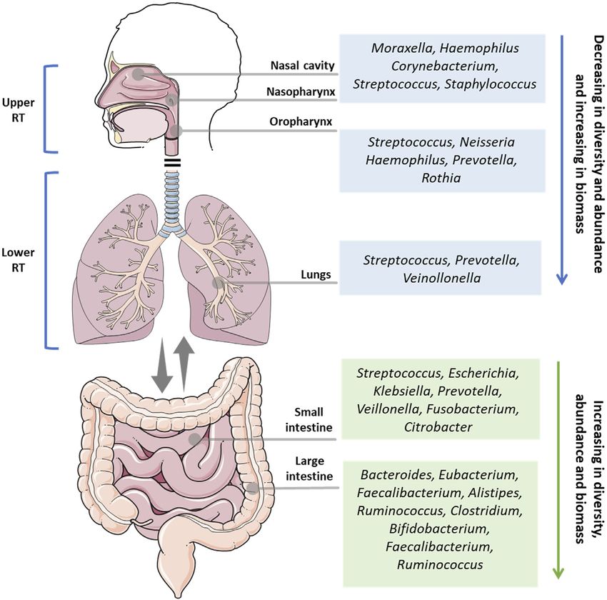
Cruz et al.                                                                                                                             Gut-Lung Axis and Infectious Diseases

  FIGURE 1 | Overview of the main microbial genus in the health upper respiratory tract (nasal cavity, nasopharynx, oropharynx), lower respiratory tract (lungs), small
  intestine, and large intestine. RT: respiratory tract.

contribute to the interaction between these two organs (Faure and                          oropharynx, while the lower respiratory tract comprises the
de Santa Barbara, 2011). Respiratory tract infections (RTI) are a                          trachea, bronchi, bronchioles, and alveoli. These organs make
global health concern. Approximately 2.38 million deaths were                              up one of the largest surface areas in the human body, that from
attributed to RTI in 2016 alone, making it the sixth leading cause                         the nostrils to the lungs, is colonized by a symbiotic and diverse
of mortality among all ages and the leading cause of death among                           community of microorganisms (Figure 1).
children under 5 years (Ferkol and Schraufnagel, 2014; Troeger                                The microbiota of the lungs and gut of healthy individuals differ
et al., 2018). Given the importance of the gut-lung axis, our review                       significantly in taxonomic composition, diversity, and function. In
summarized the latest experimental and clinical studies on this                            contrast to the thriving resident microbiota in the gut, the lung
topic and showed that modulation of the gut-lung axis with                                 microbiota is composed of transient microorganisms mainly derived
probiotics, prebiotics, and synbiotics, could be an important                              from URT. While Bacteroidetes and Firmicutes are the most
therapeutic target for preventing and treating lung infections                             abundant bacterial phyla in both microbiotas, the lung and gut
caused by bacteria, viruses, fungi, and parasites.                                         microbiota are very different at the species level. In the lungs, the
                                                                                           genera Streptococcus spp., Veillonella spp., and Prevotella were the
                                                                                           most abundant, whereas Bacteroides, Faecalibacterium, and
GUT-LUNG AXIS IN RESPIRATORY TRACT                                                         Bifidobacterium are more prevalent in the gut (Sender et al.,
INFECTIONS                                                                                 2016). In a disease or dysbiotic state, other organisms are present
                                                                                           in the lung, such as viruses, including human rhinovirus, human
The respiratory system is composed of different organs, and is                             bocavirus, polyomaviruses, human adenovirus, and human
divided into two main parts: the upper respiratory tract (URT)                             coronavirus, and fungi such as Aspergillus spp., Penicillium spp.,
and the lower respiratory tract (LRT). The URT comprises the                               Candida spp., and Alternaria spp. (Papadopoulos and Skevaki, 2006;
nostrils, nasal passages, paranasal sinuses, nasopharynx, and                              Limon et al., 2017).

Frontiers in Pharmacology | www.frontiersin.org                                        2                                        September 2021 | Volume 12 | Article 724033
Cruz et al. Gut-Lung Axis and Infectious Diseases
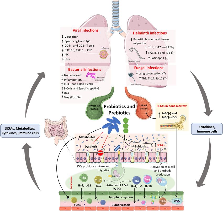
The immune responses in the gut-lung axis depend on the Probiotics are considered important tools for the modulation of
balance of microbiota composition, particularly in the gut. The microbiota in the gut-lung axis, with their benefits on the gut-
regulated interaction between the metabolites and antigens of lung axis dependent on the strains used (Figure 2). However,
symbiotic microbiota with the host is crucial for the activation of common mechanisms have been reported between species, such
pattern recognition receptors (PRRs) and metabolic sensor receptors as –1) colonization of the respiratory and intestinal tracts, 2)
such as G-protein-coupled receptors (GPCRs), and the production production of SCFAs and antimicrobial peptides, 3) maintenance
of inflammatory mediators, which are necessary for the migration, of the integrity of the intestinal and pulmonary mucosa, and –4)
activation, and proliferation of innate and adaptive immune cells stimulation of the innate and adaptive immune system
responsible for the production of pro-and anti-inflammatory (Bermudez-Brito et al., 2012; Salminen et al., 2021). The
cytokines, immunoglobulins, and antimicrobial peptides (Fan and benefits of probiotics have been shown in animal models and
Pedersen, 2021). These cells and molecules can move bidirectionally clinical studies in many disease conditions, such as post-
between the lungs and the gut through the bloodstream and antibiotic-associated diarrhea, allergies and inflammatory
lymphatic system and regulate immune and inflammatory bowel diseases, and respiratory tract infections (Vieira et al.,
responses (Marsland et al., 2015; Dang and Marsland, 2019). 2013). For a given microorganism to be assessed as a probiotic,
Intestinal dysbiosis is responsible for increasing the biosafety criteria and scientific evidence regarding its biological
susceptibility of the host to lung disease, as evidenced by the benefits must be considered (Harzallah and Belhadj, 2013).
high prevalence of asthma in patients with irritable bowel Lactobacillus and Bifidobacterium species are more commonly
syndrome (Yazar et al., 2001). Experimentally, mice treated used as probiotics; however, yeasts, certain Streptococcus spp.
with antibiotics are more susceptible to lethal infection by the strains, and Bacillus spp. are also used as probiotics, but less
influenza virus (IFV) (Ichinohe et al., 2011; Pang et al., 2018). frequently (Fijan, 2014). The use of inactivated probiotics is also
Furthermore, infections in the lungs are also linked to dysbiosis in of great interest because live probiotic microorganisms may cause
the gut; mice infected with IFV displayed a significant increase in systemic infections, excessive immune stimulation, and antibiotic
Enterobacteriaceae and decreased diversity of Lactobacillus and resistance gene transfer (Doron and Snydman, 2015). Taking this
Lactococcus (Wang et al., 2018). Influenza infection also affects into consideration, the term postbiotics was proposed as
the production of short-chain fatty acids (SCFAs) and impairs the preparation for inanimate microorganisms and/or their
gut barrier properties thereby increasing susceptibility to second components that confer a health benefit on the host (Salminen
bacterial infections (Sencio et al., 2020, 2021). et al., 2021).
SCFAs, such as butyrate, propionate, and acetate derived from Prebiotics are dietary fibers, such as inulin,
the fermentation of dietary fibers by the microbiota, are involved fructooligosaccharides, and galactooligosaccharides, which are
in regulating the inflammatory process and pulmonary immune fermented in the gut and promote an increase in the diversity
response (Fukuda et al., 2011; Trompette et al., 2014). SCFAs and activity of specific symbiotic microorganisms (Salminen
activate GPCRs and inhibit histone deacetylases, thus et al., 2021). The activity of prebiotics also leads to an
contributing to the reduction of inflammation in the gut-lung enhancement of immune response, decrease in colon pH, local
axis by inhibiting the NF-κB signaling pathway, increasing induction of reactive oxygen species (ROS), trophic effects on
regulatory T (Treg) cells, and decreasing T helper 1 (Th1) and enterocytes, and anti-inflammatory responses (Vieira et al.,
Th17 cells (Maslowski et al., 2009; Kim et al., 2013; Li et al., 2018). 2013). In addition, the SCFAs butyrate and propionate,
SCFAs can also reach the bone marrow and influence the derived from the metabolism of prebiotics, can increase
generation and development of immune cells such as Ly6C- miRNAs through the inhibition of histone deacetylases,
and Ly6C + monocytes and dendritic cells, which can be leading to improved antibody class switching and local and
recruited into the lungs and modulate the immune response systematic impact on the T-dependent and T-independent
against pathogens (Trompette et al., 2014, 2018; Kopf et al., 2015). immunoglobulin production (Sanchez et al., 2020).
Our research group has also demonstrated that activation of the Synbiotics consist of probiotics and prebiotics to achieve
GPR43 receptor in neutrophils and alveolar macrophages by synergistic and complementary effects on their functions
acetate is essential for modulating the inflammatory response (Salminen et al., 2021). A recent meta-analysis of randomized
and controlling pulmonary infection by Klebsiella pneumoniae controlled clinical trials involving over 10,000 individuals showed
(Galvão et al., 2018) and Streptococcus pneumoniae serotype 1 in the effectiveness of synbiotic interventions in reducing the rate of
mice (Sencio et al., 2020). In another study, activation of GPR43 respiratory tract infections (Chan et al., 2021). Understanding the
in pulmonary epithelial cells induced interferon (IFN)-β in the specific mechanisms of interaction between probiotics and
lungs and increased the protection of mice infected with prebiotics and their modulation of the gut environment and
respiratory syncytial virus (RSV) (Antunes et al., 2019). immune response will lead to better utilization of the synbiotics to
treat infections and metabolic diseases.
PROBIOTICS, PREBIOTICS, AND
Gut-Lung Axis Modulation in the Context of
SYNBIOTICS
Bacterial Lung Infections
Probiotics are live microorganisms that confer benefits to the host The lung is highly vulnerable to bacterial infections due its
when administered in adequate amounts (Salminen et al., 2021). constant exposure to environment agents. One of the most
Frontiers in Pharmacology | www.frontiersin.org 3 September 2021 | Volume 12 | Article 724033Cruz et al. Gut-Lung Axis and Infectious Diseases FIGURE 2 | Effects and mainly mechanisms of probiotics and prebiotics in the gut-lung axis and context of respiratory infections. Probiotics and prebiotics administered orally can improve dysbiosis and induce eubiosis in the host, leading to an increase in SCFAs directly (produced by probiotics) or indirectly (produced by commensal microbiota). Furthermore, probiotics can also reduce the burden and epithelial damage induced by intestinal parasites. The uptake of probiotics by DCs in the intestinal submucosa, and their migration to lymph nodes, induces the activation and proliferation of Th1, Th2, Th17, Treg, and B cells. Activated T cells and B cells produce cytokines and antibodies, enter the circulatory and lymphatic systems, and reach the lungs, where they will increase resistance to infections caused by viruses, bacteria, and fungi. The fermentation of prebiotics and production of SCFAs increases the number of DCs precursors in the bone marrow and increases CD8+ T cells activity, that confer protection against infections in the lung. The immunomodulation demonstrated after the administration of probiotics and prebiotics may be linked to the reduced viral titer, bacterial colonization, parasite load, and migration in the lungs. Probiotic-induced immunomodulation can increase the frequency of dendritic cells and CD4+ and CD8+ T cells in the lungs against infections by viruses and bacteria and can increase specific IgG and IgM antibodies to these pathogens. Also, the increase in Treg cells may be related to the reduction of inflammation-induced lung damage. In parasitic infections, probiotics have been linked with increased frequency of Th1 and concentration of IL-12 and IFN-γ, which may justify the reduction in the parasite load and larvae migration in the lung. Because there are no scientific studies that demonstrate the reduction of lung colonization by fungi after oral administration of probiotics, is still unknow if the antimycotic potential from probiotics metabolites, as shown in vitro, could be applied in an in vivo system. common diseases caused by bacteria in the lungs is pneumonia CRL 423 and Streptococcus thermophilus CRL 412 (Villena et al., that is characterized by alveolar infection and intense 2006), L. casei CRL 431 (Villena et al., 2005, 2009), L. fermentum inflammatory response that ranges from mild to severe and (Cangemi De Gutierrez et al., 2001), and L. rhamnosus CRL 1505 can affect both the right and left lobes and may impair the (Barbieri et al., 2017) causes: 1) increased resistance to infection, gaseous exchange. The most common causes of bacterial 2) decreased number of bacteria in the lungs, and 3) increased pneumonia in immunocompetent hosts include S. survival of mice infected with S. pneumoniae. In general, these pneumoniae, Haemophilus spp., and Mycobacterium articles, associated this protection with an increase in neutrophils, tuberculosis. In immunocompromised hosts the number of lymphocytes, macrophages, phagocytic activity, and levels pathogens that cause pneumonia is much larger, and in specific anti-S. pneumoniae IgG and IgA in the lungs. The general those individuals are more vulnerable and have worse increase in phagocytic activity and the number of neutrophils outcomes (van der Poll and Opal, 2009) (Table 1). in the lower respiratory tract is the first line of defense against Several studies have shown that the oral administration of invading pathogens, and the increase in regulatory cells and different strains of probiotics, such as Lactobacillus bulgaricus cytokines contributes to the reduction of the inflammatory Frontiers in Pharmacology | www.frontiersin.org 4 September 2021 | Volume 12 | Article 724033
Cruz et al. Gut-Lung Axis and Infectious Diseases
TABLE 1 | Pre-clinical studies on the modulation of the microbiota for treatment of bacterial and viral lung infections.
Strategy for Dose and Experimental Pathogen Main outcomes References
microbiota modulation route of model
administration
Effects on bacterial pathogen
Lactobacillus bulgaricus CRL 2 × 10⁸ CFU, via oral Malnourished, Swiss Streptococcus Reduced bacterial load in the lungs; Villena et al. (2006)
423 and Streptococcus albino mice pneumoniae increased bactericidal function of
thermophilus CRL 412 bronco-alveolar phagocytes;
reduced tissue inflammation;
increased neutrophils in blood; and
increased level of lung anti-
pneumococcal IgA and IgG
Lactobacillus casei CRL 431 1 × 10⁹ CFU, via Malnourished, Male, Streptococcus Increased the bacteria lung Villena et al. (2009)
intranasal 3-week-old Swiss pneumoniae clearance; improved production of
albino mice TNF-α; increased activity of
phagocytes in the respiratory tract;
increased IL-4, IL-10, and
Pneumococcus-specific IgG
Lactobacillus casei CRL 431 1 × 10⁹ CFU, via oral Male 6-week-old Streptococcus Increased pathogen clearance from Villena et al. (2005)
Swiss albino pneumoniae blood; lower lung damage;
improved number of leukocytes and
neutrophils; and increased levels of
antipneumococcic IgA in BAL.
Lactobacillus fermentum 1 × 10⁷, via intranasal Adult, BALB/c mice Streptococcus Increased the number of activated Cangemi De
pneumoniae macrophages and lymphocytes; Gutierrez et al.
and increased anti-S.pneumoniae (2001)
antibodies
Lactobacillus rhamnosus 1 × 10⁸ CFU, via Malnourished, Male, Streptococcus Changed the quantitative and Barbieri et al.
CRL1505 intranasal 3-week-old Swiss- pneumoniae qualitative alterations of CD4+ T cells (2017)
albino mice in the bone marrow, thymus, spleen
and lung induced by malnutrition
and infection; and increased IL-10
and IL-4 in respiratory and systemic
compartments
Lactobacillus casei CRL 431, 1 × 10⁹ CFU, via oral 3-week-old, Swiss Pseudomonas Enhanced lung clearance of P. Alvarez et al.
Lactobacillus delbrueckii albino mice aeruginosa aeruginosa; increased phagocytic (2001)
subsp. bulgaricus and activity of alveolar macrophages;
Streptococcus thermophilus and increased IgA and IgM levels
in BAL.
Lactobacillus rhamnosus GG 4 × 10⁸ CFU, via oral 5 to 8-week-old, FVB/ Pseudomonas Mice treated had improved survival; Meitert et al.
N mice aeruginosa reduced bacterial counts in BAL; (2013)
decreased the levels of IL-6 and
increased levels of IL-10 mRNA;
improved lung pathology; and
increased levels of Treg cell marker
Foxp3
Lactobacillus fermentum 9 × 10⁶ CFU, via 6 to 8-week-old, Pseudomonas Decreased secretion in BAL of IL-6 Fangous et al.
K.C6.3.1E, Lactobacillus zeae intratracheal C57BL/6 mice aeruginosa and TNF-α (2019)
Od.76, and Lactobacillus
paracasei ES.D.88
Bifidobacterium longum 51A 1 × 10⁸, via oral 8 to 12-week-old, Klebsiella pneumoniae Reduced bacterial burden; faster Vieira et al. (2016)
C57BL/6 WT and resolution of inflammation;
Mal/TIRAP-/- mice decreased lung damage; increased
production of IL-10; and increased
alveolar macrophages ROS
associated with Mal/TIRAP
activation
Effects on viral pathogen
Lactobacillus casei Shirota 1 × 10⁸ CFU, via oral Neonatal and infant, Influenza A/PR/8/34 Higher survival rate, reduced titer of Yasui et al. (2004)
BALB/c mice (H1N1) virus in the nasal washings; greater
pulmonary NK cell activity; and
increased IL-12 production by
mediastinal lymph nodes
Lactobacillus rhamnosus M21 1 × 10⁹ CFU, via oral Female, specific Influenza A/NWS/33 Increased IL-2 and IFN-γ; increased Song et al. (2016)
pathogen-free, BALB/ (H1N1) sIgA levels; reduced inflammatory
c mice cells in BAL.
(Continued on following page)
Frontiers in Pharmacology | www.frontiersin.org 5 September 2021 | Volume 12 | Article 724033Cruz et al. Gut-Lung Axis and Infectious Diseases
TABLE 1 | (Continued) Pre-clinical studies on the modulation of the microbiota for treatment of bacterial and viral lung infections.
Strategy for Dose and Experimental Pathogen Main outcomes References
microbiota modulation route of model
administration
Lactobacillus pentosus S-PT84 Heat-killed L. pentosus Female, BALB/c mice Influenza A/PR/8/34 Increased survival rates; reduced Izumo et al. (2010)
S-PT84, via intranasal (H1N1) titer of influenza virus in BAL;
increased IL-12 and IFNγ
production in mediastinal lymph
node cells; increased IL-12 and IFN-
α in BAL; and increased NK cell
activity
Lactobacillus plantarum and 1 × 10⁹ CFU, via oral Female, 5-week-old, Influenza rK09 (H1N1) Restrained viral replication; and Bae et al. (2018)
Leuconostoc mesenteroides BALB/c mice increased rates of survival of
infected mice
Lactobacillus rhamnosus GG 200 µg of L. Female, 7-week-old Influenza A⁄ PR⁄ 8⁄ 34 Higher survival rate; increased cell- Harata et al.
rhamnosus GG BALB/c mice (H1N1) killing activity of lung cells; and (2010)
lyophilized, via increased mRNA expression of
intranasal interleukin IL-1 beta, TNF and
MCP-1
Lactobacillus acidophilus L-92 4 × 10¹⁰ CFU, via oral Female, 4-weeks-old Influenza A/PR/8/34 Reduced Virus titers in the lung; Goto et al. (2013)
BALB/c mice (H1N1) increased NK cells activity;
decreased the number of
neutrophils; increased eotaxin,
MCSF, IL-1β, RANTES and IFN-α in
the lung; and increased IL-17 levels
in Peyer’s patches
Lactobacillus plantarum DK119 1 × 10⁸ or 1 × 10⁹ CFU, Female, BALB/c mice Influenza A/PR8 Reduced lung viral loads; increased Park et al. (2013)
via oral levels of cytokines IL-12 and IFN-γ in
BAL; and reduced degree of
inflammation
Lactococcus lactis subsp. 1 mg of heat-killed L. Female, DBA/2jjcl Murine Parainfluenza Increased survival rate; prevention Jounai et al.
lactis JCM5805 lactis subsp. lactis mice virus (mPIV1) of weight loss; reduced lung (2015)
JCM5805, via oral histopathology scores; increased
activation of Peyer’s patches (PP)
and PP pDCs; increased levels of
type I IFNs; and increased
expressions of anti-viral factors such
as Isg15, Oasl2, and Viperin, at lung
Lactobacillus paracasei CNCM 2 × 10⁸ CFU, via oral Female, 6-week-old Influenza A/Scotland/20/ Reduced weight loss; and increased Belkacem et al.
I-1518 BALB/c mice 74 (H3N2) recruitment of inflammatory myeloid (2017)
cells, such as interstitial monocytes
and dendritic cells, to the lungs
Bacillus subtilis 3 (UCM B- 1 × 10⁷ CFU, via oral Four-week-old BALB/ Influenza A/FM/1/47 Prevented influenza infection Starosila et al.
5007) c mice (H1N1) (2017)
Lactobacillus rhamnosus 1 × 10⁸ CFU, via oral Male, 6-week-old Respiratory Syncytial Reduced lung immune-coagulative Zelaya et al.
CRL1505 BALB/c mice Virus strain A2 and reaction triggered by TLR3 (2014)
Influenza virus A/PR/8/34 activation
(H1N1)
Lactobacillus plantarum 1 × 10⁹ CFU, via BALB/c and C57BL/6 Pneumonia Virus of mice Protection against lethal infection; Gabryszewski
NCIMB 8826 and Lactobacillus intranasal MyD88−/− mice (PVM) strain J3666 reduced granulocyte recruitment; et al. (2011)
reuteri F275 reduced expression of
proinflammatory cytokines CXCL10,
CXCL1, CCL2, and TNF.
Lactobacillus rhamnosus 1 × 10⁸ CFU, via Female, 3-week-old Human RSV strain A Increased levels of IFN-α, IFN-β, Tomosada et al.
CRL1505 and CRL1506 intranasal BALB/c mice IFN-γ, IL-6 and IL-10; increased (2013)
levels of CD4+ Tregg cells and
CD11c+CD103+ DCs; reduced viral
replication and lung damage
response and to the maintenance of tolerance to symbiotic Similar results were observed in mice infected with
microorganisms, which is necessary to reduce damage Pseudomonas aeruginosa, treated orally with the probiotics L.
associated with infections by pathogens (Martin and Frevert, casei CRL 431, L. delbrueckii subsp. bulgaricus, S. thermophilus
2005). (Alvarez et al., 2001) and L. rhamnosus GG (Meitert et al., 2013).
Frontiers in Pharmacology | www.frontiersin.org 6 September 2021 | Volume 12 | Article 724033Cruz et al. Gut-Lung Axis and Infectious Diseases
TABLE 2 | Clinical studies on the modulation of the microbiota for treatment of bacterial and viral lung infections.
Strategy for Dose and Study design Pathogen and Outcome References
microbiota modulation route of and subjective disease
administration
Effects on bacterial pathogen
Lactobacillus casei 1 × 10⁹ CFU, via oral Prospective, randomized, Pseudomonas aeruginosa Reduction in the occurrence of Forestier et al.
rhamnosus Lcr35 double-blind, placebo- P. aeruginosa respiratory (2008)
controlled pilot study with colonization and/or infection in
patients aged 18-91 the probiotic group. Reduction
in the frequency of VAP to P.
aeruginosa
Lactobacillus rhamnosus GG 2 × 10⁹ CFU, via oral Prospective, randomized, VAP by Gram-positive and Reduction in the development Morrow et al.
double-blind, placebo- Gram-negative pathogens of microbiologically confirmed (2010)
controlled trial with patients VAP. Patients treated with
at high risk of probiotics had fewer days of
developing VAP antibiotics prescribed for VAP.
Bifidobacterium breve Yakult, 1 × 10⁸ CFU of B. breve and Randomized controlled trial Patients with more than Reduced incidence of VAP. Shimizu et al.
Lactobacillus casei Shirota, L. casei and Galacto- with patients with more 16 years old, placed on a Increased number of (2018)
and galacto-oligosaccharide oligosaccharides in a 10 g/ than 16 years old ventilator, and who were Bifidobacterium and
day formula, via oral diagnosed as having Lactobacillus. Increased
sepsis concentration of acetate in the
feces
Effects on viral pathogens
Lactobacillus rhamnosus GG 1 × 10⁹ CFU/day for Randomized, double- Rhinovirus-associated Reduced respiratory tract Luoto et al.
ATCC 5310 and galacto- 1–30 days and 2 × blind, placebo-controlled respiratory tract infection infections. Reduced (2014)
oligosaccharide and 109 CFU/day for study with preterm infants rhinovirus-induced episodes
polydextrose mixture 31–60 days, via oral
Lactobacillus brevis KB290 6 × 10⁹, via oral Open-label, parallel-group Influenza infection Reduced incidence of Waki et al.
trial with elementary influenza infections (2014)
schoolchildren
Lactobacillus rhamnosus GG 1 × 10⁹, via oral Randomized, double- Influenza infection Reduced laboratory- Wang et al.
blind, placebo-controlled confirmed respiratory viral (2018)
study with nursing home infections
residents aged 65 and
older
Streptococcus thermophilus 2.4 × 10⁹, via oral Retrospective, COVID-19 pneumonia by Increased survival rates of Ceccarelli
DSM 32245, Bifidobacterium observational cohort study SARS-CoV-2 patients that received BAT et al. (2021)
lactis DSM 32246, with adults plus oral bacteriotherapy
Bifidobacterium lactis DSM
32247, Lactobacillus
acidophilus DSM 32241,
Lactobacillus helveticus DSM
32242, Lactobacillus
paracasei DSM 32243,
Lactobacillus plantarum DSM
32244, and Lactobacillus
brevis DSM 27961
In addition, the administration of L. rhamnosus GG induces an macrophages through activation of the mal/TIRAP signaling
anti-inflammatory response by increasing the levels of regulatory pathway (Vieira et al., 2016). There was a concomitant
T cells (Treg) Foxp3+ and decreasing the production of the reduction in the inflammatory process and concentration of
proinflammatory cytokine IL-6. This anti-inflammatory profile cytokines TNF-α and IL-6 and an increase in IL-10 in the
was also observed in mice infected with P. aeruginosa with lungs of mice. However, only viable probiotics were able to
intratracheal administration of probiotics L. fermentum increase the levels of IL-10 in the lungs of mice. Viable B.
K.C6.3.1E, L. zeae Od.76, and L. paracasei ES.D.88, longum 51A produces SCFA acetate in large quantities, and
demonstrated by the reduction of lung inflammation and acetate administration in mice before respiratory infection by
decreased production of IL-6 and tumor necrosis factor K. pneumoniae induced increased production of IL-10 in animals.
(TNF)-α (Fangous et al., 2019). The authors demonstrated that acetate might be the primary
In an experimental lung infection by K. pneumoniae, the inducer production of IL-10 in this model (Vieira et al., 2016). In
administration of viable or inactivated probiotic addition, intestinal colonization of germ-free mice with B. longum
Bifidobacterium longum 51A induced pulmonary clearance of 51A restored the ability of these mice to decrease infection by
K. pneumoniae by increasing ROS production in alveolar increasing the production of CXCL1 and the recruitment of
Frontiers in Pharmacology | www.frontiersin.org 7 September 2021 | Volume 12 | Article 724033Cruz et al. Gut-Lung Axis and Infectious Diseases
neutrophils (Vieira et al., 2016). In mice infected with IFV, the coronavirus SARS-CoV-2 has emerged as a pandemic that has
lower quantity of acetate was also related to increased caused more than 3.5 million deaths (Zhu et al., 2020) (Table 1).
susceptibility to secondary respiratory pneumococcal infection, Several studies have demonstrated the potential for oral and
mainly due to the impaired bactericidal activity of alveolar intranasal administration of probiotics such as L. casei Shirota
macrophages, and this detrimental effect was restored after (Yasui et al., 2004), L. rhamnosus M21 (Song et al., 2016), L.
acetate supplementation (Sencio et al., 2020). pentosus S-PT84 (Izumo et al., 2010), and L. plantarum and
The potential of probiotics to protect the host from pulmonary Leuconostoc mesenteroides (Bae et al., 2018) to protect and
infections has also been assessed in several clinical studies, increase the survival of IFV-infected animals, mainly by
including diverse patients, methodological designs, and inducing anti-viral immune responses with the activation of
inclusion criteria (Table 2). Most of these studies focused on NK cells and increased production of cytokines such as IL-12
probiotics for prevention and treatment of nosocomial and IFN-γ, increased production of IgA in the respiratory
pulmonary infections in patients admitted to intensive care mucosa, and reduction of polymorphonuclear inflammatory
units (ICUs). Two prospective, randomized, double-blind, and infiltrate in the lung tissue. In addition to these protective
placebo-controlled studies showed that the probiotics L. casei effects, L. rhamnosus GG administered intranasally (Harata
rhamnosus Lcr35 and L. rhamnosus GG, administered orally or et al., 2010), and L. acidophilus L-92 (Goto et al., 2013) also
oropharyngeally, resulted in decreased colonization and infection demonstrated the ability to increase the levels of proinflammatory
of the LRT by P. aeruginosa or related gram-positive and gram- cytokines, such as IL-1β, monocyte chemotactic protein 1 (MCP-
negative pathogens in patients admitted to the ICU using 1), and chemokines such as eotaxin and M-CSF.
mechanized pulmonary ventilation (Forestier et al., 2008; Dendritic cells are crucial for developing immune responses
Morrow et al., 2010). Only one study showed that because of their ability to detect pathogens through TLRs and
administration of a synbiotic consisting of B. breve Yakult, L. create a link between innate and adaptive immune responses.
casei Shirota, and galactooligosaccharides decreased the incidence Mice with depleted alveolar macrophages lost the anti-viral
of ventilator-associated pneumonia (VAP) in patients diagnosed protection against IFV infection conferred by increasing IL-12
with sepsis admitted to the ICU (Shimizu et al., 2018). and IFN-γ levels after oral administration of the probiotic L.
In general, we can conclude that the modulation of the plantarum DK119 (Park et al., 2013). In addition, the importance
intestinal microbiota, mainly with probiotics, is an exciting of dendritic cells was demonstrated after oral administration of
alternative for treating lung diseases caused by bacteria. the probiotic Lactococcus lactis subsp. lactis JCM5805 in mice
Although it is already well established in the literature that infected with murine parainfluenza virus. The authors showed
probiotic species and strains behave differently according to that the probiotic was incorporated into CD11c+ immune cells in
their metabolic pathways and their interaction with the host, Peyer’s patches and activated plasmacytoid dendritic cells that
the probiotic species used against lung alterations attract produce type I IFNs at draining mucosal sites. The authors also
attention to those from the Lactobacillus genus. Most observed an increase in IFN-related genes, such as lsg15, Oasl2,
articles demonstrated that non-specific immune responses and Viperin in the lungs, suggesting that the type I IFN produced
mediated by probiotics, prebiotics, and symbionts are the by plasmacytoid dendritic cells could reach systemic levels and
principal host protection against lung bacteria. Generally, it induce anti-viral activity in the lungs. In addition, ex vivo
seemed more significant phagocytic activity of lung stimulation with murine parainfluenza virus of lung
macrophages, reduced lung bacterial load, and less tissue lymphocytes from mice treated with JCM5805 demonstrated
inflammation, associated with increased levels of IL-4 and high expression of IFN-α and IFN-β (Jounai et al., 2015).
IL-10, increased frequency of Treg cells, increased Determining the taxonomic composition and function of the
production of IgA and IgG, and reduced of IL-6 and TNF-α microbiota is crucial for understanding the impact of probiotics
levels. This demonstrates a more resolving anti-inflammatory on the protective response against pathogens. The oral
profile after modulation of the host’s intestinal microbiota. administration of L. paracasei CNCM I-1518 did not modify
Despite the benefits, these articles use different study designs, the gut microbiota structure in mice infected with IFV; however,
experimental models, doses, and routes of administration, it conferred protection against the virus (Belkacem et al., 2017).
making it challenging to translate the results obtained in Diets rich in inulin and SCFAs improve mice lung pathology
animal models to humans and thus develop more specific after infection with IFV by promoting the differentiation of
therapies with probiotics. alternatively activated macrophages (AAMs) from circulating
Ly6C- monocytes and decreasing the immunopathological
effects of neutrophils. Also SCFAs increases anti-IFV
Gut-Lung Axis Modulation in the Context of immunity by enhancing the CD8+ T cells activity by serving
Lung Viral Infections as a substrate for fatty acid oxidation and by specifically
Viral infections generally cause common cold, bronchiolitis, and interacting with the receptor GPR41 (Trompette et al., 2018).
pneumonia and vary widely in severity depending on age, One study showed that activation of GPR43 and interferon-α/β
immune and nutritional status, genetics, and use of antibiotics. receptor (IFNAR) in pulmonary epithelial cells by SCFA acetate
IFV, RSV, and rhinovirus are the most abundant and common induced increased levels of IFN-β in the lungs and increased
causes of lung infections (Jain, 2017). IFV is well known to cause protection of mice in an experimental model of RSV infection
outbreaks of varying severity every year, but recently the novel (Antunes et al., 2019).
Frontiers in Pharmacology | www.frontiersin.org 8 September 2021 | Volume 12 | Article 724033Cruz et al. Gut-Lung Axis and Infectious Diseases Other probiotic-derived metabolites also require further preterm infants showed that a synbiotic composed of L. investigations. A peptide P18 produced by the probiotic rhamnosus GG ATCC5310 and galactooligosaccharides and Bacillus subtilis 3 (UCM B-5007) share high structural polydextrose reduced the rate of rhinovirus infection homology with IFV neutralizing antibody, and it is capable of compared to the placebo group (Luoto et al., 2014). In inhibit IFV replication in vitro and protect 80% of mice from school-aged children, consumption of L. brevis KB290 lethal IFV infection when administered in a therapeutic regimen. during the influenza season was associated with a reduction This protection is superior to that observed using the anti-viral in the clinical diagnosis of IFV infection (Waki et al., 2014). In drug oseltamivir (approximately 70%) (Starosila et al., 2017). a randomized, double-blind, placebo-controlled pilot study, Double-stranded RNA intermediates from IFV and RSV are the probiotic L. rhamnosus GG was also associated with a associated with changes in the host’s coagulation process by reduction in the occurrence of influenza infections (Wang activation of receptors such as TLR-3, and retinoic acid- et al., 2018). inducible gene I (RIG-I). The activation of these receptors by As demonstrated, the most used probiotics in studies of these viruses, increases the expression of coagulation factors in pulmonary diseases caused by viruses are also those of the endothelial cells and monocytes and inhibits fibrinolysis, Lactobacillus genus. The effects of this genus related to inducing a prothrombotic state in the hosts, leading to fibrin increased protection against viruses are linked to increased deposition in the pulmonary alveoli and exacerbation of tissue production of IFN types I and II, proinflammatory cytokines inflammation. In order to address this issue one study such as IL-12 and IFN-γ, or even increased expression of genes demonstrated in a murine model of IFV and RSV infection, encoding anti-viral factors. Unfortunately, a mechanistic basis for that oral administration of L. rhamnosus CRL 1505 in mice the observed beneficial effects of probiotics in combating viral increases the clearance of both viruses and controls immune- lung infections is often not well defined. This knowledge gap is coagulative responses initiated by the activation of TLR-3 in the mainly because most experiments using probiotics for viral lungs, in a process dependent on IL-10 (Zelaya et al., 2014). treatment use different study designs and experimental Intranasal administration of viable or heat-killed L. plantarum models, doses, times, and routes of administration. Therefore, NCIMB 8826 and L. reuteri F275 protected mice from lethal more research is needed to understand better the role of pneumonia virus of mice (PVM) infection. The lungs showed probiotics in our immune system in fighting viral pulmonary minimal inflammation, with fewer granulocytes and an increased infections. number of lymphocytes, correlated with a reduction in proinflammatory cytokines CXCL10, CXCL1, CCL2, and TNF-α. The Gut-Lung Axis During SARS-CoV-2 Infection Evaluation of the lymphocyte populations demonstrated that The severe acute respiratory syndrome coronavirus 2 (SARS- treatment did not result in changes in the relative proportions of CIoV-2), which causes coronavirus disease 2019 (COVID-19), CD4+ T cells (CD3+CD4+CD8−), CD8+ (CD3+CD4−CD8+), or has spread around the world since 2019 and has been declared a B cells (B220+). In contrast, the fraction of NK cells (CD3−DX5+) pandemic that continues to spread with devastating decreased. The authors demonstrated that these results are not consequences to public health. As of July 2021, there were specific for L. plantarum NCIMB 8826 and L. reuteri F275, as approximately 190, 600, 300 global confirmed cases and the same protection was observed when using the non-pathogenic 4,130,000 confirmed deaths. gram-positive bacteria Listeria innocua. These probiotics also SARS-CoV-2 can invade human cells by binding its spike increased the survival of mice infected with the PVM, with the protein to a variety of receptors, such as angiotensin-converting deleted MyD88 (TLRs adapter protein) gene (MyD88−/−), thus enzyme 2 (ACE2), neuropilin-1, tyrosine-protein kinase receptor demonstrating that this induced protection can be TLR- (AXL), and antibody–FcγR complexes (V’kovski et al., 2021). The independent (Gabryszewski et al., 2011). However, other studies current evidence suggests that the severity of COVID-19 is a have determined that the anti-viral activity of probiotics is related to consequence of a hyperinflammatory immune response the activation of TLRs. This was demonstrated in mice with L. culminating in a ‘cytokine storm’, with markedly increased rhamnosus probiotics CRL1505 and CRL1506 that differentially levels of proinflammatory cytokines such as IL-1, IL-6, IL-12, activate the TLR3/RIF-I pathway to inoculate with poly (I:C) (a IFN-γ, and TNF-α, which elicit extensive local and systemic tissue molecular pattern associated with viruses). The activation of TLR3/ damage (Coperchini et al., 2020). RIF-I leads to increased production of IFN-γ, IFN-β, TNF-α, IL-6, Recent studies have revealed that patients infected with SARS- and IL-10, the frequency of CD3+CD4+IFN-γ+, CD3+CD4+IL-10+, CoV-2 demonstrate intestinal dysbiosis, which correlates with the and the dendritic cells D11c+CD11blowCD103+ and susceptibility and severity of COVID-19 (Zuo et al., 2020; Yeoh CD11c+CD11bhighCD103, in the lungs. Additionally, this study et al., 2021). Though some studies detected SARS-CoV-2 RNA in showed an increase in MHC-II levels in both populations of the feces of patients, the activity and infectivity of SARS-CoV-2 in dendritic cells. The authors also demonstrated that this protective the GI tract are still largely unknown (Zuo et al., 2021). However, response and modulation of the immune response were similar to as ACE2 is highly expressed in the intestinal epithelia, this those observed in mice infected with the human RSV strain receptor may be involved in gastrointestinal symptoms that (Tomosada et al., 2013). are common in severe cases (Chen et al., 2020; Villapol, 2020; Clinical studies showed that probiotics have general effects Koester et al., 2021). Interestingly, in a study with gnotobiotic on viral infections of the respiratory tract (Table 2). A rats, researchers demonstrated that the gut microbiota regulates randomized, double-blind, placebo-controlled study, on the colonic mRNA of ACE2 (Yang et al., 2020). Frontiers in Pharmacology | www.frontiersin.org 9 September 2021 | Volume 12 | Article 724033
Cruz et al.                                                                                                      Gut-Lung Axis and Infectious Diseases

   Another study demonstrated that patients with severe                      to cellular metabolism, which culminated in the complete
COVID-19 had a significant decrease in the abundance of                       inhibition of spore germination and development of the germ
butyrate-producing bacteria, such as Faecalibacterium                        tubes and hyphae of the pathogen A. fumigatus (Crowley et al.,
prausnitzii, Clostridium butyricum, Clostridium leptum, and                  2013). In one in vivo study using the probiotics Saccharomyces
Eubacterium rectale, and an increased number of common                       boulardii, L. paracasei ST-11, and L. rhamnosus GG, mice were
opportunistic pathogens, Enterococcus and Enterobacteriaceae                 not protected against lung infection caused by the pathogen
(Tang et al., 2020). In non-human primates infected with                     Cryptococcus gattii (Oliveira et al., 2017).
SARS-CoV-2, 16S rRNA analysis of the microbial gut                              The recognition of lectins in the fungal cell wall by PRRs is
community showed changes in the taxonomic composition,                       crucial for the activation of dendritic cells and macrophages and
with the relative abundance of Acinetobacter and                             the activation of T cells, including Th1 and Th17, which are the
Ruminococcaceae being positively correlated with the presence                best defense strategies against fungal infections, as they help
of SARS-CoV-2 in the URT. In addition, SARS-CoV-2 infection                  promote the clearance of fungi through innate effectors such
significantly alters the metabolite composition with a reduction in           as neutrophils and macrophages. The activation of Treg cells and
the levels of SCFAs, bile acids, and tryptophan metabolites (Sokol           anti-inflammatory cytokines is also fundamental to the anti-
et al., 2021).                                                               fungal immune response, as these cells and molecules are
   The impact of probiotics in COVID-19 and in the cytokine                  essential for controlling the inflammatory response. The
storm can be deduced by their known mechanisms in modulating                 immune response against fungi has already been extensively
immune response and inflammation (He et al., 2020; de Oliveira                reviewed by other authors, such as Lionakis et al. (2017).
et al., 2021), but more basic and clinical research is needed to                Among the opportunistic species that affect the lungs,
show their benefits. In a retrospective study of ICU patients                 Aspergillus spp. are the primary etiologic agents of invasive
diagnosed with pneumonia caused by SARS-CoV-2, the                           lung diseases and mainly affect transplant patients
association of the best available therapy with the probiotics S.             (Kontoyiennis et al., 2010; Pappas et al., 2010). Evidence also
thermophilus DSM 32245, B. lactis DSM 32246, B. lactis DSM                   suggests that COPD patients are at a high risk of developing
32247, L. acidophilus DSM 32241, L. helveticus DSM 32242, L.                 invasive aspergillosis, although this association is poorly explored
paracasei DSM 32243, L. plantarum DSM 32244, and L. brevis                   (Hammond et al., 2020). An experimental study demonstrated
DSM 27961 showed a positive association with reduced mortality               the importance of intestinal microbiota in structuring the
(Ceccarelli et al., 2021).                                                   pulmonary anti-Aspergillus immune response. During infection
                                                                             by A. fumigatus, the administration of antibiotics decreased the
                                                                             number of Th17 cells and IL-17 in the lungs, which correlated
Gut-Lung Axis Modulation in the Context of                                   with a decrease in intestinal colonization by segmented
Fungal Lung Infections                                                       filamentous bacteria (SFB). By investigating how commensal
Among the wide variety of respiratory pathogens, fungi are                   SFBs were linked to this phenotype, the authors, through
responsible for only a small proportion of nosocomial or                     serum transfer experiments from mice colonized by SFB to
community-acquired pneumonia. However, these species are                     negative SFB mice, demonstrated that SFBs contribute to the
of relevant medical interest, as fungi cause high morbidity and              accumulation of Th17 cells in the lung by inducing an increase in
mortality especially when they affect immunosuppressed                       IL-1. This was confirmed when mice that received serum pre-
patients, or patients with chronic lung diseases, such as                    incubated with an IL-1 antagonist attenuated the response of
chronic obstructive pulmonary disease (COPD) (Charlson                       Th17 cells in the lungs (McAleer et al., 2016). Germ-free mice
et al., 2012; Delhaes et al., 2012).                                         (GF) infected with C. gattii showed greater susceptibility to lung
    Although some studies showed that the microbiota in the gut-             colonization, mortality, correlated with reduced levels of IFN-γ,
lung axis is fundamental to the host response to lung infections by          IL-1β, and IL-17 and reduced phagocytosis and ROS production
fungi, most studies that have demonstrated antimycotic action                than conventional mice. After restoring the intestinal microbiota
against respiratory pathogens have been carried out in vitro. In             those mice mounted a stronger response to infection by C. gattii,
vitro experiments showed that the bacterium Bacillus safensis can            associated with prolonged survival rates and higher levels of
block, in a contact-dependent manner, several Cryptococcus                   inflammatory mediators (Costa et al., 2016).
neoformans virulence factors including melanin, antiphagocytic                  Pneumocystis jirovecii is another opportunistic fungus that
capsule, and biofilm formation (Mayer and Kronstad, 2017).                    causes pneumonia, particularly in HIV-positive patients, with an
Many microorganisms of the genus Bacillus are characterized                  inverse relationship between the CD4+ T cell count in the blood
as probiotics (Hong et al., 2005); however, B. safensis has not yet          and the risk of infection by P. jirovecii (Dunbar et al., 2020).
been characterized as such. B. safensis is phylogenetically close to         When investigating the diversity of the intestinal microbial
the probiotic B. pumilus (Satomi et al., 2006), and its anti-fungal          community between immunocompetent mice and mice
activity has been described (Pandya and Saraf, 2015), making B.              depleted of CD4+ T cells, with pneumonia caused by
safensis an exciting candidate for studies of biological safety and          Pneumocystis murina, there was a significant change in alpha
probiotic activity. Another study demonstrated in vitro that                 and beta diversity and a change in the taxonomic abundance of
treatment with concentrated cell-free supernatant from the                   the intestinal microbiota among these groups, suggesting that the
culture of L. plantarum 16 altered the transcription of genes                loss of CD4+ T cells affects the intestinal microbiota and the
involved in a variety of cellular functions, especially those related        response to P. murina. P. murina infection was also found to

Frontiers in Pharmacology | www.frontiersin.org                         10                                September 2021 | Volume 12 | Article 724033
Cruz et al. Gut-Lung Axis and Infectious Diseases
increase the expression of genes in the intestinal microbiota and survival of mice. This was demonstrated experimentally
related to carbohydrate energy metabolism, xenobiotic with the probiotic L. casei ATCC7469 in infection with
degradation, and signal transduction pathways (Samuelson Trichinella spiralis (Bautista-Garfias et al., 2001), B.
et al., 2016a). animalis 04450B against infection by S. venezuelensis
Another study demonstrated that vaccination, using a prime- (Oliveira-Sequeira et al., 2014), and S. boulardii in mice
boost vaccination strategy, with live P. murina induced protection infected with T. canis (Avilada et al., 2012). However,
against subsequent lung infection with the same pathogen in researchers have already reported the beneficial effect of
immunocompetent mice and even in mice depleted of CD4+ probiotics in reducing the parasitic burden of larval stages
T cells. In immunocompetent mice, this immunization increased during T. canis infection in mice. In vitro experiments
the number of CD4+ T cells, CD8+ T cells, CD19+ B cells, and demonstrated a reduction in the viability of T. canis larvae
CD11b+ macrophages in the lungs after a respiratory infection after direct incubation with live cells or cell-free supernatant
and increased the levels of IgG and IgA specific for P. murina. A of the probiotic Enterococcus faecalis CECT712. The same
significant reduction in the lung load of P. murina was observed study also showed that oral administration of E. faecalis
in serum transfer experiments from non-infected and immunized CECT712 significantly reduced the number of T. canis
mice to infected mice. The beta diversity of the intestinal larvae found in the lungs of these animals (Chiodo et al.,
microbial community in mice immunized with P. murina was 2010). L. rhamnosus (Walcher et al., 2018) and L. acidophilus
also altered, suggesting that the effectiveness of this ATCC 4356 (Cadore et al., 2020) were also able to reduce the
immunization may be partly related to the modification of the parasitic larval burden of T. canis in the lungs of mice, but had
microbiota; however, further studies are needed to determine no antiparasitic action against the larvae in vitro, which
whether changes in the microbiota participate in the induction of indicates the indirect action of these two probiotics on T.
immunological memory in P. murina (Samuelson et al., 2016b). canis, probably related to the stimulation of the protective
Despite the importance of the intestinal microbiota and its immune response. The administration of S. boulardii in mice
metabolites for the development of anti-fungal immune infected with T. canis increased the transcription of genes
responses, and in vitro studies demonstrate that probiotics have encoding IL-12 and IFNγ, which correlated with a decrease in
an action against fungi that cause lung infections, there have been no the number of larvae in the lungs and other tissues (de Avila
reports of robust studies aiming to assess the effectiveness of the et al., 2016).
modulation of in vivo intestinal microbiota for the prevention and/or Some probiotics, mainly from the Lactobacillus genera, showed
treatment of pulmonary fungal infections. Thus, with the action in vitro and in vivo against helminths that cycle through the
significance of lung infections in mind, more researchers urgently lungs. However, differences in the efficacy of species and strains used
need to turn their attention to this broad and promising field. can be attributed to variability in the experimental models, the
probiotic dose, and the administration route. However, data are
insufficient to determine the molecular mechanisms by which
Gut-Lung Axis Modulation in the Context of probiotics act on helminths that cycle through the lungs.
Parasitic Lung Infection Furthermore, further studies on host-microbiota-helminth
Many helminths cause disease, but they have been shown to also interaction mechanisms are needed to validate the actions of
influence the pulmonary immune response. Similar to bacteria, probiotics in clinical studies with humans.
they co-evolved with the host’s immune system to maintain a
mutually beneficial relationship (Schwartz et al., 2018). Some
helminths also share the same niche, the intestinal lumen, with STUDIES PERSPECTIVES AND
bacteria belonging to the microbiota, and some studies have CONCLUSION
shown that there are complex interactions between the two
(Leung et al., 2018). The intestinal microbiota acts as one of Although studies have shown that probiotics, prebiotics, and
the main inducers of the activation and function of local and synbiotics have prophylactic and therapeutic effects against lung
systemic antiparasitic responses, such as the activation of Th2 infections caused by bacteria, viruses, fungi, and helminths,
cells and eosinophils (Jiménez-Saiz et al., 2020). further studies are needed to better understand the
During the larval phase of their life cycle, different species of mechanisms of action and molecular pathways involved in
helminths, such as Ascaris lumbricoides, Toxocara sp., Necator these strategies. It is necessary to favor the translational use of
americanus, Ancylostoma duodenale, and Strongyloides sp., gut microbiota modulation strategies as a therapeutic approach to
migrate through the lungs and induce pathological immune human lung diseases. In addition, since the effect of these
responses and cause tissue damage, such as eosinophilic strategies is highly linked to the strains of the microorganism
pneumonia, which is characterized mainly by increased and the dose and route of administration, more in-depth
infiltration of eosinophils in the lung parenchyma and blood investigations should be performed, considering well-defined
eosinophilia (Akuthota and Weller, 2012). experimental protocols. Considering the complexity of the
Some studies involving the modulation of the microbiota in microbiota and its interaction with the host, it is also
spite of helminth infections have focused only on intestinal important to determine whether this strategy acts in synergy
pathology and on the parasitological aspects of the infection, with the microbiota or has another mechanism involving direct
such as intestinal parasitic load, release of eggs in the feces, action against the lung pathogen or modulation of the host
Frontiers in Pharmacology | www.frontiersin.org 11 September 2021 | Volume 12 | Article 724033Cruz et al. Gut-Lung Axis and Infectious Diseases
immune system. In addition, it is important to emphasize the FUNDING
therapeutic window necessary to restore the gut microbiota to re-
establish homeostasis after lung infection control. This work was supported by Conselho Nacional de
Desenvolvimento Científico e Tecnológico (CNPq) (402530/
2020-9) and (420350/2018-7) to ATV and to CSC Fellowship,
AUTHOR CONTRIBUTIONS Coordenação de Aperfeiçoamento de Pessoal de Nível Superior
(CAPES), Fundação de Amparo à Pesquisa de Minas Gerais
CC wrote the manuscript, tables, and made the figures; MR and (FAPEMIG) (APQ-03328-18), Pro-Reitoria de Pesquisa da
AV contributed to the design, writing, and review of the Universidade Federal de Minas Gerais -PRPq and Instituto
manuscript. The authors read and approved the final manuscript. Serrapilheira (Serra-1912-31552).
and Meta-Analysis of Randomized Controlled Trials. Adv. Nutr. 11, 979–988.
REFERENCES doi:10.1093/ADVANCES/NMAA003
Charlson, E. S., Diamond, J. M., Bittinger, K., Fitzgerald, A. S., Yadav, A., Haas, A.
Akuthota, P., and Weller, P. F. (2012). Eosinophilic Pneumonias. Clin. Microbiol. R., et al. (2012). Lung-Enriched Organisms and Aberrant Bacterial and Fungal
Rev. 25, 649–660. doi:10.1128/CMR.00025-12 Respiratory Microbiota after Lung Transplant. Am. J. Respir. Crit. Care Med.
Alvarez, S., Herrero, C., Bru, E., and Perdigon, G. (2001). Effect of Lactobacillus 186, 536–545. doi:10.1164/rccm.201204-0693OC
Casei and Yogurt Administration on Prevention of Pseudomonas aeruginosa Chen, R., Yu, Y. L., Li, W., Liu, Y., Lu, J. X., Chen, F., et al. (2020). Gastrointestinal
Infection in Young Mice. J. Food Prot. 64, 1768–1774. doi:10.4315/0362-028X- Symptoms Associated With Unfavorable Prognosis of COVID-19 Patients: A
64.11.1768 Retrospective Study. Front. Med. (Lausanne). 7, 608259–9. doi:10.3389/
Antunes, K. H., Fachi, J. L., de Paula, R., da Silva, E. F., Pral, L. P., dos Santos, A. Á., fmed.2020.608259
et al. (2019). Microbiota-Derived Acetate Protects Against Respiratory Chiodo, P. G., Sparo, M. D., Pezzani, B. C., Minvielle, M. C., and Basualdo, J. A.
Syncytial Virus Infection Through a GPR43-Type 1 Interferon Response. (2010). In Vitro and In Vivo Effects of Enterococcus faecalis CECT7121 on
Nat. Commun. 10, 3273–3317. doi:10.1038/s41467-019-11152-6 Toxocara Canis. Mem. Inst. Oswaldo Cruz. 105, 615–620. doi:10.1590/S0074-
Avilada, L. F. d. C. d. C. de., Conceição, F. R., Telmo, P. d. L., Dutra, G. F., Santos, D. 02762010000500003
G. d. l., Martins, L. H. R., et al. (2012). Saccharomyces Boulardii Reduces Coperchini, F., Chiovato, L., Croce, L., Magri, F., and Rotondi, M. (2020). The
Infection Intensity of Mice With Toxocariasis. Vet. Parasitol. 187, 337–340. Cytokine Storm in COVID-19: An Overview of the Involvement of the
doi:10.1016/j.vetpar.2012.01.002 Chemokine/Chemokine-Receptor System. Cytokine Growth Factor. Rev. 53,
Bae, J. Y., Kim, J. I., Park, S., Yoo, K., Kim, I. H., Joo, W., et al. (2018). Effects of 25–32. doi:10.1016/j.cytogfr.2020.05.003
Lactobacillus Plantarum and Leuconostoc Mesenteroides Probiotics on Human Costa, M. C., Santos, J. R., Ribeiro, M. J., Freitas, G. J., Bastos, R. W., Ferreira, G. F.,
Seasonal and Avian Influenza Viruses. J. Microbiol. Biotechnol. 28, 893–901. et al. (2016). The Absence of Microbiota Delays the Inflammatory Response to
doi:10.4014/jmb.1804.04001 Cryptococcus Gattii. Int. J. Med. Microbiol. 306, 187–195. doi:10.1016/
Barbieri, N., Herrera, M., Salva, S., Villena, J., and Alvarez, S. (2017). Lactobacillus j.ijmm.2016.03.010
Rhamnosus CRL1505 Nasal Administration Improves Recovery of T-Cell Crowley, S., Mahony, J., Morrissey, J. P., and van Sinderen, D. (2013).
Mediated Immunity Against Pneumococcal Infection in Malnourished Mice. Transcriptomic and Morphological Profiling of Aspergillus Fumigatus Af293
Benef. Microbes. 8, 393–405. doi:10.3920/BM2016.0152 in Response to Antifungal Activity Produced by Lactobacillus Plantarum 16.
Bautista-Garfias, C. R., Ixta-Rodríguez, O., Martínez-Gómez, F., López, M. G., and Microbiology (Reading). 159, 2014–2024. doi:10.1099/mic.0.068742-0
Aguilar-Figueroa, B. R. (2001). Effect of Viable or Dead Lactobacillus Casei Dang, A. T., and Marsland, B. J. (2019). Microbes, Metabolites, and the Gut-Lung
Organisms Administered Orally to Mice on Resistance against trichinella Axis. Mucosal Immunol. 12, 843–850. doi:10.1038/s41385-019-0160-6
Spiralis Infection. Parasite. 8, S226–S228. doi:10.1051/parasite/200108s2226 de Avila, L. F., de Leon, P. M., de Moura, M. Q., Berne, M. E., Scaini, C. J., and
Belkacem, N., Serafini, N., Wheeler, R., Derrien, M., Boucinha, L., Couesnon, A., et al. Leivas Leite, F. P. (2016). Modulation of IL-12 and IFNγ by Probiotic
(2017). Lactobacillus Paracasei Feeding Improves Immune Control of Influenza Supplementation Promotes protection Against Toxocara canis Infection in
Infection in Mice. PLoS One. 12, e0184976. doi:10.1371/journal.pone.0184976 Mice. Parasite Immunol. 38, 326–330. doi:10.1111/pim.12314
Bermudez-Brito, M., Plaza-Díaz, J., Muñoz-Quezada, S., Gómez-Llorente, C., and de Oliveira, G. L. V., Oliveira, C. N. S., Pinzan, C. F., de Salis, L. V. V., and Cardoso,
Gil, A. (2012). Probiotic Mechanisms of Action. Ann. Nutr. Metab. 61, 160–174. C. R. d. B. (2021). Microbiota Modulation of the Gut-Lung Axis in COVID-19.
doi:10.1159/000342079 Front. Immunol. 12, 635471. doi:10.3389/fimmu.2021.635471
Budden, K. F., Gellatly, S. L., Wood, D. L. A., Cooper, M. A., Morrison, M., Delhaes, L., Monchy, S., Fréalle, E., Hubans, C., Salleron, J., Leroy, S., et al. (2012).
Hugenholtz, P., et al. (2017). Emerging Pathogenic Links Between Microbiota The Airway Microbiota in Cystic Fibrosis: a Complex Fungal and Bacterial
and the Gut-Lung Axis. Nat. Rev. Microbiol. 15, 55–63. doi:10.1038/ Community-Iimplications for Therapeutic Management. PLoS One. 7, e36313.
nrmicro.2016.142 doi:10.1371/journal.pone.0036313
Cadore, P. S., Walcher, D. L., Sousa, N. F., Martins, L. H., Hora, V. P., Groll, A. Dickson, R. P., Erb-Downward, J. R., Freeman, C. M., McCloskey, L., Beck, J. M.,
Von., et al. (2020). Protective Effect of the Probiotic Lactobacillus Acidophilus Huffnagle, G. B., et al. (2015). Spatial Variation in the Healthy Human Lung
ATCC 4356 in BALB/c Mice Infected with Toxocara Canis. Rev. Inst. Med. Microbiome and the Adapted Island Model of Lung Biogeography. Ann. Am.
Trop. Sao Paulo. 63, 1–6. doi:10.1590/S1678-9946202163009 Thorac. Soc. 12, 821–30. doi:10.1513/AnnalsATS.201501-029OC
Cangemi De Gutierrez, R., Santos, V., and Nader-Macías, M. E. (2001). Protective Doron, S., and Snydman, D. R. (2015). Risk and Safety of Probiotics. Clin. Infect.
Effect of Intranasally Inoculated Lactobacillus Fermentum against Dis. 60 Suppl 2, S129–S134. doi:10.1093/cid/civ085
Streptococcus Pneumoniae challenge on the Mouse Respiratory Tract. FEMS Dunbar, A., Schauwvlieghe, A., Algoe, S., van Hellemond, J. J., Reynders, M.,
Immunol. Med. Microbiol. 31, 187–195. doi:10.1111/j.1574- Vandecasteele, S., et al. (2020). Epidemiology of Pneumocystis Jirovecii
695X.2001.tb00519.x Pneumonia and (Non-)use of Prophylaxis. Front. Cel. Infect. Microbiol. 10,
Ceccarelli, G., Borrazzo, C., Pinacchio, C., Santinelli, L., Innocenti, G. P., Cavallari, 224–227. doi:10.3389/fcimb.2020.00224
E. N., et al. (2021). Oral Bacteriotherapy in Patients with COVID-19: A Fan, Y., and Pedersen, O. (2021). Gut Microbiota in Human Metabolic Health and
Retrospective Cohort Study. Front. Nutr. 7, 1–8. doi:10.3389/fnut.2020.613928 Disease. Nat. Rev. Microbiol. 19, 55–71. doi:10.1038/s41579-020-0433-9
Chan, C. K. Y., Tao, J., Chan, O. S., Li, H.-B., and Pang, H. (2020). Preventing Fangous, M. S., Alexandre, Y., Hymery, N., Gouriou, S., Arzur, D., Blay, G. L., et al.
Respiratory Tract Infections by Synbiotic Interventions: A Systematic Review (2019). Lactobacilli Intra-Tracheal Administration Protects from Pseudomonas
Frontiers in Pharmacology | www.frontiersin.org 12 September 2021 | Volume 12 | Article 724033You can also read