Ecotoxicology and Environmental Safety - Fluoride Action Network
←
→
Page content transcription
If your browser does not render page correctly, please read the page content below
Ecotoxicology and Environmental Safety 215 (2021) 112108

                                                                    Contents lists available at ScienceDirect

                                                       Ecotoxicology and Environmental Safety
                                                           journal homepage: www.elsevier.com/locate/ecoenv

Probiotic alleviate fluoride-induced memory impairment by reconstructing
gut microbiota in mice
Jinge Xin a, 1, Hesong Wang b, 1, Ning Sun a, Shamsuddin Bughio c, Dong Zeng a, Lianxin Li a,
Yanyan Wang a, Abdul Khalique a, Yan Zeng a, Kangcheng Pan a, Bo Jing a, Hailin Ma d, e,
Yang Bai b, *, Xueqin Ni a, **
a
  Animal Microecology Institute, College of Veterinary Medicine, Sichuan Agricultural University, Chengdu, Sichuan, China
b
  Guangdong Provincial Key Laboratory of Gastroenterology, Department of Gastroenterology, Institute of Gastroenterology of Guangdong Province, Nanfang Hospital,
Southern Medical University, Guangzhou, China
c
  Department of Veterinary Pharmacology, Sindh Agriculture University Tandojam, Pakistan
d
  Plateau Brain Science Research Center, South China Normal University, Guangzhou 510631, China
e
  Tibet University, Lhasa 850012, China

A R T I C L E I N F O                                      A B S T R A C T

Edited by: Professor Bing Yan                              Fluoride which is widespread in our environment and food due to its geological origin and industrial pollution
                                                           has been identified as a developmental neurotoxicant. Gut-brain axis provides new insight into brain-derived
Keywords:                                                  injury. We previously found the psychoactive effects of a probiotic strain, Lactobacillus johnsonii BS15 against
Fluoride                                                   fluoride-induced memory dysfunction in mice by modulating the gut-brain axis. In this study, we aimed to detect
Lactobacillus johnsonii BS15
                                                           the link between the reconstruction of gut microbiota and gut-brain axis through which probiotic alleviate
Memory dysfunction
                                                           fluoride-induced memory impairment. We also added an hour of water avoidance stress (WAS) before behavioral
Gut–brain axis
Hippocampus                                                tests and sampling, aiming to demonstrate the preventive effects of the probiotic on fluoride-induced memory
Gut microbiota                                             impairment after psychological stress. Mice were given fluoridated drinking water (sodium fluoride 100 ppm,
                                                           corresponding to 37.8 ± 2.4 ppm F‾) for 70 days and administered with PBS or a probiotic strain, Lactobacillus
                                                           johnsonii BS15 for 28 days prior to and throughout a 70 day exposure to sodium fluoride. Results showed that
                                                           fluoride increases the hyperactivity of hypothalamic-pituitary-adrenal (HPA) axis and reduces the exploration
                                                           ratio in novel object recognition (NOR) test and the spontaneous exploration during the T-maze test in mice
                                                           following WAS, which were significantly improved by the probiotic. 16S rRNA sequencing showed a significant
                                                           separation in ileal microbiota between the fluoride-treated mice and control mice. Lactobacillus was the main
                                                           targeting bacteria and significantly reduced in fluoride-treated mice. BS15 reconstructed the fluoride-post
                                                           microbiota and increased the relative abundance of Lactobacillus. D-lactate content and diamine oxidase
                                                           (DAO) activity, two biomarkers of gut permeability were reduced in the serum of probiotic-inoculated mice. ZO-
                                                           1, an intestinal tight junction protein was reduced by fluoride in mRNA, and its protein levels were increased by
                                                           the probiotic treatment. Moreover, the hippocampus which is essential to learning and memory, down-regulated
                                                           mRNA level of both the myelin-associated glycoprotein (MAG), and protein levels of brain-derived neurotrophic
                                                           factor (BDNF), including the improvement of cAMP response element-binding protein (CREB) by BS15 in
                                                           fluoride-exposed mice after WAS. Via spearman correlation analysis, Lactobacillus displayed significantly positive
                                                           associations with the behavioral tests, levels of nerve development related factors, and intestinal tight junction
                                                           proteins ZO-1, and negative association with TNF-α of the hippocampus, highlighting regulatory effects of gut
                                                           bacteria on memory potential and gut barrier. These results suggested the psychoactive effects of BS15 on
                                                           fluoride-induced memory dysfunction after psychological stress. In addition, there may be some correlations
                                                           between fluoride-induced memory dysfunction and reconstruction of gut microbiota.

     * Correspondence to: 1838 Guangzhou Avenue, Guangzhou 510000, Guangdong, China.
    ** Correspondence to: 211 Huimin Road, Chengdu 610400, Sichuan, China.
       E-mail addresses: 13925001665@163.com (Y. Bai), xueqinni@foxmail.com (X. Ni).
     1
       These authors are equal contributors.

https://doi.org/10.1016/j.ecoenv.2021.112108
Received 10 October 2020; Received in revised form 23 February 2021; Accepted 24 February 2021
Available online 30 March 2021
0147-6513/© 2021 The Authors.              Published by Elsevier Inc.        This is an open                             access    article   under    the    CC     BY-NC-ND   license
(http://creativecommons.org/licenses/by-nc-nd/4.0/).
J. Xin et al. Ecotoxicology and Environmental Safety 215 (2021) 112108
Availability of data and materials: 16S rRNA sequencing reads have uploaded to NCBI. The accession code of 16S
rRNA sequencing reads in the National Center for Biotechnology Information (NCBI) BioProject database:
PRJNA660154.
1. Introduction communication between the gut and brain. Recently, Mao et al. (2020)
observed increased levels of lactate in the fecal and brains of mice
Fluoride is a widespread environmental pollution, and groundwater inoculated with Lactobacillus, and consequently, the mice had an
is the major source of exposure in which the fluoride concentration can improved memory. High levels of GABA are linked to novel object
be as high as 35 ppm (Petrone et al., 2013). Fluorosis induced by recognition and improved working memory and are consumed and
geological origin is a serious public health concern in 28 nations produced by the gut microbiota, which influences circulating GABA
particularly in India and China (Rafique et al., 2015). In India, 230 levels (Strandwitz, 2018). Damage to gut integrity can cause bacteria
districts of 20 states are at risk of a high level of fluoride in drinking and harmful metabolites to enter the brain. Recently, Emery et al.
water (Srivastava and Flora, 2020). In China, almost all the provinces (2017) found evidence for microbiological incursion into the brain.
have reported fluorosis except for Shanghai and Hainan (Sun, 2010; Sun Zhan et al. (2016) found increased levels of Escherichia coli K99 and
et al., 2009). Moreover, social modernization results in fluoride pollu lipopolysaccharide (LPS) in Alzheimer’s disease (AD) brain and sug
tion because of industrial production, the mechanical processing of food, gested that Gram-negative bacteria-derived LPS induced AD neuropa
and the use of fluorine-containing crop protection. Based on previous thology in an ischemia–hypoxia rat model. Collectively, these studies
reports, fluoride concentration in canned meat and brick tea is under 1 indicate a link between the gut microbiota and memory potential.
to more than 8.6 mg/kg (Fein and Cerklewski, 2001) and 600–2800 Luo et al. (2016) found that Lactobacillus spp. remarkably decreased
mg/kg (Fung et al., 1999), respectively. Fluoride accumulation in our and E. coli and Enterococcus spp. increased in fluoride-treated broiler.
body can damage both bone (Petrone et al., 2013) and non-bone tissues Thus, studies based on gut-brain axis hypothesis may be effective in
(Yan et al., 2019; Qian et al., 2013), such as the liver, kidney, spleen, and preventing the fluoride-associated memory impairment through the
brain. The neurotoxic effects of fluorine must not be ignored because modulation of gut environment by probiotics. Lactobacillus. johnsonii
such effects can affect brain health in rodents at levels below those that BS15 (CCTTCC M2013663) was isolated from homemade yogurt
induce dental lesions (Grandjean, 2019). To date, fluoride has been collected from Hongyuan Prairie, Aba Autonomous Prefecture, China. L.
identified as a developmental neurotoxicant (Grandjean and Landrigan, johnsonii BS15 showed a steady effect on adjusting the gut environment
2014). and lowering the intestinal permeability of mice with high-fat diet,
Recently, there is increasing evidence on fluoride-induced brain thereby preventing non-alcoholic fatty liver disease (Xin et al., 2014). L.
damage by focusing on learning and memory dysfunction. An epide johnsonii BS15 could also be considered as a potential “psychobiotic” as
miological study from Hulunbuir, Inner Mongolia of China based on 331 it was found to prevent psychological stress-induced memory dysfunc
children aged 7–14 from four schools with the same teaching quality tion in mice by modulating the gut environment (Wang et al., 2020).
demonstrated that fluoride exposure even in low levels had negative Therefore, we selected L. johnsonii BS15 to regulate the gut environment,
effects on children’s memory (Ding et al., 2011). Similar studies were aiming to demonstrate the relationship between the intestinal environ
also found in other countries (Green et al., 2020; Bashash et al., 2017). ment and the memory impairment in fluoride-exposed mice. The psy
Moreover, Liu et al. (2010) found that a rat exposed to fluoride (50 ppm choactive effect of L. johnsonii BS15 against fluoride-exposed memory
NaF) for 6 months showed prolonged escape latency in the Morris water dysfunction through gut-brain axis was revealed in our previous study
maze test. Other rodent experiments also demonstrated that fluoride (Xin et al., 2020). However, to further understand the mechanism un
exposure could cause microtubule lesions; thickened postsynaptic den derlying the psychoactive effect, it is still important to establish the
sity; pathologic, indistinct, and short synaptic cleft; and myelin damage microbiome-gut-brain axis by detecting the reconstruction of gut
(Niu et al., 2018). Furthermore, brain-derived neurotrophic factor microbiota. Though the underlying mechanism remains elusive, a close
(BDNF) and cAMP/Ca2+-responsive element-binding protein (CREB), relationship between psychological stress and intestinal inflammation
which have been identified to be involved in hippocampal plasticity and has been widely accepted (Wu et al., 2014). Moreover, according to our
hippocampus-dependent memory based on considerable evidence, were previous study, memory dysfunction was found in mice subjected to
decreased in mice exposed to fluoride (100 ppm NaF) for 60 days (Niu 7-day water-avoidance stress (WAS), and the gut-brain axis was also
et al., 2018). significantly influenced (Wang et al., 2020). As part of our project to
Moreover, fluoride-exposed mice were not only confronted by demonstrate the mechanism of fluoride-induced memory dysfunction
memory impairment but also accompanied by intestinal inflammation and the effects of modulating gut-brain axis, we were also interested in
and increased gut permeability. It is well accepted that intestinal determining whether or not the probiotic could alleviate
microbiota highly shapes the intestinal microenvironment including fluoride-induced memory impairment after psychological stress.
intestinal barrier function and intestinal inflammation (Xin et al., 2020). Therefore, we used 16S rRNA gene sequencing to detect the feature
Interestingly, increasing evidence has demonstrated that the gut of the gut microbiota in fluoride-treated mice and BS15-treated mice in
microbiota is associated with mood and memory disturbances, and the present study. In addition, we assessed the difference in memory
improving the gut microbiota is a potential method to treat such dis ability between untreated and treated individuals by T-maze test and
eases. For example, patients with colitis characterized by disordered gut novel object recognition (NOR) test after psychological stress. We
microbiota have a high risk for anxiety (21%) and depression (15%) selected 1 h of WAS as the psychological stress since a single 1 h of WAS
(Neuendorf et al., 2016). Different colitis models represent the human did not result in a measurable change in memory ability but might
behavioral phenotype. Zhao et al. (2020) found that the depression and aggravate the memory impairment when other stressor was given
anxiety-like behavior in dextran sulfate sodium-induced colitis model (Gareau et al., 2011). In addition to evaluating the intestinal integrity
could be improved by lycopene through increasing the relative abun and permeability, hippocampal inflammation and memory-associated
dance of Bifidobacterium and Lactobacillus. Similarly, Jang et al. (2018) protein were detected in this study given the importance of the hippo
inoculated Lactobacillus johnsonii to 2,4,6-trinitrobenzenesulfonic campus on memory function. The present study aimed to provide evi
acid-induced colitis model, which improved memory impairment by dence to answer the following two research questions: 1) Was the gut
restoring the disturbed gut microbiota composition. Neuroactive me microbiota reconstructed by L. johnsonii BS15 when using the probiotic
tabolites and gut integrity are the main mechanisms underlying the to modulate the gut-brain axis and alleviate fluoride-induced memory
2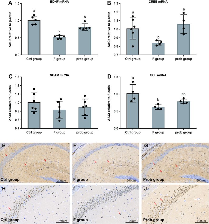
J. Xin et al. Ecotoxicology and Environmental Safety 215 (2021) 112108
impairment? 2) Did the preventive effects of L. johnsonii BS15 still exist hippocampus can generally solve very difficult reference memory
on fluoride-induced memory impairment after psychological stress? problems, as long as there is no spatial component (Deacon and Rawlins,
2006). T-maze test which represents spatial working memory makes up
2. Materials and Methods for it (Deacon and Rawlins, 2006). Enclosed T-maze, an apparatus with
10 cm-wide floor and 20 cm-high walls in the form of a “T” placed
2.1. Culture and treatment with BS15 horizontally, was used. The stem of the two goal arms and a start arm
was all 30 cm long. A central partition was placed in the middle of two
L. johnsonii BS15 was grown under anaerobic condition in de Man goal arms extending into the start arm (7 cm). Every arm had a guillotine
–Rogosa–Sharpe broth (Qingdao Rishui Bio-technologies Co., Ltd., door. Schematic outline of T-maze apparatus was displayed in Fig. A.1B
Qingdao, China) at 37 ◦ C for 24 h. The number of bacterial cells was (Supplementary materials). The apparatus and operating steps were
assessed by heterotrophic plate counts. L. johnsonii BS15 cells were then consistent with Deacon and Rawlins (2006). First, the central partition
centrifuged (10,000×g, 10 min at 4 ◦ C), washed three times by was placed in the T-maze with all the doors open. Then, the mouse,
phosphate-buffered saline (PBS), and re-suspended in PBS (pH 7.0) at a directly from its home cage or flowing exposure to WAS, was placed in
density of 1 × 10⁹ cfu cells/mL (daily consumption dose: 0.2 mL/mice). the start area and allowed to select the left or right arm. The mouse was
L. johnsonii BS15 was gavaged to mice once a day at 9:00 am. kept in the chosen arm by quietly sliding the door down. After 30 s, the
mouse and central partition were removed, and the mouse was returned
2.2. Water avoidance stress (WAS) to its holding cage. After a retention interval of 1 min, the mouse was
placed in the start area for a second trial with all the doors open. There is
Water avoidance stress, a well-established model of psychological no habituation to the maze during spontaneous alternation due to the
stress in mice, was used in our study as a psychological stressor. Mice novelty of the maze is involved to drive the spontaneous alternation
were exposed to WAS, as Gareau et al. (2011) described with minor (Deacon and Rawlins, 2006). To avoid that the mouse felt sated with
modifications. In brief, mice were placed on a small platform sur exploring the maze, each rodent was tested for 5 days with two trials per
rounded by room-temperature water (1 cm below the platform) in the day. If the mouse selected the other goal arm in consecutive trials, then
middle of the home cage for 1 h. All the WAS and behavioral tests were this trial was marked as “correct.” Each exploration should take no more
carried out between 7:00 am–11:30 am. The schematic outline of WAS than 2 min. If one mouse fails to run within 90 s, a reasonable criterion at
was shown in Fig. A.1A (Supplementary materials). which to abort the trial, it was removed and tested again after resting
(Deacon and Rawlins, 2006).
2.3. Behavioural tests
2.4. Establishment of an animal model and study design
Two observers simultaneously recorded the results of the T-maze and
NOR tests to eliminate the influence of concentration loss. Observers A total of 108 male ICR mice (3 weeks old, Chengdu Dashuo Bio
were blind to the treatment conditions of each mice and remained un logical Institute, Chengdu, China) were given 1 week to adapt to the new
changed in the same behavioural test. environment. After the adaptation period, mice were randomly divided
NOR test: The novel object recognition test was used to assess the into three groups and administered with either PBS (control group, F
ability of rodents to recognize a novel object in the environment. It is a group) or L. johnsonii BS15 (prob group; 0.2 mL/day) for 28 days prior to
widely used method for the detection of working memory alterations and throughout a 70 day exposure to sodium fluoride. Mice were pro
based on nature propensity for novel objects displayed by rodents. In the vided fluoridated drinking water (100 ppm NaF; corresponding to 37.8
NOR test, memory formation mainly depends on dorsal hippocampus ± 2.4 ppm F‾) from 28 days to 98 days, except for the control group. The
which plays an important role, especially when spatial or contextual method of actual concentration detection of sodium fluoride in drinking
information is a relevant factor (Goulart et al., 2010). The task pro water can be found in supplementary (Supplementary Note 1). The se
cedure included three phases (Antunes and Biala, 2012): habituation, lection of fluoride dose was based on (i) documented human exposures
familiarization, and test phase. Schematic outline of NOR test was dis (The range of fluoride dose in adult is 0.84–27.1 mg per day, 0.19–6.02
played in Fig. A.1C (Supplementary materials). In brief, in the habitu ppm when converted to ppm [based on a 55 kg person drinking 4.5 L of
ation phase, each mouse was placed into the empty open-field arena (l × water per day]; Supplementary Note 2 and Table A.1), (ii) a dose
b × h = 40 cm × 40 cm × 45 cm) for 1 h for habituation. The mouse was equivalent equation adjusting for surface area differences between mice
then removed from the arena and placed in its home cage. During the and humans (Coryell et al., 2018) (Supplementary Note 2), (iii) the use
familiarization phase, one mouse, following 1 h WAS, was placed in the of similar exposures in studies researching fluoride-associated brain
arena containing two different objects (#A+#B) for 5 min. These two lesion in mice (Table A.2), and (iv) high dose for the potential damage in
objects were placed in opposite corners of the cage. Then, the mouse was a shortened test period. Mice were housed in a constant-temperature
removed from the arena and returned to its cage for a 20 min rest. (20–22 ◦ C) room with a 12 h light/dark cycle (lights on from 06:00 to
During the test phase, the mouse was returned to the arena and con 18:00) and given free access to water and normal chow diet (Chengdu
fronted with object B and a novel object (object #C, distinguishable from Dashuo Biological Institute). Each group contains six cages with six in
object #A). Exploration ratio (F#C/(F#C + F#B) × 100, F#C = fre dividuals per cage. All animal experiments were performed according to
quency of exploring the object #C, F#B = frequency of exploring the the guidelines for the care and use of laboratory animals approved by the
object #B), which was used to assess the memory, was calculated. Institutional Animal Care and Use Committee of Sichuan Agricultural
The tendency of mice to explore novelty indicated that the presen University (approval number: SYXKchuan2019-187). At the end of the
tation of the familiar object existed in their memory. The objects used experiment, ten mice (one or two mice per cage) from each group were
included a green bottle cap (#A), an orange bottle cap (#B), and a small used for behavioral testing. Another twenty mice in each group were
smooth stone (#C). To avoid object bias, our preliminary studies used for sampling. The extra mice were kept on feeding until they die.
revealed that mice showed equal preference for #A, #B and #C.
T-Maze test: T-maze alternation has been used for decades in 2.5. Biochemical evaluation
academia and industry for its sensitivity in assessing cognitive
dysfunction and its simplicity of construction (Deacon and Rawlins, On day 98 of the experiment, six mice (one mouse per cage) from the
2006). Spontaneous alternation of T-maze task relies on the novelty of control, F, and prob groups were selected and sacrificed by cervical
the maze and does not need food restriction. Mice with removed or dislocation for sampling following exposure to 1 h WAS. Blood was
damaged (e.g., by surgical removal or genetic modification) sampled from the mice orbit, and serum was separated by incubation at
3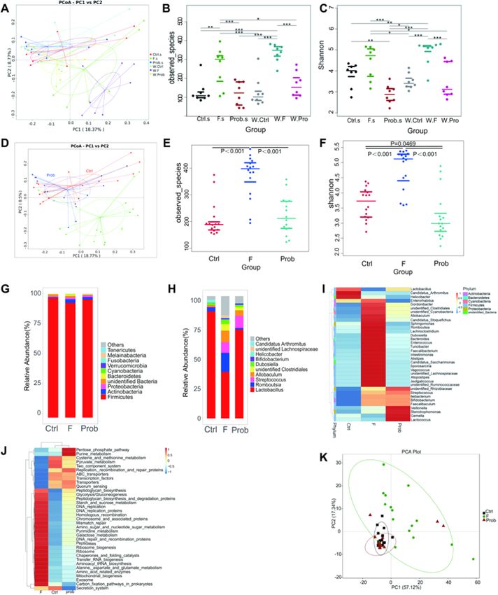
J. Xin et al. Ecotoxicology and Environmental Safety 215 (2021) 112108
4 ◦ C for 30 min, followed by centrifugation at 2000 × g for 20 min, and by shaking for 5 min, then sealed for 30 min by 3% bull serum albumin,
stored at − 30 ◦ C. The ileal tissue and right hippocampus were ground and incubated with monoclonal rabbit anti-BDNF (1:400), polyclonal
(pH 7.4) into 10% or 5% homogenate with PBS, respectively, and then rabbit anti-CREB (1:500), anti-Occludin, anti-Claudin-1, or anti-ZO-1
centrifuged (12,000 × g for 5 min at 4 ◦ C). The obtained liquid super (1:200) antibodies at 4 ◦ C overnight. Species-specific biotinylated anti-
natant was used for biochemical detection. The D-lactate and diamine rabbit immunoglobulin (horseradish peroxidase labeled) was used for
oxidase (DAO) activity, contents of corticosterone (CORT) in the serum, immunodetection. Following the second antibody incubation, the 3,3′ -
the inflammatory cytokines in the liquid supernatant of the ileum and diaminobenzidine staining kit was used to complete the reaction ac
hippocampal homogenate, and apoptosis-regulated proteins in the cording to the manufacturer’s instructions. Hematoxylin staining was
liquid supernatant of hippocampal homogenate were measured by the performed to re-stain the nucleus.
commercial enzyme-linked immunosorbent assay (ELISA) kit for mice
according to the manufacturer’s instructions. The inflammatory cyto 2.8. 16S rRNA gene sequencing
kines included tumor necrosis factor-alpha (TNF-α), interleukin-1β (IL-
1β), IL-6, interferon-gamma (IFN-γ), and IL-10 (only detects ileal tissue). The ileal content of the abovementioned mice and another ten mice
All the commercial ELISA reagent kits were obtained from Enzyme- (one mouse per cage) from each group without acute stress was collected
linked Biotechnology Company (Shanghai Enzyme-linked Biotech and selected enough eight samples for 16S rRNA gene sequencing.
nology Co., Ltd., China). Bacterial genomic DNA was extracted using the E.Z.N.A.TM stool
DNA isolation kit (Omega Bio-Tek, Doraville, CA). The final elution
2.6. Real-time quantitative polymerase chain reaction (qPCR) analysis of volume was 100 µL, and the integrity, purity, fragment size, and con
gene expression centration were determined by electrophoresis with 1% agarose gel. The
16S V3-V4 was amplified by PCR using the primer 515F/806R of the
The left hippocampus and partial ileal tissue were removed and 16SrRNA gene. Then, the purified PCR products were formed into a li
washed with ice-cold sterilized saline without RNA enzyme (RNase) and brary with Ion Plus Fragment Library Kit 48 rxns (Thermofisher, USA)
then immediately frozen in liquid nitrogen for gene expression analysis. and sequenced in the Ion S5™XL platform (Thermofisher, USA) using
Total hippocampal RNA and ileal RNA were isolated using E.Z.N.A.® the single-end sequencing. The primer contained adapter sequences and
Total RNA Kit (OMEGA Bio-Tek) according to the manufacturer’s in single-end barcodes, allowing pooling and direct sequencing of PCR
structions. Isolated RNA was assessed from the ratio of absorbance at products. Cutadapt (V1.9.1, http://cutadapt.readthedocs.io/en/stable/)
260/280 nm and agarose gel electrophoresis for quantitative and qual was applied to the resulting high-quality 16SrRNA gene reads. The 16S
itative analyses. The isolated RNA was transcribed into first-strand rRNA gene read pairs were demultiplexed based on the unique molec
complementary DNA with PrimeScript RT reagent kit with gDNA ular barcodes, and reads were merged using VSEARCH (Rognes et al.,
Eraser (Thermo Scientific, Waltham, Massachusetts, USA) according to 2016). Sequences were clustered into operational taxonomic units
the manufacturer’s instructions. qPCR was performed with SYBR green (OTUs) at a similarity cutoff value of 97%. Then, OTU representative
and primers (details previously published) (Xin et al., 2014). The PCR sequences were produced using the Uparse v7.0.1001. Species annota
conditions were as follows: 1 cycle at 95 ◦ C for 5 min, followed by 40 tion analysis was performed on the OTU representative sequences
cycles at 95 ◦ C (10 s) and optimum temperature (30 s), and then a final through the SILVA Database. Homogenized data of each sample were
melting curve analysis to monitor the purity of the PCR product. The constructed using the sample with the least amount of data as the
optimum temperature of each gene was shown in Table A.3. β-Actin was standard. This sample was used for downstream analyses of
used as reference genes to normalize the relative mRNA expression alpha-diversity, beta-diversity, phylogenetic trends, and functional
levels of target genes with values presented as 2− ΔΔCq. Primer sequences prediction.
and optimum annealing temperatures were shown in Table A.3. The
relative expression levels of neurogenesis-related factors [BDNF, CREB, 3. Statistical analysis
stem cell factor (SCF), and neural cell adhesion molecule (NCAM)],
molecular myelin structure [myelin oligodendrocyte glycoprotein The 16S rRNA sequencing data were analyzed by Wilcoxon rank-sum
(MOG), proteolipid protein (PLP), myelin basic protein (MBP), and test to detect significant differences among the different groups.
myelin-associated glycoprotein (MAG)], apoptosis-related proteins All other results were reported by mean ± standard deviation.
[Bcl2-associated X protein (Bax), Bcl-xL/Bcl-2 asociated death promoter Normality was evaluated using the Shapiro-Wilk normality test. If data
(Bad), B-cell lymphoma-2 (Bcl-2), B-cell lymphoma-extra large (Bcl-xl), were not normally distributed they were log transformed for analysis.
caspase9, and caspase3), cytokines [interferon-γ (IFN-γ), tumor necrosis Data that remained not normally distributed were analyzed by Kruskal-
factor-α (TNF-α), interleukin (IL)-1β, IL-6, and IL-10] in the hippocam Wallis test followed by Wilcoxon rank-sum test. If data were normally
pus, and tight junction (TJ) proteins [zonula occludens protein 1 (ZO-1), distributed they were analyzed by one-way analysis of variance
claudin-1, and occludin) in ileal tissue were detected. (ANOVA) followed by post hoc Duncan’s multiple range test. In short,
All biochemical parameters were assessed by one-way ANOVA followed
2.7. Immunohistochemistry by post hoc Duncan’s multiple range test except for BDNF using Kruskal-
Wallis test followed by Wilcoxon rank-sum test. Differences of p < 0.05
Another four mice (one mouse per cage) in each group were sacri were considered statistically significant. Data were analyzed with IBM
ficed following exposure to 1 h WAS, and their brain and ileum were SPSS Statistics 25.
removed, fixed in 4% paraformaldehyde solution, and stored in 4 ◦ C for
immunohistochemical assay. Tissues were embedded by paraffin and cut 4. Results
by a microtome. Slices were submerged in citrate antigen retrieval so
lution and heated at medium heat until boiling using a microwave oven 4.1. L. johnsonii BS15 improved the memory ability and serum CORT
(model: P70D20TL-P4; Galanz, Guangdong, China). The temperature level in fluoride-treated mice exposed to WAS
was ceased, and the tissues were kept warm for 8 min. Then, the tissues
were heated at medium–low heat for 7 min. After free cooling, the slices As shown in Fig. 1A and B, the spontaneous exploration in the T-
were placed into PBS (pH 7.4) and shaken for 5 min for decoloration, maze test and the exploration ratio in the NOR test were significantly
which was repeated three times. Afterward, the sections were incubated reduced in the F group compared with the control and prob groups (T-
in 3% oxydol for 25 min at room temperature and away from the light to maze: control vs. F, P < 0.001; F vs. prob, P = 0.008; NOR test: control
block endogenous peroxidase. The slices were washed three times in PBS vs. F, P < 0.001; F vs. prob, P = 0.001). No significant difference was
4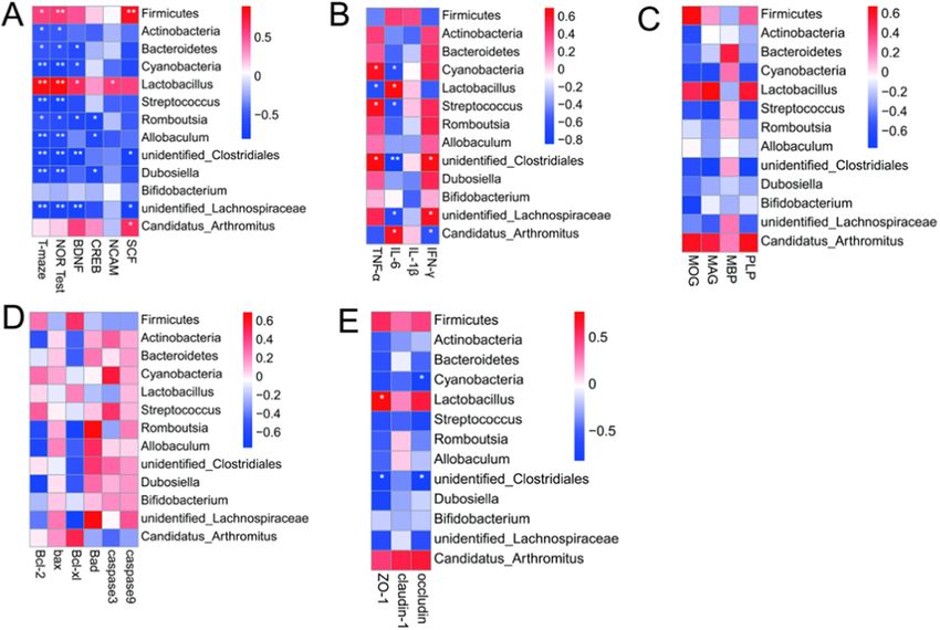
J. Xin et al. Ecotoxicology and Environmental Safety 215 (2021) 112108
Fig. 1. Results of behavioral tests and the serum CORT level. (A) T-Maze (n = 8; one-way ANOVA, F2, 21 = 10.407, P = 0.001), (B) NOR preference tests (n = 10;
one-way ANOVA, F2, 27 = 16.685, P < 0.001), and (C) the serum CORT level (n = 4–5; one-way ANOVA, F2, 10 = 15.748, P = 0.001). Data are presented as the means
± standard deviation. Bars with different letters indicate significant difference based on Duncan’s multiple range test (P < 0.05).
observed in the abovementioned indexes between the control and prob sharply decreased compared with that in the control group. The PLP in
groups (T-maze: P = 0.127; NOR test: P = 0.068). Fig. 1C shows that the the prob group was slightly higher than that in the F group (P = 0.125).
F group exhibited a remarkably higher serum CORT level than the other No significant difference in the MOG was observed between the F and
two groups (control vs. F, P < 0.001; F vs. prob, P = 0.002), but no prob groups (P = 0.994). No changes in the MBP mRNA expression level
significant difference was found between the other two groups. were observed among the three groups (control vs. F, P = 0.594; control
vs. prob, P = 0.278; F vs. prob, P = 0.609). As shown in Fig. 3A, the F
4.2. Effect of L. johnsonii BS15 on BDNF, CREB, NCAM, and SCF group presented a significantly lower MAG mRNA level than the other
expressions in the hippocampus of fluoride-treated mice two groups (control vs. F, P < 0.001; F vs. prob, P < 0.001). Compared
with the control group, MAG was remarkably reduced in the prob group
Fig. 2A and B shows a sharp decrease in mRNA expression levels of (P = 0.01).
BDNF (control vs. F, P = 0.011; F vs. prob, P = 0.011) and CREB (con No significant difference in the Bcl-2 mRNA level was found among
trol vs. F, P = 0.026; F vs. prob, P = 0.011) in the F group compared the three groups (control vs. F, P = 0.637; control vs. prob, P = 0.113; F
with the other two groups. The mRNA level of BDNF in the control group vs. prob, P = 0.052), whereas the prob group showed the highest among
was higher than that in the prob group (P = 0.036) (Fig. 2A). No dif the three groups (Fig. 3B). In Fig. 3B, the F group presented a lower Bcl-
ference was observed in the CREB mRNA level between the control and xl mRNA level than the other two groups (control vs. F, P = 0.042; F vs.
prob groups (P = 0.448) (Fig. 2B). Moreover, no significant difference in prob, P = 0.021), but no difference was observed in the other two
the NCAM was found among the three groups (control vs. F, P = 0.058; F groups (control vs. prob, P = 0.8). As shown in Fig. 3B, the Bax mRNA
vs. prob, P = 0.058; control vs. prob, P = 0.058) (Fig. 2C). Compared level in the F group was slightly increased compared with that in the
with the control group, the SCF mRNA level was remarkably reduced in other two groups, but no significant difference was observed among the
the F group and slightly decreased in the prob group (control vs. F, three groups (control vs. F, P = 0.734; control vs. prob, P = 0.772; F vs.
P = 0.005; control vs. prob, P = 0.062) (Fig. 2D). By contrast, the prob prob, P = 0.531). Fig. 3B shows a remarkably increased Bad mRNA level
group presented a slightly higher SCF mRNA level than the F group in the F group compared with the other two groups (control vs. F,
(P = 0.164) (Fig. 2D). As shown in Fig. 2E–J, the protein expression P = 0.047; F vs. prob, P = 0.012), whereas no difference was observed
levels of BDNF and CREB were significantly reduced in the F group in the other two groups (control vs. prob, P = 0.445). As shown in
compared with that in the other two groups. Fig. 3B, no significant difference in the caspase3 and caspase9 mRNA
level was observed among the three groups (casepase3: control vs. F,
P = 0.139; control vs. prob, P = 0.195; F vs. prob, P = 0.902; casepase9:
4.3. L. johnsonii BS15 alleviated the myelin damage and improved the
control vs. F, P = 0.256; control vs. prob, P = 0.476; F vs. prob,
apoptosis-related proteins in fluoride-treated mice exposed to WAS
P = 0.62), but caspase3 in the control group and caspase9 in the F group
were the lowest and highest, respectively, among the three groups.
Fig. 3A shows that the mRNA expression levels of PLP (control vs. F,
P < 0.001; control vs. prob, P = 0.002) and MOG (control vs. F,
P = 0.007; control vs. prob, P = 0.005) in the F and prob groups were
5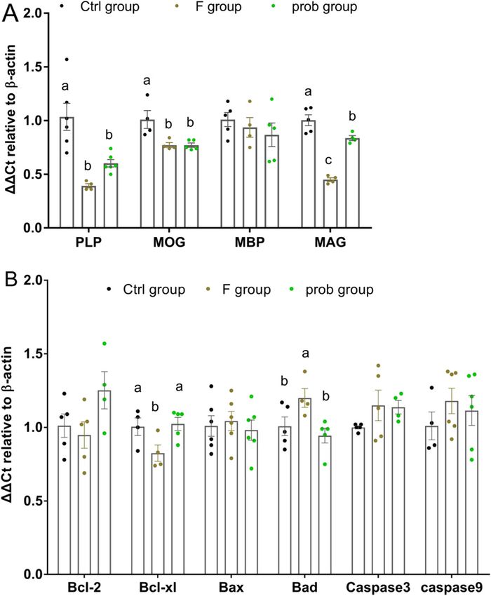
J. Xin et al. Ecotoxicology and Environmental Safety 215 (2021) 112108
Fig. 2. Expressions of neurogenesis-related factors in the hippocampus. Relative expression levels of (A) BDNF (Kruskal–Wallis test, H2 = 11.622, P = 0.003;
Wilcoxon test, control vs. F, P = 0.011; F vs. prob, P = 0.011; control vs. prob, P = 0.036), (B) CREB (one-way ANOVA, F2, 11 = 5.229, P = 0.025), (C) NCAM (one-
way ANOVA, F2, 15 = 1.144, P = 0.345), and (D) SCF (one-way ANOVA, F2, 9 = 6.691, P = 0.017). Immunohistochemistry of BDNF (E-G) and CREB (H-J) expression
in the hippocampus of mice. The BDNF- and CREB-positive cells are brown like the arrow indication. Data are presented as the means ± standard deviation
(n = 4–6). Bars with different letters indicate significant difference on the basis of Duncan’s multiple range test (CREB, NCAM, SCF) or Wilcoxon test (BDNF)
(P < 0.05). (For interpretation of the references to colour in this figure legend, the reader is referred to the web version of this article.)
4.4. L. johnsonii BS15 reduced the intestinal permeability in fluoride- immunohistochemistry, and the results showed the same trend (Fig. 4C).
treated mice exposed to WAS As shown in Fig. 4B, compared with the control group, the serum
DAO content was significantly and slightly increased in the F and prob
As shown in Fig. 4A, the F group shows a markedly lower ZO-1 groups (control vs. F, P = 0.021; control vs. prob, P = 0.182), respec
mRNA level than the other two groups (control vs. F, P = 0.001; F vs. tively. The control group showed a remarkably lower serum D-lactate
prob, P = 0.028), but significant difference was not observed in the activity than the other two groups (control vs. F, P < 0.001; control vs.
other two groups (control vs. prob, P = 0.059). Compared with the prob, P = 0.016). By contrast, the D-lactate activity in the F group was
control group, the mRNA expression levels of claudin-1 (control vs. F, significantly higher (P = 0.018) than that in the prob group.
P = 0.025; control vs. prob, P = 0.032) and occludin (control vs. F,
P < 0.001; control vs. prob, P = 0.002) were sharply decreased in the F 4.5. L. johnsonii BS15 prevented disorder in the gut microbiota induced
and prob groups. No significant difference was observed in claudin-1 by fluoride
mRNA level between the F and prob groups (P = 0.878). The occludin
mRNA level in the prob group was slightly higher than that in the F The gut microbiota with or without stress were observed in each
group (P = 0.241). The protein expressions of TJs were also detected by group to determine the influence of fluoride and acute stress on the gut
6
J. Xin et al. Ecotoxicology and Environmental Safety 215 (2021) 112108
unidentified_Clostridiales, Dubosiella, Bifidobacterium, and unidentified_
Lachnospiraceae compared with the control group. However, L. johnsonii
BS15 suppressed these alterations except for Streptococcus, unidenti
fied_Clostridiales, Bifidobacterium, and Candidatus_Arthromitus. Compared
with the control group, more genera bloomed or diminished in the ileal
lumen of the F group than those in the prob group (Fig. 5I). Moreover,
compared with the control group, multiple microbial pathways were
altered in the F group, which were reversed in the prob group (Fig. 5J).
As shown in Fig. 5K, PCA showed significant separation between the
control and F groups in the function of the microbiome in KEGG path
ways but was not observed between the control and prob groups.
4.6. Changes of dominant bacteria genus and correlation between
bacteria and clinical data
As shown in Fig. 6A and B, compared with control and F groups, the
significant discriminative taxa in the control group were Firmicutes,
Bacilli, Lactobacillales, Lactobacillaceae, Lactobacillus, Lactobacillus_tai
wanensis, Lactobacillus_reuteri, Lactobacillus_intestinalis, and in F group
were Clostridia, Erysipelotrichia, Clostridiales, Erysipelotrichales, Peptos
treptococcaceae, Erysipelotrichaceae, Streptococcaceae, unidentified_Clos
tridiales, Lachnospiraceae, Romboutsia, Streptococcus, Allobaculum,
Streptococcus_hyointestinalis, Streptococcus_hyointestinalis. Compared with
F and prob groups, the significant discriminative taxa in F group were
Erysipelotrichia, Clostridia, Erysipelotrichales, Clostridiales, unidentified_
Clostridiales, Lachnospiraceae, Erysipelotrichaceae, Erysipelotrichaceae,
Fig. 3. mRNA expression levels of myelin- and apoptosis-associated proteins in unidentified_Lachnospiraceae, Dubosiella, unidentified_Clostridiales, Allo
the hippocampus. Relative expression levels of (A) PLP (one-way ANOVA, F2, 13 baculum, Romboutsia, as well as Clostridium_sp_ND2, and in prob group
= 13.906, P = 0.001), MOG (one-way ANOVA, F2, 10 = 8.091, P = 0.008), MBP were Bacilli, Lactobacillales, Lactobacillaceae, Lactobacillus (Fig. 6C and
(one-way ANOVA, F2, 11 = 0.651, P = 0.541), and MAG (one-way ANOVA, F2, D). Compared with control and prob groups, the key genera in prob
10 = 56.933, P < 0.001). Relative expression levels of (B) Bcl-2 (one-way
group were Streptococcaceae, Streptococcus, Allobaculum, unidentified_
ANOVA, F2, 11 = 2.559, P < 0.112), Bcl-xl (one-way ANOVA, F2, 10 = 4.269,
Clostridiales, Streptococcus_hyointestinalis, as well as Clostridium_sp_ND2,
P = 0.046), Bax (one-way ANOVA, F2, 15 = 0.206, P = 0.816), Bad (one-way
ANOVA, F2, 11 = 4.704, P = 0.033), caspase-3 (one-way ANOVA, F2, 11 = 1.528,
and in control group were Lactobacillus_taiwanensis, Lactobacillus_reuteri,
P = 0.260), and caspase-9 (one-way ANOVA, F2, 13 = 0.705, P = 0.512). Data Lactobacillus_intestinalis (Fig. 6E and F).
are presented as mean ± standard deviation (n = 4–6). Bars with different Fig. 7A shows that bacteria from phylum (Firmicutes, Actinbacteria,
letters indicate significant difference on the basis of Duncan’s multiple range Bacteroidetes, Cyanobacteria) and genus (Lactobacillus, Streptococcus,
test (p < 0.05). Romboutsia, Allobaculum, unidentified_Clostridiales, Dubosiella, Bifi
dobacterium, unidentified_Lachnospiraceae, Candidatus_Arthromitus)
microbiota. Through 16s rRNA gene sequencing, we did not find sig in top ten level with significant difference among groups displayed
nificant alterations in the gut microbiota of mice exposed to WAS versus significant associations with spontaneous exploration (T-maze), explo
non-stressed animals (Fig. 5A-C and Table A.4). Thus, the data with or ration ratio (NOR test), mRNA levels of BDNF, CREB, NCAM, and SCF of
without WAS were combined in each group. hippocampus except for Bifidobacterium. Firmicutes and Lactobacillus
As shown in Fig. 5D and Fig. A.2A, principal coordinate analysis showed a positive regulation to these indexes. Fig. 7B shows that Cya
(PCOA) based on unweighted UniFrac distances and principal compo nobacteria, Streptococcus, and unidentified_Clostridiales were observ
nent analysis (PCA) showed a clear separation between the ileal ably positive associated with TNF-α and negative associated with IL-6.
microbiota of mice in the F group and those in the other groups, whereas Moreover, IL-6 was also significantly and positively correlated with
no significant separation was observed between the other two groups. Lactobacillus and Candidatus_Arthromitus, and negatively correlated
Moreover, the observed species (Fig. 5E) and Shannon index (Fig. 5F) in with unidentified_Lachnospiraceae. IFN-γ was significantly positively
the ileal lumen microbiome of mice in the F group were significantly correlated with unidentified_Clostridiales and unidentified_Lachnospir
increased compared with that in the control group, whereas significant aceae, and negatively correlated with Candidatus_Arthromitus. A sig
increases were not observed in the prob group. The ileal microbial nificant association between myelin-associated protein, apoptosis-
communities among the three groups were dominated by Firmicutes in related proteins in hippocampus, and gut microbiota was not observed
the phylum level (Fig. 5G). Relative abundance of Firmicutes in the F (Fig. 7C and D). Intestinal tight junction proteins were significantly
group was significantly reduced compared with that in the control and associated with Cyanobacteria, Lactobacillus, and unidentified_Clos
prob groups (Fig. 5G and Fig. A.2B). On the contrary, the relative tridiales (Fig. 7E).
abundances of Actinobacteria (Fig. A.2C), Bacteroidetes (Fig. A.2D), and
Cyanobacteria (Fig. A.2E) increased markedly in the F group compared 5. Discussion
with the control and prob groups. The alteration of these common phyla
induced by fluoride was significantly inhibited by L. johnsonii BS15. At 5.1. Behavioral phenotypes and neuroplasticity
the genus level (Fig. 5H), Lactobacillus was the important bacterium in
the control, F, and prob groups, and the relative abundance of the nine A 70-day exposure to sodium fluoride was found to induce memory
main genera found in the three groups was different. As shown in dysfunction in mice after 1 h exposure to WAS according to the results of
Fig. A.2F–N, the F group presented a sharply reduced relative abundance behavioral tests in our present study. Lower memory ability was indi
of Lactobacillus and Candidatus_Arthromitus and a significantly increased cated by the low spontaneous exploration in T-maze test and low
relative abundance of Streptococcus, Romboutsia, Allobaculum, exploration ratio in NOR test in the fluoride-exposed group. Studies
found that an hour of WAS exposure could not induce detectable
7
J. Xin et al. Ecotoxicology and Environmental Safety 215 (2021) 112108
Fig. 4. Results of intestinal permeability. (A) mRNA expression levels of ZO-1 (one-way ANOVA, F2, 10 = 11.689, P = 0.002), claudin-1 (one-way ANOVA, F2, 9
= 4.535, P = 0.043) and occludin (one-way ANOVA, F2, 11 = 16.702, P < 0.001) in the ileum, (B) Serum DAO activity (one-way ANOVA, F2, 15 = 3.333, P = 0.063)
and D-lactate concentration (one-way ANOVA, F2, 15 = 14.382, P < 0.001). (C) Immunohistochemistry of TJs protein expressions in ileum of mice. The ZO-1-,
claudin-1- and occludin-positive cells are brown like the arrow indication. Data are presented as mean ± standard deviation (n = 4–6). Bars with different letters
indicate significant difference on the basis of Duncan’s multiple range test (p < 0.05). (For interpretation of the references to colour in this figure legend, the reader is
referred to the web version of this article.)
behavioral changes in the T-maze and NOR tests but could cause a caused an impairment of spatial and reference memory (Mizuno et al.,
compounding effect on memory ability when combined with other 2000). CREB, the transcriptional regulator of BDNF, has been considered
stressors (e.g., endotoxin exposure and bacterial infection) by enhancing as the central to AD pathology by similar genomic network analysis
the HPA axis responsiveness (Gareau et al., 2011; Walker et al., 2008). (Jeong et al., 2001). In addition, many studies using genetically modi
We found that fluoride exposure resulted in HPA hyper-responsiveness fied mice revealed that a growing number of genes, such as CREB, BDNF,
to WAS given the significantly elevated CORT in the serum in F group. NCAM, and SCF, were involved in the regulation of neurogenesis.
This result was consistent with that found by Gareau et al. (2011). Consistent with our previous study Xin et al. (2020) and Niu et al.
Neuronal plasticity is the basis of learning and memory and occurs by (2018), fluoride showed adverse effects on neuroplasticity as indicated
neurogenesis, synaptic-dependent activity, cellular apoptosis, and by the remarkable decrease in the expressions of BDNF, CREB, and SCF
reorganization of neuronal networks (Johnston, 2004, 2009). The hip on the mRNA and/or protein levels. Buffington et al. (2016) reported
pocampus, a highly plastic region, is critical for learning and memory that the behavioral deficits and disordered gut microbiota in maternal
(Nakazawa et al., 2004). BDNF is one of the important modulators of high-fat diet offspring could be recovered by co-housing the offspring of
neuroplasticity because of its multiple effects, such as increasing hip mothers on a regular diet. Kumar et al. (2017) suggested that L. johnsonii
pocampal neurogenesis, dendritic branching, cell proliferation, and could increase the concentrations of acetate and butyrate in feces.
promoting hippocampal long-term potentiation (Pang and Lu, 2004; Butyrate, a short-chain fatty acid, could decrease BDNF methylation and
Cassilhas et al., 2016). Blocking the hippocampal BDNF expression consequently cause overexpression of BDNF by decreasing 10 to 11
8J. Xin et al. Ecotoxicology and Environmental Safety 215 (2021) 112108
Fig. 5. Effects of L. johnsonii BS15 on the gut microbiome structure in fluoride-treated mice. (A) Principal coordinate analysis (PCoA) of unweighted UniFrac dis
tances among groups with and without WAS. (B) Gut microbiome richness (observed species) in ileal luminal samples of each group. Significance: Wilcoxon. (C) Gut
microbiome community diversity (Shannon) in each group. significance: Wilcoxon. (D) Principal coordinate analysis (PCoA) of unweighted UniFrac distances among
the groups after combing data with and without WAS. (E) Gut microbiome richness (observed species) in ileal luminal samples of each group. Significance: Wilcoxon.
(F) Gut microbiome community diversity (Shannon) in each group. Significance: Wilcoxon. (G) Relative abundance (%) at the phylum level of each group. (H)
Relative abundance (%) at the genus level of each group. (I) Genera that are markedly altered by excess fluoride intake compared with the control group and reverted
by L. johnsonii BS15. (J) Same as (I) but for KEGG pathways level 3. (K) Same as (A) PCA based on KEGG pathways level 3. Significance: Wilcoxon. *p < 0.05,
**p < 0.01, ***p < 0.001. Ctrl.s: control group without WAS; F.s: F group without WAS; Prob.s: Prob group without WAS; W.Ctrl: control group with WAS; W.F: F
group with WAS; W.Prob: Prob group with WAS.
9J. Xin et al. Ecotoxicology and Environmental Safety 215 (2021) 112108
Fig. 6. LEfSe analysis of discriminative taxa of gut microbiota in mice. The linear discriminant analysis (LDA) score (A, C, and E) and cladogram (B, D, and F) were
generated from LDA effect size (LEfSe). Only taxa meeting the LDA significance thresholds (> 4) are shown.
10J. Xin et al. Ecotoxicology and Environmental Safety 215 (2021) 112108
Fig. 7. Correlations analysis. (A) Correlations between intestinal flora and spontaneous exploration (T-maze), exploration ratio (NOR test), mRNA levels of BDNF,
CREB, NCAM, and SCF of hippocampus. (B) Correlations between intestinal flora and protein expression levels of inflammatory cytokines in hippocampus. (C)
Correlations between intestinal flora and mRNA expression levels of myelin-associated proteins of hippocampus. (D) Correlations between intestinal flora and
apoptosis-related proteins of hippocampus. (E) Correlations between intestinal flora and intestinal tight junction proteins. The analysis method was spearman rank
analysis. Bacteria were selected from phylum and genus in top ten level with significant difference. Heatmap showed that microbial taxa was positively or negatively
related to behavioral phenotype and brain chemistry. *P < 0.05 and **P < 0.01 denoted statistical significance between bacterial taxa and biochemical parameters;
Wilcoxon tests.
translocation methylcytosine dioxygenase 1, which was the enzyme fluoride exposure on hippocampal inflammation in mice after WAS
responsible for catalyzing the conversion of DNA methylation to (Fig. A.3), which was consistent with our previous study (Xin et al.,
hydroxymethylation (Wei et al., 2014). Moreover, Luo et al. (2016) 2020). In addition, we observed the mRNA expression of
found that Lactobacillus spp. was significantly reduced in myelin-associated protein to assess the effect of L. johnsonii BS15 on
fluoride-treated broiler. Thus, in the present study, L. johnsonii BS15 was hippocampal impairment of fluoride-treated mice under psychological
supplemented to fluoride-treated mice to investigate whether L. john stress. Previous studies have demonstrated that fluoride could induce
sonii BS15 could alleviate memory impairment. Based on our results, the demyelination (Niu et al., 2018). In our previous study, we also found
L. johnsonii BS15-inoculated mice showed a significant memory ten-week fluoride exposure induced significant decrease in PLP and
improvement compared with fluoride-treated, stressed mice. Further MOG (Xin et al., 2020). After adding one hour WAS to fluoride-treated
more, the negative effects of fluoride treatment on the HPA response, mice, not only the PLP and MOG, but also the decreased mRNA level
BDNF, and CREB have been reversed by L. johnsonii BS15, indicating the of MAG was observed in this study. The down-regulated MAG induced
psychoactive effect of the probiotic on memory impairment simulta by WAS in fluoride-treated mice was significantly increased by BS15
neously induced by fluoride exposure and acute psychological stress. supplement. Notably, myelin sheath constituted by MOG, PLP, MBP, and
MAG was important for axonal protection and interneuronal commu
5.2. Neuroinflammation, myelin-associated protein and neuron apoptosis nication (Nguyen et al., 2009). MAG located in the innermost lamellae of
myelin sheaths, as an inhibitor of mature axonal regeneration, could also
Chronic neuroinflammation attracts public attention for its role in promote stability and survival of myelinated axons (Nguyen et al.,
mental health and diseases. However, considerable evidence proved that 2009). A local downregulation of MAG is the critical signal for CNS
fluoride accumulation could activate microglia, a resident macrophage injury (Nguyen et al., 2009). Treatment with L. johnsonii BS15 effec
in the central nervous system (CNS), and lead to the production of tively inhibited the reduction of MAG in fluoride-treated, stressed mice.
proinflammatory cytokines. Prior studies have noted the importance of Moreover, L. johnsonii BS15 upregulated the mRNA expression of Bcl-xl
aberrant intestinal microbiota, altered intestinal immune response, and (anti-apoptotic) and downregulated the mRNA expression of Bad
impaired intestinal barrier on neuroinflammation (Salehipour et al., (pro-apoptotic) in the hippocampus of fluoride-treated and stressed
2017). In this study, we examined inflammatory cytokines in both mice, indicating that hippocampal apoptosis was linked to
mRNA and protein levels in the hippocampus to explore the influence of fluoride-induced memory impairment, whereas L. johnsonii BS15
11J. Xin et al. Ecotoxicology and Environmental Safety 215 (2021) 112108
improved hippocampal apoptosis. supported by the comparison between the CORT results in two studies.
We evaluated the CORT level in the serum in all three groups collected
5.3. Intestinal integrity and stored in our previous work (Xin et al., 2020). Unlike the signifi
cantly higher level of CORT shown in F group in this study, no significant
On the basis of recent reports, which associated bacteria and their change of CORT level was observed according to the result we obtained
products with memory in human (Emery et al., 2017; Zhan et al., 2016) (Fig. A.5). No significant change of claudin-1 was found in
(e.g., E coli K99, LPS), we focused on understanding the alterations of gut fluoride-treated mice without WAS (Xin et al., 2020), but the claudin-1
inflammation and intestinal integrity underlying the improvement of level was reduced in fluoride-treated, stressed mice in the present study.
memory. We observed increased pro-inflammatory cytokines (TNF-α, In addition, the probiotic did not show the same ability to reverse the
IL-1β, and IFN-γ) and decreased anti-inflammatory cytokine (IL-10) decreased level of occludin in mice exposed by both fluoride and WAS as
which was in accordance with our previous study (Xin et al., 2020) it was reported in the previous study (Xin et al., 2020). Therefore, based
(Fig. A.4). The improvement of BS15 on intestinal inflammation was on the changes of tight junction proteins in this study, especially
also observed in fluoride-treated, stressed mice. TJ proteins (ZO-1, claudin-1 and occludin, an hour of WAS possibly aggravated the impact
claudin-1, and occludin) in the mRNA and/or protein levels in the ileum of fluoride intake on gut-brain axis and thus enhanced the memory
of the F group were significantly reduced, indicating a damaged intes impairment.
tinal epithelial integrity. The increased serum DAO content and
D-lactate activity in the fluoride-treated, stressed mice also indicated 5.5. Gut microbiota
that fluoride increased gut permeability. Serum DAO activity and
D-lactate content, indicators of mucosal integrity, would be increased The gut microbiota is a key modulator of the bidirectional signaling
when the intestinal mucosal integrity was destroyed (Luk et al., 1980; pathways between the gut and brain. We conducted 16S rRNA gene
Ewaschuk et al., 2005). BS15 was detected to improve gut permeability sequencing on the ileal contents of mice to identify whether the recon
of fluoride-treated mice under psychological stress according to the struction of gut microbiota was a potential mechanism underlying the
present results, which was consistent with the previous finding that hypothesis that BS15 protected mice from memory impairment induced
specific probiotic administration could improve gut inflammation and by fluoride. According to the results shown in PCoA of unweighted
intestinal mucosa integrity (Mujagic et al., 2017). Damaged intestinal UniFrac distances of microbiome communities structure before and after
mucosal integrity and some pro-inflammatory factors not only caused WAS and Table A.4, an hour of WAS exposure was not detected to
peripheral immune activation but also crossed the blood–brain barrier significantly change the gut microbiota structure. We substantiated the
to aggravate neuroinflammation in the CNS under pathological condi composition differences of the gut microbiota in fluoride-treated mice
tions. Ait-Belgnaoui et al. (2012) also found that probiotic treatment through Adonis testing (Table A.5) and unweighted analyses of UniFrac
could prevent leaky gut, thereby attenuating HPA response to acute distances, and the administration of L. johnsonii BS15 reversed those
psychological stress in rats. Hence, these results indicated that gut differences. Firmicutes, which accounted for up to 90% of the total se
inflammation and intestinal mucosa integrity might be involved in the quences, was the dominant phylum in each group. Disordered gut
pathogenesis of neurotoxic effects of fluoride and might be the primary microbiota in fluoride-exposed mice was primarily manifested by the
reason that BS15 improved memory deficit in fluoride-treated mice relative abundance decrease of Firmicutes in the phylum level and
under acute stress. Lactobacillus in the genus level. Lactobacillus is an important genus in
Firmicutes. The relative abundance of Lactobacillus in the control, F, and
5.4. The psychological stress prob groups was 87.7%, 38.1%, and 72.7%, respectively.The reduction
of Lactobacillus induced by fluoride was consistent with the results of Luo
An hour of WAS exposure was added in this study to find whether or et al. (2016).
not the probiotic could alleviate fluoride-induced memory impairment Spearman correlation analysis in the genus level of the control group
after psychological stress through the gut-brain axis. Comparing to our showed that Lactobacillus was negatively associated with most of the
previous work, several lines of evidence supported that WAS made the bacteria (66.7%; Fig. A.6A). In addition to the reduction of Lactobacillus,
potential memory impairment more evident. It significantly aggravated the above mentioned negative association between Lactobacillus and
the impact of fluoride exposure by influencing the gut-brain axis, which most bacteria was weakened and shifted to a positive association in the F
was most importantly supported by the result of NOR test in the present group (Fig. A.6B), indicating that the inhibiting effect of Lactobacillus on
study. In this study, the exploration ratio was significantly lower in F other bacteria was weakened. The above observation also explains the
group while it was not changed by single fluoride exposure in our pre increases in community diversity (Shannon index) and richness indices
vious work (Xin et al., 2020), directly indicating the impact of acute (number of observed features) in fluoride-treated mice. The adminis
psychological stress in fluoride-treated mice. Apart from the behavioral tration of L. johnsonii BS15 reversed these changes (Fig. A.6C). This
phenotypes, differences between the changes of the expression levels of finding was in accordance with that of previous studies, which have
memory-associated and myelin-associated proteins in two studies also suggested that Lactobacillus could suppress the growth of other bacteria,
support the aggravation induced by WAS. Especially, the probiotic could particularly harmful bacteria, because of its enzymatic, fibrinolytic, and
not reverse the expression level of BDNF to baseline level in broad-spectrum antimicrobial activity (Eom et al., 2015). New evidence
BS15-treated mice in the present study, and the expression level of SCF by 16S rRNA gene sequencing of the gut microbial community in the
in fluoride-treated, stressed mice significant decreased in F group while genetically defined collaborative cross mouse cohort with different
no change was observed in our previous study (Xin et al., 2020). In memory potentials showed that higher relative abundances of Lactoba
addition, the improvement of BS15 on the reduced hippocampal PLP in cillus indicated higher memory potential (Mao et al., 2020). This result
fluoride-treated mice (Xin et al., 2020) was not observed in was in accordance with that of Gareau et al. (2011), who showed that
fluoride-treated, stressed mice according to the results in this study. We BDNF could reduce the risk of disorder and increase the stability of the
speculated that hyper-responsiveness to stress in fluoride-treated mice gut microbiota. The function of the microbiome in KEGG pathways also
further inhibited the expression levels of these memory-associated mirrored the disorder induced by fluoride exposure and the reversion of
proteins and thus make the potential memory impairment more L. johnsonii BS15.
evident in the present study. A previous study showed that repeated The core genera in the control group compared with F group
CORT injections reliably increased depression-like behavior on the including Firmicutes, Bacilli, Lactobacillales, Lactobacillaceae, and Lacto
forced-swim test in rats, suggesting that high levels of CORT contribute bacillus were health-associated bacterial communities, while most of
to the etiology of depression. Our hypothesis could therefore be core genera in F group were associated with disease (Griffen et al.,
12You can also read