DJ-1 (Park7) affects the gut microbiome, metabolites and development of Innate Lymphoid cells (ILCs) - bioRxiv
←
→
Page content transcription
If your browser does not render page correctly, please read the page content below
bioRxiv preprint first posted online Sep. 19, 2019; doi: http://dx.doi.org/10.1101/776005. The copyright holder for this preprint
 (which was not peer-reviewed) is the author/funder, who has granted bioRxiv a license to display the preprint in perpetuity.
                                    All rights reserved. No reuse allowed without permission.

         DJ-1 (Park7) affects the gut microbiome, metabolites and development of
                               Innate Lymphoid cells (ILCs)

     Yogesh Singh1*, Christoph Trautwein2, Achal Dhariwal3, Madhuri S Salker4, Mohammed
     Alauddin4, Laimdota Zigmare2, Lisan Pelzl5,6, Martina Feger7, Jakob Matthes1, Nicolas
     Casadei1, Michael Föller7, Vivek Pachauri8, David S Park9, Tak W Mak10, Julia S Frick11,
     Diethelm Wallwiener4, Sara Y Brucker4, Florian Lang5#, Olaf Riess1#
     1
       Institute of Medical Genetics and Applied Genomics, Calwerstraße 7, 72076, Tübingen
     University, Tübingen, Germany
     2
       Werner Siemens Imaging Centre (WSIC), Department of Radiology and Preclinical Imaging,
     Röntengweg 10, 72076, Tübingen University, Tübingen, Germany
     3
       Deptartment of Oral Biology, University of Oslo, Norway
     4
       Department of Women Health, Calwerstraße 7, 72076, Tübingen University, Tübingen,
     Germany
     5
       Department of Vegetative Physiology, Wilhelmstraße 56, 72076, Tübingen University,
     Tübingen, Germany
     6
       Clinical Transfusion Medicine Centre, Otfried-Müller straße 4/1, 72076, Tübingen University,
     Tübingen, Germany
     7
       Physiology Department, University of Hohenheim, Stuttgart, Germany
     8
       Institute for Materials in Electrical Engineering 1, RWTH Aachen University, Aachen,
     Germany
     9
       Health Research Innovation Centre, Hotchkiss Brain Institute, 3330 Hospital Drive NW
     Calgary, Alberta, T2N 4N1, Canada
     10
        Campbell Family Institute for Breast Cancer Research, Ontario Cancer Institute, UHN, 620
     University Ave Toronto, M5G 2C1, Canada
     11
        Institute for Medical Microbiology and Hygiene, Elfriede-Aulhorn-Strasse 6, 72076,
     Tübingen University, Tübingen, Germany

     # senior authors

     *Address for correspondence

     Yogesh Singh, PhD
     Institute of Medical Genetics and Applied Genomics,
     Calwerstraße 7, 72076,
     Tübingen University,
     Tübingen,
     Germany
     Phone: +49 7071 29 72287
     Fax: +49 7071 29 25098
     Email: ysinghbt@gmail.com

     Key words: Parkinson’s Disease, Gut microbiome, DJ-1, Innate lymphoid cells (ILCs),
     Metabolites

     Short title: DJ-1 deficiency leads to gut dysbiosis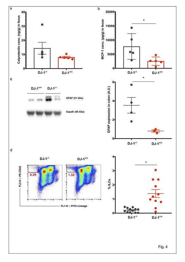
bioRxiv preprint first posted online Sep. 19, 2019; doi: http://dx.doi.org/10.1101/776005. The copyright holder for this preprint
 (which was not peer-reviewed) is the author/funder, who has granted bioRxiv a license to display the preprint in perpetuity.
                                    All rights reserved. No reuse allowed without permission.

     Abstract:
     The proper communication between gut and brain is pivotal for maintenance of health and
     dysregulation of the gut-brain axis can lead to several clinical disorders. Also, in Parkinson’s
     disease (PD) 85% of all patients experienced constipation long before showing any signs of
     motor phenotypes. For differential diagnosis and when it comes to preventive treatment there
     is an urgent need for the identification of biomarkers indicating early disease stages long before
     the disease phenotype manifests. DJ-1 is a chaperon protein involved in the protection against
     PD and genetic mutations in this protein have been shown to cause familial PD. However, how
     the deficiency of DJ-1 modifies the PD risk remains incompletely understood. In the present
     study we provide evidence that DJ-1 is implicated in shaping the gut microbiome including
     their metabolite production or innate immune cells (ILCs) development. We revealed that in 4
     months old mice genetic deficiency of DJ-1 leads to significantly decrease in several bacterial
     genera and significantly increase in two specific genera, namely Alistipes and Rikenella. DJ-1
     deficient mice have a higher production of calprotectin/MCP-1 inflammatory protein - a known
     protein involved in colonic inflammation – and significantly higher expression of glial fibrillary
     acidic protein (GFAP) than control littermates. Expression of a-Synuclein, a key protein in
     Lewy bodies, in the colon was not significantly different between genotypes. Metabolic profiles
     of feces extracts analysed by H1-NMR spectroscopy showed increased short chain fatty acids
     (SCFAs) and decreased amino acid levels, suggesting a general switch from protein towards
     fibre degrading strains in DJ-1 deficient mice. We observed that Malonate - which is known to
     influence the immune system – has significantly higher concentrations in DJ-1 deficient mice.
     Moreover, DJ-1 deficient mice have high levels of the phenol derivate 3-(3-Hydroxyphenyl)
     propanoic acid (3-HPPA) which is a breakdown product of aromatic substrates like tyrosine,
     phenylalanine and polyphenols. DJ-1 deficient mice also showed significantly reduced
     percentage of ILCs. Thus, our data suggests that absence of DJ-1 leads to increase in gut
     inflammatory bacteria composition, deregulated metabolites and dysregulated innate immunity
     which could be a key factor in the initiation of PD disease in the gut, and potentially also in
     brain during disease progression.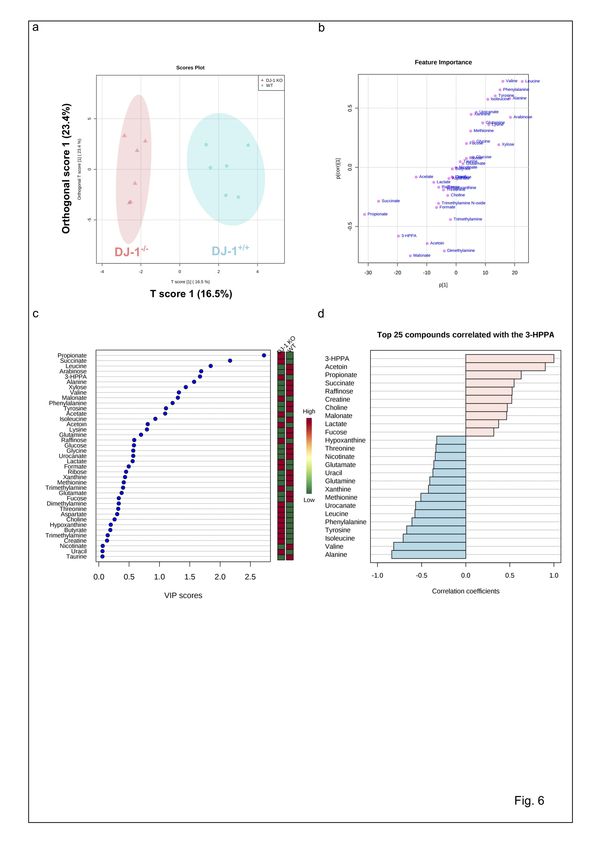
bioRxiv preprint first posted online Sep. 19, 2019; doi: http://dx.doi.org/10.1101/776005. The copyright holder for this preprint
 (which was not peer-reviewed) is the author/funder, who has granted bioRxiv a license to display the preprint in perpetuity.
                                    All rights reserved. No reuse allowed without permission.

     Introduction:

     Parkinson’s disease (PD) is the most common movement disorder and the second most
     prevalent neurodegenerative disease in humans (1). Clinically, PD patients suffer from resting
     tremor, rigidity, bradykinesia and altered gait (1). PD is an incurable neurodegenerative disease
     distinguished by the loss of neurons predominantly in the substantia nigra pars compacta
     (SNpc) region in the brain and the presence of Lewy bodies in the surviving neurons, however
     the exact cause of PD and how the disease process is triggered remained incompletely
     understood (2). Most of the PD cases are sporadic, however, several rare genetic forms of the
     disease have been identified that have contributed prominently to our understanding of the
     mechanisms underlying disease pathogenesis (3). In the Lewy bodies, presence of these
     intracellular protein inclusions is mainly comprised of misfolded alpha-Synuclein (α-Syn),
     which has also been shown to be genetically linked to familial and sporadic forms of PD (4, 5).

     In addition to α-Syn-related genetic links with PD, several other mutations such as PARK7 (DJ-
     1), Parkin, UCH-L1, Pink1 and dardarin genes account for sporadic cases with early-onset
     recessive PD (6, 7). DJ-1 is a small ubiquitously expressed protein implicated in several
     pathways associated with PD pathogenesis (3). DJ-1 protein is encoded by the PARK7 gene
     and comprises of 189 amino acid long (8). DJ-1 is localized primarily in the cytoplasm, however
     it can also be found in the nucleus and linked with mitochondria in the cell (8). DJ-1 is involved
     in several cellular functions, serving as an oxidative stress sensor (via a cysteine residue at
     position 106, C106), a protein chaperone, a protease, an RNA-binding protein, a transcription
     regulator, a regulator of mitochondria function and a regulator of autophagy (9). It is still not
     clear which of these processes are responsible for DJ-1-dependent pathogenesis in PD.

     Previous studies revealed that DJ-1 is involved in the modulation, aggregation and toxicity of
     α-Syn (8, 10-12). Double transgenic mice for DJ-1 deficiency and α-Syn expression (expressing
     pathogenic Ala53Thr human a-Syn) called M83-DJ-1 null mice, revealed that onset of disease
     and pathological changes were not different when compared with single transgenic M83 mice
     line and concluded that α-Syn and DJ-1 mutation may lead to PD via independent mechanisms
     (13). However, a recent in vitro study revealed that DJ-1 directly binds monomeric/oligomeric
     α-Syn and showed that DJ-1 interacts with α-Syn in living cells (8). Nevertheless,
     overexpression of DJ-1 protects against neurodegeneration in the yeast and Drosophila models
     in vivo (8). DA neurons number in the SNpc region and fibre densities and dopamine levels in
     the striatum were reported to be normal in DJ-1-/- mice. Enhanced striatal denervation and
     dopaminergic neuron loss was induced by 1-methy-4-phenyl-1,2,3,6-tetrahydropyridine
     (MPTP) together with amphetamine in DJ-1-/- mice (7). Additionally, DJ-1-/- mice, which were
     backcrossed with C57BL/6 mice (known as DJ-1C57-/-) suffered from early-onset unilateral
     loss of dopaminergic (DA) neurons in their SNpc, progressing to bilateral degeneration of the
     nigrostriatal axis with aging as well as mild motor behaviour deficits at aged time points (14).
     The authors suggested that the DJ1-C57 model effectively recapitulates the early stages of PD
     and allowing to study the preclinical aspects of neurodegeneration (14). Our own studies with
     DJ-1-/- mice models also suggested that DJ-1 protein is also involved in the maintenance the
     physiology of adaptive immune CD4+ T cells and their development and functions by regulating
     the sodium hydrogen exchanger 1 (NHE1) and ROS formation (15, 16). Keeping in mind that
     different DJ-1-/- mice lines with different background could prone to develop neurodegeneration
     and some are resistance then it is possible that in addition to genetic changes additional
     environment and unknown factors may play a crucial role for the disease development in DJ-1-
     /-
        mice. One of factor could be the gut microbiome which could be governing the resistant
     phenotype. Thus, studying the role of gut microbiome in the context of disease development in
bioRxiv preprint first posted online Sep. 19, 2019; doi: http://dx.doi.org/10.1101/776005. The copyright holder for this preprint
 (which was not peer-reviewed) is the author/funder, who has granted bioRxiv a license to display the preprint in perpetuity.
                                    All rights reserved. No reuse allowed without permission.

     DJ-1-/- mice model is warranted to understand the PD pathophysiology to develop novel tools
     for pre-clinical studies.

     Trillions of bacteria live and reside in the gut named jointly as the gut microbiome. They are
     important for normal functioning of the intestine (17). Recent advances in metagenomics
     techniques leads to step into this new world of research to uncover the profound impacts that
     the microbiota may have on neurodevelopment and diseased of the central nervous system
     (CNS) (18, 19). An essential function of the gastrointestinal tract (GIT) is to perceive and react
     to external signals such as environment, food, and xenobiotics (20). Studies from germ-free
     mice (GF) and antibiotic-treated mice suggested that bacteria are pivotal in hippocampal
     neurogenesis maintenance as well as spatial and object recognition (21). In mice, antibiotics
     treatment changed transiently the microbiota, and as a result increased expression of the brain-
     derived neurotropic factor (BDNF) in the hippocampus and enhanced exploratory behavior was
     observed (22). Further studies suggested that the microbiota promotes enteric and circulating
     serotonin (5-hydroxytryptamine, 5-HT) production from colonic enterochromaffin cells,
     modulates the GIT mobility, platelet functions (23), affects anxiety, hyperactivity, and
     cognition (24, 25). Dysbiosis (alterations to the microbial composition) of the human
     microbiome has not only been described in mice, but also in persons with several neurological
     diseases (26). For example, fecal and mucosa-associated gut microbes are different between
     individuals with PD and healthy controls (27-31). Thy1-α-Syn [ASO] transgenic PD mouse
     model study also suggested that PD derived microbiota have adverse effect on the Parkinson’s
     pathogenesis including the accumulation of α-Syn and change in the motor phenotype (32).
     Several other mouse studies also suggested the gut dysbiosis in chemically induced toxins and
     other PD models (32-38). Human studies from fecal metabolites suggested that PD patients
     have reduced short chain fatty acids (SCFAs), which are the metabolic products of certain gut
     bacteria (31). However, how bacterial produced gut metabolites are affecting the
     neurodegenerative diseases are not understood and an area of active research.

     Immune cells are capable of engaging in direct communication with enteric neurons (18, 20).
     The extent of the functional impact of neuro-immune synapses is not clear yet however
     published studies advocated that activated immune cells can temper neuronal activity via the
     release of neurotransmitters, metabolites and cytokines (19, 39, 40). Based on the common
     occurrence of GIT symptoms in PD, dysbiosis among PD patients, and evidence that the
     microbiota impacts CNS function, we hypothesized that DJ-1 protein could also be involved in
     the regulation of the gut microbiome and inflammation. Herein, we report that the microbiota
     composition is dysregulated, change in the innate immunity, increased inflammation in the
     colon and feces and dyregulated metabolites in the absence of DJ-1 in young adult mice.

     Results

     Gut microbiome dysbiosis (dysregulation of intestinal bacterial community signatures) in
     young DJ-1-/- mice

     Recent studies in PD patients have described the potential link with gut microbial abundance
     and Parkinson’s pathogenesis (28, 29, 31). Further findings suggested that the brain-gut axis
     interactions are controlled by the gut microbiome through immunological, neuroendocrine and
     direct neural mechanisms, respectively (19, 39). Therefore, a clear understanding of the
     microbiota-gut-brain axis interaction could bring a new insights in the pathophysiology of PD
     and allow an earlier diagnosis with a focus on peripheral biomarkers within the enteric nervous
     system (41). However, how the DJ-1 deficiency has any potential effects on the gut microbiome
     is not known yet. To understand this process in more detail, we used 16S rRNA sequencing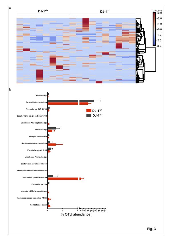
bioRxiv preprint first posted online Sep. 19, 2019; doi: http://dx.doi.org/10.1101/776005. The copyright holder for this preprint
(which was not peer-reviewed) is the author/funder, who has granted bioRxiv a license to display the preprint in perpetuity.
All rights reserved. No reuse allowed without permission.
method and characterized the gut microbiome from DJ-1 deficient (DJ-1 KO or DJ-1-/-) and
control littermate wild-type (WT) fecal samples from 4 months old animals. Both the WT and
DJ-1-/-, were kept in the same cage and animals were kept in 5-6 different cages to nullify the
cage effect as mice are coprophagic in nature. Heterozygous mothers were mated to obtain DJ-
1-/- and WT (DJ-1+/+) animals to minimize the effect of the maternal microbiome. Data analysis
of 16S rRNA sequencing reads were performed using the MEGAN-CE microbiome analyzer
software (42) as well as MicrobiomeAnalyst tool: a web-based tool for comprehensive
statistical, visual and meta-analysis of microbiome data (43) as described in the materials and
methods sections in detail.
Total number of reads were significantly less in DJ-1-/- samples compared with WT control
littermate samples (Fig. 1a). Our microbiome data at phylum level analysis suggested that 4
months old DJ-1 deficient mice tended to have higher alpha diversity Chao 1 and lower
Shannon-Weaver index: species richness within a single microbial ecosystem (44) of gut
microbiome compared with the control littermate WT, but it did not reach significance level
(Fig. 1b, c). Similarly, beta diversity: diversity in microbial community between different
environments (44) tended to be reduced (UniFrac: both weighted and Unweighted), a
difference, however, again not reaching statistical significance. DJ-1-/- mice have significantly
higher abundance of Bacteroidetes and significantly less abundance of Firmicutes and
Cyanobacteria compared with WT in an aged matched control littermate (Fig. 1d). Further,
when we mined the data for the overall composition of the gut bacterium at the phylum level,
we found that indeed several bacterial phyla tended to be different, but the difference again did
not reach statistical significance (Fig. 1e). As earlier studies suggested that
Firmicutes/Bacteroidetes (F/B) could help to predict the functionality of the microbiome, hence,
we calculated the F/B ratio and found that in DJ-1-/- mice the F/B ratio was statistically
significantly decreased when it was compared with control WT littermates (Fig. 1e).
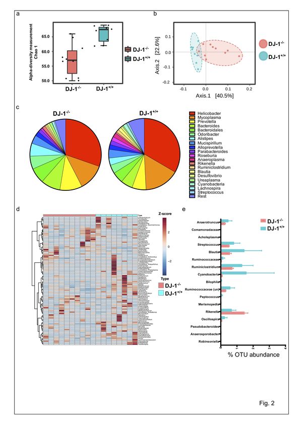
We further calculated the alpha and beta diversities at the genera level and found that both the
alpha diversity (Chao1 and Shannon-Weaver index) was significantly reduced in DJ-1-/- mice
compared with control littermate WT animals (Fig. 2a, b). Furthermore, analysis using Bray-
Curtis principal component analysis (PCoA) with MEGAN-CE and UniFrac to differentiate
between the two different mice lines (WT and DJ-1-/-) and found that indeed, at a younger age
both the animals were different in their bacteria abundance and clustering. Beta diversity was
also calculated using MicrobiomeAnalyst tool (43) (PERMANOVA). As a result, WT and DJ-
1-/- were different at both at the genus (p value=0.03) and species level (p value=0.006)
respectively. More than 1% bacterial genera were represented in a pie chart and clustering of
bacterial genera were shown as heat map (Fig. 2c, d). In total 17 bacterial genera were
significantly changed in the DJ-1 deficient animals (Fig. 2e). Most of the bacterial genera were
significantly downregulated (Anaerotruncus, Comamonadaceae, Acholeplasma,
Streptococcus, Blautia, Ruminococcaceae, Cyanobacteria, Bilophila, Ruminococcaceae-
uncultured, Peptococcus, Merismopedia, Oscillospira, Pseudobacteroides,
Anaerosporobacter, and Robinsoniella), only the opportunistic symbionts Rikenlla and
Alistipes were significantly upregulated (Fig. 2e). The abundance analysis at species levels
confirmed that both the pathogenic bacteria Alistipes sp and Rikenella sp were significantly
higher in DJ-1 deficient mice with the WT littermate controls (Fig. 3a, b). Although 16S rRNA
sequencing is not specific enough to characterize the bacterium at species/strain level, however,
it is sensitive enough to get an approximate idea which species could be present. Henceforth,
we mined our 16S rRNA data and found that indeed both unknown Rikenella sp. and Alistipes
timonesis were significantly upregulated. The bacterial Prevotella sp was also significantly
more abundant in DJ-1-/- mice.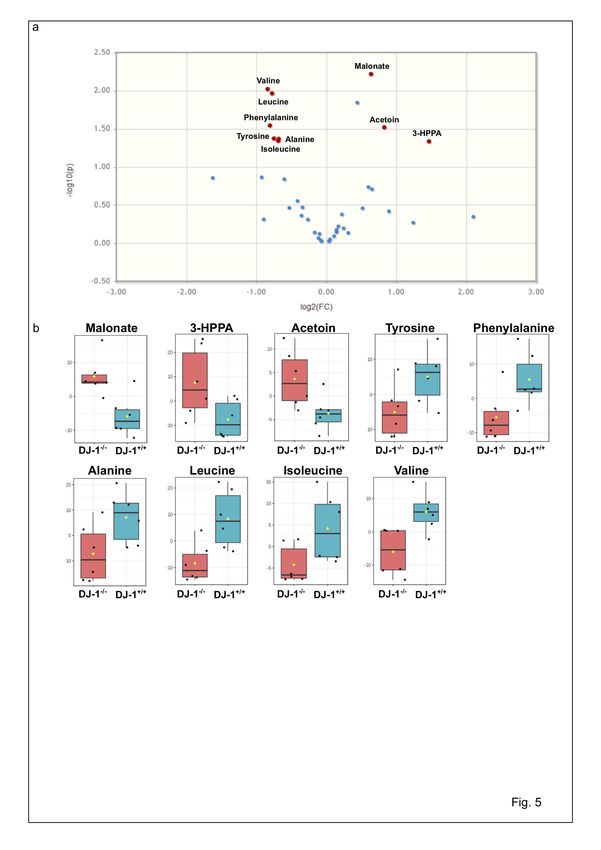
bioRxiv preprint first posted online Sep. 19, 2019; doi: http://dx.doi.org/10.1101/776005. The copyright holder for this preprint
 (which was not peer-reviewed) is the author/funder, who has granted bioRxiv a license to display the preprint in perpetuity.
                                    All rights reserved. No reuse allowed without permission.

     We next used the functional gene prediction profiling through Tax4Fun from our 16s rRNA
     data set (45) and output of these functional profile at KEGG metabolism level was visualized
     in SDP module of MicrobiomeAnalyst tool (43). The association analysis was performed using
     Global test algorithm between the two groups (DJ-1-/- and DJ-1+/+ mice). We observed that most
     of genes were predominantly associated with the pathways of amino acid, carbohydrate, energy,
     vitamins, cofactors and nucleotides metabolisms in WT and DJ-1 deficient mice (Suppl. Fig.
     1). Overall, our data of the 16S rRNA bacterial sequencing analysis suggested that genetic
     deficiency of DJ-1 affects the bacterial composition and functions in the gut, even before these
     animals develop any disease phenotype.

     Increased inflammation in the feces and colonic tissues, compromised innate immune system
     in DJ-1-/- mice

     The dysregulated immune system could lead to inflammation and increased permeability of the
     gut (46, 47). Thus, we further characterized the inflammatory protein calprotectin in the feces.
     Due to leukocytes shedding in the intestinal lumen, pro-inflammatory proteins such as
     calprotectin (S100A8/S100A9) can be detected and measured in the stool by ELISA (48). The
     concentration of calprotectin is directly proportional to the intensity of the neutrophil infiltrate
     in the gut mucosa. Calprotectin is released from neutrophils, monocytes, macrophages and
     epithelial cells in the case of the gastrointestinal tract inflammation (48); another study
     suggested that PD patients have higher expression of fecal calprotectin and zonulin (marker of
     increased gut permeability) proteins (47) and other study also suggested enhanced inflammation
     in PD patients (49). To validate whether increased gut dysbiosis in DJ-1-/- mice have higher
     inflammation in the gut or not, we measured the calprotectin levels. In DJ-1-/- mice calprotectin
     appeared to be higher than in WT, a difference, however, not reaching statistical significance
     (Fig. 4a). Moreover, we also measured other inflammatory cytokines in the feces and found
     that the monocyte chemotactic protein-1 (MCP-1) was significantly upregulated in DJ-1
     deficient mice compared with littermate WT control (p =0.034) (Fig. 4b). Other pro-
     inflammatory cytokines (IL-12p70, TNF-α, IL-17A, IFN-γ, IL-23 and IL-6) were tended to be
     upregulated in the feces from DJ-1-/- compared with WT mice, however, did not reach at
     significance level (Suppl. Fig. 2). The cytokines such as GM-CSF, IFN-β and IL-27 were
     significantly down regulated in DJ-1-/- mice compared to WT (Supp. Fig. 2). Additionally,
     previous studied suggested that pro-inflammatory cytokines are able to increase glial fibrillary
     acidic protein (GFAP) expression in enteric glia (50) and PD patients have also enhanced
     inflammation (49), therefore, we explored the quantification of GFAP from the colon tissue.
     Our Immunoblotting data suggested that DJ-1-/- colon have significantly higher GFAP protein
     compared with WT control littermate mice (Fig. 4c).

     Unwanted activation of the innate immune system from small intestinal bacterial overgrowth
     leads to increased intestinal permeability and may cause systemic inflammation (41). In the
     same perspective, unwanted activation of enteric neurons and enteric glial cells could follow
     the initiation of α-Syn accumulation and misfolding in the gut and the brain (51). In addition to
     the innate immune system, the adaptive immune system could also be affected and changed by
     bacterial proteins cross-reacting with host antigens and could lead to severe inflammation of
     the host tissues (52). Elevated α-Syn expression impairs innate immune cells function (53). Our
     previous studies suggested that DJ-1-/- mice have less induced regulatory T cells (Tregs) (16).
     We speculated that the defect could be in the adaptive immune cell development or functions
     in the DJ-1-/- mice. However, involvement of innate lymphoid cells (ILCs) in the pathogenesis
     of PD has not been described yet. To delineate the role of ILCs, we first characterized these
     cells using Flow cytometry and found that CD45+Lineage- (CD3, CD5, CD3, CD11b, CD11c,
bioRxiv preprint first posted online Sep. 19, 2019; doi: http://dx.doi.org/10.1101/776005. The copyright holder for this preprint
(which was not peer-reviewed) is the author/funder, who has granted bioRxiv a license to display the preprint in perpetuity.
All rights reserved. No reuse allowed without permission.
F4/80, Gr-1, B220 or CD19, and Ter119) were significantly less abundant in DJ-1-/- mice
compared with WT control littermates in the spleen (Fig. 4d).
Bacterial metabolites in the feces of DJ-1-/- mice are dysregulated
Our results suggested that DJ-1-/- mice suffer from gut dysbiosis and as a result could have
differences in their fecal metabolite production resulting in changed physiology and disease
outcome. Several previous studies have identified that gut bacteria control different metabolites
which are involved in neurodegenerative diseases (19, 54-56). Therefore, we used 1H-NMR
based metabolomics for the identification and quantification of fecal metabolites as described
elsewhere in detail (57, 58). The detected compounds cover a comprehensive range of
metabolite classes such as amino acids, SCFAs, phenols, amines, carbohydrates, purines,
alcohols and others.
Our extraction procedure yielded very rich and high-quality 1H-NMR spectra with final TSP
linewidths < 1 Hz. A total of 40 metabolites could be annotated and quantified in all samples,
mainly amino acids, short chain fatty acids, carbohydrates and nucleotides. The clustering and
non-clustering of all metabolites is shown in the heatmap (Supp. Fig. 3). Aside from known
metabolites we identified in half of the samples high levels of 3-(3-Hydroxyphenyl)propanoic
acid (3-HPPA) (CAS 621-54-5), a phenol derivative which has been shown to be able to readily
cross the gut epithelium (59) into the blood and brain (60). 3-HPPA has been shown to be
formed mainly by Clostridium, Escherichia and Eubacteria species (61, 62).
Using classical volcano plot analysis with a fold change threshold of 1.5 and student’s t-test we
identified 9 metabolites as highly discriminating the two groups (Fig. 5a) with Malonate (p =
0.006) being top for DJ-1-/- and Valine (p = 0.008) for WT (Fig. 5b). Further multivariate
statistics using orthogonal T-score (Partial Least Squares Discriminant Analysis: oPLS-DA)
was used to find out the similarity between DJ-1-/- and WT feces samples (Fig. 6a,b). Here, we
found a strong separation of both groups and the corresponding s-plot demonstrates that SCFAs
and amino acids are the most discriminating metabolites for clustering. Further, we applied
variable importance of projection (VIP) PLS-DA analysis to identify correlation patterns and
found again high significance of SCFAs, amino acids as well as carbohydrates and aromatic
compounds metabolism with VIPs-scores > 1 (Fig. 6c). Interestingly the high abundance of
SCFAs in DJ-1-/- shows a high correlation with the newly identified metabolite 3-HPPA (Fig.
6d and Suppl. Fig. 4). Thus, overall data suggested that DJ-1-/- mice have impaired protein
degradation -low levels of amino acids while fiber digestion was enhanced - high SCFAs
production and could potentially trigger immune response and gut epithelial dysbiosis.
Discussion
Over the past years, our understanding of human-associated microorganisms has been vastly
improved beyond that of a certain species toward an appreciation of the diverse and niche-
specialized microbial communities that develop in the human host (63). Our intestinal tract
especially the colon contains a largest reservoir of different microorganisms such as bacteria,
viruses, fungi etc. interacting with the host epithelial and immune cells. These microorganisms
orchestrate the primary and secondary metabolism on the gut mucosal surface and influence the
other body organs, mucosal and hematopoietic immune functions (63). Thus, it is not surprising
that modulation in the composition and function of the gut bacteria has been linked with several
chronic diseases including gastrointestinal inflammation, metabolic disorders as well as
cardiovascular and neurological diseases (41, 64-66). Recent discoveries in the field of the gut
microbiome and their role in the neurodegeneration in patients and animal models highlightsbioRxiv preprint first posted online Sep. 19, 2019; doi: http://dx.doi.org/10.1101/776005. The copyright holder for this preprint
(which was not peer-reviewed) is the author/funder, who has granted bioRxiv a license to display the preprint in perpetuity.
All rights reserved. No reuse allowed without permission.
an important link among each other (67). As most of the common neurodegenerative diseases
such as Alzheimer’s disease and PD occurs late in the life (40, 68) biomarkers allowing early
diagnosis would be invaluable.
Bacterial composition and dysbiosis
Animal models are useful tools to understand the pathophysiological functions of disease as
these models allow us to manipulate the course of the disease (69). To understand the impact
of the gut microbiome in the pathophysiology of PD in early life prior to development of any
phenotype or symptoms, we used DJ-1-/- animal models. The DJ-1 mutation has been found in
1-2% early-onset recessive PD and PD patients containing DJ-1 mutation are normally
responsive to levodopa (8). We found that two novel bacteria such as Alistipes and Rikenella
genera were highly abundant in DJ-1-/- mice compared with WT control littermate animals.
Both the alpha and beta diversity analysis suggested that both genotypes of animals have a
distinct microbiota diversity and community. Alistipes and Rikenella sp have been described
earlier to be involved in the inflammatory bowel disease as well as colon cancer development
(70). In humans (colorectal cancer patients) a number of Bacteroides and Parabacteroides
species, along with Alistipes putredinis, Bilophila wadsworthia, Lachnospiraceae bacterium
and Escherichia coli were enriched in carcinoma samples compared with both healthy and
advanced adenoma samples (71). Gut commensals such as Bifidobactium animalis and
Streptococcus thermophilus, on the other hand, decreased in faeces from adenoma or carcinoma
patients, consistent with deviation from a healthy microbiome (71). Alistipes and Ruminococcus
are shown to be positively correlated with TNF-α production after anti-IL-10R/CpG
oligonucleotide immunotherapy in C57BL/6 mice suffering from MC38 colon carcinoma (72).
Chondroitin Sulfate has been shown to increase the abundance of Rikenella, a genus of sulphate
reduced bacteria, but did not significantly change the abundance of Akkermansia muciniphila
(73). Surprisingly, Rikenella was exterminated by cephalosporin whereas Rikenella blossomed
on exposure to berberine. However, A. muciniphila was eliminated by this antibiotic
compound. Cephalosporin significantly reduced colonic mucus lesions and delayed the early
pathogenesis of dementia, steatohepatitis, and atherosclerosis. Berberine significantly
aggravated colonic mucus lesions and enhanced multi-systemic pathogenesis (73). Thus,
Rikenella could be involved in sulphate reduction in DJ-1-/- mice and could be a triggering
factor to induce colonic mucus lesions and PD disease progression.
Intestinal inflammation regulation by inflammatory bacteria and immune cells
Intestinal inflammation has been linked with gut permeability and induction of immune
response as well as function of gut neurons (19, 63). LPS challenged DJ-1-/- mice IFN-γ and
interferon-inducible T-cell α chemoattractant were enhanced in the SNpc and microglial cells
compared with WT animals (74). These results suggested that interaction of genetic defect with
inflammatory mediators could participate in the development of PD. Our data also suggested
that DJ-1-/- mice suffer from higher fecal inflammation as well as neuronal inflammation
(GFAP), which could be linked with neurodegeneration. However, the DJ-1-/- mice do not
develop any phenotype in unchallenged conditions. It is worth to note that the study was
performed at early stage before any neurological symptom develops in the brain. Recent studies
suggested that DJ-1C57-/- with a different genetic background (it is backcrossed 14 generations
with C57BL/6 mice), is more penetrant in disease phenotype development, as these animals
develop the phenotype within 3 months of age and progress to by 12 months (14). The exact
mechanism is unknown, but it could possibly be correlated with the genetic backgroung
composition in an interplay with the gut microbiome development. However, further studies,bioRxiv preprint first posted online Sep. 19, 2019; doi: http://dx.doi.org/10.1101/776005. The copyright holder for this preprint
(which was not peer-reviewed) is the author/funder, who has granted bioRxiv a license to display the preprint in perpetuity.
All rights reserved. No reuse allowed without permission.
in particularly also of the microbiome, are warranted to understand the disease penetration in
DJ-1C57-/- mice and DJ-1-/- animals. DJ-1-/- mice have no appreciable change in α-Syn
expression in the colon. Henceforth, absence of DJ-1 protein may trigger mechanisms which
are not dependent upon α-Synucleinopathy. Interestingly, we found that ILCs are reduced in
DJ-1-/- mice. Thus, innate immunity is severely compromised such as high abundance of
Alistipes and Rikenella. Previous studies suggested that Alistipes sp thrived in the absence of
lipocalin 2 and IL-10-/- mice and induced the intestinal inflammation and cancer progression in
those animals (70). Moreover, NOD2 or RIP2 deficiency resulted in pro-inflammatory
microenvironment that enhanced epithelial dysplasia following chemical injury and causes gut
dysbiosis probably due to higher abundance of Rikenella bacterium in these mice (75).
Interestingly, Rikenella was also shown to flourish in MyD88-deficient mice as these mice also
lacks suitable innate immune system (76). Gender-specific differences found in the immune
system and gut microbiome composition in males and females apparently fosters the expansion
of Alistipes, Rikenella and Porphyromonadaceae in the absence of innate immune defence
mechanism in male mice (77). These bacterial groups were linked with induction of weight
loss, inflammation and DNA damage upon transfer of the male microbiota to germ-free female
recipients (77). This study points out that it could be the case as DJ-1-/- mice have lower
numbers of ILCs and increased inflammation as well as higher abundance of Alistipes and
Rikenella.
Dysregulation of metabolites and neurodegeneration
The gut microbiome is a complex biological system and exhibits various tasks for the host
including digestion, degradation of macromolecules, vitamin production and educating of the
host innate and adaptive immune system (78). The gut microbiome composition affects the
health of host via bacterial metabolites including SCFAs, TMAO and other metabolites,
respectively (66). Bacterial metabolites appear to have diverse effects on metabolism and
immune response and are considered as biomarkers for disease risk factor, whereas bacterial
components cause an innate inflammatory response (66). Our metabolites data suggested that
amino acids including valine, leucine, phenylalanine, alanine, tyrosine, and isoleucine were
downregulated, whereas SCFAs including malonate, dimethyamine and Acetoin are
upregulated in DJ-1-/- mice. Defects in mitochondrial energy metabolism have been involved
in the pathology of several neurodegenerative diseases (79). Metabolites generated during the
bacterial metabolism and oxidation of the neurotransmitter dopamine (DA) are considered to
damage the neurons of the basal ganglia (79). Infusion of metabolic inhibitor malonate into the
striatum of mice or rats produced generation of DA nerve terminals and malonate induces a
substantial increase in DA efflux in awake, behaving mice as quantified by in vivo
microdialysis (79). Decreased SCFAs butyrate/propionate concentrations suggest loss of lactate
utilizing bacterial strains (80). In PD patients fecal SCFAs were reduced compared with healthy
controls (31). However, in DJ-1-/- mice SCFAs were similar to those of WT mice. Furthermore,
mitochondrial respiratory complex II (CII) is a protein complex located in the inner membrane
of mitochondria and it forms part of the electron transport chain and is involved in succinate
signalling and reactive oxygen species (ROS) (81). Malonate is a competitive inhibitor of the
CII and reduces the cellular respiration, whereas succinate is rather drives the CII activity in
macrophages (82, 83). Thus, metabolic stress induced by malonate, dimethyamine and acetoin
could be involved in the neurodegenerative process in DJ-1-/- mice potentially to be generated
from bacterial metabolism or colon tissues.
The majority of the amino acids in the intestines derive from the metabolism of ingested dietary
proteins, host tissue proteins or the conversion of other nitrogenous substances, whereas a small
amount of amino acids is de novo synthesized by the gut bacteria (84). Amino acids, such as L-bioRxiv preprint first posted online Sep. 19, 2019; doi: http://dx.doi.org/10.1101/776005. The copyright holder for this preprint
(which was not peer-reviewed) is the author/funder, who has granted bioRxiv a license to display the preprint in perpetuity.
All rights reserved. No reuse allowed without permission.
glutamine function as a double-edged sword for gut health as they can sponsor to the expression
of pathogenic virulence genes as well as protect against disease (84). Most of the amino acids
including valine, leucine, isoleucine and alanine were downregulated in the feces of DJ-1-/-
mice. Our results corroborate findings on PD patients samples derived from cerebrospinal fluid
(CSF) for the amino acids including valine, leucine, isoleucine (85). PD patients might suffer
from dysfunction of the transport of neutral and basic amino acids across the blood-brain
barriers (85). Changes in amino acid metabolism in plasma/CSF of the patients, highlighting
the role of altered amino acid metabolism and PD pathology (86). Decreased amino acid
concentrations could be involved in loss of microbial proteases/peptide catabolism (80).
Apparently, there is a switch from protein to fibre utilization under DJ-1 deficient conditions
(87). PD patients who are not treated with levodopa or with dopamine agonists were reported
to have higher CSF tyrosine and phenylalanine levels than those not treated with these drugs
and also than controls (85). In this study, we found less tyrosine and phenylalanine in the feces
of DJ-1-/- mice compared with WT control group. Aromatic amino acids tyrosine and
phenylalanine could be the most important amino acids as they are involved in L-DOPA
synthesis by tyrosine hydroxylase (TH), which is further converted into dopamine,
norepinephrine (noradrenaline), and epinephrine (adrenaline) (88). It is shown that 3-HPPA is
formed through fermentation of tyrosine by Clostridium species (61, 62). Reduced levels of
microbial tyrosine and phenylalanine point out that decreased production of dopamine
precursors while enhanced levels of 3-HPPA which are able to cross into the brain might have
a directive effect on dopamine synthesis e.g. as competitive inhibitor. However, further studies
are warranted to understand the role of amino acid metabolism by gut bacteria in PD
pathophysiology.
Conclusion:
Our study suggests that DJ-1-/- mice present with gut dysbiosis, reduced innate lymphoid cells
numbers or development, increased inflammation in the colon and feces, and dysregulated
metabolites production already at an early disease stage (4 months) which could be toxic to
colon tissues or neurons. Therefore, these disease markers could subsequently be explored for
biomarker development in the PD patients.
Material and Methods:
Animal breeding and ethical permission and materials collection
DJ-1-/- mice were described earlier (7) and obtained from Prof. Tak W Mak, Canada. As
described earlier, F1 progeny were backcrossed for seven generation to C57BL/6 mice and
heterozygous animals (male and female) were used to set up the breeding to obtain homozygous
for the targeted DJ-1 allele. Genotypes of mice was performed using PCR (WT DJ-1 forward
primer, TGC TGA AAC TCT GCC ATG TGA ACC; WT DJ-1 reverse primer, CCT GCT TGC
CGA ATA TCA T; and Neo, AGG TGA CAC TGC CAG TTG CTA GTC). PCR conditions
were used as follow: 95°C for 30 sec, 64°C for 30 sec, and 72°C for 1 min (40 cycles). Age
matched 3-4 months old DJ-1 deficient mice and control WT littermates from the same cohort
were used for the feces collection for 16S rRNA microbiome analysis and stored at -80 0C until
use. Some animals were sacrificed using CO2 methods and colons (3-4 cm long piece from at
the junction of ceacum) and were collected in 4% PFA and snap frozen in liquid N2. All the
animals were kept in the open cage in a standard environment mice facility. All the experiments
were performed according to the EU Animals Scientific Procedures Act and the German law
for the welfare of the animals. All the procedure and methods were approved by the localbioRxiv preprint first posted online Sep. 19, 2019; doi: http://dx.doi.org/10.1101/776005. The copyright holder for this preprint
(which was not peer-reviewed) is the author/funder, who has granted bioRxiv a license to display the preprint in perpetuity.
All rights reserved. No reuse allowed without permission.
government authorities (Regierungspräsidium, Tübingen according to §4 animal welfare act on
20/07/2017) of the state of Baden-Württemberg, Germany.
Bacterial DNA isolation from the feces
Frozen feces were weighted (50mg/sample) on dry ice and hammered to break into powder
form and transferred into a 2.0 ml Eppendorf tube and kept on dry ice. Once all the samples
were measured, all the weighted sample tubes were transferred to ice until the samples were
thawed and added 1.0 ml of lysis buffer from QIAamp Fast DNA Stool Mini Kit (Cat no.
#51604; Qiagen, Germany). All the procedures were followed as recommended by
manufacturer’s guidelines for bacterial DNA isolation and DNA was dissolved in 100 µl instead
of 200 µl DNA buffer.
16S rRNA sequencing
For 16S rRNA amplification, 12.5 ng of DNA was amplified using 0.2 μM of both forward
primer
(TCGTCGGCAGCGTCAGATGTGTATAAGAGACAGCCTACGGGNGGCWGCAG,
Metabion)
and reverse primer
(GTCTCGTGGGCTCGGAGATGTGTATAAGAGACAGGACTACHVGGGTATCTAATC
C, Metabion).
KAPA HiFi HotStart Ready Mix (KK2601; KAPABiosystems) was used for the PCR
amplification. PCR was performed using a first denaturation of 95°C for 3 minutes (min),
followed by 25 cycles of amplification at 95°C for 30 s, 55°C for 30 s and 72°C for 30s, final
elongation at 72°C for 5 min and the amplified DNA was stored at 4°C. DNA gel
electrophoresis of all the samples was performed to verify the amplicon specificity.
Further, samples were then purified (Agencourt AMPure XP, Beckman Coulter) and PCR
amplicons were indexed using Nextera XT index and KAPA HiFi HotStart ReadyMix. PCR
was performed using a first denaturation of 95°C for 3 min, followed by 8 cycles of
amplification at 95°C for 30 s, 55°C for 30 s and 72°C for 30, final elongation at 72°C for 5
min. Samples purified were then validated using BioAnalyzer (Bioanalyzer DNA 1000,
Agilent) and 4 nM of each library pooled using unique indices before sequencing on a MiSeq
(Illumina) and paired 300-bp reads.
Sequence Analysis and Statistics
Available sequence data has been trimmed and filtered using SeqPurge (89). Trimming
parameters demanded a minimum quality at 3’ end of q=35 (parameter qcut=35). Processed
sequence data has been aligned using MALT (version 0.3.8; https://ab.inf.uni-
tuebingen.de/software/malt) against the 16S database SILVA SSU Ref Nr 99 (https://www.arb-
silva.de/documentation/release-128/) and classified using NCBI taxonomy. Alignment has
been performed using semi-global alignment and a minimum sequence identity of 90%
(parameter minPercentIdentity=90). Further analysis and visualization were performed using
MEtaGenome Analyzer-Community Edition (MEGAN-CE) version 6.14.2, built 23 Jan 2019
(42) as describer earlier (90).
Preparation of tissue for histological analysisbioRxiv preprint first posted online Sep. 19, 2019; doi: http://dx.doi.org/10.1101/776005. The copyright holder for this preprint
(which was not peer-reviewed) is the author/funder, who has granted bioRxiv a license to display the preprint in perpetuity.
All rights reserved. No reuse allowed without permission.
Colon samples prepared on ice were fixed for 24 h in 4% paraformaldehyde (PFA), stored at
4°C in 0.4% PFA for a maximum of 4 weeks prior embedding in paraffin. Fixed samples were
then alcohol-dehydrated and embedded in paraffin. Samples were embedded in paraffin blocks
using a tissue embedding station and stored at room temperature until use. Paraffin blocks
containing colon tissues were cut into 7 μm thick sections using a microtome. Section were
placed in 45°C water bath for flattening, collected on a glass slide, dried in an incubator at 50°C
for 1-2 h and stored at room temperature.
Immunohistochemistry
First slides were deparaffinize using autostained program 6 for a run for 51 minutes and slides
were kept in TBS until antigen retrieval step. Antigen retrieval was done using sodium citrate
buffer (1x) and citric acid (1x) method. Slides were boiled in the citric acid + sodium citrate
buffer for 5 minutes for 3 times (3x) and slides were allowed to cool down for 15 minutes in
TBS. After cooling slides were blocked of endogenous peroxidase and incubated for 20 minutes
at room temperature (RT) and washed quickly 3x with TBS. Further slides were blocked for
unspecific bindings using 5% normal goat serum in 0.3% Triton X-100 TBS and incubate for
1 hour at RT on slow rotation shaker. After incubation with unspecific binding, slides were
washed for 3x3 minutes with TBS. Antibody staining was performed using α-Synuclein
antibody (# 610786 BD Biosciences, Netherlands) for overnight (1:1000) dilution at 4 0C.
Slides were washed next day with 0.025% Triton X-100 TBS for 3x5minutes. Secondary
antibody was used (1:1000) dilution for 1 hour at RT. Further slides were stained with ABC
complex-DAB for detection of primary antibody staining.
Tissue lysate preparation for WB
Colon tissue were weighted frozen and lysed with 10 volumes of RIPA buffer (50 mM Tris,
150 mM NaCl, 1.0% NP-40, 0.5% sodium deoxycholate, 0.1% SDS, pH 8.0) supplemented
with protease inhibitor (Complete; Roche Diagnostics). Colon tissues were homogenized for 1
minute using a disperser (T10 ULTRA-TURRAX; VWR) on ice. After the homogenization,
samples were incubated for 30 min at 4°C and spun for 20 min at 12,000 g at 4 0C. Proteins
lysate supernatants were supplemented with 10% glycerol before long storage at -80°C. Protein
concentration was determined using BCA method (#23225; Thermofisher, Germany).
Immunodetection
Samples were prepared by diluting protein lysates in PAGE buffer (0.2 M glycine, 25 mM Tris,
1% SDS), followed by a denaturation at 95°C for 10 min in loading buffer (80 mM Tris, 2%
SDS, 5% 2-mercaptoethanol, 10% glycerol, 0.005% bromophenol blue, pH 6.8) and a short
centrifugation 30 sec at 400 g. Proteins were separated by electrophoresis using 12% SDS-
PAGE gel. Gels containing proteins were washed for 5 minutes in transfer buffer (0.2 M
glycine, 25 mM Tris, 10–20% methanol) and transferred to membranes equilibrated in transfer
buffer. Transfer was performed for 90 min at 80 V at 4°C on nitrocellulose membranes (88018,
Life Technology). Immunoblot were washed 5 min in TBS buffer, fixed with 4% PFA for 1
hour (only for a-Syn detection) and blocked using 5% non-fat milk (Slim Fast) in TBS.
Membranes were then washed twice 5 min in TBST and incubated with the primary antibody
over night at 4°C (human and mouse a-syn: 610786 BD Biosciences; GAPDH: #5174, Cell
Signaling and GFAP #MAB360, Merck Millipore, Germany). After incubation with the first
antibody, membranes were washed four times (5 min each) with TBST. Membranes were then
incubated for 75 min with the secondary antibody coupled to horseradish peroxidase (GE
Healthcare). After four washing steps with TBST (5 min each), bands were visualized using thebioRxiv preprint first posted online Sep. 19, 2019; doi: http://dx.doi.org/10.1101/776005. The copyright holder for this preprint
(which was not peer-reviewed) is the author/funder, who has granted bioRxiv a license to display the preprint in perpetuity.
All rights reserved. No reuse allowed without permission.
enhanced chemiluminescence method (ECL+; GE Healthcare). Light signal was detected using
LI-COR Odyssey and were quantified using Odyssey software.
Calprotectin ELISA and LEGENplex inflammatory cytokines
To measure the calprotectin/MRP 8/14 from the fecal samples, S100A8/S100A9 ELISA kit
rat/mice (#K6936, Immundiagnostik AG, Germany) as well as LEGENplex™ (#740150 or
#740446, Biolegend, Germany) mouse inflammation panel (13-plex) were used according to
the manufacture’s guidelines. Fecal samples were measured (weight between 50±5.0 mg) and
dissolved in 500 µl of extraction buffer supplied by the kit, mixed by vortexing and then
centrifuged for 10 minutes at 3000 g. Supernatant was taken and transferred to a new
microcentrifuge tube and 100 µl of sample was used for measuring the protein for Calprotectin,
however, for LEGENDplex inflammatory panel 25 µl of sample was used respectively. The
data was analyzed using 4 parameters algorithm and the concentration of
calprotectin/inflammatory cytokines was normalized with feces weight and data are presented
in ng/g or pg/g.
Flow cytometry
Splenocytes from WT and DJ-1-deficient mice were characterised by using surface and
intracellular staining with relevant antibodies. In brief, splenocytes were collected and used for
surface staining for dump-FITC lineage markers - CD3, CD5, CD3, CD11b, CD11c, F4/80, Gr-
1, B220 or CD19, and Ter119 and CD45-PE (eBioscience, Germany) washed with PBS. Cells
were fixed with Foxp3 fixation/permeabilization buffer (eBioscience, Germany) for
intracellular staining and incubated for 30 minutes. After incubation, cells were washed with
1x permeabilization buffer, exposed to added intracellular monoclonal antibodies for RoRgt-
PerCP-Cy5.5, Eomes-APC, Tbet-PerCP-Cy5.5 and GATA3-APC and incubated for an
additional 45 minutes. Cells were washed again with permeabilization buffer and PBS was
added to acquire the cells on a flow cytometer (FACS-calibur™ from Becton Dickinson;
Heidelberg, Germany).
Feces sample prep for the metabolite detection using 1H-NMR
For metabolite extraction, 50 mg of deep-frozen feces sample were transferred into 2 mL AFA
glass tubes (Covaris Inc, Woburn, USA) and mixed with 400 µL of ultrapure methanol and 800
µL of MTBE (solvent grade). The mixture was manually dispersed with a disposable plastic
spatula, then vortexed and transferred to a focused ultrasonicator (Covaris E220evolution,
Woburn, USA). Feces metabolites were extracted with a 5 min lasting ultrasonication program
in a degassed water bath at 7° C. After extraction, the metabolite suspension was separated into
a polar and lipid phase by adding 400 µL of ultrapure water. In order to remove and remaining
solids from the samples, the glass tubes were centrifuged for 5 min at 4000 rpm. 700 µL of each
phase were then transferred to a fresh 1.5 mL Eppendorf cup. The polar phase was subject to a
2nd centrifugation step for 5 min at 12,000 rpm and 600 µL of the supernatant were transferred
to a new 1.5 mL Eppendorf cup and evaporated to dryness over night with a vacuum
concentrator (Eppendorf Speedvac).
1
H-NMR for metabolites and data analysis
For NMR analysis, the dried pellets were resuspended with 60 µL of deuterated phosphate
buffer (200 mM K2HPO4, 200 µM NaN3, pH 7.4) containing 1 mM of the internal standard
TSP (trimethylsilylpropanoic acid). In order for a maximum dissolution, the plastic tubes werebioRxiv preprint first posted online Sep. 19, 2019; doi: http://dx.doi.org/10.1101/776005. The copyright holder for this preprint
(which was not peer-reviewed) is the author/funder, who has granted bioRxiv a license to display the preprint in perpetuity.
All rights reserved. No reuse allowed without permission.
quickly sonicated and then centrifuged 5 min at 14,000 rpm. 50 µL of the supernatant were
transferred with gel loading pipette tips into 1.7 mm NMR tubes (Bruker BioSpin, Karlsruhe,
Germany) and a 96 well rack placed into the cooled (4° C) NMR autosampler.
Spectra were recorded on a 600 MHz ultra-shielded NMR spectrometer (Avance III, Bruker
BioSpin GmbH) equipped with a triple resonance (1H, 13C, 31P) 1.7 mm room temperature
probe at 298 K. For optimum water suppression and shim adjustment a quick simple ZG
experiment was performed followed by a 1h lasting CPMG (Carr-Purcell-Meiboom-Gill)
experiment in order to suppress residual background signals from macromolecules such as
bilirubin (time domain = 64k points, sweep width = 20 ppm, 512 scans). The recorded free
induction decays (FIDs) were fourier-transformed and spectra properly phase- and baseline
corrected. Metabolite annotation and quantification was performed with ChenomX NMR Suite
8.3 and statistical analysis with MetaboAnalyst 4.0.
The phenol derivative 3-(3-Hydroxyphenyl) propanoic acid 3-HPPA was identified by selective
TOCSY experiments followed by purchasing possibly fitting reference standards of different
phenols with substituted groups in the meta position. Reference spectra of those were recorded
in the used feces extract phosphate buffer and by applying spiking experiments.
Statistical analysis
MEGAN-CE (version 6.14.2, built 23 Jan 2019) and MicrobiomeAnalyst were used for data
acquisition and analysis. GraphPad and Inkscape were used for the final figure preparation.
One-way ANOVA or Student’s t-test was used for statistical analysis using GraphPad wherever
it was appropriate and described in the figure legend. Data shown in either Means±SD or SEM.
The p value (≤0.05) considered significant. Metabolite concentrations from 1H-NMR analysis
were exported as comma separated value spreadsheet file to MetabAnalyst, normalized with
PQN (probabilistic quantile normalization) and range scaled and then analyzed with student’s
t-test, oPLSDA and VIP analysis.
Fig. legends
Fig. 1: Gut dysbiosis is prevalent in four months old DJ-1-/- mice.
(a) Total number of sequences read obtained from sequencing of colon fecal samples from DJ-
1+/+ (n=8) and DJ-1-/- (n=10) mice. (b) Measurement of alpha diversities (Chao1 and Shannon-
Weaver index) for WT and DJ-1-/- mice. (c) Bacteroidetes, Firmicutes and Cyanobacteria were
significantly different between WT and DJ-1-/- mice. (d) Bacterial abundance data (mean±SD)
presentation at phylum level in WT and DJ-1-/- mice. (e) Firmicutes/Bacteroidetes ratio in WT
and DJ-1-/- mice, it was significantly reduced in DJ-1-/- mice compared with WT. (f)
Firmiuctes/Bacteroiidetes (F/B) ratio was decreased in DJ-1-/- compared with WT mice. *
represents the p value ofbioRxiv preprint first posted online Sep. 19, 2019; doi: http://dx.doi.org/10.1101/776005. The copyright holder for this preprint
(which was not peer-reviewed) is the author/funder, who has granted bioRxiv a license to display the preprint in perpetuity.
All rights reserved. No reuse allowed without permission.
Fig. 3: Significant increase in Rikenella sp. and Alistipes timonensis in DJ-1-/- mice compared
with WT.
(a) Clustering of all bacteria at species level. (b) Bar diagram (mean±SD) shows all the
significantly different bacteria at species level in WT and DJ-1-/- mice. *represents the p value
ofbioRxiv preprint first posted online Sep. 19, 2019; doi: http://dx.doi.org/10.1101/776005. The copyright holder for this preprint
(which was not peer-reviewed) is the author/funder, who has granted bioRxiv a license to display the preprint in perpetuity.
All rights reserved. No reuse allowed without permission.
organization Deutschland, Bundesministerium für Bildung und Forschung (BMBF, FKZ). This
research was supported by the Deutsche Forschungsgemeinschaft, DFG through the funding of
the NGS Competent Centre Tübingen (NCCT-DGF). We thank Prof Bernd Pichler for allowing
to have access to the 1H-NMR for metabolic studies. Funders have no role in the study design
and data analysis. We acknowledge support by Deutsche Forschungsgemeinschaft and Open
Access Publishing Fund of University of Tübingen.
Ethics statement
All animal experimental protocols used in this study is strictly adhered to the international
standards for the care and use of laboratory animals and were approved by the local Animal
Welfare and Ethics committee of the Country Commission Tübingen, state of Baden-
Württemberg, Germany (§4 animal welfare act on 20/07/2017).
Author’s role
YS: Study design, performed the research and managed the overall project, involved in entire
study, analysed the data, made the figures and wrote the manuscript
CT: Study design, performed the metabolites study, data analysis, made the figures, wrote the
manuscript
AD, JM: Helped with 16S rRNA data analysis bioinformatics meta data analysis and made the
figures
MA, MSS: study design, performed the research, helped with animal experiments
LZ: Helped with the identification of 3-HPPA, did reference standards and spiking experiments
LP: performed the animal experiments
MF, MF: Provided/tools provided for the study, performed and help with the animal
experiments
VP: Helped with study design and data analysis
DSP, TWM: Materials/tools provided for the study
JSF: Helped with study design and discussion of the 16S rRNA data
NC: Study design, substantial discussion for 16S rRNA data
MSS, DW, SYB, FL, OR: Study design, funding generation, wrote the manuscript
All authors read the manuscript and approved to be co-authors on the manuscript and have
substantial contribution in the manuscript.
Reference:
1. L. M. L. de Lau, M. M. B. Breteler, Epidemiology of Parkinson's disease. The Lancet
Neurology 5, 525-535 (2006).
2. L. V. Kalia, A. E. Lang, Parkinson's disease. The Lancet 386, 896-912 (2015).
3. D. G. Hernandez, X. Reed, A. B. Singleton, Genetics in Parkinson disease: Mendelian
versus non-Mendelian inheritance. J Neurochem 139 Suppl 1, 59-74 (2016).
4. S. M. Spillantini MG, Lee VM, Trojanowski JQ, Jakes R, Goedert M., Alpha-synuclein in
Lewy bodies. Nature 28, 839-840 (1997).
5. R. A. C. Maria Grazia Spillantini, Ross Jakes, Masato Hasegawa, and Michel Goedert, α-
Synuclein in filamentous inclusions of Lewy bodies from Parkinson’s disease and
dementia with Lewy bodies. PNAS 95, 6469-6473 (1998).
6. V. Bonifati, Autosomal recessive parkinsonism. Parkinsonism & Related Disorders 18,
S4-S6 (2012).You can also read