Neoadjuvant Chemotherapy for Breast Cancer: Moving Beyond Pathological Complete Response in the Molecular Age - Acta Medica ...
←
→
Page content transcription
If your browser does not render page correctly, please read the page content below
Clinical Science
Narrative Review
Acta Medica Academica 2021;50(x):xx-xx
DOI: 10.5644/ama2006-124.XX
Neoadjuvant Chemotherapy for Breast Cancer: Moving Beyond Pathological
Complete Response in the Molecular Age
Elena Provenzano
Department of Histopathology, Addenbrookes Hospital, Cambridge, UK
Correspondence: elena.provenzano@addenbrookes.nhs.uk; Tel.: + 44 1223 256154; Fax.: + 44 1223 257003
Received: 14 January 2021; Accepted: 2 February 2021
Abstract
This review focuses on neoadjuvant chemotherapy for breast cancer which introduces practical issues for pathologists, including
predicting response, optimising specimen handling, size measurement and assessment of residual disease, and recent advances
in management of the axilla. The role of neoadjuvant chemotherapy in breast cancer is increasing, and it has become standard
of care for high risk Human Epidermal Growth Factor Receptor 2 positive and triple negative breast cancers. The benefits of the
neoadjuvant approach extend beyond pathological complete response to tumour downstaging permitting conservative surgi-
cal options in the breast and axilla, and assessment of response provides valuable prognostic information to enable escalation
and de-escalation of adjuvant therapy to optimise oncological outcomes. Hence histopathologists play a vital role in patient
management in the neoadjuvant setting. Optimal patient selection for neoadjuvant chemotherapy requires consideration of pre-
treatment histopathological and molecular tumour characteristics. Post chemotherapy, tumour staging can be challenging, and
changes in criteria for measurement of primary tumour and metastases in the 7th and 8th editions of the TNM have led to confu-
sion amongst pathologists. This review offers practical guidance on specimen handling and measurement of lesion size. Mov-
ing forwards more detailed information on degree of response will be required for adjuvant therapy decision making, and the
Residual Cancer Burden is emerging as the preferred method for quantifying residual disease not just within clinical trials but
in routine practice. Recent advances in management of the axilla are discussed, including the significance of minimal residual
disease in the form of isolated tumour cells and micrometastases which portend a worse prognosis in the neoadjuvant setting.
Conclusion. Neoadjuvant chemotherapy now forms part of routine breast cancer management, and detailed histopathologi-
cal assessment and an understanding of the importance of molecular tumour biology is essential for clinical decision making.
Key Words: Breast Cancer Neoadjuvant Therapy Staging Grading Response.
Introduction the potential for breast conservation surgery (BCS)
in patients that would have required mastectomy
Neoadjuvant chemotherapy (NACT) has evolved
pre-treatment (2). There is also a growing body of
from treatment of locally advanced breast cancer to
evidence to support the role of sentinel node biop-
routine management of biologically aggressive dis-
sy (SLNB) following NACT in both node negative
ease, particularly oestrogen receptor negative (ER-
) and/ or human epidermal growth factor 2 posi- and node positive patients, leading to avoidance
tive (HER2+) cancers. The neoadjuvant approach of axillary clearance (ALND) following a com-
shows similar survival outcomes to adjuvant thera- plete response in the axilla (3-5). Interestingly, in
py, but offers potential advantages in both standard our own multidisciplinary meetings, it is now the
clinical care and clinical trial settings (1). Firstly, surgeons rather than the oncologists driving deci-
response to neoadjuvant therapy with complete sions regarding NACT. Tumour downstaging to
eradication of disease or a reduction of tumour vol- enable conservative procedures can reduce surgical
ume enables less aggressive surgical options, with morbidity without compromising oncological out-
Copyright © 2021 by the Academy of Sciences and Arts of Bosnia and Herzegovina.
Acta Medica Academica 2021;50(x):xx-xx

comes, however NACT is not the correct approach            Tumour staging post NACT also shows a strong
for all cases and careful patient selection based on   association with survival outcomes (16, 17). Mea-
clinical features and histological and molecular tu-   suring residual tumour size can be challenging,
mour subtypes is essential to optimise results.        particularly when there has been patchy response
    Perhaps even more importantly, assessment of       across the tumour bed. Definitions of size mea-
response to NACT provides valuable prognostic          surements used for staging in both the breast and
information that is increasingly used to guide fur-    axillary lymph nodes have evolved across the 6th, 7th
ther adjuvant therapy (6). Complete pathological       and 8th editions of the TNM, leading to confusion
response (pCR) shows an association with survival      amongst pathologists (18-20). Accurate staging is
outcomes across all molecular subtypes, although       essential not only in determining patient progno-
this is strongest for ER- and/or HER2+ disease (1,     sis, but to generate reliable population based data
7). As a result, pCR has been approved by the U.S.     from cancer registries around the world.
Food and Drug Administration (FDA) as a sur-               Hence, the pathologist plays a key role in deter-
rogate outcome to survival for neoadjuvant clini-      mining optimal patient care in the neoadjuvant set-
cal trials in high risk breast cancer (8). The neo-    ting. This review will focus on some of the key prac-
adjuvant context provides faster results in smaller    tical issues for pathologists, including predictors of
cohorts of patients, and alongside novel adaptive      response, optimising specimen handling, size mea-
trial designs such as ISPY provides exciting poten-    surement and assessment of residual disease, and
tial to screen new agents resulting in more rapid      recent advances in management of the axilla.
introduction of effective drugs into clinical prac-
tice (9, 10). Furthermore, patients who experience
a pCR may not benefit from further adjuvant ther-      Predictors of Response to NACT
apy, and there are trials looking at de-escalation     Response to NACT, including the likelihood of
of adjuvant therapy in complete responders (11).       achieving pCR and its association with progno-
Hence accurate identification of pCR is vital for      sis, is strongly linked to tumour biology (1, 21-
ongoing patient management, and requires careful       25). This has important implications for clinical
and methodical histological assessment beginning       decisions regarding whether to give neoadjuvant
with gross specimen handling.                          versus adjuvant therapy, particularly if the goal is
    At the other end of the spectrum, patients who     tumour downsizing to enable conservation. Breast
show a limited response to NACT have a poor            cancer is generally divided into 3 broad molecu-
prognosis. Recent trials, including the KATHER-        lar groups; luminal (ER+/HER2-), HER2+ and
INE and CREATE-X trials, have shown improved           TNBC (25). HER2+ and TNBC show the greatest
survival outcomes with additional adjuvant ther-       response to NACT, but even these tumour types
apy in incomplete responders with HER2+ and            contain subgroups with different behaviour.
triple negative breast cancer (TNBC) respectively          NACT response of HER2+ breast cancers large-
(12, 13). However, non-pCR encompasses a wide          ly depends on ER status. ER+/HER2+ cancers giv-
variation of response from almost complete re-         en standard chemotherapy without HER2 targeted
sponse with minimal residual disease (MRD), to         agents show a pCR rate of 18%, rising to 31% with
minimal or absent response with significant re-        the addition of trastuzumab (1). In contrast, ER-/
sidual tumour. Some series have shown similar          HER2+ tumours have a much higher pCR rate of
survival outcomes for patients with MRD to those       30% without trastuzumab and 50% with trastu-
who undergo a pCR, however the impact of resid-        zumab; the addition of pertuzumab gives pCR rates
ual disease volume on survival outcomes varies by      as high as 80% (26). The association between pCR
molecular tumour subtype (14, 15). Assessment of       and survival outcomes is also much stronger for
the degree of response beyond pCR will form an         ER-/HER2+ cancers (HR 0.29; 95% CI 0.17-0.50)
integral part of patient care moving forwards.         without trastuzumab and HR 0.08(95% CI 0.03-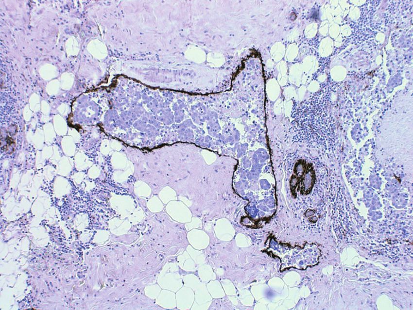
Elena Provenzano: Breast Cancer Neoadjuvant Therapy Beyond pCR

0.22) with trastuzumab) than for ER+/HER2+             study found androgen receptor (AR) positivity
cancers where it does not reach significance (HR       was associated with improved response to NACT
0.57(95% CI 0.31-1.04) without trastuzumab and         with trastuzumab, and better survival outcomes in
HR 0.56 (95% CI 0.23-1.37) with trastuzumab).          ER- disease (36). Other tumour features that have
ER+/HER2+ tumours also show a different pattern        been associated with response to NACT in HER2+
of recurrence with late relapses, in comparison to     disease include higher levels of tumour infiltrating
ER-/HER2+ disease where the majority of relapses       lymphocytes (TILs), and presence of PIK3CA al-
occur within the first 5 years after diagnosis (27).   terations has been associated with lower pCR rates
    Similar differences are seen when clinically       and poorer survival (31).
defined HER2+ tumours are classified as HER2-              TNBC form an even more heterogeneous
Enriched or luminal subtypes by gene expression        group, perhaps unsurprising given they encompass
profiling (28). Within the NOAH trial only 55%         several histological subtypes including salivary
of tumours were HER2-Enriched, with 21% lumi-          type and metaplastic carcinomas. Overall, TNBC
nal, 7% basal and 18% normal-like. The pCR rate        show a pCR rate of 34% with a very strong asso-
was significantly higher in HER2-Enriched com-         ciation between pCR and survival outcomes (HR
pared with luminal HER2+ tumours (53% versus           0.16; 95% CI 0.11-0.25) (1). Modern chemotherapy
29% respectively), and there was a larger improve-     regimens with inclusion of platinum agents have
ment in event free survival with the addition of       increased the pCR rate to over 50% (37). Gene ex-
trastuzumab indicating greater benefit from HER2       pression analysis identified six different subtypes of
pathway blockade (29). These findings have been        TNBC which was revised to four subgroups; two
confirmed in a meta-analysis of 16 neoadjuvant         basal-like, a mesenchymal, and a luminal AR group
trials which showed a significant association with     (38). The luminal AR group has high expression of
HER2-Enriched subtype and pCR in both ER+              genes related to AR signalling, and a response pat-
and ER- disease (30). Recent reviews suggest in-       tern similar to ER+ cancers with a relatively low
trinsic subtype as defined by PAM50 is a valuable      pCR rate (29%) but better survival outcomes than
adjunct to clinical receptor status in making de-      other TNBC subtypes (39). The basal-like 1 group
cisions about NACT (27, 31). Studies have also         has a signature enriched for genes involved in pro-
suggested a relationship between higher HER2           liferation and DNA damage repair and shows the
protein expression, gene copy number >10 and           highest pCR rate (49%) with intermediate survival
HER2:CEP17 ratio >4.5 and improved pCR rates           outcomes, whilst the basal-like 2 group driven by
following NACT with trastuzumab (32-34). Can-          growth factor receptor signalling has a low pCR
cers that are HER2 3+ on immunohistochemistry          rate (18%) and poor survival.
show higher pCR rates than those that are 2+ with          The original 6 types included an immune
HER2 gene amplification on FISH (35). Presence         modulatory group with a pCR rate of 30% and a
of intratumoural heterogeneity for HER2, more          relatively good prognosis; this signature is now be-
commonly found in association with equivocal           lieved to reflect infiltration with TILs which is asso-
cases and polysomy/ co-amplification of the HER2       ciated with chemotherapy response and improved
and CEP17 probe sites, is also associated with low-    outcomes in TNBC (38, 40). A recent meta-analy-
er pCR rates and poorer survival outcomes; in one      sis confirmed the relationship between increasing
series 10% of cases showed HER2 heterogeneity of       levels of TILs with pCR, disease free survival (DFS)
which none went on to pCR (31). Newer drug con-        and overall survival (OS) in TNBC (41).
jugates which use the HER2 receptor to enter cells         Metaplastic carcinoma is a subtype of TNBC as-
and have a bystander effect, such as trastuzumab-      sociated with poor response to NACT and adverse
deruxtecan, may prove to be an effective treatment     survival outcomes. This reflects the difference in
option in these difficult cases. Approximately one     molecular profile compared to NST TNBC, with
third of apocrine carcinomas are HER2+; a recent       lower levels of genomic instability and a higher
Acta Medica Academica 2021;50(x):xx-xx

rate of EGFR and PI3K and Wnt signalling abnor-
malities (42, 43). In one single institution series of
18 patients, 7 showed no response or progressed
whilst on treatment, and only 2 had a pCR (44). In
another single institution series, there were 29 cas-
es of metaplastic carcinoma that received NACT
with a pCR rate of 17% (45). Interestingly, 4 of the
5 cases that had a pCR were matrix-producing
metaplastic carcinomas with a pCR rate of 24%
for this subtype, although pCR or tumour type
were not associated with survival. There are sev-
eral special types of TNBC associated with good                                                                   A
prognosis, including adenoid cystic carcinoma, se-
cretory carcinoma, the recently described tall cell
carcinoma with reversed polarity (TCCRP), and
low grade adenosquamous and fibromatosis-like
variants of metaplastic carcinoma, where systemic
therapy is not indicated (Figure 1). These tumours
do not have the genomic instability typical of NST
type TNBC, with adenoid cystic and secretory car-
cinomas characterised by translocations of MYB-
NFIB and ETV6-NTRK3 genes respectively, and
TCCRP with mutations in the IDH2 gene (46). The
important thing is to recognise these cancers on
core biopsy to prevent the patient from receiving                                                                 B
unnecessary NACT. If the diagnosis is uncertain
then primary surgery should be recommended.
    Luminal, or ER+, breast cancers are generally
associated with low pCR rates of 0-16% (1). In the
intrinsic subtype classification, they are divided
into luminal A with low proliferation and high
expression of ER signalling genes, and luminal B
cancers with high proliferation and/ or HER2 pos-
itivity (47, 48). Low grade ER+ tumours with low
proliferation have a very low pCR rate (2-7%) but
retain an excellent prognosis due to their response
to endocrine therapy, and do not derive any ad-
ditional benefit from chemotherapy (1, 48-52).                                                                    C
Many invasive lobular cancers fall into the luminal      Figure 1. Special types of triple negative breast cancer as-
A or low risk subtypes on gene expression profil-        sociated with good prognosis: A) Classical adenoid cystic
ing, and several studies have shown poor response        carcinoma (× 20); B) Tall cell carcinoma with reversed polar-
                                                         ity (×10); C) Low grade adenosquamous carcinoma (×20).
to NACT with lower pCR rates than grade and ER
matched ductal NST cancers, as well lower rates
of tumour downstaging and BCS (53-57). In one               However, there is a subset of ER+/HER2- breast
study, lobular histological type predicted absence       cancers with a worse prognosis in which chemo-
of response to NACT (58).                                therapy is indicated; features associated with in-
Elena Provenzano: Breast Cancer Neoadjuvant Therapy Beyond pCR

creased responsiveness to NACT include grade 3,          fixation is vital for subsequent histological inter-
PR negativity and a high Ki67 labelling index (55).      pretation, and specimens should be sliced when
In the Cortazar analysis, grade 3 ER+ tumours had        fresh if possible to ensure formalin penetration.
a pCR rate of 16%, with pCR showing a significant        When delays are likely, one option is to instruct
association with improved OS with a HR of 0.29           surgeons on how to slice larger specimens such as
(95% CI 0.13-0.65) (1). High Ki67 has been shown         mastectomies to aid fixation without compromis-
to predict pCR in ER+/HER2- cancers, however             ing subsequent pathological evaluation.
there are difficulties interpreting the literature due       Residual tumour is often more ill-defined and
to differences in methodology and variation in           softer post NACT, especially if there has been a
cut points (59-61). The most recent ASCO-CAP             good response to treatment, making it more chal-
guidelines recognise a Low Positive ER group with        lenging to detect on gross assessment. Textural
nuclear staining in 1-10% of cells, representing         changes may be found on palpation, even if there
less than 5% of ER+ cancers (62); many of these          is no visible tumour bed. Placement of fiducial
tumours have a basal-like gene expression profile        marker clips at the time of diagnosis is extremely
(63, 64). In one trial, 18% of ER+/HER2- cancers         helpful in localising the tumour bed when there is
were of basal intrinsic subtype, and these tumours       no gross residual lesion, and is recommended even
had a pCR rate of 32% (65). This reinforces data         in patients planned for mastectomy to aid locali-
from HER2+ cancers that intrinsic subtype pro-           sation of the tumour bed (67). Gel foam or larger
vides additional information regarding benefit of        metallic clips may be seen on slicing; gel foam clips
NACT.                                                    appear as a cyst filled with gelatinous substance
                                                         (68). Alternatively, the markers can be identified
                                                         on x-ray of the specimen slices. Where the tumour
Specimen Handling                                        was associated with malignant calcification this
Surgical excisions post NACT are becoming in-            can also be identified on specimen x-ray, although
creasingly common, and represent the most com-           calcifications can increase or decrease with NACT,
plex breast specimens handled by histopathology          and the presence of residual calcification does not
laboratories. Methodical detailed gross specimen         show a good correlation with pathological tumour
handling is essential for accurate determination of      response (69).
pCR, assessment of response and tumour staging.              As residual tumour is harder to delineate mac-
For this to occur, communication between pathol-         roscopically, it is typically necessary to take more
ogists and the multidisciplinary team, with provi-       sections than in the adjuvant setting. Blocks should
sion of adequate clinical information on pathology       include any gross residual disease and/ or marker
request forms is vital (66). At a minimum, the clin-     clips, and adjacent uninvolved tissue to encompass
ical notes need to state neoadjuvant therapy has         the extent of the tumour on pre-treatment imag-
been given and it’s nature, with a clear description     ing (67, 70). For small wide local excision (WLE)
of the number of tumour foci and their location          specimens it is prudent to submit the entire speci-
within the breast; a schematic diagram indicating        men for histological evaluation. For larger WLE or
the site of tumour/s is very helpful. Where avail-       mastectomy specimens, close clinical-pathological
able, details of tumour size on pre-treatment im-        correlation guided by the imaging findings to lo-
aging should also be provided, as sampling should        calise the site of the tumour bed is preferable to
include the area of the original pre-treatment tu-       exhaustive blind sampling. There is some guidance
mour bed, which may extend beyond macroscopi-            on the number of blocks required for diagnosis of
cally detectable residual disease.                       pCR and assessment of response. The US FDA have
    Basic principles of specimen handling also ap-       recommended taking one block for every cm of tu-
ply in the neoadjuvant setting. Where national           mour size on pre-treatment imaging, or at least 10
guidelines exist these should be followed. Good          tumour blocks, whichever is greater (8). In guid-
Acta Medica Academica 2021;50(x):xx-xx

ance published in 2015, the international Residual        tumour response in the breast. Several series have
Disease Working Group advised taking blocks rep-          shown number of involved nodes and size of larg-
resenting the full face of the pre-treatment tumour       est metastasis post NACT to be the strongest de-
area from every 1-2 cm slice of the specimen, up          terminants of overall survival (72-74). Hence, cur-
to a maximum of 25 blocks (70). To determine the          rently accepted definitions of pCR require absence
Residual Cancer Burden (RCB), described below,            of residual disease in the axilla also. Importantly,
five sections representing the maximum cross sec-         whilst ITCs are staged as ypN0(i+), their presence
tion of the tumour bed is sufficient to estimate re-      indicates treatment resistant residual disease and
sidual tumour cellularity (71). If clip site or tumour    is not regarded as pCR (19).
bed changes are not present in the initial sections, it       Whether the presence of residual DCIS should
may be necessary to review the specimen and take          be considered pCR is controversial. A pooled anal-
further blocks. Additional routine blocks, such as        ysis found no difference in survival outcome with
those for assessment of margins, should also be           residual DCIS alone (1), however in a cumulative
taken as per the adjuvant setting.                        analysis of their trials the German Breast Group
    Precise description of where blocks have been         found residual DCIS was associated with worse
taken is essential for reconstruction of the speci-       DFS but not OS (7). This may be due to increased
men from the glass slides to enable size and cel-         local recurrence risk with incompletely excised
lularity estimates. A visual annotation of the posi-      DCIS, although a differential response in DCIS
tion of blocks on sketched diagrams, photographs          and invasive components has been reported in
of specimen slices, or copies of specimen radio-          HER2+ disease (75). The histopathology report
graphs is the best way to do this, and is invalu-         should include a comment on the presence of re-
able in subsequent reporting of the microscopic           sidual DCIS in the breast regardless of the defini-
findings. Where available, large tissue cassettes or      tion of pCR used, along with measurement of its
‘megablocks’ are helpful for measurement of lesion        extent and proximity to margins as per the mini-
size and assessment of margins.                           mum dataset in the adjuvant setting.
                                                              A rare but challenging scenario with respect to
                                                          staging is the presence of lymphovascular invasion
Defining pCR                                              (LVI) in the absence of a residual invasive tumour
The ultimate goal of NACT is the attainment of            focus. Firstly, ensure that the tumour bed has been
pCR, i.e. the complete eradication of invasive dis-       adequately sampled and invasive tumour has not
ease. The broadest definition of pCR is the absence       been missed. An alternative possibility is invasive
of residual invasive disease in the breast and axilla     disease or DCIS with retraction artefact; immu-
(ypT0 ypN0 and ypTis ypN0 – the y prefix indicat-         nostaining for a lymphatic marker such as D2-40
ing post NACT). The overall rate of pCR decreases         (podoplanin) may be helpful in distinguishing the
according to the stringency of definition used; in        two (Figure 2) (70). When presence of residual LVI
a pooled analysis the rate of pCR was 22% for no          alone is confirmed, although this is strictly staged
invasive tumour in breast only, 18% for no inva-          as ypN0, it should not be regarded as pCR, similar
sive tumour in breast and axilla, and only 13% for        to the scenario with ITCs above. If the area of LVI
no invasive tumour or DCIS in the breast and no           is localised, the LVI itself can be measured and cel-
disease in the axilla (1). Early clinical trials con-     lularity assessed to quantify residual disease and
sidered pCR in the breast only, however up to 4%          calculate the RCB. This pattern of residual disease
of patients who have a pCR in the breast will have        has been associated with poor survival outcomes
residual disease in the axilla (72). Residual disease     in small series (76, 77), although one slightly larger
in the axilla, including the presence of isolated tu-     study suggested that pre and post treatment nodal
mour cells (ITCs) and micrometastases, is associ-         involvement are also important (78).
ated with worse survival outcomes independent of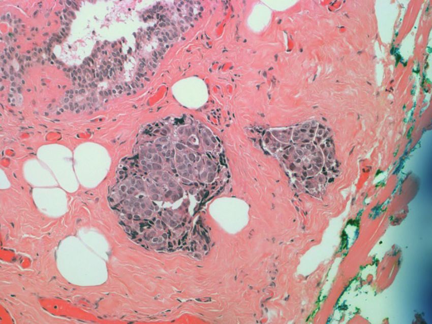
Elena Provenzano: Breast Cancer Neoadjuvant Therapy Beyond pCR

                                                     A                                                            B

                                                     C                                                            D
Figure 2. Residual invasive carcinoma predominantly in the form of LVI (A); D2-40 immunohistochemical staining distin-
guishing tumour in lymphatic spaces from invasive disease (B); Tumour adjacent to a margin confirmed as LVI on D2-40
staining (C+D).

TNM Staging                                                 ing systems incorporating both molecular tumour
                                                            characteristics and tumour response are required.
Traditional staging systems, such as the TNM and                There are two main patterns of response seen
Nottingham Prognostic Index (NPI), retain prog-             on serial imaging in patients receiving NACT (81,
nostic significance following NACT (16, 79). Path-          82). The first is concentric shrinking, where there is
ological TNM staging post NACT is given a y pre-            a single tumour mass that progressively decreases
fix. There have been modifications to how primary           in size. Measurement of tumour size in this situa-
invasive tumour and metastases are measured for             tion is relatively straight forward as there is a single
staging purposes in the 7th and 8th editions (18, 19),      invasive tumour focus. Tumour bed changes may
which are summarised in Table 1. Although only              extend beyond the invasive carcinoma, however it
currently applied following primary surgery, there          is the maximum residual invasive cancer size that
is emerging evidence that the AJCC prognostic               is measured; surrounding stroma without invasive
stage incorporating grade and receptor status in-           tumour is excluded (Figure 3) (66, 70, 71).
troduced in the 8th edition is also predictive of out-          The second pattern is the scatter or Swiss
come post NACT and may provide better discrim-              cheese pattern, where there is a patchy response
ination of prognostic groups (17, 80). Future stag-         with scattered foci of residual enhancement across
                                                            the tumour bed. This pattern is a reflection of in-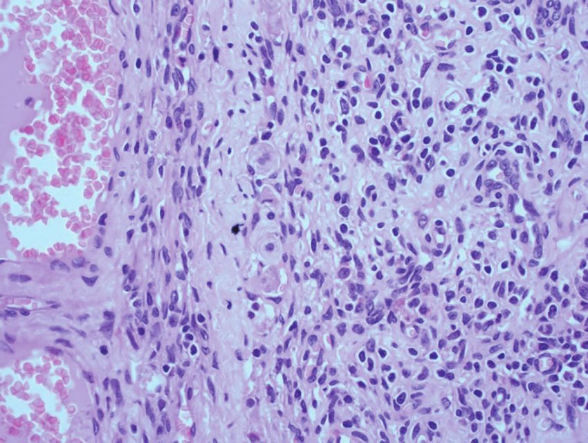
Acta Medica Academica 2021;50(x):xx-xx

Table 1. Definitions Used for Primary Tumour and Metastasis Measurement in Residual Cancer Burden and Subsequent
Editions of the TNM Staging System

 Staging system       Size measurement breast                                         Size measurement nodal metastases
 Residual             Maximum size residual invasive disease in two dimensions.       Maximum dimension metastatic focus including
 Cancer Burden        Scattered foci measured as a single lesion including areas of   associated fibrosis. ITCs regarded as positive.
                      intervening fibrosis.
 AJCC/ UICC 6th       Maximum size residual invasive disease in one dimension.        Maximum dimension metastatic focus including
 edition              Scattered foci measured as a single lesion including areas of   associated fibrosis. ITCs regarded as negative.
                      intervening fibrosis.
 AJCC/ UICC 7th       Measurement largest contiguous tumour focus, with use of        Maximum dimension size of metastatic focus
 edition              (m) classifier if multiple deposits present across the tumour   including associated fibrosis. ITCs regarded as
                      bed                                                             negative.
 AJCC/ UICC 8th       Measurement largest contiguous tumour focus, with use of        Maximum dimension of largest contiguous tumour
 edition              (m) classifier if multiple deposits present across the tumour   cell deposit excluding associated fibrosis. ITCs
                      bed                                                             regarded as negative.
ITCs=Isolated tumour cells.

                                                                  A                                                                     B

                                                                          Figure 3. Schematic diagrams illustrating measurement
                                                                          of tumour size post neoadjuvant chemotherapy. Hatched
                                                                          area is stromal reaction: a) is maximum size measurement
                                                                          according to 7th/8th edition TNM; b) is maximum size mea-
                                                                          surement used for RCB. A) Concentric shrinking pattern.
                                                                          Size of residual invasive tumour is measured excluding
                                                                          tumour bed extending beyond the invasive focus: a and b
                                                                          are the same. B) Scatter pattern with even distribution of
                                                                          tumour islands across tumour bed, measured as a single fo-
                                                                          cus: a and b are the same. C) Scatter pattern with unevenly
                                                                          scattered tumour foci: a is the largest individual focus (black
                                                                          line); b is the size of the entire lesion including all foci and
                                                                  C       intervening fibrosis (red line).

tratumoural heterogeneity leading to a differential                       dispersed within an ill-defined background of re-
response to NACT. At the histological level, this is                      active fibrous stroma (Figure 4).
seen as separate nests and islands of tumour cells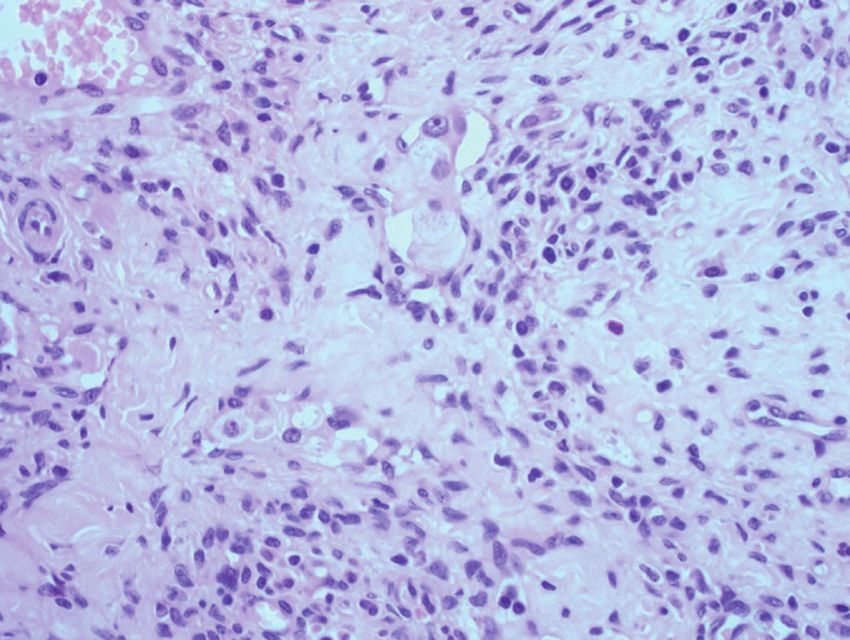
Elena Provenzano: Breast Cancer Neoadjuvant Therapy Beyond pCR

                                                  A                                                               B

                                                  C                                                               D
                                                        Figure 4. Example of tumour showing scatter pattern of
                                                        response. Pre treatment MRI showed a single tumour (A).
                                                        Post treatment histology showed widely dispersed residual
                                                        tumour foci with intervening fibrosis (B-E). For TNM stag-
                                                        ing the largest single focus is measured, however to calcu-
                                                        late the RCB the entire size including intervening fibrosis is
                                                        measured in 2 dimensions.

                                                        tern of response (53%, versus 11% and 29% re-
                                                        spectively) (83). On closer analysis, the HER2+
                                                        tumours differed by ER status, with 78% of ER+/
                                                        HER2+ tumours showing the scatter pattern com-
                                                        pared with 53% of ER-/HER2+ cancers. Of interest,
                                                  E     presence of macrophages in the tumour bed was
                                                        also associated with TNBC, whereas elastosis and
                                                        myxoid change was more common in ER+/HER2-
   Perhaps unsurprisingly, the pattern of response      cancers. In contrast, the study of Ballesio et al looked
has been shown to correlate with molecular sub-         solely at MRI patterns of response and found that
type. In one series looking at histological findings,   ER-/HER2+ showed a concentric pattern, whilst
TNBC was more likely to show the concentric             TNBC showed a multinodular pattern (82).
shrinking pattern, whilst ER+/HER2- and HER2+              The scatter pattern has been associated with
tumours more commonly showed the scatter pat-           a higher locoregional recurrence (LRR) rate post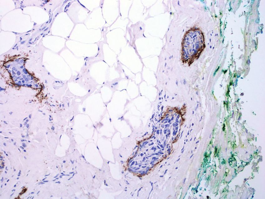
Acta Medica Academica 2021;50(x):xx-xx

breast conservation surgery and increased risk of        by the U.K. Royal College of Pathologists (87, 88),
positive margins. Standard definitions of clear mar-     and has been shown to correlate with survival (16).
gins as ‘tumour at ink’ are likely to be inadequate          The method of size measurement was amend-
in this context, and if residual invasive tumour lies    ed in 7th edition TNM, whereby if the residual
in close proximity to the margin with transection        tumour consists of multiple nests in a fibrotic
of the tumour bed consideration should be given          stroma, the largest contiguous focus of invasive
to re excision (2). The MD Anderson group identi-        carcinoma is measured and used for ypT staging,
fied four features associated with increased risk of     with the ‘m’ modifier to indicate multiple tumour
LRR post NACT; clinical nodal stage 2/3, residual        foci are present (18, 19). So in simple terms, the
invasive tumour size >2 cm, scatter/ multifocal pat-     largest single tumour focus is measured and this
tern of residual disease and presence of LVI (84).       is used for TNM staging; other foci and the as-
A recent study found no difference in LRR rates          sociated stromal background are NOT included.
between a margin
Elena Provenzano: Breast Cancer Neoadjuvant Therapy Beyond pCR

influence decisions to give adjuvant regional radio-            associated with survival (71, 72, 74). The 8th edi-
therapy (70). If a node was clipped pre-treatment,              tion TNM has changed the method of measuring
then presence of the clip site should be document-              metastases to the size of the largest contiguous fo-
ed and specific comment made as to presence of                  cus in the node not including tumour associated
residual disease and treatment effect in the clipped            fibrosis (19). According to the definition in adju-
node. As with the primary tumour, there is a lack               vant disease, a contiguous focus is tumour cells
of agreement in how to measure disease in this                  directly in contact with one another without inter-
setting that generates confusion amongst patholo-               vening lymphocytes. When there has been good
gists. In the 6th and 7th edition TNM the approach              response to NACT, residual metastatic disease is
was to measure the size of the entire area involved             often present as scattered single cells within a reac-
by metastatic tumour including intervening fibro-               tive fibrous background and this is now defined as
sis (18, 20); as in the breast, this is the distance be-        ITCs under the 8th edition (Figure 5). This could
tween tumour cells, and fibrous tissue extending                potentially downstage nodal involvement in a sig-
beyond metastatic tumour cells is excluded. This                nificant number of patients, and again an element
is also the maximum metastasis size measure-                    of clinical judgement is required. My personal ap-
ment used for calculating the RCB, and has been                 proach, as with the primary tumour, is to look at

                                                         A                                                               B

                                                         C                                                               D
Figure 5. Lymph node post chemotherapy showing an area of fibrosis containing scattered single cells and small clusters,
classified as isolated tumour cells in the 8th edition TNM: A) Low power H&E showing area of fibrosis; B) Low power cytokera-
tin stain highlighting distribution of residual tumour cells; C-D) Higher power images of residual tumour cells (bold arrows)
on H&E (×40 magnification).
Acta Medica Academica 2021;50(x):xx-xx
ITCs are handled the same way as in the adjuvant
setting in the TNM Staging System and are classed
as node negative [ypN0(i+)] (19), whereas in the
UK reporting guidelines nodes containing ITCs
should be counted as positive (87). Regardless of
whether they are considered positive or negative,
ITCs post NACT represent tumour cells that have
persisted despite systemic therapy and have dif-
A ferent significance to the adjuvant setting and it
is agreed they should not be regarded as axillary
pCR. There is considerable evidence that the pres-
ence of any residual tumour cells in the axillary
lymph nodes following NACT, even in the form of
ITCs, is associated with worse prognosis (72-74).
In a recent series examining a US National Can-
cer Database (NCD) cohort, ITCs were associated
with poorer survival outcomes with 83% 5 year
B
OS compared with 89% for ypN0; this was present
in patients that were cN0 and cN1 pre-treatment
(66% and 81% increase in mortality respectively),
with the greatest impact on TNBC (89).
Assessing Response
Whilst early clinical trials showed a drop in pro-
portion of cases classified as pCR following cen-
C
tral histology review compared with local reports
(90), our own experience with the ARTEMIS trial
Figure 6. Schematic diagram illustrating measurement of
metastases post neoadjuvant chemotherapy. Hatched area
showed excellent agreement between source labo-
represents associated fibrosis. A) Single metastatic focus ratory reports and central review with respect to
within area of fibrosis. Measure extent of tumour only, not pCR (91). However, in an audit of local pathology
background fibrosis extending beyond tumour. B) If mul- reports as part of the trial, only 45% of reports in-
tiple separate foci of tumour, with or without fibrosis, them
cluded an assessment of tumour response in the
measure the largest single focus. C) Scattered metastatic
foci in a single area of fibrosis: a) size of largest single contig- breast, dropping to 30% for response in the axil-
uous cluster of cells excluding background fibrosis used for lary lymph nodes (92). A similar review of exter-
TNM staging (black line); b) maximum size including all foci nal pathology reports is being undertaken as part
and intervening fibrosis (red line) used to calculate the RCB. of the UK multicentre PARTNER trial, and whilst
most reports now include a general comment on
the pattern of spread of the tumour cells across the presence or absence of response in both the pri-
metastatic deposit; if they form discrete foci a dis- mary tumour and axillary nodes, the majority still
tance apart then these should be measured indi- do not incorporate formal grading of response
vidually, however if they are evenly dispersed cells/ (unpublished data).
foci across the area of fibrosis I measure the entire There are two main approaches to assessment
lesion as a single deposit (Figure 6). of residual disease post NACT. The first examines
The interpretation of ITCs is a particular source actual response by comparing tumour cellularity
of controversy in the neoadjuvant setting (70). before and after treatment. Response to NACT isElena Provenzano: Breast Cancer Neoadjuvant Therapy Beyond pCR
often accompanied by a reduction in tumour cel- bed area; ‘tumour bed area’ refers to the size of
lularity, and this is associated with improved sur- the residual invasive cancer, i.e. the greatest dis-
vival outcomes. Comparison between pre and post tance between invasive tumour cell foci (Figure
treatment cellularity forms the basis for several 3). Background stromal changes such as reactive
grading systems of response, including the Che- fibrosis or DCIS that extends outside the limit of
vallier, Sataloff, Miller-Payne and Pinder systems the invasive tumour are not included. It is not nec-
(93-96). The second approach is quantification of essary to measure the area of stromal change, just
residual disease post NACT by looking at inva- the dimensions of the residual invasive disease.
sive tumour size and cellularity, the main example Second, the invasive tumour dimension for the
of which is the RCB proposed by Symmans et al. RCB includes intervening background stroma, i.e.
(71). Newer systems such as the Neo-Bioscore include fibrosis between invasive tumour cell foci.
have been developed that incorporate tumour If there are scattered islands of tumour cells across
molecular profile and biomarkers such as Ki67, al- the tumour bed you measure the total size across
though these are currently not in widespread use all the islands as a single lesion, unless there are
(60, 97, 98). The different systems have advantages multiple separate primary tumours. This is differ-
and disadvantages and at present there is no one ent to the size measurement for TNM staging from
universally agreed system; readers are referred to the 7th edition onwards, described above (Table 1,
review articles comparing the different systems Figure 3). Similarly, when evaluating cellularity,
(99-101). The important thing for pathologists is the entire tumour area including intervening fields
to work closely with their oncology colleagues to with no tumour should be assessed to calculate the
agree which system to use. average, not just fields that contain tumour cells.
There are similar caveats in evaluating the
nodal disease. The total nodal count includes all
Residual Cancer Burden nodes that contain tumour cells including nodes
The RCB is presently the most widely used system with ITCs only, although these are not regarded
and will be described in more detail; it has been as positive nodes for TNM staging. As with inva-
well validated, is simple and reproducible (91, 102, sive tumour, the size of the largest metastasis is the
103), and shows a strong association with survival greatest distance between tumour cells within a
outcomes across all molecular subtypes. As a re- lymph node including background reactive fibro-
sult the RCB has been incorporated in the soon to sis between metastatic tumour cell islands, but not
be released International Collaboration on Cancer fibrosis that extends outside the metastasis. Again,
Reporting (ICCR) minimum dataset for breast pa- this is different to how metastases are measured
thology reporting post NACT. The RCB website according to the 8th edition of the TNM (Table 1,
provides detailed instructions on how to assess the Figure 6)
RCB score, including macroscopic specimen han- These values are combined in an algorithm
dling, a visual guide to estimating the percentage available online that calculates a continuous nu-
of residual tumour cells, and an online calculator merical score, and places residual tumour in 3
that provides both the numerical RCB score and classes with class I representing MRD and class III
RCB class (88). extensive residual disease. Although class I cor-
The RCB incorporates four variables; maxi- relates with excellent response and class III with
mum invasive tumour size measured in two di- poor response, this system is not strictly a measure
mensions, average residual invasive tumour cel- of response as cellularity in this case is absolute
lularity, number of positive lymph nodes and size cellularity post treatment rather than the change
of largest metastasis. There are several important in cellularity. Indeed low cellularity post-treat-
things to note when making measurements for ment does not necessarily equate with response as
the RCB. The website refers to primary tumour some cancers, e.g. lobular cancers, are hypocellu-Acta Medica Academica 2021;50(x):xx-xx
lar to begin with. Cellularity is heavily weighted in tumour with high cellularity will be low AJCC
the algorithm, so small tumours with a high cel- stage but RCB class II, and conversely a large tu-
lularity will often end up as RCB II, whereas larger mour with low cellularity will have a higher stage
tumours with low cellularity can still be RCB I. but a relatively low RCB score. For discrepant cas-
As mentioned, both RCB class and the RCB es, if residual disease was RCB or ypAJCC stage 3
score as a continuous variable show an associa- there was a poor outcome suggesting the two sys-
tion with survival outcomes across all molecular tems are complementary.
subtypes, although the nature of the relationship
varies by subtypes (14). Early data suggested for
TNBC, patients that achieve RCB I have an excel- Management of the Axilla
lent prognosis similar to that of pCR. A more re- In patients that are axillary node negative pre-
cent multicentre pooled analysis with larger num- treatment, the safety and accuracy of SLNB post
bers has shown a linear relationship between RCB NACT has now been established in several large
and BCSS, with a small but significant difference series with identification (IR) and false negative
between pCR and RCB I (15). In contrast, for ER-/ rates (FNR) comparable to the adjuvant setting. A
HER2+ patients the curve has a slightly different meta-analysis found IR of 93-97%, a FNR of 6%
shape with a steeper rise at low levels of residual and axillary recurrence rates of 2% (105). In pa-
disease that plateaus out across higher RCB scores tients with proven positive axillary lymph nodes
suggesting even small volumes of residual disease pre-NACT surgical management of the axilla is
has an adverse prognosis for this subtype. ER+/ still subject to debate. Early series showed huge
HER2- cancers had the opposite profile with the variation in results, with one meta-analysis find-
curve rising slowly across low volumes of residual ing an IR of 68-100% with a pooled FNR of 11%,
disease and a steeper rise beginning in RCB II. The although the FNR was as high as 33% in individual
relationship between residual disease and survival studies (106). However, the reliability of SLNB in
in ER+/HER2- cancers has been a source of con- node positive patients has now been examined in
troversy, with these tumours having a relatively several prospective clinical trials, and with better
favourable prognosis despite low pCR rates and a patient selection and the evolution of targeted axil-
poorer correlation between residual disease and lary sampling techniques is yielding more promis-
survival outcomes; this data confirms the prognos- ing results.
tic relevance of RCB for this subtype, and molecu- Early evidence came from the NSABP-B27 tri-
lar type-specific RCB class cut-offs could improve al, where a subset of 428 patients underwent SLNB
clinical accuracy. This highlights the importance followed by ALND; the SLN was positive in 36%,
in considering molecular subtype when assessing and in 56% was the only positive node (107). The
residual disease, and the future need for a com- FNR was 12% for patients that were cN1-2, and
bined system including anatomical residual dis- in patients with breast pCR this fell to 2%. Sub-
ease extent and tumour biological characteristics. sequently, the ACOSOG Z1071 trial specifically
Whilst ypAJCC and RCB staging both provide addressed the question of post NACT SLNB in
a quantitative assessment of residual disease and patients with biopsy proven axillary metastases
show an association with survival outcomes, an with no prior axillary surgery (108). Patients un-
analysis of cases from the I-SPY-1 trial showed a derwent SLNB followed by completion ALND; the
discrepancy in classification in up to one third of overall nodal pCR rate was 41%, rising to 49% in
cases using 7th edition TNM (104). Of 55 discrep- TNBC and 65% for HER2+ disease, and in 21%
ant cases, 36 had a higher RCB class, and 19 had a residual nodal disease was confined to the SLN.
higher ypAJCC stage. The source of discrepancy The overall FNR was 12.6% which fell short of the
was weighting of lymph node involvement and tu- study target of 10%, however if dual mapping with
mour cellularity in the RCB. For example, a small blue dye and radioisotope was used the FNR fellElena Provenzano: Breast Cancer Neoadjuvant Therapy Beyond pCR
to 11%, and if 3 or more nodes were sampled the The Mayo Clinic group reported 38 SLNB af-
FNR was only 9% compared with 21% for 2 nodes ter clipping the biopsied node; 25 had a 125I seed
and 31% if one node was removed. The SN FNAC placed in the clipped node, 9 had no preoperative
study looked at SLNB in node positive patients, localisation of the clip, and 4 had no documenta-
with immunohistochemistry (IHC) undertaken tion (115). In the 25 patients with the 125I seed,
on all negative nodes; the FNR was 13%, which the labelled node was successfully identified pre-
fell to 8% when nodes with ITCs were regarded as operatively in 20; in the remaining 14 where the
positive (109). A subset analysis of Z1071 utilizing seed was not localised or localisation was not at-
IHC found a similar FNR of 9%. tempted, the clipped node was found in 11. Over-
The Europe-based SENTINA study had a more all, the IR for the clip was 78% with a FNR of 3%.
complex design including both cN0 and cN1 dis- The same group had trialled using HydroMARK
ease. Patients that were cN1 proceeded directly to gel clips to mark the biopsied node but found these
post NACT SLNB, with an IR of 80% and a FNR of were no longer visible following NACT, however
14%. cN0 patients had a pre chemotherapy SLNB, in the Spanish ILINA trial placement of Hydro-
and if positive had a second attempt at SLNB post MARK clips with intraoperative US successfully
NACT with ALND (110). The second line SLNB localised the clipped node in 96% of cases, with a
had an IR of 60% and a FNR of 52%, showing re- 4% FNR when combined with SLNB; in all cases
peat SLNB has a poor success rate. the clipped node was positive except one false neg-
Within Z1071, a substudy of 170 patients ex- ative case where both clipped node and SLN were
amined the role of clipping the biopsied node and negative (116).
identifying the clip at the time of SLNB. The clip In a separate single institution series of 630 cN1
was present in a SLN in 76% of cases with a FNR patients without clipping of the positive node, 91%
of 7%, however in the remaining 24% where the converted to cN0 post NACT and proceeded to
clip was in a non-SLN the FNR was 19% (111). In SLNB (117, 118). Three or more SLNs were mapped
41% the clipped node was the only positive node. in 93% of cases, regarded as adequate mapping,
This has led to the evolution of targeted axillary with 7% having less than 3 nodes identified and
sampling techniques, where the biopsied node is complete failure in 2%. Unsuccessful mapping was
clipped or otherwise labelled and localised at the associated with high body mass index and pres-
time of surgery and/ or at least 3 SLN are removed ence of LVI. In patients with successful mapping,
following dual localisation, achieving acceptably 41% had nodal pCR and were able to avoid ALND,
low FNR. by molecular subtype 20% of HR+/HER2-, 44%
In a series of 12 patients, Caudle et al. clipped of TNBC, 55% of HR+/HER2+ and 78% of HR-/
the positive biopsied node then placed a radioac- HER2+. Of note, 43% of patients with unsuccess-
tive 125I seed before surgery to localise the clipped ful mapping also achieved an axillary pCR. Other
node; five underwent SLN, which included the predictors for avoiding ALND included ductal or
clipped node in 4 patients (112). A total of 9 pa- apocrine histological subtype (44% and 50% ver-
tients including the SLN group had an ALND; 4 sus 17% for lobular cancers), grade 3 cancers (54%
had residual metastatic disease and the clipped versus 24% for grade 2 and 14% for grade 1) and
node was positive in all cases. In a larger follow up absence of LVI (78% versus 22%). Grade 3, molec-
series, removing the clipped node gave a FNR of 4%, ular subtype and presence of LVI remained signifi-
which fell to 2% when combined with SLNB (113). cant predictors of ALND on multivariate analysis.
In a similar approach, a Dutch group used 125I seed This supports the conclusion from an earlier study
to label the positive node at the time of diagnosis, that clipping the biopsied node is not required if
termed the MARI procedure (Marking the Axillary there is thorough SLN technique with dual label-
lymph node with Radioactive I) then removed the ling and removal of 3 or more nodes at the time of
labelled node alone with a FNR of 7% (114). SLNB (119).Acta Medica Academica 2021;50(x):xx-xx
The ISPY-2 trial group have published guid- in those that had ALND (122). There is still limited
ance on surgical management of the axilla for use data on LRR rates in patients who achieve axillary
in clinical trials that is generalizable to routine pCR post NACT. The study of Pitilin et al. found
practice (3). For cN0 patients, SLN with removal 17 LRR in 602 patients after 34 months of follow
of at least 2 nodes is advised. For proven node pos- up, 3 in patients that were ypN0; of interest none of
itive patients, the biopsied node should be marked the 9 patients with ITCs had a LRR (123).
at the time of diagnosis with SLN or ALND after Results of two ongoing clinical trials are await-
completion of NACT. Where SLN is performed, ed. The NSABP B-51/RTOG 1304 trial is looking
dual tracer mapping of the SLN is required with at the oncological safety of SLNB alone in node
identification and removal of the clipped node. If positive patients that revert to node negative post
the node was not clipped, a minimum of 2 SLNs NACT, and is randomising patients to regional
must be removed. If the SLN is positive ALND is nodal irradiation versus no further axillary thera-
advised but not mandated; however, if RCB calcu- py. In contrast, the Alliance 11202 trial will exam-
lation is part of the trial then completion ALND is ine the group of women with positive SLNB post
needed to determine the RCB score. In multidis- NACT and randomise them to nodal radiotherapy
ciplinary UK guidelines, in patients with a posi- versus ALND. Of note, both trials regard women
tive axilla SLN may be considered post NACT but with ITCs as node negative so will not provide
dual mapping with removal of four nodes is ad- direct evidence as to the need for further axillary
vised (120). If any residual disease including ITCs therapy in this important subset of patients.
is identified then ALND is recommended. A rare clinical scenario is presentation with
Of note, a recent review of the US NCD has axillary nodal disease and no identifiable primary
shown an increase in adoption of SLNB for cN+ breast tumour. A recent study looking at 28 wom-
patients post NACT from 32% in 2012 to 49% in en with occult primary breast cancer found a pCR
2015, with SLNB more frequent in younger pa- rate of 80%, 93% in those with cN1 disease, sug-
tients, TNBC or HER2+ disease, and following gesting that SLN alone post NACT may be an op-
BCS (5). Of concern, follow up ALND was not tion for these patients (124). Interestingly, looking
performed for 37% of patients with ITCs (21% in at the molecular subtypes the pCR rate was 50%
2012 increasing to 49% in 2015), 24% with mi- in ER+/HER2- tumours, 88% for TNBC and 100%
crometastases (19% in 2012 to 31% in 2015) and for HER2+ disease, higher than for women with
13% with ypN1 macrometastases. This is despite an identifiable breast primary in most series. One
clinical guidelines recommending ALND for any proposed theory is this represents a subset of tu-
residual nodal disease including ITCs post NACT mours that invoke a strong immune response with
due to a lack of clinical evidence on safety of omis- regression of the primary disease, and immune
sion of ALND. Studies post NACT show higher therapies may be a future treatment option for
FNR with additional non-SLN positivity in 17% these patients.
of cases with ITCs, 64% with micrometastases
and 62% with macrometastases (121). There is Conclusion
evidence showing worse DFS for ypN0(i+) and
ypN1(mic) (1.9 and 2.2 times increased mortality In conclusion, NACT is now routine breast cancer
respectively); this was true for both cN0 and cN1 management. Assessment of response is becoming
disease, with the greatest impact of low volume re- increasingly important in adjuvant therapy deci-
sidual nodal disease in TNBC and HER2+ cancers sions, and more than ever the pathologist plays a
(89). Using NCD data, Almahariq et al. showed vital role in patient care. Management of the ax-
inferior survival outcomes in ypN1 patients that illa remains controversial but there is growing evi-
underwent SLNB alone with regional nodal irra- dence supporting the safety of SLNB in previously
diation, with 71% 5 year OS compared with 77% node positive patients, however even minimal re-Elena Provenzano: Breast Cancer Neoadjuvant Therapy Beyond pCR
sidual nodal involvement in the form of ITCs and 9. Barker AD, Sigman CC, Kelloff GJ, Hylton NM, Berry
micrometastases has adverse prognostic signifi- DA, Esserman LJ. I-SPY 2: an adaptive breast cancer trial
design in the setting of neoadjuvant chemotherapy. Clin
cance and clinical evidence for the safety of omit- Pharmacol Ther. 2009;86(1):97-100.
ting ALND in these patients is currently lacking. 10. Esserman LJ, Woodcock J. Accelerating identification
and regulatory approval of investigational cancer drugs.
Conflict of Interest: The author declares that she has no con- JAMA. 2011;306(23):2608-9.
flict of interest.
11. File D, Curigliano G, Carey LA. Escalating and De-esca-
lating Therapy for Early-Stage HER2-Positive Breast Can-
Acknowledgement: This research was supported by the
cer. Am Soc Clin Oncol Educ Book. 2020;40:1-11.
NIHR Cambridge Biomedical Research Centre (BRC-1215-
20014). The views expressed are those of the authors and not 12. Masuda N, Lee SJ, Ohtani S, Im YH, Lee ES, Yokota I, et al.
necessarily those of the NIHR or the Department of Health Adjuvant Capecitabine for Breast Cancer after Preopera-
and Social Care tive Chemotherapy. N Engl J Med. 2017;376(22):2147-59.
13. von Minckwitz G, Huang CS, Mano MS, Loibl S, Ma-
mounas EP, Untch M, et al. Trastuzumab Emtansine for
References Residual Invasive HER2-Positive Breast Cancer. N Engl J
Med. 2019;380(7):617-28.
1. Cortazar P, Zhang L, Untch M, Mehta K, Costantino JP, 14. Symmans WF, Wei C, Gould R, Yu X, Zhang Y, Liu M, et
Wolmark N, et al. Pathological complete response and al. Long-Term Prognostic Risk After Neoadjuvant Che-
long-term clinical benefit in breast cancer: the CTNeoBC motherapy Associated With Residual Cancer Burden and
pooled analysis. Lancet. 2014;384(9938):164-72. Breast Cancer Subtype. J Clin Oncol. 2017;35(10):1049-
2. King TA, Morrow M. Surgical issues in patients with 60.
breast cancer receiving neoadjuvant chemotherapy. Nat 15. Yau C, Van der Noordaa M, Wei J, Osdoit M, Reyal F,
Rev Clin Oncol. 2015;12(6):335-43. Hamy AS, et al. Residual cancer burden after neoad-
3. Boughey JC, Alvarado MD, Lancaster RB, Fraser Sym- juvant therapy and long-term survival outcomes in
mans W, Mukhtar R, Wong JM, et al. Surgical Standards breast cancer: A multi-center pooled analysis. Cancer
for Management of the Axilla in Breast Cancer Clinical Res. 2020;80(4 Suppl):GS5-01. doi: 10.1158/1538-7445.
Trials with Pathological Complete Response Endpoint. SABCS19-GS5-01.
NPJ Breast Cancer. 2018;4:26. 16. Carey LA, Metzger R, Dees EC, Collichio F, Sartor CI,
4. Morrow M, Dang CT. Sentinel node biopsy after neoadju- Ollila DW, et al. American Joint Committee on Cancer
vant chemotherapy: a new standard for patients with axil- tumor-node-metastasis stage after neoadjuvant chemo-
lary metastases? JAMA. 2013;310(14):1449-50. therapy and breast cancer outcome. J Natl Cancer Inst.
5. Wong SM, Weiss A, Mittendorf EA, King TA, Golshan M. 2005;97(15):1137-42.
Surgical Management of the Axilla in Clinically Node- 17. Kantor O, Bao J, Jaskowiak N, Yao K, Tseng J. The Prog-
Positive Patients Receiving Neoadjuvant Chemotherapy: nostic Value of the AJCC 8th Edition Staging System for
A National Cancer Database Analysis. Ann Surg Oncol. Patients Undergoing Neoadjuvant Chemotherapy for
2019;26(11):3517-25. Breast Cancer. Ann Surg Oncol. 2020;27(2):352-8.
6. Pelizzari G, Gerratana L, Basile D, Fanotto V, Bartoletti M, 18. Edge SB, Byrd DR, Compton CC, Fritz AG, Greene FL,
Liguori A, et al. Post-neoadjuvant strategies in breast can- Trotti I, editors. Cancer Staging Handbook From the
cer: From risk assessment to treatment escalation. Cancer AJCC Cancer Staging Manual. 7th ed. New York: Spring-
Treat Rev. 2019;72:7-14. er-Verlag; 2010.
7. von Minckwitz G, Untch M, Blohmer JU, Costa SD, Ei- 19. Hortobagyi GN, Connolly JL, D’Orsi CJ, Edge SB, Mit-
dtmann H, Fasching PA, et al. Definition and impact of tendorf EA, Rugo H, et al. Breast. AJCC Cancer Staging
pathologic complete response on prognosis after neoad- Manual. 8th ed. New York: Springer; 2017. p. 589-628.
juvant chemotherapy in various intrinsic breast cancer 20. Greene FL, Page DL, Fleming ID, Fritz AG, Balch CM,
subtypes. J Clin Oncol. 2012;30(15):1796-804. Haller DG, et al., editors. AJCC Cancer Staging Manual.
8. U.S. Food and Drug Administration. Guidance for In- 6th ed. New York: Springer; 2002.
dustry: Pathological Complete Response in Neoadjuvant 21. Boughey JC, Ballman KV, McCall LM, Mittendorf EA,
Treatment of High-Risk Early-Stage Breast Cancer: Use Symmans WF, Julian TB, et al. Tumor Biology and Re-
as an Endpoint to Support Accelerated Approval. [cited sponse to Chemotherapy Impact Breast Cancer-spe-
26/12/2020] Available from: http://www.fda.gov/down- cific Survival in Node-positive Breast Cancer Patients
loads/Drugs/GuidanceComplianceRegulatoryInforma- Treated With Neoadjuvant Chemotherapy: Long-term
tion/Guidances/UCM305501.pdf2014. Add a date of cita- Follow-up From ACOSOG Z1071 (Alliance). Ann Surg.
tion in the red parentheses 2017;266(4):667-76.You can also read