The pathogenesis and diagnosis of sepsis post burn injury
←
→
Page content transcription
If your browser does not render page correctly, please read the page content below

Burns & Trauma, 2021, 9, tkaa047
                                                                                                                    doi: 10.1093/burnst/tkaa047
                                                                                                                                        Review

Review

The pathogenesis and diagnosis of sepsis post

                                                                                                                                                                          Downloaded from https://academic.oup.com/burnstrauma/article/doi/10.1093/burnst/tkaa047/6128653 by guest on 14 November 2021
burn injury
Pengju Zhang1 , Bingwen Zou2 , Yih-Cherng Liou3 ,* and Canhua Huang1 ,*
1
 State Key Laboratory of Biotherapy and Cancer Center, West China Hospital, and West China School of Basic
Medical Sciences & Forensic Medicine, Sichuan University, and Collaborative Innovation Center for Biotherapy,
No.17 People’s South Road, Chengdu, 610041, China, 2 Department of Thoracic Oncology and Department of Radiation
Oncology, Cancer Center, West China Hospital, Sichuan University, No.37 Guoxue Alley, Wuhou District, Chengdu,
610041, China and 3 Department of Biological Sciences, Faculty of Science, National University of Singapore,
14 Science Drive 4, 117543, Singapore

*Correspondence. Canhua Huang, Email: hcanhua@hotmail.com; Yih-Cherng Liou, Email: dbslyc@nus.edu.sg

Received 10 September 2020; Revised 20 October 2020; Editorial decision 27 November 2020

Abstract
Burn is an under-appreciated trauma that is associated with unacceptably high morbidity and
mortality. Although the survival rate after devastating burn injuries has continued to increase in
previous decades due to medical advances in burn wound care, nutritional and fluid resuscitation
and improved infection control practices, there are still large numbers of patients at a high risk of
death. One of the most common complications of burn is sepsis, which is defined as “severe organ
dysfunction attributed to host’s disordered response to infection” and is the primary cause of death
in burn patients. Indeed, burn injuries are accompanied by a series of events that lead to sepsis and
multiple organ dysfunction syndrome, such as a hypovolaemic state, immune and inflammatory
responses and metabolic changes. Therefore, clear diagnostic criteria and predictive biomarkers
are especially important in the prevention and treatment of sepsis and septic shock. In this review,
we focus on the pathogenesis of burn wound infection and the post-burn events leading to sepsis.
Moreover, the clinical and promising biomarkers of burn sepsis will also be summarized.

Key words: Burn, Infection, Sepsis, Septic shock, Multiple organ dysfunction syndrome, Immune dysregulation, Hypermetabolism,
Trauma, Biomaker, Inflammation

Highlights
•   Sepsis is one of the most common and severe complications of severe burns.
•   Immunosuppression and hypermetabolism play key roles in the development of burn sepsis.
•   Recent diagnostic tools and potential biomarkers are discussed.

Background                                                                               (friction, high temperature, cold, radiation and electricity)
Burn injuries cause unpredictable and devastating trauma                                 and chemical factors [1]. Nevertheless, thermal injury caused
and are associated with high morbidity and mortality. There                              by hot liquids, solids or fire makes up the majority of
are numerous causative mechanisms, including physical                                    burn injuries [2]. According to a report from the World

© The Author(s) 2021. Published by Oxford University Press.
This is an Open Access article distributed under the terms of the Creative Commons Attribution License (http://creativecommons.org/licenses/by/4.0/), which permits   1
unrestricted reuse, distribution, and reproduction in any medium, provided the original work is properly cited.
2                                                                                                                   Burns & Trauma, 2021, Vol. 9, tkaa047

                                                                                                                                                                        Downloaded from https://academic.oup.com/burnstrauma/article/doi/10.1093/burnst/tkaa047/6128653 by guest on 14 November 2021
Figure 1. Zones of burn injury for different depths. First-degree burns only involve the epidermis (the uppermost layer of the skin); the skin becomes red and
painful, but this is limited in duration. Burns that affect the dermis (the underlying skin layer) are classed as partial-thickness burns, which are frequently
accompanied by the formation of painful blisters that increase the risk of infection. Partial-thickness burns can be divided into superficial partial-thickness burns,
which are painful, moist, hyperemic and blanch, and deep partial-thickness burns which are less sensate, drier and do not blanch. Full-thickness burns extend
through the full dermis and require surgical management due to high risk of infection. Burns extending into deeper tissues (such as muscle or even bone) are
defined as fourth-degree burns and are usually blackened and often result in loss of the burned tissues

Health Organization in 2018, about 11 million burn cases                             considered the first line of defense against microbial invasion
occur annually worldwide, with burn injuries claiming as                             in hosts [11]. Under the conditions of a dysregulated host
many as 180,000 lives [3]; looking back to almost a decade                           response to an infection, burn patients may develop sepsis
ago, mortality from burns has decreased from the 300,000                             syndrome characterized by fever, increased fluid require-
deaths recorded in 2011 [4]. The significant improvement                             ments, decreased urinary output and even MODS [12–14].
in the survival rate of burn patients is in part attributed                          In 2016, the Third International Consensus Definition for
to advances in intensive care unit treatment and improved                            Sepsis and Septic Shock (Sepsis-3) redefined sepsis as life-
wound management, infection control practices and control                            threatening organ dysfunction caused by a dysregulated host
of hemodynamic disorders [5, 6]. The mortality rate, however,                        response to infection [15]. This new sepsis definition places
remains unacceptably high, particularly in patients with                             more emphasis on the process of organ dysfunction, and
severe burns. The severity and prognosis of burn injuries                            the Sequential Organ Failure Assessment score (Table 1) is
depends principally on the depth (Figure 1) and size (Figure 2)                      used to define sepsis severity, including septic shock [15, 16].
of the burn site. Most patients who suffer from severe burn                          Indeed, the incidence of sepsis in burn patients can range
injuries require rapid and specialized emergency burn care                           between 3–30% for burns of more than 20% of the total
to reduce morbidity and mortality. The high fatality rate                            body surface area (TBSA) [17]. Even more concerning is that
of severe burns is due to not only hypovolaemic shock and                            approximately 54% of burn-related deaths in modern burn
vascular leak, but also abnormal body responses, including                           units occur due to septic shock and MODS instead of osmotic
immunosuppression [6, 7], excessive inflammation [8] and                             shock and hypovolemia [18, 19]. A recent autopsy study
hypermetabolism [9]. These responses that accompany severe                           showed that over 60% of deaths in burn patients resulted
burn injury will result in increased incidence of infection,                         from infectious complications and MODS, which is a direct
sepsis and multiple organ dysfunction syndrome (MODS),                               consequence and poor outcome of sepsis [20]. Therefore, the
which are the leading causes of death in severe burn patients                        early diagnosis and effective treatment of sepsis would benefit
[10].                                                                                burn patients, especially those with severe burns. Although
   Burn wound infection is one of the most common and                                the Surviving Sepsis Campaign has put in immense effort to
severe complications of severe burns, and occurs due to pro-                         drive the improvement of survival in sepsis and septic shock
found hypermetabolic response and the loss of skin, which is                         patients, burn wound sepsis is distinguished from general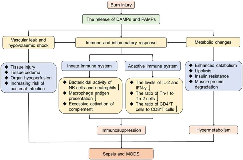
Burns & Trauma, 2021, Vol. 9, tkaa047                                                                                                                            3

                                                                                                                                                                      Downloaded from https://academic.oup.com/burnstrauma/article/doi/10.1093/burnst/tkaa047/6128653 by guest on 14 November 2021
Figure 2. Lund and Browder diagrams for estimation of total burned surface area (TBSA). The “rule of nines” (using multiples of 9) is frequently used to assess the
proportion of TBSA affected in adults and to help guide immediate treatment decisions based on burn size. However, the rule of nines is inaccurate in children
due to different head-to-body size ratios at different ages. Lund and Browder diagrams are therefore more suitable for assessing of the proportion of TBSA
affected in both children and adults. The body areas are separated into different regions (including anterior and posterior) by dashed lines and the numbers are
percentages of the TBSA. For instance, 19 in the diagram for children aged 1–4 years relates to the face, neck and head that make up 19% of the TBSA

sepsis because of skin loss that suggests the risk of infection is                  to have more complications related to infection [28, 29]. For
present as long as the burn wounds have not healed [21, 23].                        example, AIDS patients have a higher incidence of sepsis and
   In this review, we seek to address the major pathogenesis                        a longer period of hospitalization than HIV-negative patients,
of current categories of infection, sepsis and septic shock in                      although data on the reported outcome are limited due to the
patients with burn injury. In addition, the recent diagnostic                       small number of AIDS patients [30].
tools and potential biomarkers, including C-reactive protein                            Indeed, burn wound infection can be considered a series of
(CRP), procalcitonin (PCT) and cytokines, are intensively                           dynamic pathophysiological processes, including microbial
discussed.                                                                          colonization, biofilm formation and invasive burn wound
                                                                                    infection. Microorganisms can rapidly colonize the burn
                                                                                    wounds due to thermal destruction of the skin barrier. Burn
Review                                                                              eschar (avascular necrotic tissue) caused by deep partial-
Burn wound infections                                                               thickness and full-thickness burns provides a protein-rich
Burn wound infection, one of the most important causes                              niche for bacterial colonization and proliferation [31, 32]. In
of sepsis, is associated with high fatality rates in patients                       addition, burn eschar may also increase the risk of infection
with burn injury. The occurrence of burn wound infection                            by inhibiting early healing via basic fibroblast growth
often surfaces during the acute post-injury period and exhibits                     factor-induced endothelial cell proliferation and sprouting
considerable differences among burn patients of different ages                      [33]. However, eschar factors can inhibit hypertrophic scar
[21, 24]. Young children (under the age of 4 years) and                             formation of full-thickness burn wounds by preventing
elderly adults (over the age of 55 years) have a higher risk                        excessive granulation tissue formation [33].
of being infected, with higher fatality rates compared with                             Once planktonic (free-living) organisms form aggregates
other age groups [25, 26]. A possible cause is that infants,                        and attach to burn wounds, the formation of biofilm is initi-
young children and the elderly have an increased inclination                        ated. Biofilms are defined as structured communities encased
for deep burn injury due to their much thinner dermal layer                         in a self-produced extracellular polysaccharide matrix, or
[27]. Another reason may be the poor compliance of these                            slime [34, 35]. A mature biofilm provides efficient barriers
patients with early medical care and drug regimens. Apart                           for microorganisms against the host immune system and
from the above, some special populations, including obese                           antimicrobial agents, including biocides, antibiotics, oxidiz-
adults, diabetes patients and AIDS patients, have a higher                          ing agents and nano-drugs [36]. For example, the microor-
incidence of burn wound infection and have also been shown                          ganisms within biofilms have an increased capacity to tolerate
4                                                                                                                 Burns & Trauma, 2021, Vol. 9, tkaa047

Table 1. Sequential organ failure assessment (SOFA) scoring system [37]

Six organ systems           SOFA score 0              SOFA score 1               SOFA score 2               SOFA score 3              SOFA score 4

Respiratory system:         ≥53.3
Burns & Trauma, 2021, Vol. 9, tkaa047                                                                                             5

   In the first 5–7 days after injury, burn wounds are occupied      the inflammatory response (Figure 4) is uncontrolled and
by other microorganisms, such as gram-negative bacteria,             leads to vascular endothelium dysfunction, delayed healing,
fungi and viruses. For example, Pseudomonas aeruginosa               immune suppression and systemic inflammatory response
(P. aeruginosa), a gram-negative organism, is a common               syndrome [63]. Metabolically, inflammation also causes an
culprit of burn wound infection in the intensive care unit           enhanced catabolic state that is associated with an increased
due to their multi drug resistances and multiple virulence           incidence of sepsis and multiple organ failure. Compared with
factors [53, 54]. In a study of P. aeruginosa prevalence in          patients with only burns, the level of catabolism in septic
Chinese burn wards from 2007 to 2014, Dou and coworkers              burn patients is more than doubled, as measured using stable
showed that the detection rate of P. aeruginosa in hospitalized      isotope perfusion [64].

                                                                                                                                       Downloaded from https://academic.oup.com/burnstrauma/article/doi/10.1093/burnst/tkaa047/6128653 by guest on 14 November 2021
burn patients increased from 10.20% in 2007 to 26.16% in
2014 [55]. The main cause of this growing trend may be the
metabolic versatility of P. aeruginosa, its ability to colonize of   Vascular leak and hypovolemic state
a wide range of ecological niches and its low outer membrane         Interconnected microvessels are crucial for substance
permeability, which can resist antiseptics and antibiotics [56].     exchange (nutrients, oxygen and metabolic waste) between
Similar to S. aureus, P. aeruginosa also has quite a lot of vir-     blood and surrounding tissues through the regulation of
ulence factors, including adhesins, lipopolysaccharides, elas-       local hydrostatic and oncotic pressures [65]. Under the
tases, exoenzyme S, exotoxin A, leukocidins and proteases.           regulation of physical structure and chemical messenger,
These make P. aeruginosa a major cause of bloodstream inva-          the vascular barrier function maintains tissue perfusion and
sion, sepsis and poor prognosis in severely burned patients          homeostasis to adapt to physiological stimuli. The vascular
[57, 58]. It remains unclear whether the formation of biofilm        barrier function maintains tissue perfusion and homeostasis
or invasive burn wound infection is the important inducer            to adapt to physiological stimuli under the regulation of
for sepsis. Therefore, the prevention of burn wound infection        physical structure and chemical messenger. However, in
remains the better choice to diminish the incidence of sepsis        some traumatic injuries, particularly burn injuries, structural
and septic shock.                                                    disruption and inflammatory mediators lead to increased
                                                                     vascular permeability, which contributes to leakage of the
                                                                     intravascular fluids into the interstitial space, leading to
Events leading to sepsis and septic shock following                  further profound tissue edema and even hypovolemic shock
burn injury                                                          [66].
Sepsis and MODS are common complications of invasive                     Multiple mediators of barrier function, including his-
burn infection and are responsible for a significant proportion      tamine, bradykinin, platelet-activating factor, leukotrienes,
of the mortality in patients with burn injuries, particularly        vascular endothelial growth factor (VEGF) and VEGF
severe burns. Williams et al. performed statistical analysis         receptors, affect vascular hyperpermeability by inducing
on patients with burn injury and found that 55% of males             cellular signaling and structural alterations. Histamine is
and 54% of females died from sepsis and infections between           released mainly from mast cells and increases vascular
1989 to 2009, but the data short of highlighting the global          permeability through vascular dilation, increasing of blood
prevalence of this trend [59]. The limitation is attributed          flow and endothelial barrier disruption. There is increasing
mainly by neglecting low-income and middle-income coun-              evidence that nitric oxide (NO) and RhoA/Rho-associated
tries, which may have a higher incidence of sepsis in burn           protein kinase (ROCK) play an important role in histamine-
patients. Therefore, clear diagnostic criteria for burn sepsis       induced hyperpermeability. Ashina and coworkers found
are necessary to minimize and prevent septic complications.          that inhibiting NO synthesis with a nitric oxide synthase
The updated criterion in the Sepsis-3 consensus definition           (NOS) inhibitor, Nitro-L-arginine methyl ester (L-NAME)
established in 2016 focuses more on multiple organ dysfunc-          could alleviate the histamine-induced blood flow increase and
tion than on signs of inflammation [15], compared with the           hyperpermeability [67]. In addition, the authors also found
American Burn Association (ABA) sepsis criteria (2007) [60]          that histamine disrupted the endothelial barrier due to the
and the Mann-Salinas novel burn-specific sepsis predictors           localization of vascular endothelial cadherin (VE-cadherin)
(2013) [61, 62] (Table 3). This is particularly discerning,          (a cadherin maintaining the junction of adjacent endothelial
considering that a series of pathophysiological events can           cells) at the endothelial cell junction being changed [67].
lead to sepsis and multiple organ failure, including inflam-         Consistent with this observation, Mikelis et al. suggested that
matory response, hypovolaemic shock and vascular leak,               histamine induced the localization of VE-cadherin adhesion
immune dysregulation and hypermetabolism (Figure 3), with            complexes to focal adherens junctions through Rho/ROCK
inflammation present almost throughout the whole process             and further led to the formation of gaps in the endothelial
from initial injury to burn wound healing. Inflammation              barrier [68].
behaves like a double-edged sword in burn injuries: immedi-              Bradykinin, like histamine, activates RhoA and induces the
ately following minor burn injuries, inflammatory responses          rearrangement of the cytoskeleton and disassembly of tight
are initiated to activate the cascade of signals required for        junction (TJ) proteins. These changes can cause vascular leak-
wound healing [8]; however, in patients with severe burns,           age, although vascular hyperpermeability was observed in
6 Burns & Trauma, 2021, Vol. 9, tkaa047
Table 3. Different criteria for sepsis
Consensus definitions Criteria Predictors
ABA Sepsis Criteria [59] At least one or more of the following 1) Positive culture
2) Pathologic tissue source identified
3) Clinical response to antimicrobial agents
AND at least three of the following 1) Temperature >39 ◦ C or 110 bpm)
3) Progressive tachypnoea
Downloaded from https://academic.oup.com/burnstrauma/article/doi/10.1093/burnst/tkaa047/6128653 by guest on 14 November 2021
4) Thrombocytopenia
5) Hyperglycaemia 6) Inability to continue enteral
feedings 24 hours
Mann-Salinas et al. Novel burn-specific sepsis Predictors 1) Tachycardia >130 bpm
predictors [60, 61] 2) MAPBurns & Trauma, 2021, Vol. 9, tkaa047 7
Downloaded from https://academic.oup.com/burnstrauma/article/doi/10.1093/burnst/tkaa047/6128653 by guest on 14 November 2021
Figure 3. A series of pathogenic events responsible for sepsis post burn injury. Following severe burn injury, damaged tissues lead to the release of endogenous
DAMPs (such as double-stranded RNA and mitochondrial DNA) and exogenous PAMPs (such as lipopolysaccharides and peptidoglycans). Subsequently, PAMPs
can result in vascular leak and hypovolemic shock, immune and inflammatory responses and metabolic changes. Vascular leak causes tissue edema, organ
hypoperfusion and increasing risk of bacterial infection. Meanwhile, the excessive inflammatory response leads to immunosuppression by inhibition of the
innate and adaptive immune systems. Moreover, hypermetabolism emerges in the form of enhanced catabolism, lipolysis, insulin resistance and muscle protein
degradation. These events contribute to the susceptibility of the burn patients to sepsis and MODS. DAMPs damage-associated molecular patterns, PAMPs
pathogen-associated molecular pattern molecules, NK natural killer, IL-2 interleukin 2, IFN-γ interferon γ , Th-1 helper T lymphocyte 1, Th-2 helper T lymphocyte 2,
MODS multiple organ dysfunction syndrome
including monocytes, macrophages, dendritic cells, natural In addition to impairing the function of the innate
killer (NK) cells and neutrophils, are among the first immune immune system, severe burns reduce the total numbers of
cells to respond to wounds and coordinate the wider immune T lymphocytes, which play dominant roles in the adaptive
response. Following burn injuries, the antimicrobial actions immune system [93, 94]. Surprisingly, not all T lymphocytes
of neutrophils and NK cells are impaired [81–83]. Coinciden- are diminished, with helper T lymphocyte 2 (Th-2) present
tally, the phagocytic capacity of macrophages is also dimin- in increased numbers due to the increased levels of IL-4
ished in severe burn [84]. In addition, increased apoptosis and IL-10 [95]. Moreover, the reduced levels of IL-2 and
of conventional and plasmacytoid dendritic cells has been interferon-γ also cause the increase of Th-2 following burn
observed in patients with sepsis [85]. The complement system injuries [96]. The depressed levels of IL-2 and interferon-
represents an evolutionarily conserved and important element γ and high levels of IL-4 and IL-10 simultaneously inhibit
of the innate immune system [85]. According to the severity of the activity of helper T lymphocyte (Th-1) that support cell-
burns, the levels of complement decrease to different extents mediated immune responses [95]. The decreased ratio of Th-1
at the beginning of burn injury and subsequently rise to to Th-2 is an important etiologic factor in the suppression of
unprecedented levels [87]. The increased complements, such adaptive immune responses [27]. Furthermore, the ratio of
as C3a, C3b and C5a, may suppress immune response directly CD4-positive T helper cells to CD8-positive T suppressor
by impairing the function of leukocytes and lymphocytes cells also declines after severe burn [97]. Similarly, burn injury
[88, 89]. Intriguingly, multiple interleukins (IL, a kind of results in immune dysregulation by destabilizing the balance
lymphokine), such as IL-4 and IL-10, can significantly inhibit between helper T lymphocyte 17 (Th-17) and regulatory
the antigen presentation of macrophage and the bactericidal T cell, which plays prominent roles in protection against
activity of NK cells and neutrophils [90–92]. bacterial infections. On the one hand, Th-17 responses have8 Burns & Trauma, 2021, Vol. 9, tkaa047
Downloaded from https://academic.oup.com/burnstrauma/article/doi/10.1093/burnst/tkaa047/6128653 by guest on 14 November 2021
Figure 4. The host response to infection and during sepsis. After infection, PAMPs are released and interact with cell-surface, intracellular and even secreted PRRs,
including toll-like receptors, nucleotide-binding oligomerization domain-like receptors, retinoic acid-inducible gene-like receptors and C-type lectin receptors.
The interaction between PAMPs and PRRs can result in cytokine secretion, immune cell apoptosis and the activation of the complement system. In some
burn patients, these events lead to the simultaneous imbalanced activation of proinflammatory response (excessive inflammation) and anti-inflammatory
response (immune suppression). Excessive inflammation can result in the dysfunction of the endothelial barrier, microvascular thrombi and further injuries.
Immunosuppression causes decreased bactericidal activity of neutrophils and NK cells, decreased phagocytosis and antigen presentation of macrophages and
impaired innate immune system response. Taken together, excessive inflammation and immunosuppression contribute to a greatly increased risk for sepsis
and organ dysfunction. PAMPs pathogen-associated molecular pattern molecules, PRR pattern recognition receptors, DAMPs damage-associated molecular
patterns, NK natural killer, Th-2 helper T lymphocyte 2
been shown to be elicited in murine models of burn injury adaptive immune responses result in enhanced susceptibility
[98]. The perturbation of Th-17 cytokines IL-17 and IL-22 to infection, sepsis and multiple organ failure.
may further delay wound healing and promote burn sepsis
[99]. On the other hand, the proportion of Treg cells is Hypermetabolic state in bury injury
increased in patients with burn injury and this may decrease In addition to hypovolemic response and immune dys-
effector T cell function and further contribute to sepsis [100]. function, the hypermetabolic state following burn trauma
Taken together, the compromised alterations in innate and is another primary contributor to multiple organ failureBurns & Trauma, 2021, Vol. 9, tkaa047 9 and sepsis. Hypermetabolism (increased metabolic rate) is patients is attributed to these ATP consuming reactions [111]. characterized by an elevated (>10% above normal) resting Mitochondria, as the powerhouse of cells, play an important energy expenditure [101] and is more likely to occur in role in ATP production, mainly via the coupling of oxidative severe burns (>20% TBSA). Studies have demonstrated that phosphorylation. However, the coupling of mitochondrial there is an increase of 40–80% in resting energy expenditure respiration to adenosine diphosphate phosphorylation is during the acute phase of post-burn injury in patients with significantly attenuated in patients with burns [101]. On the burns of more than 40% TBSA [102, 103]. Generally, the contrary, the uncoupling of oxidative phosphorylation, which metabolic level is attenuated in the early stage of burn contributes to proton conductance via uncoupling proteins (
10                                                                                           Burns & Trauma, 2021, Vol. 9, tkaa047

aggravate hypotension and accelerate the occurrence of septic       Procalcitonin PCT, as a popular biomarker in bacterial infec-
shock. Both thrombosis and hypotension also impair tissue           tions and sepsis, has been studied extensively and utilized
oxygenation and aggravate organ dysfunction. In addition,           clinically [136, 137]. Several studies have compared PCT to
mitochondrial dysfunction, caused by oxidative stress and the       CRP in the diagnosis of sepsis, and most of the evidence
uncoupling mitochondria respiration, impairs cellular               suggests that PCT is superior to CRP [138–140]. PCT, the
oxygen use and the normal operation of vital organs                 prohormone of calcitonin, is a 116-amino acid polypeptide
[125]. For instance, in a septic mouse model, there is              encoded by the CALC-1 gene [141]. PCT is mainly produced
overproduction of reactive oxygen species and reactive              by neuroendocrine cells of the thyroid and its expression is
nitrogen species, which have been shown to impact negatively        inhibited in non-endocrine tissues under normal physiological

                                                                                                                                        Downloaded from https://academic.oup.com/burnstrauma/article/doi/10.1093/burnst/tkaa047/6128653 by guest on 14 November 2021
on myocardial mitochondrial function and cardiomyocyte              conditions [142]. Bacterial infection facilitates the transcrip-
contractility [126]. The contractility of cardiomyocytes is         tion of CALC-1 gene in non-endocrine cells and increases
also impaired by other mechanisms, such as the cell-surface         PCT levels to a peak during the first 20 hours after infection
adhesion molecule ICAM-1 [127], small calcium-regulated             [143]. The increasing serum levels of PCT in patients with
molecules (S100A8 and S100A9) [128] and inappropriate               burn was first reported by Assicot and colleagues, who con-
mitochondrial autophagy (mitophagy) [129]. In short, septic         jectured that levels of PCT are associated with the progression
cardiomyopathy exacerbates hypoperfusion of other organs            of infections, sepsis and septic shock [136]. Consistent of this
and accelerates the progression of sepsis, ultimately resulting     hypothesis, Brunkhorst et al. showed that levels of PCT were
in septic shock.                                                    proportional to the severity of sepsis in critically ill patients
                                                                    [144]. Conversely, recent studies by Seoane et al. and Paratz
                                                                    et al. found no association between PCT levels and sepsis in
Diagnosis of sepsis after burn trauma                               adult burn patients [145, 146]. Therefore, like CRP, PCT is
Due to the high lethality of sepsis, it is particularly important   not specific in early diagnosis of sepsis in burn patients.
to choose an appropriate definition and criteria for early
diagnosis and prediction of sepsis in burn patients. Yan and        Cytokines Major burn injuries are often accompanied by
coworkers compared the sensitivity of the sepsis criteria of        an inflammatory response that results in the activation of
the ABA definition, the Mann-Salinas definition, and the            inflammatory pathways and the augmentation of various
new Sepsis-3 consensus definition in patients with burn and         cytokines, including proinflammatory cytokines (TNF-α,
found Sepsis-3 to have a higher sensitivity (85%) than the          IL-6, IL-8) and anti-inflammatory cytokines (IL-10) [147].
ABA (60%) and Mann-Salinas (20%) definitions. The major             Recently, the potential of these cytokines in the early diagnosis
advantage of Sepsis-3 is that it argues that the multiple           of sepsis post burn injury has been investigated. Compared
organ dysfunction is more sepsis-specific than inflammation         with the burn patients without signs of sepsis, higher levels of
[15]. Despite this, it remains challenging to differentiate sep-    TNF-α were observed in burn patients with sepsis [148]. This
sis from systemic inflammatory response syndrome because            difference also appears in serum IL-6 values between the burn
they have similar clinical manifestations in multiple aspects,      patients with and without sepsis [147]. In addition, a clinical
including core body temperature, respiratory rate, heart rate       study, in which 468 children with burn injuries were divided
and hyperglycemia [101, 130]. Thus, the importance of more          into 2 groups based on IL-8 levels, has shown the positive
specific diagnostic and prognostic tools of burn sepsis cannot      correlation between the serum levels of IL-8 and sepsis in
be overemphasized. Several utilized clinical and promising          pediatric patients with elevated IL-8 [149]. Interestingly, IL-
predictors associated with burn sepsis are presented in this        10, an anti-inflammatory cytokine, has a negative impact on
section.                                                            the production of proinflammation cytokines, whereas the
                                                                    elevation of serum IL-10 levels is also correlated with the
C-reactive protein CRP is an evolutionarily conserved pro-          development of sepsis and even the risk of mortality in burn
tein and is produced primarily by hepatocytes following             patients [150, 151]. Taken together, these findings indicate
induction by inflammatory cytokines, such as IL-6 [131]. This       that cytokines hold great early diagnostic potential in sepsis
biomarker of inflammation in acute-phase responses has been         and further studies will be needed to verify this.
widely adopted in clinical settings. In healthy individuals, the
levels of CRP in plasma are almost undetectable, while more         Promising biomarkers Presepsin, a glycoprotein fragment
than 500 mg/l can be observed in patients with burn trauma          produced by monocytes and macrophages, is a soluble
[132]. Its levels may further increase in burn patients with        subtype of the cluster of differentiation 14 [152]. This
infection or sepsis [133], thus previous studies have suggested     glycoprotein recognizes and interacts with endotoxin
CRP as a good predictor of sepsis in burn patients. However,        complexes for the activation of systemic inflammatory
recent evidence has shown that CRP has drawbacks in the             signaling pathways [153]. There is mounting evidence
specific diagnosis of sepsis in severely burned patients [134,      to indicate that presepsin is a promising biomarker for
135]. Taken together, CRP may not be a specific biomarker           diagnosing sepsis in burn patients, although it cannot be
of sepsis, but its levels have important reference value in con-    used alone to confirm or exclude the presence of sepsis in
junction with other tools, such as PCT and some cytokines.          burn patients [154–157]. Mid-regional pro-atrial natriuretic
Burns & Trauma, 2021, Vol. 9, tkaa047 11
peptide is another promising biomarker, and Gille et al., Abbreviations
in a prospective observational study of 42 burn patients, MODS: multiple organ dysfunction syndrome; TBSA: total body
found that burn patients with sepsis have higher levels of surface area; Sepsis-3: Third International Consensus Definition for
this peptide and PCT [158]. Moreover, Hampson et al. found Sepsis and Septic Shock; CRP: C-reactive protein; PCT: procalcitonin;
that neutrophil function, immature granulocyte count and VEGF: vascular endothelial growth factor; ROCK: Rho-associated
plasma cell-free DNA levels showed significant potential protein kinase; NO: nitric oxide; VE-cadherin: vascular endothelial
for the early diagnosis of sepsis in burn patients [159]. cadherin; TNF: tumor necrosis factor; NK: natural killer; WAT:
Especially interesting w that micro RNA can also serve white adipose tissue; ABA: American Burn Association; ABA:
as a diagnostic biomarker. An example of this is miR- American Burn Association; ATP: adenosine triphosphate; CRP:
Downloaded from https://academic.oup.com/burnstrauma/article/doi/10.1093/burnst/tkaa047/6128653 by guest on 14 November 2021
C-reactive protein; DAMPs: damage-associated molecular patterns;
495, which is significantly downregulated in patients with
ER: endoplasmic reticulum; IL: interleukins; MAP: mean arterial
sepsis and negatively correlated with CRP and PCT [160].
pressure; MODS: multiple organ dysfunction syndrome; NK:
Although numerous promising biomarkers of sepsis have natural killer; NO: nitric oxide; PAMPs: pathogen-associated
been discovered, none of them alone can diagnose sepsis post molecular pattern molecules; PCT: procalcitonin; ROCK: Rho-
burn injury, and their values must be interpreted with caution associated protein kinase; Sepsis-3: Third International Consensus
to ensure accurate diagnosis. Definition for Sepsis and Septic Shock; SOFA: Sequential Organ
Failure Assessment; TBSA: total body surface area; Th-1: helper
Conclusions T lymphocyte; Th-2: helper T lymphocyte 2; Th-17: helper T
lymphocyte 17; TNF: tumor necrosis factor; TJ: tight junction; VE-
Sepsis and septic complications not only account for the poor cadherin: vascular endothelial cadherin; VEGF: vascular endothelial
outcomes in burn patients, but also prolonged hospital stays growth factor; WAT: white adipose tissue.
and higher medical costs. Burn wound infection is a major
cause of sepsis development in patients with severe burns.
Moreover, other events following burn injury play impor- References
tant roles in the occurrence of sepsis, such as vascular leak,
1. Jeschke MG, van Baar ME, Choudhry MA, Chung KK, Gibran
hypovolemia, hypermetabolism and immune dysregulation.
NS, Logsetty S. Burn injury. Nat. Rev. Dis. Primers. 2020.
Integrated management, including, but not limited to, fluid https://doi.org/10.1038/s41572-020-0145-5.
resuscitation, nutritional support, antimicrobial therapy and 2. National Burn Repository 2019 Update, Report of data
vasoactive medications is beneficial for the prevention and from 2009–2018 ameriburn.site- ym.com [Internet]. 2019.
prognosis of sepsis by targeting the events leading to sepsis Available from: https://ameriburn.site-ym.com/store/ViewPro
following burn injury. However, the prediction and diagnosis duct.aspx?id=14191872.
of sepsis or infection remains an ongoing challenge in burn 3. Pereira RF, Barrias CC, Granja PL, Bartolo PJ. Advanced
patients, although numerous predictors for burn sepsis have biofabrication strategies for skin regeneration and repair.
been reported. More investigations are needed to explore Nanomedicine (Lond.) 2013;8:603–21.
novel diagnostic tools of burn sepsis due to the unreliability 4. Peck MD. Epidemiology of burns throughout the world. Part
I: distribution and risk factors. Burns 2011;37:1087–100.
and limitation of the established biomarkers (CRP, PCT and
5. Cioffi WG. deLemos RA, Coalson JJ, Gerstmann DA, Pruitt
cytokines). Meanwhile, Sepsis-3, which can be applied for
BA Jr. decreased pulmonary damage in primates with inhala-
analysis or research purposes, appeared to be a better defi- tion injury treated with high-frequency ventilation. Ann. Surg.
nition of sepsis. Based on this definition and patient-specific 1993;218:328–35 discussion 35-7.
molecular and biochemical profiles, clinicians can design an 6. Finnerty CC, Herndon DN, Jeschke MG. Inhalation injury
individualized management strategy which may improve the in severely burned children does not augment the systemic
prognosis of burn patients with sepsis. inflammatory response. Crit. Care 2007. https://www.doi.o
rg/10.1186/cc5698.
Acknowledgements 7. Xiao W, Mindrinos MN, Seok J, Cuschieri J, Cuenca AG, Gao
H, et al. A genomic storm in critically injured humans. J. Exp.
We are grateful to Dr Yew Mun Lee for his helpful discussion and
Med. 2011;208:2581–90.
suggestions.
8. Stanojcic M, Abdullahi A, Rehou S, Parousis A, Jeschke MG.
Pathophysiological response to burn injury in adults. Ann.
Funding Surg. 2018;267:576–84.
9. Jeschke MG, Gauglitz GG, Kulp GA, Finnerty CC, Williams
This work was supported by grants from the National Natural
FN, Kraft R, et al. Long-term persistence of the pathophysio-
Science Foundation of China (81821002, 81790251 and 81672381),
logic response to severe burn injury. PLoS One 2011;6:e21245.
the Guangdong Basic and Applied Basic Research Foundation
https://www.doi.org/10.1371/journal.pone.0021245.
(2019B030302012), the Science and Technology Department of
10. Martin GS, Mannino DM, Eaton S, Moss M. The epidemiology
Sichuan Province (2018RZ0133) and the Chengdu Science and
of sepsis in the United States from 1979 through 2000. N. Engl.
Technology Program (2019-YF05–00715-SN).
J. Med. 2003;348:1546–54.
11. Rodríguez-Luna A, Ávila-Román J, González-Rodríguez ML,
Conflicts of interest Cózar MJ, Rabasco AM, Motilva V, et al. Fucoxanthin-
The authors declare no conflict of interest. containing cream prevents epidermal hyperplasia and UVB-12 Burns & Trauma, 2021, Vol. 9, tkaa047
induced skin erythema in mice. Mar. Drugs 2018. https://www. 30. Salehi SH, As’adi K, Tabatabaeenezhad SA, Naderan M,
doi.org/10.3390/md16100378. Shoar S. Prevalence of HIV infection among burn patients: is
12. Bone RC, Sibbald WJ, Sprung CL. The ACCP-SCCM there a relationship with patients’ outcomes? Int. Wound J.
consensus conference on sepsis and organ failure. Chest 2017;14:85–8.
1992;101:1481–3. 31. Barret JP, Herndon DN. Effects of burn wound excision
13. Neely AN, Fowler LA, Kagan RJ, Warden GD. Procalcitonin on bacterial colonization and invasion. Plast. Reconstr. Surg.
in pediatric burn patients: an early indicator of sepsis? J. Burn 2003;111:744–50.
Care Rehabil. 2004;25:76–80. 32. Evdokiou A, Kanisicak O, Gierek S, Barry A, Ivey MJ, Zhang
14. Stanojcic M, Vinaik R, Jeschke MG. Status and challenges of X, et al. Characterization of burn eschar pericytes. J. Clin. Med.
predicting and diagnosing sepsis in burn patients. Surg. Infect. 2020;9.
Downloaded from https://academic.oup.com/burnstrauma/article/doi/10.1093/burnst/tkaa047/6128653 by guest on 14 November 2021
2018;19:168–75. 33. Monsuur HN, van den Broek LJ, Jhingoerie RL, Vloemans A,
15. Singer M, Deutschman CS, Seymour CW, Shankar-Hari M, Gibbs S. Burn eschar stimulates fibroblast and adipose mes-
Annane D, Bauer M, et al. The third international consen- enchymal stromal cell proliferation and migration but inhibits
sus definitions for sepsis and septic shock (Sepsis-3). JAMA endothelial cell sprouting. Int. J. Mol. Sci. 2017. https://www.
2016;315:801–10. doi.org/10.3390/ijms18081790.
16. Li W, Wang M, Zhu B, Zhu Y, Xi X. Prediction of median 34. Costerton JW, Stewart PS, Greenberg EP. Bacterial biofilms:
survival time in sepsis patients by the SOFA score combined a common cause of persistent infections. Science (New York,
with different predictors. Burns Trauma 2020;8:tkz006. N.Y.) 1999;284:1318–22.
17. Jeschke MG, Patsouris D, Stanojcic M, Abdullahi A, Rehou S, 35. Flemming HC, Wingender J, Szewzyk U, Steinberg P, Rice SA,
Pinto R, et al. Pathophysiologic response to burns in the elderly. Kjelleberg S. Biofilms: an emergent form of bacterial life. Nat.
EBioMedicine 2015;2:1536–48. Rev. Microbiol. 2016;14:563–75.
18. Chai J, Sheng Z, Diao L, Yang H, Gao J, Xu M. Effect of 36. Koo H, Allan RN, Howlin RP, Stoodley P, Hall-Stoodley L. Tar-
extensive excision of burn wound with invasive infection on geting microbial biofilms: current and prospective therapeutic
hypermetabolism in burn patients with sepsis. Zhonghua Wai strategies. Nat. Rev. Microbiol. 2017;15:740–55.
Ke Za Zhi [Chinese journal of surgery]. 2000;38:405–8. 37. Vincent JL, Moreno R, Takala J, Willatts S, De Mendonça
19. Fitzwater J, Purdue GF, Hunt JL, O’Keefe GE. The risk factors A, Bruining H, et al. The SOFA (Sepsis-related Organ Failure
and time course of sepsis and organ dysfunction after burn Assessment) score to describe organ dysfunction/failure. On
trauma. J. Trauma 2003;54:959–66. behalf of the Working Group on Sepsis-Related Problems of the
20. Gomez R, Murray CK, Hospenthal DR, Cancio LC, Renz EM, European Society of Intensive Care Medicine. Intensive Care
Holcomb JB, et al. Causes of mortality by autopsy findings of Med 1996;22:707–10.
combat casualties and civilian patients admitted to a burn unit. 38. Rumbaugh KP, Sauer K. Biofilm dispersion. Nat. Rev. Micro-
J. Am. Coll. Surg. 2009;208:348–54. biol. 2020;18:571–86.
21. Greenhalgh DG. Sepsis in the burn patient: a different problem 39. Edwards R, Harding KG. Bacteria and wound healing. Curr.
than sepsis in the general population. Burns Trauma. 2017. Opin. Infect. Dis. 2004;17:91–6.
https://www.doi.org/10.1186/s41038-017-0089-5. 40. Harrison-Balestra C, Cazzaniga AL, Davis SC, Mertz PM.
22. Atiyeh BS, Gunn SW, Hayek SN. State of the art in burn A wound-isolated Pseudomonas aeruginosa grows a biofilm in
treatment. World J. Surg. 2005;29:131–48. vitro within 10 hours and is visualized by light microscopy.
23. Ren C, Yao RQ, Ren D, Li Y, Feng YW, Yao YM. Comparison Dermatol Surg. 2003;29:631–5.
of clinical laboratory tests between bacterial sepsis and SARS- 41. Kennedy P, Brammah S, Wills E. Burns, biofilm and a new
CoV-2-associated viral sepsis. Mil Med Res 2020;7:36. appraisal of burn wound sepsis. Burns 2010;36:49–56.
24. Warner PM, Coffee TL, Yowler CJ. Outpatient 42. Erol S, Altoparlak U, Akcay MN, Celebi F, Parlak M. Changes
burn management. Surg. Clin. North Am. 2014;94: of microbial flora and wound colonization in burned patients.
879–92. Burns 2004;30:357–61.
25. Lionelli GT, Pickus EJ, Beckum OK, Decoursey RL, Korentager 43. Norbury W, Herndon DN, Tanksley J, Jeschke MG, Finnerty
RA. A three decade analysis of factors affecting burn mortality CC. Infection in Burns. Surg. Infect. 2016;17:250–5.
in the elderly. Burns 2005;31:958–63. 44. Vollmer W, Blanot D, de Pedro MA. Peptidoglycan structure
26. Thombs BD, Bresnick MG. Mortality risk and length of stay and architecture. FEMS Microbiol. Rev. 2008;32:149–67.
associated with self-inflicted burn injury: evidence from a 45. Scott JR, Barnett TC. Surface proteins of gram-positive
national sample of 30,382 adult patients. Crit. Care Med. bacteria and how they get there. Annu. Rev. Microbiol.
2008;36:118–25. 2006;60:397–423.
27. Church D, Elsayed S, Reid O, Winston B, Lindsay R. Burn 46. Salgado-Pabón W, Breshears L, Spaulding AR, Merriman JA,
wound infections. Clin. Microbiol. Rev. 2006;19:403–34. Stach CS, Horswill AR, et al. Superantigens are critical for
28. Kraft R, Herndon DN, Williams FN, Al-Mousawi AM, Staphylococcus aureus infective endocarditis, sepsis, and acute
Finnerty CC, Jeschke MG. The effect of obesity on adverse kidney injury. MBio 2013;4. https://www.doi.org/10.1128/
outcomes and metabolism in pediatric burn patients. Int J Obes mBio.00494-13.
(Lond). 2012;36:485–90. 47. Lin YC, Peterson ML. New insights into the prevention of
29. Duke JM, Randall SM, Fear MW, Boyd JH, Rea S, Wood staphylococcal infections and toxic shock syndrome. Expert.
FM. Diabetes mellitus after injury in burn and non-burned Rev. Clin. Pharmacol. 2010;3:753–67.
patients: a population based retrospective cohort study. Burns 48. Dayan GH, Mohamed N, Scully IL, Cooper D, Begier E, Eiden
2018;44:566–72. J, et al. Staphylococcus aureus: the current state of disease,Burns & Trauma, 2021, Vol. 9, tkaa047 13
pathophysiology and strategies for prevention. Expert Rev. 65. Kottke MA, Walters TJ. Where’s the leak in vascular barriers?
Vaccines 2016;15:1373–92. A review. Shock (Augusta, Ga) 2016;46:20–36.
49. Maresso AW, Schneewind O. Sortase as a target of anti- 66. Arbuthnot MK, Garcia AV. Early resuscitation and man-
infective therapy. Pharmacol. Rev. 2008;60:128–41. agement of severe pediatric burns. Semin. Pediatr. Surg.
50. Foster TJ, Geoghegan JA, Ganesh VK, Höök M. Adhe- 2019;28:73–8.
sion, invasion and evasion: the many functions of the sur- 67. Ashina K, Tsubosaka Y, Nakamura T, Omori K, Kobayashi K,
face proteins of Staphylococcus aureus. Nat. Rev. Microbiol. Hori M, et al. Histamine induces vascular hyperpermeability
2014;12:49–62. by increasing blood flow and endothelial barrier disruption
51. Kwiecinski J, Jin T, Josefsson E. Surface proteins of Staphy- in vivo. PLoS One 2015. https://www.doi.org/10.1371/journa
lococcus aureus play an important role in experimental skin l.pone.0132367.
Downloaded from https://academic.oup.com/burnstrauma/article/doi/10.1093/burnst/tkaa047/6128653 by guest on 14 November 2021
infection. APMIS. 2014;122:1240–50. 68. Mikelis CM, Simaan M, Ando K, Fukuhara S, Sakurai
52. McAdow M, Kim HK, Dedent AC, Hendrickx AP, Schneewind A, Amornphimoltham P, et al. RhoA and ROCK medi-
O, Missiakas DM. Preventing Staphylococcus aureus sep- ate histamine-induced vascular leakage and anaphylactic
sis through the inhibition of its agglutination in blood. shock. Nat. Commun. 2015. https://www.doi.org/10.1038/nco
PLoS Pathog. 2011. https://www.doi.org/10.1371/journal.ppa mms7725.
t.1002307. 69. Ma T, Liu L, Wang P, Xue Y. Evidence for involve-
53. MacVane SH. Antimicrobial resistance in the intensive care ment of ROCK signaling in bradykinin-induced increase in
unit: a focus on gram-negative bacterial infections. J. Intensive murine blood-tumor barrier permeability. J. Neuro-Oncol.
Care Med. 2017;32:25–37. 2012;106:291–301.
54. Kaye KS, Pogue JM. Infections caused by resistant gram- 70. Liu LB, Xue YX, Liu YH, Wang YB. Bradykinin increases
negative bacteria: epidemiology and management. Pharma- blood-tumor barrier permeability by down-regulating the
cotherapy 2015;35:949–62. expression levels of ZO-1, occludin, and claudin-5 and rear-
55. Dou Y, Huan J, Guo F, Zhou Z, Shi Y. Pseudomonas aerug- ranging actin cytoskeleton. J. Neurosci. Res. 2008;86:1153–68.
inosa prevalence, antibiotic resistance and antimicrobial use 71. Duah E, Adapala RK, Al-Azzam N, Kondeti V, Gombedza F,
in Chinese burn wards from 2007 to 2014. J. Int. Med. Res. Thodeti CK, et al. Cysteinyl leukotrienes regulate endothelial
2017;45:1124–37. cell inflammatory and proliferative signals through CysLT2
56. Chevalier S, Bouffartigues E, Bodilis J, Maillot O, Lesouhaitier and CysLT1 receptors. Sci. Rep. 2013. https://www.doi.o
O, Feuilloley MGJ, et al. Structure, function and regulation rg/10.1038/srep03274.
of Pseudomonas aeruginosa porins. FEMS Microbiol. Rev. 72. Lee KS, Kim SR, Park HS, Jin GY, Lee YC. Cysteinyl leukotriene
2017;41:698–722. receptor antagonist regulates vascular permeability by reducing
57. Tredget EE, Shankowsky HA, Rennie R, Burrell RE, Logsetty S. vascular endothelial growth factor expression. J. Allergy Clin.
Pseudomonas infections in the thermally injured patient. Burns Immunol. 2004;114:1093–9.
2004;30:3–26. 73. Antonetti DA, Barber AJ, Hollinger LA, Wolpert EB, Gardner
58. Dzvova N, Colmer-Hamood JA, Griswold JA, Hamood TW. Vascular endothelial growth factor induces rapid phos-
AN. Heparinase is essential for Pseudomonas aeruginosa phorylation of tight junction proteins occludin and zonula
virulence during thermal injury and infection. Infect. Immun. occluden 1. A potential mechanism for vascular perme-
2018. https://www.doi.org/10.1128/IAI.00755-17. ability in diabetic retinopathy and tumors. J. Biol. Chem.
59. Williams FN, Herndon DN, Hawkins HK, Lee JO, Cox RA, 1999;274:23463–7.
Kulp GA, et al. The leading causes of death after burn injury 74. Chen XL, Nam JO, Jean C, Lawson C, Walsh CT, Goka E, et al.
in a single pediatric burn center. Crit. Care 2009. https://www. VEGF-induced vascular permeability is mediated by FAK. Dev.
doi.org/10.1186/cc8170. Cell 2012;22:146–57.
60. Greenhalgh DG, Saffle JR, Holmes JH, Gamelli RL, Palmieri 75. Vasile E, Qu H, Dvorak HF, Dvorak AM. Caveolae and
TL, Horton JW, et al. American burn association consensus vesiculo-vacuolar organelles in bovine capillary endothelial
conference to define sepsis and infection in burns. J Burn Care cells cultured with VPF/VEGF on floating Matrigel-collagen
Res. 2007;28:776–90. gels. J. Histochem. Cytochem. 1999;47:159–67.
61. Mann-Salinas EA, Baun MM, Meininger JC, Murray CK, 76. Feng D, Nagy JA, Hipp J, Dvorak HF, Dvorak AM. Vesiculo-
Aden JK, Wolf SE, et al. Novel predictors of sepsis out- vacuolar organelles and the regulation of venule permeability
perform the American burn association sepsis criteria in the to macromolecules by vascular permeability factor, histamine,
burn intensive care unit patient. J Burn Care Res. 2013;34: and serotonin. J. Exp. Med. 1996;183:1981–6.
31–43. 77. van der Flier M, van Leeuwen HJ, van Kessel KP, Kimpen JL,
62. Yan J, Hill WF, Rehou S, Pinto R, Shahrokhi S, Jeschke Hoepelman AI, Geelen SP. Plasma vascular endothelial growth
MG. Sepsis criteria versus clinical diagnosis of sepsis in factor in severe sepsis. Shock (Augusta, Ga) 2005;23:35–8.
burn patients: a validation of current sepsis scores. Surgery 78. Zang Q, Maass DL, White J, Horton JW. Cardiac mito-
2018;164:1241–5. chondrial damage and loss of ROS defense after burn injury:
63. Jeschke MG, Chinkes DL, Finnerty CC, Kulp G, Suman OE, the beneficial effects of antioxidant therapy. J Appl Physiol.
Norbury WB, et al. Pathophysiologic response to severe burn 2007;102:103–12.
injury. Ann. Surg. 2008;248:387–401. 79. Willis MS, Carlson DL, Dimaio JM, White MD, White DJ,
64. Hart DW, Wolf SE, Mlcak R, Chinkes DL, Ramzy PI, Obeng Adams GA, et al. Macrophage migration inhibitory factor
MK, et al. Persistence of muscle catabolism after severe burn. mediates late cardiac dysfunction after burn injury. Am. J.
Surgery 2000;128:312–9. Physiol. Heart Circ. Physiol. 2005;288:H795–804.14 Burns & Trauma, 2021, Vol. 9, tkaa047
80. Nielson CB, Duethman NC, Howard JM, Moncure M, Wood 100. Venet F, Pachot A, Debard AL, Bohe J, Bienvenu J, Lep-
JG. Burns: pathophysiology of systemic complications and ape A, et al. Human CD4+CD25+ regulatory T lympho-
current management. J Burn Care Res. 2017;38:e469–81. cytes inhibit lipopolysaccharide-induced monocyte survival
81. Griswold JA. White blood cell response to burn injury. Semin. through a Fas/Fas ligand-dependent mechanism. J. Immunol.
Nephrol. 1993;13:409–15. 2006;177:6540–7.
82. Grogan JB. Altered neutrophil phagocytic function in burn 101. Porter C, Tompkins RG, Finnerty CC, Sidossis LS, Suman OE,
patients. J. Trauma 1976;16:734–8. Herndon DN. The metabolic stress response to burn trauma:
83. ter Haar NM, Oswald M, Jeyaratnam J, Anton J, Bar- current understanding and therapies. Lancet (London, Eng-
ron KS, Brogan PA, et al. Recommendations for the man- land) 2016;388:1417–26.
agement of autoinflammatory diseases. Ann. Rheum. Dis. 102. Porter C, Herndon DN, Børsheim E, Bhattarai N, Chao T,
Downloaded from https://academic.oup.com/burnstrauma/article/doi/10.1093/burnst/tkaa047/6128653 by guest on 14 November 2021
2015;74:1636–44. Reidy PT, et al. Long-term skeletal muscle mitochondrial dys-
84. Schildt BE. Function of the RES after thermal and mechanical function is associated with hypermetabolism in severely burned
trauma in mice. Acta Chir. Scand. 1970;136:359–64. children. J Burn Care Res. 2016;37:53–63.
85. Qiu T, Li M, Tanner MA, Yang Y, Sowers JR, Korthuis RJ, 103. Porter C, Herndon DN, Børsheim E, Chao T, Reidy PT, Borack
et al. Depletion of dendritic cells in perivascular adipose tissue MS, et al. Uncoupled skeletal muscle mitochondria contribute
improves arterial relaxation responses in type 2 diabetic mice. to hypermetabolism in severely burned adults. Am. J. Physiol.
Metab. Clin. Exp. 2018;85:76–89. Endocrinol. Metab. 2014;307:E462–7.
86. Vignesh P, Rawat A, Sharma M, Singh S. Complement 104. Auger C, Samadi O, Jeschke MG. The biochemical alter-
in autoimmune diseases. Clinica Chimica Acta. ations underlying post-burn hypermetabolism. Biochim. Bio-
2017;465:123–30. phys. Acta Mol. basis Dis. 2017;1863:2633–44.
87. Hammad A, Westacott L, Zaben M. The role of the 105. Wieser V, Moschen AR, Tilg H. Inflammation, cytokines and
complement system in traumatic brain injury: a review. insulin resistance: a clinical perspective. Arch. Immunol. Ther.
J. Neuroinflammation 2018. https://www.doi.org/10.1186/ Exp. 2013;61:119–25.
s12974-018-1066-z. 106. Houstis N, Rosen ED, Lander ES. Reactive oxygen species have
88. Hugli TE. Complement and cellular triggering reactions. Intro- a causal role in multiple forms of insulin resistance. Nature
ductory remarks. Federation proceedings. 1984;43:2540–2. 2006;440:944–8.
89. Conde P, Rodriguez M, van der Touw W, Jimenez A, Burns M, 107. Vandanmagsar B, Youm YH, Ravussin A, Galgani JE, Stadler
Miller J, et al. DC-SIGN(+) macrophages control the induction K, Mynatt RL, et al. The NLRP3 inflammasome instigates
of transplantation tolerance. Immunity 2015;42:1143–58. obesity-induced inflammation and insulin resistance. Nat. Med.
90. Donnelly RP, Fenton MJ, Kaufman JD, Gerrard TL. IL- 2011;17:179–88.
1 expression in human monocytes is transcriptionally 108. Herndon DN, Hart DW, Wolf SE, Chinkes DL, Wolfe RR.
and posttranscriptionally regulated by IL-4. J. Immunol. Reversal of catabolism by beta-blockade after severe burns. N.
1991;146:3431–6. Engl. J. Med. 2001;345:1223–9.
91. Fiorentino DF, Zlotnik A, Mosmann TR, Howard M, 109. Wang Y, Viscarra J, Kim SJ, Sul HS. Transcriptional reg-
O’Garra A. IL-10 inhibits cytokine production by activated ulation of hepatic lipogenesis. Nat. Rev. Mol. Cell Biol.
macrophages. J. Immunol. 1991;147:3815–22. 2015;16:678–89.
92. Oswald IP, Wynn TA, Sher A, James SL. Interleukin 10 inhibits 110. Jeschke MG, Herndon DN, Wolf SE, DebRoy MA, Rai J,
macrophage microbicidal activity by blocking the endogenous Thompson JC, et al. Hepatocyte growth factor modulates the
production of tumor necrosis factor alpha required as a costim- hepatic acute-phase response in thermally injured rats. Crit.
ulatory factor for interferon gamma-induced activation. Proc. Care Med. 2000;28:504–10.
Natl. Acad. Sci. U. S. A. 1992;89:8676–80. 111. Yu YM, Tompkins RG, Ryan CM, Young VR. The metabolic
93. Heideman M, Bengtsson A. The immunologic response to basis of the increase of the increase in energy expenditure
thermal injury. World J. Surg. 1992;16:53–6. in severely burned patients. JPEN J. Parenter. Enteral Nutr.
94. Sheridan RL, Weber JM, Pasternak MM, Mulligan JM, Tomp- 1999;23:160–8.
kins RG. A 15-year experience with varicella infections in a 112. Matthias A, Ohlson KB, Fredriksson JM, Jacobsson A, Neder-
pediatric burn unit. Burns 1999;25:353–6. gaard J, Cannon B. Thermogenic responses in brown fat cells
95. Gosain A, Gamelli RL. A primer in cytokines. J. Burn Care are fully UCP1-dependent. UCP2 or UCP3 do not substitute for
Rehabil. 2005;26:7–12. UCP1 in adrenergically or fatty acid-induced thermogenesis. J.
96. Schwacha MG. Macrophages and post-burn immune dysfunc- Biol. Chem. 2000;275:25073–81.
tion. Burns 2003;29:1–14. 113. Jeschke MG, Finnerty CC, Herndon DN, Song J, Boehning D,
97. Burleson DG, Mason AD, Jr, Pruitt BA, Jr. Lymphoid Tompkins RG, et al. Severe injury is associated with insulin
subpopulation changes after thermal injury and thermal resistance, endoplasmic reticulum stress response, and unfolded
injury with infection in an experimental model. Ann. Surg. protein response. Ann. Surg. 2012;255:370–8.
1988;207:208–12. 114. Wang C, Huang Z, Du Y, Cheng Y, Chen S, Guo F.
98. Rendon JL, Choudhry MA. Th17 cells: critical mediators of ATF4 regulates lipid metabolism and thermogenesis. Cell Res.
host responses to burn injury and sepsis. J. Leukoc. Biol. 2010;20:174–84.
2012;92:529–38. 115. Kraft R, Herndon DN, Finnerty CC, Hiyama Y, Jeschke MG.
99. Deitch EA, Bridges RM, Dobke M, McDonald JC. Burn wound Association of postburn fatty acids and triglycerides with clin-
sepsis may be promoted by a failure of local antibacterial host ical outcome in severely burned children. J. Clin. Endocrinol.
defenses. Ann. Surg. 1987;206:340–8. Metab. 2013;98:314–21.You can also read