Transneuronal delivery of hyper-interleukin-6 enables functional recovery after severe spinal cord injury in mice - Nature
←
→
Page content transcription
If your browser does not render page correctly, please read the page content below
ARTICLE
                  https://doi.org/10.1038/s41467-020-20112-4                 OPEN

                  Transneuronal delivery of hyper-interleukin-6
                  enables functional recovery after severe spinal
                  cord injury in mice
                  Marco Leibinger1, Charlotte Zeitler1, Philipp Gobrecht1, Anastasia Andreadaki1, Günter Gisselmann1 &
                  Dietmar Fischer 1 ✉
1234567890():,;

                  Spinal cord injury (SCI) often causes severe and permanent disabilities due to the regen-
                  erative failure of severed axons. Here we report significant locomotor recovery of both
                  hindlimbs after a complete spinal cord crush. This is achieved by the unilateral transduction
                  of cortical motoneurons with an AAV expressing hyper-IL-6 (hIL-6), a potent designer
                  cytokine stimulating JAK/STAT3 signaling and axon regeneration. We find collaterals of
                  these AAV-transduced motoneurons projecting to serotonergic neurons in both sides of the
                  raphe nuclei. Hence, the transduction of cortical neurons facilitates the axonal transport and
                  release of hIL-6 at innervated neurons in the brain stem. Therefore, this transneuronal
                  delivery of hIL-6 promotes the regeneration of corticospinal and raphespinal fibers after
                  injury, with the latter being essential for hIL-6-induced functional recovery. Thus, trans-
                  neuronal delivery enables regenerative stimulation of neurons in the deep brain stem that are
                  otherwise challenging to access, yet highly relevant for functional recovery after SCI.

                  1 Department   of Cell Physiology, Ruhr University of Bochum, Universitätsstraße 150, 44780 Bochum, Germany. ✉email: dietmar.fischer@rub.de

                  NATURE COMMUNICATIONS | (2021)12:391 | https://doi.org/10.1038/s41467-020-20112-4 | www.nature.com/naturecommunications                      1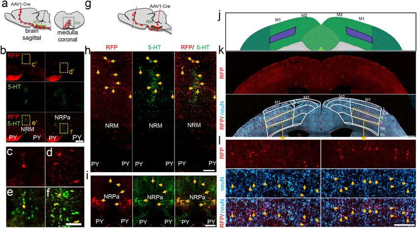
ARTICLE                                                                 NATURE COMMUNICATIONS | https://doi.org/10.1038/s41467-020-20112-4

N
         eurons of the adult mammalian central nervous system                regeneration associated signaling pathways in motoneurons, we
         (CNS) do not naturally regenerate injured axons. This               applied either AAV2-hIL-6 (GFP coexpression), AAV2-Cre (GFP
         regenerative failure often causes severe and permanent              coexpression), or AAV2-GFP into the sensorimotor cortex of
disabilities, such as para- or tetraplegia after spinal cord injury.         adult PTEN-floxed mice. Viral transduction affected mainly layer
To date, no cures are available in the clinic, underscoring the need         V neurons in the primary motor cortex, adjacent to the injection
for novel therapeutic strategies enabling functional recovery in             sites (Fig. 1a–c and Fig. S1a–e). Moreover, only AAV-hIL-6
respective patients.                                                         transduced cells showed the hIL-6 protein (Fig. S1f, g). Western
   Besides an inhibitory environment for axonal growth cones                 blot analysis verified these results, detecting hIL-6 in respective
caused by myelin or the forming glial scar, the lack of CNS                  tissues of AAV-hIL-6 treated animals only (Figs. S1h, i, S15d, e,
regeneration is mainly attributed to a developmental decline in              S16n, o). In contrast to AAV2-Cre (PTEN−/−) and AAV2-GFP,
the neuron-intrinsic growth capacity of axons per se1–3. Among               AAV2-hIL-6 application induced strong STAT3-phosphorylation
all descending pathways, the corticospinal tract (CST), which                as shown in sections of cortical tissue and western blot lysates
controls voluntary fine movements, is the most resistant to                   (Fig. 1b–g, k, m, n, o and Fig. S1j, k). These signals were seen in all
regeneration. Despite numerous efforts aiming to facilitate the              GFP-positive hIL-6-transduced neurons and adjacent cells, indi-
CST’s axon regrowth over the last decades, such as the delivery of           cating the paracrine effects of released hIL-6. STAT3 activation
neurotrophic factors4–6 or neutralizing inhibitory cues7–9, success          was already at its maximum 1 week after injection and remained
has remained very limited. However, the conditional genetic                  stable over at least 8 weeks (Fig. 1n, o, Fig. S14a-d, f-h; Fig. S16a),
knockout of the phosphatase and tensin homolog (PTEN−/−) in                  while total STAT3 protein expression was not significantly alter-
cortical motor neurons, which leads to an activation of the                  ed (Fig. 1n, p and Figs. S14f, i-m, Fig. S16b, d). In contrast to
phosphatidylinositol-3-kinase/protein kinase B (PI3K/AKT)/                   PTEN−/−, which induced robust phosphorylation of AKT
mTOR signaling pathway, enables some regeneration of CST                     (pAKT) and S6 (pS6), AAV2-hIL-6 had little impact on the
axons beyond the site of injury10. Although this approach                    phosphorylation of these proteins (Fig. 1h–l, n, q, r, t and
facilitated the most robust anatomical regeneration of the CST               Figs. S14n, S16c, f, g). PTEN−/−-induced phosphorylation of S6
after a spinal cord crush (SCC) so far10, it fails to improve                was restricted to GFP-positive (transduced) neurons only (Fig. 1k,
functional motor recovery11.                                                 l). Neither hIL-6 nor PTEN−/− influenced phosphorylation of
   In the optic nerve, the activation of the Janus kinase/signal             ERK1/2 (Fig. 1r, s; Fig. S14 m, o-p and Fig. S16e).
transducer and activator of transcription 3 (JAK/STAT3) path-
way stimulates the regeneration of CNS axons12,13. JAK/STAT3
activation is achieved via the delivery of IL-6- type cytokines such         Hyper-IL-6 promotes CST regeneration. Next, we tested whe-
as CNTF, LIF, IL-6, and/or the genetic depletion of the intrinsic            ther AAV2-hIL-6 application, PTEN−/−, or their combination
STAT3 feedback inhibitor suppressor of cytokine signaling 3                  affect corticospinal tract (CST) regeneration following com-
(SOCS3)12,14–17. However, the low and restricted expression of               plete spinal cord crush (SCC). As AAV2 reaches higher neuronal
the cytokine-specific α-receptor subunits in CNS neurons                      transduction rates in newborn animals10,11, and to keep the
required for signaling induction limits the pro-regenerative effects         methodology comparable to these previous studies, PTEN-floxed
of native cytokines. For this reason, a gene therapeutic approach            mice received injections of either AAV2-Cre (PTEN−/−) or
was recently developed using the designer cytokine hyper-                    AAV2-GFP (PTEN+/+) into the left sensorimotor cortex at
interleukin-6 (hIL-6), which consists of the bioactive part of the           postnatal day 1 (P1). After 7 weeks, mice were subjected to SCC
IL-6 protein covalently linked to the soluble IL-6 receptor α                (Fig. 2a). Each received a second injection of either AAV2-hIL-6
subunit18. In contrast to natural cytokines, hIL-6 can directly              or AAV2-GFP into the left sensorimotor cortex immediately
bind to the signal-transducing receptor subunit glycoprotein 130             afterward (Fig. 2a), resulting in four experimental groups: (a)
(GP130) abundantly expressed by almost all neurons19,20.                     Control animals that had received AAV2-GFP injections twice
Therefore, hIL-6 is as potent as CNTF but activates cytokine-                (PTEN+/+), (b) PTEN-floxed mice that were treated with AAV2-
dependent signaling more effectively18. In the visual system,                Cre and later with AAV2-GFP (PTEN−/−), (c) mice that received
virus-assisted gene therapy with hIL-6, even when applied only               AAV2-GFP and later, after SCC, AAV2-hIL-6 (hIL-6) and (d)
once postinjury, induces more robust optic nerve regeneration                mice that were treated with AAV2-Cre first and, after SCC, with
than a pre-injury induced PTEN knockout (PTEN−/−)18. Hence,                  AAV2-hIL-6 (PTEN−/−/hIL-6). Axonal biotinylated dextran
this treatment is the most effective approach to stimulate optic             amine (BDA)-tracing of the right CST (Fig. S2j) was performed
nerve regeneration when applied after injury.                                6 weeks after SCC, followed by the collection of the brains and
   The current study analyzed the effect of cortically applied AAV-          spinal cords for imaging 2 weeks after that (Fig. 2a). Neither
hIL-6 alone or in combination with PTEN−/− on functional                     PTEN−/−, hIL-6 expression nor their combination affected the
recovery after severe SCC. A one-time unilateral injection of AAV-           total number of BDA-positive CST axons in the medullary pyr-
hIL-6 applied after SCC into the sensorimotor cortex promoted                amid (Fig. S2a, b) or ipsilateral CST-axonal sprouting in the
regeneration of CST-axons stronger than PTEN−/−, and, addi-                  thoracic spinal cord rostral to the lesion site compared to controls
tionally, serotonergic fibers of the raphespinal tract, which enabled         (Fig. S2c–i). Likewise, the lesion size and width were constant
locomotion recovery of both hindlimbs. Moreover, we demonstrate              among all groups (Fig. S2k–m). Moreover, cortical AAV2-hIL-6
that cortical motoneurons project collaterals to serotonergic raphe          treatment did not activate or attract any macrophages or
neurons deep in the brain stem, allowing the release of hIL-6 to             microglia in the spinal cords of uninjured mice (Fig. S3a). It also
induce regenerative stimulation of serotonergic neurons. Thus,               did not increase the number of CD11b-positive cells in the lesion
transneuronal stimulation of neurons located deep in the brain stem          site (Fig. S3b–e), either. However, axonal dieback of CST-fibers
using highly potent molecules might be a promising strategy to               above the injury site, typically seen in control mice, was sig-
achieve functional repair in the injured or diseased human CNS.              nificantly reduced by PTEN−/− and reduced slightly further by
                                                                             AAV2-hIL-6 treatment (Fig. 2b, c and Fig. S2n–r). The combi-
                                                                             nation of PTEN−/− + AAV2-hIL-6 showed a slight, but sig-
Results                                                                      nificant additional effect (Fig. 2b, c).
Hyper-IL-6 and PTEN knockout activate different signaling                       We then analyzed fiber regeneration beyond the crush site.
pathways. To investigate the impact of hIL-6 and PTEN−/− on                  Contrary to controls (Fig. 2d, e, i and Fig. S4a), PTEN−/− enabled

2                NATURE COMMUNICATIONS | (2021)12:391 | https://doi.org/10.1038/s41467-020-20112-4 | www.nature.com/naturecommunications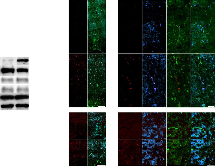
NATURE COMMUNICATIONS | https://doi.org/10.1038/s41467-020-20112-4 ARTICLE
1w 3w 5w 8w
PTEN-/-
a n
hIL-6
hIL-6
hIL-6
c pSTAT3
hIL-6
AAV-hIL-6 neuN pSTAT3
gfp
gfp
gfp
gfp
neuN kDa
wt pSTAT3 -96
b pSTAT3 STAT3 -96
GFP pSTAT3 GFP
GFP neuN
pAKT
pSTAT3 -55
GFP actin
neuN -43
d e f g o p=1.5*10-4
STAT3 (rel. intensity) pSTAT3 (rel. intensity)
25
p=1.4*10-4 p=1.5*10-4 gfp
20 p=2*10-4
hIL-6
15 PTEN-/-
10 ns
p=0.98
h i 5
0
l’
p 1w 3w 5w 8w
q
pS6
3 20
pAKT (rel. intensity)
ns
GFP
j 15 p=1.4*10-4
2
m’ 10
ns
k AAV-
l pS6 GFP
pS6
GFP
neuN
1
5
gfp
pS6
cre/gfp 0 0
1w 3w 5w 8w 1w 3w 5w 8w
cre-gfp
PTEN-/-
r s t
pERK (rel. intensity)
pS6 (rel. intensity)
PTENf/f hIL-6 2 ns
gfp
kDa 3 p=5.7*10-4
m pSTAT3
l
GFP neuN neuN pERK -43 ns
2
cre-gfp
GFP 1 p=0.95
pSTAT3 pS6
-35 1
actin
-43 0 0
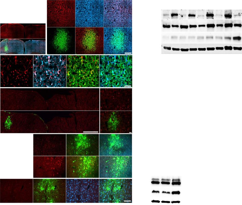
Fig. 1 Activation of signaling pathways by hIL-6 or PTEN−/−. a Schematic drawing illustrating the location of the AAV-injection sites (dashed box) shown
in (b). b Coronal section of the sensorimotor cortex from a wild-type mouse 3 weeks after intracortical injection of AAV2-hIL-6 into the left hemisphere.
The section was immunohistochemically stained for phosphorylated STAT3 (pSTAT3, red) and neuN (blue). GFP (green) was co-expressed by the AAV2-
hIL-6. Scale bar: 500 µm. c Higher magnification of dotted box shown in (b). Scale bar: 200 µm. d–g Higher magnification of dotted boxes in (c). Scale bar:
50 µm. h–j Immunohistochemical staining against phosphorylated ribosomal protein S6 (pS6, red) of sections as described in (a). Dashed boxes are
presented at higher magnification in (i, j). Scale bars: 50 µm (i, j); 500 µm (h). k Schematic drawing illustrating the injection site and location of images
shown in (l, m). l, m Cortical sections of PTENf/f mice 3 weeks after AAV2-Cre-GFP (Cre-gfp), or AAV2-GFP (gfp) injections, stained for pS6 (l, red), or
pSTAT3 (m, red). Scale bar: 50 µm. n Western blot analysis: lysates prepared from the sensorimotor cortex of PTENf/f mice 1, 3, 5, or 8 weeks (w) after
intracortical injection of either AAV2-hIL-6 (hIL-6), AAV2-GFP (gfp) or 5 weeks after AAV2-Cre application leading to PTEN−/−. AAV2-hIL-6 induced
STAT3 phosphorylation (pSTAT3) at all tested time points, while PTEN−/− only caused AKT phosphorylation. Total STAT3 protein remained mostly
unaltered. Beta-actin served as a loading control. o–q Densitometric quantifications of western blots depicted in (n). Values represent means ± SEM of
samples from 6 to 10 animals per group (gfp 1w, n = 6; hil-6 1w, n = 8; gfp 3w, n = 10; hIL-6 3w n = 10; gfp 5w, n = 7; hIL-6 5w, n = 8; gfp 8w, n = 6; hIL-6
8w, n = 6; PTEN−/−, n = 6). r Western blot analysis of cortical lysates: Phosphorylation of ERK1/2 (pERK) was not altered by AAV2-GFP-, AAV2-hIL-6, or
AAV2-Cre (PTEN−/−) 5 weeks after intracortical application. Only PTEN−/− induced significant S6-phosphorylation (pS6). s/t Densitometric
quantification of western blots depicted in (r). Values represent means ± SEM of 6 independent cortical lysates (n = 6) per group. Representative
immunohistochemical stainings shown in (b–j, l, m) were repeated four times with individual biological samples with similar results. Significances of
intergroup differences in (o–q, s, t) were evaluated using a one-way analysis of variance (ANOVA) followed by Tukey post hoc test. Statistical significance
is indicated by p-values. ns = non-significant. Dots in o-q, s, t represent values of single samples. Source data are provided as a Source data file.
the regeneration of some CST axons, most of which did, however, Hyper-IL-6 promotes functional recovery. Before tissue isolation
not exceed distances greater than 1.5 mm (Fig. 2d, f, i and and analysis, hindlimb movement had been analyzed in all four
Fig. S4a). In contrast, AAV2-hIL-6 treatment resulted in more experimental groups using open-field locomotion tests according
robust CST regeneration with the longest axon reaching up to 6 to the Basso Mouse Scale (BMS) over the postinjury period of
mm (Fig. 2d, g, i and Fig. S4a). This effect was slightly increased 8 weeks21. Consistent with previous reports21–23, the BMS score
further by the combination of PTEN−/− and AAV2-hIL-6, with dropped down to 0 in all animals 1 day after injury, also indicating
the longest axons reaching >7 mm (Fig. 2d, h, i and Fig. S4a). No the completeness of the SCC (Fig. 3a, b and Fig. S6a–e). Over time,
BDA labeled axons were detected >11 mm past the lesion site, control animals developed only active ankle movements, including
thereby excluding spared axons and verifying the completeness of spasms (Supplementary video 1)21,22, resulting in an average final
axonal injury in all animals (Fig. S5a–d). score of 2 (Fig. 3a, b and Fig. S6a). Despite the positive effect on
NATURE COMMUNICATIONS | (2021)12:391 | https://doi.org/10.1038/s41467-020-20112-4 | www.nature.com/naturecommunications 3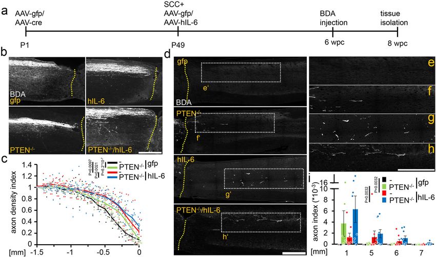
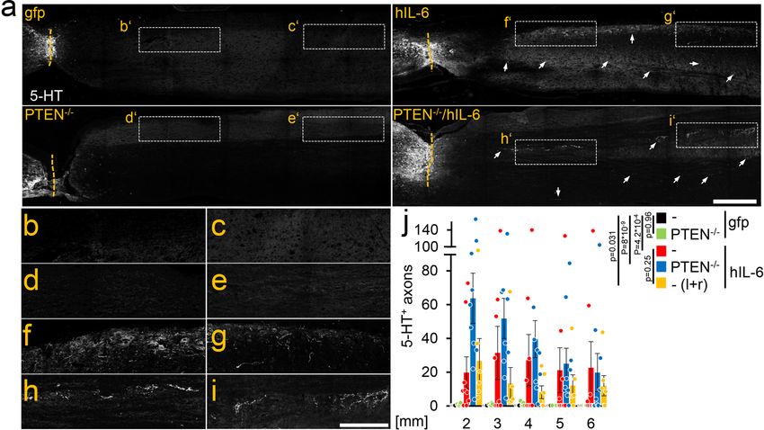
ARTICLE                                                                    NATURE COMMUNICATIONS | https://doi.org/10.1038/s41467-020-20112-4

Fig. 2 Hyper-IL-6 promotes CST axon regeneration after severe spinal cord crush. a Timeline of surgical interventions for experiments presented in (b–i)
and Figs. 3, 4. PTENf/f mice received unilateral injections of AAV2-GFP (PTEN+/+) or AAV2-Cre (PTEN−/−) at P1. After 7 weeks, PTEN+/+ and PTEN−/−
mice were then subjected to spinal cord crush (SCC) (T8) and subsequently received additional unilateral intracortical injections of either AAV2-hIL-6
(hIL-6) or AAV2-GFP (gfp). b Sagittal thoracic spinal cord sections of four differently treated groups showing BDA-labeled, retracting axons (white) of the
right main CST rostral to the lesion site (dotted line). Tissues were isolated 8 weeks after the SCC. Scale bar: 500 µm. c Axon density index of CST fibers
analyzing all spinal cord sections with the main CST of the animals described in (a, b). Values at distances from 1.5 mm rostral to the lesion site (−1.5) up
to the proximal lesion border (0) were determined. Values represent means ± SEM of 6–10 animals per group (PTEN+/+/gfp, n = 7; PTEN+/+/hIL-6, n =
9; PTEN−/−/gfp, n = 6; PTEN−/−/hIL-6, n = 10). d Representative images of BDA-labeled sagittal spinal cord sections from PTEN-floxed mice (PTENf/f)
after treatments, as described in (a, b). Tissues were isolated after 8 weeks. BDA-labeled (white) regenerating axons of the central CST beyond the lesion
site (dotted line) were seen only after PTEN−/−, hIL-6, or hIL-6/PTEN−/−-treatments. Scale bar: 500 µm. e–h Higher magnification of regenerating axons
in dotted boxes as indicated in (d). Scale bar: 500 µm. i Quantification of regenerating CST axons at the indicated distances caudal to the lesion site. Axon
numbers were divided by the total number of BDA-labeled CST fibers in the medulla (axon index) from animals as described in (a, d). Values represent the
mean ± SEM of 6–10 animals per group (PTEN+/+/gfp, n = 7; PTEN+/+/hIL-6, n = 9; PTEN−/−/gfp, n = 6; PTEN−/−/hIL-6, n = 10). Significances of
intergroup differences in (c, i) were evaluated using a three-way analysis of variance (ANOVA) with Holm Sidak post hoc tests. Statistical significance is
indicated by p-values; ns = non-significant. Dots in c and i represent values of single animals. Source data are provided as a Source data file.

CST-regeneration (Fig. 2d, f, i), PTEN−/− alone did not sig-                    allowed for the quantification of several parameters, such as the
nificantly improve the BMS score compared to AAV2-GFP treated                    area of hind paw footprints during contact with the glass plate,
controls (Fig. 3a, b and Fig. S6c; Supplementary video 2). How-                 which was similar to uninjured controls (Fig. S7b–f). Also, stride
ever, AAV2-hIL-6 treatment increased the BMS score to ∼4 by                     length (Fig. S7g, h), the base of support (BOS) (Fig. S7i), and the
restoring plantar stepping with full hindlimb weight support,                   regularity index were measured and reached about 40-60% of the
followed by lift-off, forward limb advancement, and reestablish-                pre-injury values. The latter indicates the degree of coordination
ment of weight support at initial contact in most of the animals                between hind and forelimbs (Fig. S7j and Supplementary video 5).
(Fig. 3a, b and Fig. S6b; Supplementary video 3). Combinatorial                 Thus, BMS (Fig. S7a) and Catwalk evaluation showed similar
treatment (PTEN−/− + AAV2-hIL-6) slightly enhanced this effect                  results concerning the ranking of individual animals indicated by
further (Fig. 3a, b and Fig. S6d, Supplementary video 4). These                 their linear correlation (Fig. S7k).
effects were best seen in the BMS subscore (Fig. 3e). One animal of
this group reached a coordinated movement of fore- and hin-
dlimbs (score: 7) (Fig. 3a, b and Fig. S6d). Interestingly, despite             Cortical AAV2-hIL-6 delivery promotes RpST regeneration.
only administering a unilateral AAV2-hIL-6 injection into the left              The lack of functional recovery in PTEN−/− mice suggested that
sensorimotor cortex, both hindlimbs showed similar recovery                     improved CST regeneration was not the leading cause for the
(Fig. 3a, c, d). Moreover, a bilateral application into the left and            AAV2-hIL-6 mediated functional recovery. As descending ser-
right side had no additional effect (Fig. 3f and Fig. S6e). Thus, in            otonergic (5-HT-positive) axons of the raphespinal tract (RpST)
contrast to PTEN−/−, a single unilateral postinjury application of              are reportedly relevant for locomotor recovery24–26, we stained
AAV2-hIL-6 into the sensorimotor cortex enabled both hin-                       and analyzed these axons in sagittal spinal cord sections of the
dlimbs’ locomotion after severe SCC. The effect on functional                   same animals used in the previous experiments (Figs. 2, 3).
recovery was also verified using an automated catwalk gait analysis              Control (AAV2-GFP) and PTEN−/− mice only revealed sprout-
system. To this end, we generated another cohort of injured mice                ing of 5-HT-positive axons over distances of less than 1 mm
with either unilateral AAV2-hIL-6 or AA2V-GFP application.                      beyond the crush site (Fig. 4a–e, j and Fig. S4c). Remarkably,
While all AAV2-GFP-treated mice failed to show any hind paw                     AAV2-hIL-6 treated mice showed more, and longer-distance
placement, functional restoration after AAV2-hIL-6 treatment                    regeneration of serotonergic fibers than controls or PTEN−/−

4                  NATURE COMMUNICATIONS | (2021)12:391 | https://doi.org/10.1038/s41467-020-20112-4 | www.nature.com/naturecommunications
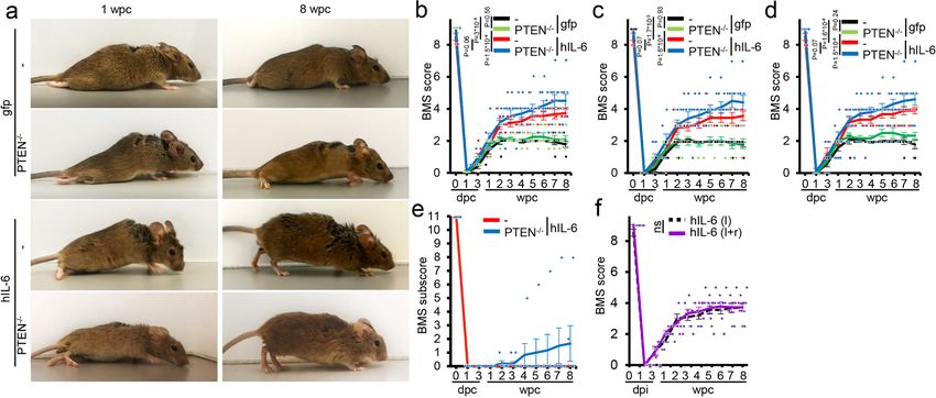
NATURE COMMUNICATIONS | https://doi.org/10.1038/s41467-020-20112-4                                                                            ARTICLE

Fig. 3 Hyper-IL-6 enables functional recovery after SCC. a Representative pictures are showing open field hindlimb movement of mice at 1 and 8 weeks
after spinal cord crush (wpc) and treatment, as described in Fig. 2. b–d BMS score of animals as described in (a). Scores were evaluated at 0, 1, 3, 7 days
post crush (dpc) and then weekly over 8 weeks after spinal cord injury. Values represent means ± SEM of 6–10 animals per group (PTEN+/+/gfp, n = 7;
PTEN+/+/hIL-6, n = 9; PTEN−/−/gfp, n = 6; PTEN−/−/hIL-6, n = 10), showing either the average score of the left and right hind paw (b) or left (c) and
right (d) side separately. e BMS subscore of hIL-6 treated PTEN+/+(−) and PTEN−/− mice as described in (a). f Average BMS score of left and right hind
paws from mice after SCC and bilateral (left and right hemisphere (l + r)) intracortical injection of AAV2-hIL-6 compared to animals that had received a
unilateral injection into the left hemisphere (l) only as presented in (b). Values represent the mean ± SEM of 9 animals per group (l, n = 9; l + r, n = 9).
Significances of intergroup differences were evaluated using a two-way analysis of variance (ANOVA) with a Tukey post hoc test (b–d) or two-sided
student’s t-test (f). P-values indicate statistical significance; ns = non-significant. Source data are provided as a Source data file.

Fig. 4 hIL-6 promotes axon regeneration of serotonergic fibers. a Sagittal thoracic spinal cord sections isolated from PTEN+/+, or PTEN−/− animals
8 weeks after spinal cord crush (SCC) and unilateral injection of AAV2-hIL-6 (hIL-6) or AAV2-GFP (gfp) (see Fig. 2). Raphe spinal tract (RpST) axons were
stained using an anti-serotonin antibody (5-HT, white). Only AAV2-hIL-6-treated mice with or without additional PTEN−/− showed significant
regeneration of serotonergic axons beyond the lesion site (dashed line). As typical for regenerating RpST axons they were located over the whole
dorsoventral width of the spinal cord. Examples are indicated by dashed boxes and white arrows. Scale bar: 500 µm. b–i Higher magnification of dashed
boxes as indicated in (a). Scale bar: 250 µm. j Quantification of regenerating 5-HT-positive axons as described in (a) at indicated distances beyond the
lesion. Values represent the mean ± SEM of 6–10 animals per group (PTEN+/+/gfp, n = 7; PTEN+/+/hIL-6, n = 9; PTEN−/−/gfp, n = 6; PTEN−/−/hIL-6,
n = 10; PTEN+/+/hIL-6 (l + r); n = 6). Significances of intergroup differences were evaluated using a two-way analysis of variance (ANOVA) with a Holm
Sidak post hoc test. Dots in j represent values of single animals. P-values indicate statistical significance; ns = non-significant. Source data are provided as a
Source data file.

NATURE COMMUNICATIONS | (2021)12:391 | https://doi.org/10.1038/s41467-020-20112-4 | www.nature.com/naturecommunications                                       5
ARTICLE NATURE COMMUNICATIONS | https://doi.org/10.1038/s41467-020-20112-4
a AAV-hlL-6/gfp DHT BDA tissue
SCC injection injection isolation
wpc 0 1 2 3 4 5 6 7 8
BMS
b Con DHT f gfp
9 hlL-6
8
DHT
7
5-HT
c’ d’
BMS score
p = 0.0056
6
p = 0.011
5 –4
p = 1.2*10
p = 5.3*10–5
4
p = 0.12
3
e Before DHT 1d after DHT
2
p = 0.79
1
Con
p = 0.79
0
1d
3d
1w
2w
3w
4w
5w
6w
1d
1w
Before
DHT
gfp
g h
SCC 6w
BMS score
BMS score
9 9
6 6
hlL-6
3 3
0 0
on
on
1d
1d
7d
7d
C
C
T
T
T
T
H
H
H
H
D
D
D
D
Fig. 5 Regeneration of serotonergic axons is essential for functional recovery. a Timeline of experiments shown in (b–f). Adult mice were subjected to
SCC and received bilateral intracortical AAV2-GFP (gfp) or AAV2-hIL-6 (hIL-6) injections. They were then scored according to the Basso Mouse Scale
(BMS) at the indicated time points (arrows) over 8 weeks post crush (wpc). Six weeks after SCC, the serotonin neurotoxin 5,7-dihydroxytryptamine (DHT)
was injected intracerebroventricularly into both hemispheres. One week before tissue isolation, BDA was intracortically injected to trace CST axons.
b Maximum intensity projection of confocal scans through 50 µm of cleared brain stem tissue from mice that received hIL-6 and DHT treatment as
described in (a) compared to a control (con) without DHT treatment. Serotonergic neurons of raphe nuclei (RN, dotted line) were visualized by 5-HT
staining (green). BDA-labeled CST axons in the pyramid (PY) are shown in red. DHT treatment eradicated almost all 5-HT-positive neurons without
affecting corticospinal neurons, indicated by intact BDA labeled axons. Scale bar: 100 µm. c, d Higher magnifications of dashed boxes (c' and d') in (b).
Scale bar: 50 µm. e Representative images of AAV2-GFP (gfp) or AAV2-hIL-6 (hIL-6)-treated mice 6 weeks post crush (wpc), or untreated mice (con)
before and 1 day after DHT injection. f BMS score of animals treated as described in (a) at indicated time points after spinal cord crush and DHT treatment.
Values represent means ± SEM of 6 animals per group (n = 6), showing the average score of left and right hind paws. g, h BMS scores (g) and subscores
(h) of untreated mice before (con) and 1 day (DHT 1d), or 1 week (DHT 7d) after DHT injection. Values represent means ± SEM of 6 animals showing the
average score of left and right hind paws. Significances of intergroup differences were evaluated using a two-way analysis of variance (ANOVA) with a
Holm Sidak post hoc test. Significance indicated by p-values within the hIL-6 group in red, within the GFP group in black, or between hIL-6 and GFP group in
blue. Source data are provided as a Source data file.
(Fig. 4a, f, g, j and Fig. S4c), with the longest axons reaching other neurons remained unaffected, indicated by the successful
distances of >7 mm. Combinatorial treatment of AAV2-hIL-6 BDA tracing of cortical motor neurons (Fig. 5b), presence of
and PTEN−/− had no additional effect (Fig. 4a, h, i, j and other neuN positive neurons next to depleted Raphe neurons in
Fig. S4c). Furthermore, as measured during functional recovery the medulla (Fig. S8k), and the almost normal open field move-
analysis (Fig. 3f), bilateral AAV2-hIL-6 treatment did not ment in uninjured mice evaluated 1 week after DHT application
increase RpST regeneration further compared to a unilateral (Fig. 5e, g, h). However, in injured animals, 1 day after DHT
application of the virus (Fig. 4j and Fig. S4c), and no 5-HT- application, the BMS of AAV2-hIL-6-treated mice dropped down
positive axons were detected >11 mm beyond the injury site in to similar levels as in AAV-GFP treated control animals, which
any of these mice verifying the absence of spared axons. themselves remained unaffected by the DHT treatment and
thereby verified the neurotoxin effect’s specificity (Fig. 5e, f and
AAV2-hIL-6-mediated recovery depends on the regeneration Supplementary video 6). Thus, AAV2-hIL-6-mediated locomo-
of serotonergic neurons. To investigate the relevance of RpST tory recovery depended on RpST regeneration, even though
regeneration for functional recovery, adult mice of the same AAV2-hIL-6 had not been applied to the brain stem where the
genetic background were subjected to SCC and received bilateral serotonergic fibers originated.
intracortical injections of either AAV2-hIL-6 or AAV2-GFP Since genetic backgrounds could potentially affect regenera-
directly afterward (Fig. 5a). At week 6, when functional recovery tion, we also tested the postinjury-applied AAV2-hIL-6 treatment
in AAV2-hIL-6 treated mice had already reached maximal levels, in non-transgenic BL6 mice, whose low potential for functional
the neurotoxin 5,7-dihydroxytryptamine (DHT) was intracer- recovery has been previously documented21. Eight weeks after
ebroventricularly injected to selectively kill serotonergic neurons SCC and unilateral intracortical AAV2-hIL-6 injection, BL-6
(Fig. 5a–d)24,27,28. The toxin almost completely abolished 5-HT mice also showed reduced axonal dieback, but less CST-
positive neurons/axons in the medulla/spinal cord as soon as regeneration caudal to the lesion site than accordingly treated
1 day after injection (Fig. S8a–j). As described previously27,29,30, PTEN-floxed OLA mice (Figs. S9a, b, S4b). Nevertheless, the
6 NATURE COMMUNICATIONS | (2021)12:391 | https://doi.org/10.1038/s41467-020-20112-4 | www.nature.com/naturecommunications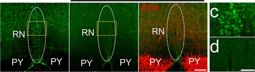
NATURE COMMUNICATIONS | https://doi.org/10.1038/s41467-020-20112-4                                                                  ARTICLE

improvement in RpST axon regeneration and functional recovery                   coronal sections showed, on average, 46% ± 4.46 SEM of the
was very similar in both mouse strains (Figs. S9c–e, S4d;                       serotonergic neurons were pSTAT3/5-HT double-positive. These
Supplementary video 7), supporting a correlation between                        neurons were close to GFP-positive axons in the ipsi- and
anatomical RpST regeneration and functional recovery.                           contralateral sides of the raphe nuclei in the medial column of the
                                                                                medulla. 50.63 ± 4.4% of double-positive neurons were identified
                                                                                in the ipsilateral side of the raphe nuclei and 49.37 ± 4.4% on the
AAV2-hIL-6 confers transneuronal stimulation of serotonergic                    contralateral side. These findings were not observed in AAV-GFP
fiber regeneration. To understand the mechanism underlying the                   treated controls (Fig. 6d–u). Moreover, intracortical AAV2-hIL-6
bilateral, RpST-dependent functional recovery by a one-time                     treatment also activated STAT3 in neurons of the red nucleus, a
unilateral AAV2-hIL-6 virus application into the sensorimotor                   known target of cortical motoneurons (Fig. S12c–e)35, thereby
cortex, we tested the hypothesis that transduced cortical moto-                 corroborating the transneuronal delivery of hIL-6.
neurons project axons to the raphe nuclei in the brain stem and                    To verify that cortical motoneurons were synaptically con-
that the release of hIL-6-protein stimulates regeneration of ser-               nected with raphe neurons, we used AAV1, which, in contrast to
otonergic neurons trans-synaptically. Starting in vitro, we used                AAV2, can trans-synaptically transduce supraspinal target
axon isolation devices to separate somata and axons of cultured                 neurons36. We injected AAV1-Cre into the left sensorimotor
sensory DRG neurons31 (Fig. S10a) and transduced these neurons                  cortex of Rosa26-tdTomato (RFP) reporter mice and isolated
by adding baculoviruses (BV)18,32 for either hIL-6 or GFP                       their brain stem tissue after 2 weeks. RFP-positive (transduced)
expression into the somal chamber. Western blot analysis                        serotonergic neurons were detected in both sides of the nucleus
detected hIL-6 protein in the medium of the axon chamber of                     raphe pallidus (NRPa) and the nucleus raphe magnus (NRM)
hIL-6 transduced neurons only, verifying its release                            (Fig. 7a–f) along their entire rostrocaudal length. Additionally, we
(Figs. S10c, S16p, q). Moreover, synaptotagmin-positive vesicles                performed a complementary experiment by injecting AAV1
containing hIL-6 were found in the axons and their tips                         directly into the raphe nuclei. As AAV1 is also retrogradely
(Fig. S10d). To test the activity of the released hIL-6, the medium             transported37,38, we found transduced neurons in the motor
of the axonal chamber was used to prepare dissociated cultures of               cortex layer V, confirming the synaptic connection to cortical
adult retinal ganglion cells (RGC) (Fig. S10b). Consistent with                 neurons (Fig. 7g–i). Thus, cortically applied AAV2-hIL-6
previous findings18, the conditioned medium from hIL-6-                          transneuronally activates regenerative signaling pathways in ipsi-
transduced sensory axons induced STAT3 phosphorylation                          and contralateral raphe neurons by releasing the protein at the
(Fig. S10e) and increased neurite outgrowth of RGCs twofold                     axon terminals.
compared to the medium from GFP controls (Fig. S10f, g). Thus,
hIL-6 is transported in axons, released at the terminals, and
remains active to stimulate axon regeneration.                                  Discussion
   To investigate whether transduced hIL-6 can also stimulate                   The current study shows significant functional recovery after a
neurons trans-synaptically in vivo, we injected either AAV2-hIL-                complete SCI in adult mammals. This recovery was achieved by
6 or AAV2-GFP into the left eye of mice to transduce RGCs.                      the one-time unilateral application of AAV2-hIL-6 into the sen-
Three weeks later, we tested for STAT3 phosphorylation in their                 sorimotor cortex. While pre-injury PTEN−/− in cortical neurons
brain targets, the lateral geniculate nucleus (LGN) and supra-                  failed to facilitate functional recovery, postinjury AAV2-hIL-6
chiasmatic nucleus (SCN) (Fig. S10h). In rodents, ~95% of the                   application enabled locomotor recovery of both hindlimbs.
optic nerve axons cross in the optic chiasm to the contralateral                Moreover, this treament promoted longer CST axon growth than
side. Consistent with a synaptic release of hIL-6, we found many                PTEN−/− and, additionally, regeneration of serotonergic axons in
pSTAT3-positive cellular nuclei near GFP-positive axon terminals                the RpST. We also provide evidence that cortical motoneurons
of AAV2-hIL-6-transduced RGCs in the LGN and SCN of the                         directly innervate raphe neurons, allowing the delivery of highly
right hemisphere, with much less found in the respective left                   potent hIL-6 to stimulate the regeneration of these serotonergic
hemispheres and no signals at all in AAV2-GFP treated controls                  brain stem neurons. Thus, the transneuronal application of highly
(Fig. S10i, j). The hIL-6 expression in RGCs did not affect axons’              active molecules has proven to be a powerful approach to activate
integrity as their number, determined after neurofilament                        regenerative signaling, particularly in neurons located in brain
staining in the optic nerve and optic tract cross-sections, was                 regions that are challenging to access but relevant for functional
not reduced compared to untreated controls, excluding any                       recovery after spinal cord injury.
uncontrolled or widespread release of the protein.                                 Incomplete, less severe hemisection- or contusion-injury
   We then investigated whether raphe neurons in the brain stem,                models permit spontaneous functional recovery with BMS
which express the hIL-6 receptor GP13033,34 receive synaptic                    scores ≥611,21,23,39,40. In contrast, the SCC, just as in a complete
input from transduced cortical motoneurons. To this end, we                     transection, severs all axonal connections between the proximal
analyzed 5-HT stained brain stem tissue from mice 8 weeks after                 and distal portions of the spinal cord, allowing only some active
SCC and cortical AAV2-hIL-6 treatment as described above                        flexion movements of hindlimbs (BMS score of ≤2)10,21–23,41.
(Fig. 2a). GFP-positive sprouts of cortical motoneurons were                    Therefore, achieving functional recovery in this injury model is
located near 5-HT-positive raphe neurons in cross-sections of the               challenging, and most previous treatment strategies tested did not
medulla (Fig. S11a–d). The GFP signal and hIL-6 in the                          reach significant effects beyond reducing the dieback of CST-
pyramidal axons were detected via high-resolution transversal                   axons42–44. Only PTEN−/− in cortical motoneurons enabled the
scans after tissue clearing (Fig. S1l–n). Western blot analyses                 regeneration of CST-axons and their synapse formation with
using brain stem lysates isolated 3 weeks after intracortical viral             interneurons, but still failed to restore locomotion10,11,41,45,46.
application showed pSTAT3 signals only in samples from AAV2-                    Therefore, we used the PTEN−/− model as a reference to
hIL-6-, but not AAV2-GFP-treated animals (Fig. 6a–c and                         validate the effects of AAV2-hIL-6. While cortical PTEN−/−
Fig. S15a-c, S16h), while total STAT3 protein expression or                     showed regenerated axons only at short distances of up to 1 mm
phosphorylation of either AKT or S6 remained unchanged                          beyond the injury site, distances were markedly longer in hIL-6
(Fig. 6b, c and Figs. S15, S16h–m). Additionally, transverse scans              treated animals. Moreover, only AAV2-hIL-6 treatment sig-
through cleared brain stem tissue (Fig. S12a, b, Supplementary                  nificantly improved serotonergic axon growth in the RpST and
video 8) from hIL-6 treated animals, and immunostaining of                      enabled locomotion recovery of both hindlimbs. These effects

NATURE COMMUNICATIONS | (2021)12:391 | https://doi.org/10.1038/s41467-020-20112-4 | www.nature.com/naturecommunications                           7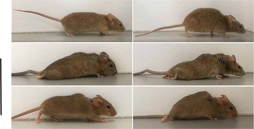
ARTICLE                                                                                                 NATURE COMMUNICATIONS | https://doi.org/10.1038/s41467-020-20112-4

                                                                                                          GFP                                                    GFP
                                                                                                         pSTAT3                        5-HT         GFP         pSTAT3
                                                                                                  pSTAT3  5-HT           pSTAT3       pSTAT3       pSTAT3        5-HT
                a                     AAV-hIL-6                               d                          L R
                                                                                                                                  e            f            g   L R
                                                                                                                                                                        h

                                                                                        gfp
                                                         RN

                                                                                                         PY   PY

                                                                                  NRM
                b                                  gfp        hIL-6   kDa
                                                                                                         L    R
                                      pSTAT3                          -96
                                                                                                                                  i            j            k   L   R
                                                                                                                                                                         l
                                       STAT3                          -96

                                                                                        hIL-6
                                           pAKT                       -70

                                            pS6                       -35
                                           GFP                        -26
                                                                                                         PY   PY
                                           actin                      -43
                                                                              m
                c                                                                                                                 n            o            p   L   R   q
                                                                                            gfp

                                                                      gfp                                 L    R
                                      15
                                            p=7*10-5                  hIL-6
                     rel. intensity

                                                                                                               PY
                                                                                       NRPa

                                                                                                         PY
                                      10
                                                                                                                                  r            s            t   L   R   u
                                                                                  hIL-6

                                       5                      p=0.93 p=0.49
                                                   p=0.06                                                 L    R
                                       0                                                                 PY    PY
                                       pSTAT3 STAT3 pAKT pS6

Fig. 6 Hyper-IL-6 transneuronally stimulates neurons of raphe and red nuclei. a Schematic of cortical AAV2-hIL-6 application and isolated brain stem
tissue (dashed box) containing the raphe nuclei (RN) for western blot analysis. b Western blots: GFP, total (STAT3) and phosphorylated STAT3 (pSTAT3),
phospho-AKT (pAKT), and phospho-S6 (pS6) were analyzed in lysates of the brain stem with raphe nuclei isolated 3 weeks after intracortical injection of
either AAV2-GFP (gfp) or AAV2-hIL-6 (hIL-6). GFP signals in lysates verified similar amounts of transduced collateral axons that projected to the brain
stem. Beta-actin served as a loading control. c Densitometric quantification of western blots from 7 to 8 animals per group (gfp, n = 7; hIL-6, n = 8) as
described in (a). Data represent means ± SEM. d, m Immunostaining of brain stem sections containing the nucleus raphe magnus (NRM, d), or nucleus
raphe pallidus (NRPa, m) of AAV2-GFP or AAV2-hIL-6 treated mice as described in Fig. 2a. Sections were stained for pSTAT3 (red), GFP (green), and
serotonin (5-HT, blue) to identify raphe neurons. The dashed yellow line indicates the midline. We observed a similar amount of pSTAT3 positive
serotonergic neurons in the left and right hemisphere of the raphe nuclei from AAV2-hIL6 treated mice (n = 6). Scale bar: 200 µm. e, f, n–u Higher
magnifications of the dotted box as indicated in (d) (e–l) or (m) (n–u). Significances of intergroup differences in (c) were evaluated using the two-sided
student’s t-test and indicated by p-values. Dots in c represent values of samples from single animals. Source data are provided as a Source data file.

were achieved in mice with different genetic backgrounds by a                                                 effects of hIL-6 on axon regeneration can be further increased in
one-time, unilateral application of AAV2-hIL-6 after the SCC,                                                 combination with a SOCS3 knockout, which usually limits the
making this gene therapeutic approach a potential strategy to                                                 activity of JAK/STAT3-signaling16,17.
facilitate spinal cord repair.                                                                                   Cortical AAV2-hIL-6 injection induced STAT3 activation and
   PTEN−/− and hIL-6 affected regenerative pathways differently.                                              promoted axon regeneration in the CST and RpST. Although
While PTEN−/− expectedly activated PI3K/AKT/mTOR only in                                                      AAV2-hIL-6 enabled longer CST-regeneration than PTEN−/−,
transduced neurons, AAV2-hIL-6 induced JAK/STAT3-signaling                                                    our data suggest that the functional recovery mostly depended on
additionally in adjacent cortical neurons (Fig. 1) and raphe neurons                                          the improved regeneration of serotonergic fibers. This is because
via the transneuronal route. Therefore, it is conceivable that the                                            (i) the PTEN−/− induced CST regeneration did not enable
combinatorial PTEN−/− and hIL-6 treatment enabeled stronger                                                   hindlimb recovery, (ii) AAV2-hIL-6 improved RpST-
CST-regeneration than each treatment by itself as previously shown                                            regeneration and functional recovery similarly in BL6 and Ola-
in the optic nerve16–18. However, these synergistic effects were                                              PTEN-floxed mice; however, the effect on CST regeneration
limited to CST-regeneration due to the restricted effect of PTEN                                              was different in these mouse strains and (iii) selective elimination
−/− on virally transduced cortical motoneurons. Moreover, the                                                 of serotonergic fibers by DHT24,27,28 abolished most of the
finding that hIL-6 alone did not measurably affect PI3K/AKT/                                                   recovered locomotion after AAV2-hIL-6 treatment. More-
mTOR activity but showed even stronger effects than PTEN−/−                                                   over, RpST regeneration is essentially involved in locomotor
suggests that extensive activation of AKT/mTOR is not essential to                                            recovery after less severe spinal cord injuries, which allow some
reduce axonal dieback or improve CST regeneration. AAV2-hIL-6                                                 endogenous sprouting of serotonergic axons24,48–51. Although
did not activate the MAPK/ERK pathway either, leading to the                                                  our data demonstrate that AAV2-hIL-6-mediated functional
conclusion that its beneficial effect on axon regeneration was                                                 recovery depends on the regeneration of serotonergic fibers, we
mediated by STAT3 activation, as previously shown in the visual                                               cannot exclude the possibility that hIL-6 might have also stimu-
system12,16,17,47. Future experiments need to address whether a                                               lated axon regeneration in other neurons that receive collateral
knockout or inhibition of STAT3 in cortical neurons compromises                                               input from cortical motoneurons. Neurons in the red nuclei are
the hIL-6 effects on CST-regeneration. It is also possible that the                                           such examples35, where we also observed STAT3 activation

8                   NATURE COMMUNICATIONS | (2021)12:391 | https://doi.org/10.1038/s41467-020-20112-4 | www.nature.com/naturecommunications
NATURE COMMUNICATIONS | https://doi.org/10.1038/s41467-020-20112-4 ARTICLE Fig. 7 CST axon collaterals project to raphe nuclei. a Schematic illustration, indicating the cortical application, anterograde axonal transport, and terminal release of AAV1-Cre into raphe nuclei (RN, green) in Rosa-tdTomato reporter mice (sagittal view). A coronal view of the medulla illustrates RFP-positive pyramidal (PY) axon sprouts from AAV1-Cre transduced cortical neurons. Some pyramidal axons project to raphe neurons (green), leading to transneuronal transduction and subsequent RFP expression. The dotted box indicates the area of tissue sections shown in (b). b Coronal medullary sections from Rosa-tdTomato mice 2 weeks after intracortical AAV1-Cre injection as described in (a). Transneuronally transduced serotonergic neurons of the nucleus raphe magnus (NRM, left) and nucleus raphe pallidus (NRPa, right) were identified by 5-HT staining (green) and RFP fluorescence (red). Scale bar: 250 µm. c–f Higher magnification of dashed yellow boxes as indicated in b. Scale bar: 100 µm. g Schematic illustration showing AAV1-Cre injection into raphe nuclei of Rosa-tdTomato reporter mice, and retrograde axonal transport to the motor cortex via pyramidal sprouts. h, i Immunohistochemical staining of brain stem sections against serotonin (5-HT, green) at the site of AAV1-Cre application as described in (g) 2 weeks after injection. Transduced cells in the nucleus raphe magnus (NRM, h), or the nucleus raphe pallidus (NRPa, I) are labeled by expression of dt tomato (RFP, red). Scale bar: 200 µm. j Reference map from the Allen Brain Atlas showing the primary motor cortex (M1) layer V in purple. k Coronal cortical section showing RFP (red) fluorescence and neuN (blue) immunostaining. The image is superimposed by the cortical map, as shown in (j), indicating the primary (M1) and secondary (M2) motor area and cortical layers 1–6. Scale bar: 500 µm. l Higher magnification of dashed boxes as indicated in (k), showing retrogradely transduced cortical layer 5 motor neurons expressing RFP. Scale bar: 250 µm. Representative immunohistochemical stainings shown in (b, h–i, k–l) were repeated three times with individual biological samples with similar results. Source data are provided as a Source data file. following intracortical AAV2 hIL-6 application. Whether the spinal cord injury models (e.g., pyramidotomy, hemisection, regeneration of fibers in the rubrospinal tracts is also contribut- or contusion). ing to the beneficial effects of AAV-hIL-6 is currently unknown. Although the collateral projection of cortical motoneurons into Similarly, it is unclear whether cortical AAV2-hIL6 application various brain areas, e.g., in the striatum, the thalamus52,55,56, the transneuronally stimulated regeneration of long and/or short red nucleus, and the reticular formation, are well documented57–59, descending propriospinal interneurons, which could facilitate the the innervation of raphe nuclei has not yet been directly shown. bridging of signals to second-order motoneurons. However, a The current study used AAV1, which contrary to AAV2 can trans- main role of these neurons in this context appears unlikely, as synaptically transduce neurons36. Cortical AAV1 application in even robust propriospinal axon regeneration does not result in transgenic reporter mice demonstrated that cortical neurons pro- functional recovery after spinal cord crush52. ject almost equally into the ipsi- and contralateral sides of the Despite PTEN−/− induced CST regeneration alone being raphe nuclei. Moreover, we observed transneuronal transduction insufficient, improved regeneration of the CST in addition to the over the whole rostrocaudal length of the raphe nuclei, even RpST could have contributed to the functional recovery effect of though the overall rate of transneuronal transduction was expect- hIL-6. This would explain the finding that the more robust CST edly low. This is due to the fact that AAVs cannot reproduce regeneration in the AAV2-hIL-6/PTEN−/− group compared to themselves without specific helper plasmid in neurons. Therefore, hIL-6 animals correlated with a slightly higher BMS although the only virus particles that remain functionally intact after entering RpST regeneration remained similar. So improved CST- the cell, being directly transported along the axon and released at regeneration might only affect subtle aspects of functional the synapses can transduce other neurons. Thus transneural recovery on top of basic, RpST dependent walking behavior. transduction is limited. Finally, the activation of STAT3 (pSTAT3) Accordingly, CST axon regeneration reportedly improves in serotonergic raphe neurons by unilateral AAV2-hIL-6 applica- voluntary movements and skilled locomotion in less severe pyr- tion in the motor cortex provides further evidence for this inner- amidotomy- and contusion-injury models, which leave other vation target. spinal tracts intact26,40,53,54. Future experiments need to investi- The current study demonstrates that virally expressed hIL-6 is gate to what extent regeneration of serotonergic fibers alone can not only released from the soma to stimulate adjacent moto- mimic the full AAV2-hIL-6 effect on functional recovery and to neurons in a paracrine fashion but that it is also transported over what extent hIL-6 improves regeneration/recovery in less severe long distances in axons of either RGCs in the visual system or NATURE COMMUNICATIONS | (2021)12:391 | https://doi.org/10.1038/s41467-020-20112-4 | www.nature.com/naturecommunications 9
ARTICLE                                                                             NATURE COMMUNICATIONS | https://doi.org/10.1038/s41467-020-20112-4

cortical neurons. In contrast to AAV1 mediated transneuronal                             for example, transduced on average 1630 (±124 SEM) cortical cells. We observed
transduction, hIL-6 is continuously expressed, transported, and                          only very rare transduction of astrocytes or neurons of other M1 layers
                                                                                         (Fig. S1a–e). All GFP- expressing neurons also expressed hIL-6 (Fig. S1f). There-
released at axon terminals to secondary neurons as reflected by a                         fore, hIL-6 transduction was identified by GFP expression during the whole study.
high percentage of pSTAT3 positive serotonergic neurons (~50%)
in all raphe nuclei. The release of active hIL-6 at axonal terminals/
                                                                                         Injection into the red nucleus. Adult wt mice were anesthetized by intraperitoneal
synapses was verified in cell culture experiments and by                                  ketamine (100 mg/kg) and xylazine (10 mg/kg) injection and then placed in a
pSTAT3 staining in visual target areas and directly in the raphe                         stereotaxic frame. A midline incision was made over the skull to reveal bregma. A
nuclei themselves. In this context, it is worth mentioning that                          micro drill with a 0.5 mm bit was used to open a 1 ×1 mm window in the skull to
hIL-6 is highly potent, so that even the smallest quantities can                         expose the cortex. Biotinylated dextran amine (BDA, 10,000 MW, 10% solution in
                                                                                         water, Invitrogen, D1956) was injected into the red nucleus through a glass pipette
activate different types of neurons expressing gp130, the receptor                       attached to a nanoliter injector (Nanoject III, Drummond). To this end, 500 nl was
to which it directly binds18. Moreover, due to the localization of                       given at the following coordinates26: 0.6 mm lateral, 3.5 mm deep, and 2.5 mm
the raphe nuclei in the medial column and, therefore, in the                             caudal to bregma. For injecting 500 nl, we applied 5 pulses of 100 nl at a rate of 5
center of the brainstem, one motor cortex hemisphere projects                            nl/s and waited for 10 s after each pulse to allow for distribution of the virus
                                                                                         solution. The needle was left in place for 1 min before removal, and the brain was
into both raphe nuclear hemispheres along the whole ros-                                 kept moist during the procedure. After surgery, the skin was closed with sutures.
trocaudal length. Together with the high potency of hIL-6, this
explains why the unilateral cortical application of AAV2-hIL-
                                                                                         Spinal cord crush. For complete spinal cord crush, adult PTEN floxed (C57BL/
6 improved both hindlimbs’ recovery to a similar degree and why                          6;129/J-TgH(Pten-flox)) or wt (C57BL/6) mice were anesthetized by intraper-
the bilateral application had no additional beneficial effect on                          itoneal injections of ketamine (120 mg/kg) and xylazine (16 mg/kg). A midline
RpST regeneration or functional recovery.                                                incision of ~1.5 cm was performed over the thoracic vertebrae. The fat and muscle
   In conclusion, our finding that the transneuronal application of                       tissue were cleared from thoracic vertebrae 7 and 8 (T7, T8). While holding onto
                                                                                         T7 with forceps, we performed a laminectomy at T8 to expose the spinal cord.
hIL-6 enables functional recovery opens possibilities to further                         Afterward, the complete spinal cord was crushed for 2 s with forceps that had been
improve the functional outcome by combining it with other                                filed to a width of 0.1 mm for the last 5 mm of the tips to generate a homo-
strategies, such as neutralizing extracellular inhibitors at the                         geneously thin lesion site. To ensure that the spinal cord’s full width was included,
lesion site8,50,60 or bridging the lesion site with permissive                           we took care to gently scrape the forceps’ tips across the bone on the ventral side of
                                                                                         the vertebral canal so as not to spare any axons ventrally or laterally. After surgery,
grafts61,62. Such combinatorial approaches, also in less severe                          the muscle layers were sutured with 6.0 resorbable sutures, and the skin was
injury models, may maximize axon regeneration and functional                             secured with wound clips. The completeness of the injury in the SCC model was
recovery after spinal cord injury, potentially also in humans.                           verified by an astrocyte free gap and the absence of any spared CST or raphe spinal
                                                                                         tract (RpST) fibers caudal to the lesion site shortly after injury (Fig. S13e–j). Two
                                                                                         animals with PTEN−/− and one control mouse which had received AAV-GFP were
Methods                                                                                  excluded because they did not meet the criteria described above.
Mouse strains. Male and female mice were used for all experiments. Wild-type
C57BL/6 and 129/Ola mice were crossbred with PTENf/f mice (C57BL/6;129) to
obtain C57BL/6;129/J-TgH (Pten-flox) animals without further crossbreeding.               CST tracing. To trace the axons of cortical motoneurons in PTEN floxed (C57BL/
C57BL/6J mice were obtained from Janvier Labs. Rosa26 loxP-stop-loxP-tdTomato            6;129/J-TgH(Pten-flox)) or wt (C57BL/6) mice, we injected the axon tracer bioti-
(Rosa-tdTomato) mice were obtained from Jackson Laboratories (Stock No:                  nylated dextran amine (BDA, 10,000 MW, 10% solution in water, Invitrogen,
007914). All animals were housed under the same conditions for at least ten days         D1956) into the sensorimotor cortex 2 weeks before the mice were sacrificed10,11,41.
before the start of experiments and generally maintained at 24 °C ambient tem-           Therefore, the skin was opened, and 490 nl BDA was applied to each injection site
perature and 60% humidity with a 12 h light/dark cycle and ad libitum access to          using the same procedure and coordinates as described for the AAV2 injections.
food and water. All experimental procedures were approved by the local animal            After surgery, the skin was closed with sutures. In groups with the PTEN knockout,
care committee (LANUV Recklinghausen) and conducted in compliance with                   we verified that 70-80% of BDA-traced neurons were also Cre-positive
federal and state guidelines for animal experiments in Germany.                          (Fig. S13a–d).

                                                                                         DHT injection. Mice received bilateral intracerebroventricular injections of 30 µg
Intracortical injection of pups. Postnatal (P1) PTEN floxed (C57BL/6;129/J-TgH
                                                                                         of the serotonin neurotoxin 5,7-dihydroxytryptamine (DHT, Biomol) dissolved in
(Pten-flox)) pups were fixed on a stereotactic frame and continuously supplied with
                                                                                         0.5 μl of 0.2% ascorbic acid in saline to deplete serotonergic inputs to the lumbar
2% isoflurane for anesthesia via a mouthpiece. A midline incision into the skin was
                                                                                         spinal cord. To this end, mice were anesthetized and fixed under a stereotactic
made to expose the skull using microscissors. Since the skulls of P1 mice are still
                                                                                         frame as described above for intracortical injections. The tip of a glass micropipette
soft, the cortex could be accessed using a 30-gauge needle to create two small holes
                                                                                         attached to a nanoliter injector (nanoject II, Drummond) was positioned at the
in the left skull hemisphere with the following coordinates: −0.2 mm and 0.3 mm
                                                                                         following coordinates: 0.6 mm posterior, 1.6 mm lateral to bregma, and 2 mm deep
anteroposterior, 1.0 mm lateral to bregma. For AAV2-GFP or AAV2-Cre appli-
                                                                                         from the cortical surface. DHT was injected at the same rate as described for the
cation, 770 nl of the virus suspensions were injected at a depth of 0.5 mm into the
                                                                                         AAV2 injections, and the pipette was left in place for 1 min before withdrawal. The
two sites using a pulled glass pipette connected to a nanoliter injector (Nanoject II,
                                                                                         monoamine uptake inhibitor, desipramine (Sigma), was administered at 25 mg/kg
Drummond). To inject 770 nl, we applied 11 pulses of 70 nl at a rate of 23 nl/s and
                                                                                         intraperitoneally 30 min in advance for all mice that received DHT to prevent the
waited for 10 s after each pulse to allow distribution of the virus solution. After
                                                                                         toxin’s uptake into noradrenergic neurons.
injection, the pipette was left in place for 1 min before being carefully withdrawn,
and the incision carefully closed with a 4-0 black silk suture.
                                                                                         Intraocular injection of AAV2. Adult mice (C57BL/6) received intravitreal AAV2
                                                                                         injections (1 µl)18,63. After 3 weeks, animals were sacrified and tissues isolated.
Intracortical injection of adult mice. For intracortical injections, adult PTEN
floxed (C57BL/6;129/J-TgH(Pten-flox)) or adult wt (C57BL/6) mice were anes-
thetized by intraperitoneal injection of ketamine (120 mg/kg) and xylazine (16 mg/       Injection into the raphe nuclei. For brainstem injections, adult Rosa26 loxP-stop-
kg) and then placed in a stereotaxic frame. A midline incision was made over the         loxP-tdTomato mice were anesthetized by intraperitoneal ketamine (120 mg/kg)
skull to open the skin and to reveal bregma. A micro drill with a 0.5 mm bit was         and xylazine (16 mg/kg) injections and then placed in a stereotactic frame. A
used to open a 2 × 2 mm window on each side of the skull to expose the sensor-           midline incision was made over the skull to reveal bregma. A micro drill with a 0.5
imotor cortex. The respective AAV2 was injected 30 min after spinal cord injury          mm bit was used to open a 2 × 2 mm window in the center of the skull ~5 mm
into the cortex layer V through a glass pipette attached to a nanoliter injector         caudal to bregma to expose the cerebellum. AAV1-Cre obtained from Addgene was
(Nanoject II, Drummond). To this end, four injections of 490 nl each were given          injected through a glass pipette attached to a nanoliter injector (Nanoject III,
either unilaterally (left hemisphere) or in both hemispheres, at the following           Drummond). To this end, 300 nl injections were given to five sites spanning the
coordinates: 1.5 mm lateral, 0.6 mm deep, and 0.5 mm anterior; 0.0 mm, 0.5 mm,           whole rostrocaudal length of the brainstem raphe nuclei. The following coordinates
and 1.0 mm caudal to bregma. For injecting 490 nl into each injection site, we           were used: 0 mm lateral, 5.2 mm deep, and 4.7 mm, 5 mm, 5.3 mm, 5.6 mm, and
applied 7 pulses of 70 nl at a rate of 23 nl/s and waited for 10 s after each pulse to   5.9 mm caudal to bregma. For injecting 300 nl into each injection site, we applied
allow distribution of the virus solution. The needle was left in place for 1 min         six pulses of 50 nl at a rate of 10 nl/s and waited for 10 s after each pulse to allow
before moving to the next site, and the brain was kept moist during the procedure        distribution of the virus solution. The needle was left in place for 1 min before
by moving the skin over the exposed area after each injection. After surgery, the        moving to the next site, and the brain was kept moist during the procedure by
skin was closed with sutures. The virus transduced mainly neurons in layer 5 of the      moving the skin over the exposed area after each injection. After surgery, the skin
primary motor cortex (M1). As determined by GFP coexpression, the AAV-hIL-6,             was closed with sutures.

10                    NATURE COMMUNICATIONS | (2021)12:391 | https://doi.org/10.1038/s41467-020-20112-4 | www.nature.com/naturecommunications
You can also read