ARID1A loss in adult hepatocytes activates b-catenin-mediated erythropoietin transcription - eLife
←
→
Page content transcription
If your browser does not render page correctly, please read the page content below
RESEARCH ARTICLE

                                    ARID1A loss in adult hepatocytes
                                    activates b-catenin-mediated
                                    erythropoietin transcription
                                    Rozenn Riou1,2,3, Meriem Ladli3, Sabine Gerbal-Chaloin4, Pascale Bossard2,3,
                                    Angélique Gougelet1,2,3, Cécile Godard1,2,3, Robin Loesch1,2,3, Isabelle Lagoutte3,5,
                                    Franck Lager3,5, Julien Calderaro6,7, Alexandre Dos Santos8, Zhong Wang9,
                                    Frédérique Verdier3, Sabine Colnot1,2,3*
                                    1
                                      INSERM, Sorbonne Université, Université de Paris, Centre de Recherche des
                                    Cordeliers (CRC), Paris, France; 2Equipe labellisée Ligue Nationale Contre le
                                    Cancer, Paris, France; 3INSERM, CNRS, Institut COCHIN, Paris, France; 4INSERM
                                    U1183, Université Montpellier, Institute for Regenerative Medicine & Biotherapy
                                    (IRMB), Montpellier, France; 5Plateforme d’Imageries du Vivant de l’Université de
                                    Paris, Paris, France; 6INSERM, Université Paris-Est UPEC, Créteil, France;
                                    7
                                      Department of Pathology, Henri Mondor Hospital, Créteil, France; 8INSERM, Paul-
                                    Brousse University Hospital, Hepatobiliary Centre, Villejuif, France; 9Department of
                                    Cardiac Surgery Cardiovascular Research Center, University of Michigan, Ann
                                    Arbor, United States

                                    Abstract Erythropoietin (EPO) is a key regulator of erythropoiesis. The embryonic liver is the
                                    main site of erythropoietin synthesis, after which the kidney takes over. The adult liver retains the
                                    ability to express EPO, and we discovered here new players of this transcription, distinct from the
                                    classical hypoxia-inducible factor pathway. In mice, genetically invalidated in hepatocytes for the
                                    chromatin remodeler Arid1a, and for Apc, the major silencer of Wnt pathway, chromatin was more
*For correspondence:                accessible and histone marks turned into active ones at the Epo downstream enhancer. Activating
sabine.colnot@inserm.fr             b-catenin signaling increased binding of Tcf4/b-catenin complex and upregulated its enhancer
Competing interests: The            function. The loss of Arid1a together with b-catenin signaling, resulted in cell-autonomous EPO
authors declare that no             transcription in mouse and human hepatocytes. In mice with Apc-Arid1a gene invalidations in single
competing interests exist.          hepatocytes, Epo de novo synthesis led to its secretion, to splenic erythropoiesis and to dramatic
Funding: See page 25                erythrocytosis. Thus, we identified new hepatic EPO regulation mechanism stimulating
                                    erythropoiesis.
Received: 12 November 2019
Accepted: 20 October 2020
Published: 21 October 2020

Reviewing editor: Irwin
Davidson, Institut de Génétique
et de Biologie Moléculaire et
                                    Introduction
Cellulaire, France                  Chromatin dynamics strongly modulates gene expression, and the liver is a prominent tissue in which
                                    chromatin opening is a pre-pattern for cell fate programming (Zaret, 2016). ARID1A, ‘AT-rich inter-
   Copyright Riou et al. This
                                    acting domain containing protein 1A’, is a BAF (BRG1-associated factors) subunit of the highly evolu-
article is distributed under the
                                    tionarily conserved SWI/SNF chromatin remodeling complexes. These complexes use the energy of
terms of the Creative Commons
Attribution License, which          ATP hydroxylation to reposition, eject, or exchange nucleosomes and thus modulate DNA accessibil-
permits unrestricted use and        ity (de la Serna et al., 2006). They are essential for the regulation of gene expression and are
redistribution provided that the    involved in several cellular functions, such as differentiation, development, proliferation, DNA repair,
original author and source are      and adaptation to the extracellular environment (Kadoch et al., 2016). Recently, mutations in chro-
credited.                           matin modifying factors have been identified in several types of cancer (Kadoch et al., 2016).

Riou et al. eLife 2020;9:e53550. DOI: https://doi.org/10.7554/eLife.53550                                                            1 of 29
Research article                                                       Chromosomes and Gene Expression Developmental Biology

                                        In the adult mouse liver, Arid1a has been shown to play a role in liver regeneration and in tumori-
                                    genesis (Sun et al., 2018; Sun et al., 2016). In human hepatocellular carcinoma (HCC), the most
                                    common primary liver cancer (Torre et al., 2016), ARID1A is the chromatin modifier gene the most
                                    frequently inactivated (>13% of HCCs). These mutations are preferentially found in HCC with activat-
                                    ing mutations of the CTNNB1 gene encoding b-catenin, accounting for one third of HCC
                                    (Guichard et al., 2012; Rebouissou et al., 2016). This suggested a potential link between Wnt/b-
                                    catenin pathway and ARID1A for the regulation of hepato-specific gene expression programs
                                    involved in liver pathophysiology.
                                        In the adult liver, the Wnt/b-catenin pathway can induce both physiological and oncogenic effects
                                    (Cavard et al., 2008; Colnot, 2016; Monga, 2015). Such signaling is restricted to the hepatocytes
                                    surrounding the central vein, the so-called pericentral hepatocytes, where it is activated by nearby
                                    endothelial Wnt and R-Spondin ligands (Planas-Paz et al., 2016; Benhamouche et al., 2006). b-cat-
                                    enin transcriptionnally patterns the liver to ensure its pericentral metabolic functions
                                    (Gougelet et al., 2014; Torre et al., 2011). A genetically engineered panlobular activation of the
                                    Wnt/b-catenin pathway quickly induced a pericentral-like liver phenotype and hepatomegaly, result-
                                    ing in mouse death (Benhamouche et al., 2006). Additionally, the focal activation of b-catenin in
                                    vivo in single murine hepatocytes is oncogenic, leading to the development of b-catenin-activated
                                    liver tumors (Colnot et al., 2004). We used transcriptomic and metabolomic approaches and
                                    showed that the genetic program expressed in b-catenin-activated liver is similar to the oncogenic
                                    signature found in human HCC harboring activating b-catenin mutations (Gougelet et al., 2014;
                                    Gougelet et al., 2019; Senni et al., 2019).
                                        When activated, b-catenin translocates into the nucleus and interacts with its co-factor Tcf4 to
                                    bind Wnt-responsive elements (WRE) located in the vicinity of target genes (Gougelet et al., 2014).
                                    Chromatin remodeling processes have been shown to unlock chromatin over WREs, allowing b-cate-
                                    nin to dictate specific transcriptomic programs (Mosimann et al., 2009). Given the frequent inactiva-
                                    tion of ARID1A in CTNNB1-mutated liver tumors, our aim was to determine in mice whether and
                                    how the loss of the chromatin remodeler Arid1a cooperates with b-catenin to impact on mouse liver
                                    pathophysiology. We used transgenic murine models in which the main brake of the Wnt/b-catenin
                                    pathway, the tumor suppressor Adenomatous polyposis coli (Apc) (Colnot et al., 2004) and/or
                                    Arid1a (Gao et al., 2008) are lost in adult hepatocytes. We unexpectedly revealed a novel major
                                    function of ARID1A and the Wnt/b-catenin pathway in regulating EPO expression and adult
                                    erythropoiesis.

                                    Results
                                    Emergence of peliosis-like regions in the liver of [Apc-Arid1a]ko-focal
                                    mice
                                    We investigated the effects of the loss of the chromatin remodeler Arid1a in a context of focal and
                                    aberrant b-catenin activation. To do so, we injected transgenic mice carrying Apc and/or Arid1a
                                    floxed genes with a low dose of Cre-expressing Adenovirus (AdCre) known to mainly target the liver
                                    (Colnot et al., 2004). In Apc-floxed mice, we previously showed that this dose was sufficient to
                                    induce b-catenin activation in single hepatocytes and promote tumorigenesis without killing the
                                    mice (Colnot et al., 2004). Accordingly, this injection in compound Apc/Arid1a-floxed mice inacti-
                                    vated both Apc and Arid1a genes in approximately 20% of hepatocytes ([Apc-Arid1a]ko-focal mice,
                                    Figure 1a, Figure 1—figure supplements 1).
                                       Surprisingly, an ultrasound follow-up showed the development of striking echogenic features in
                                    [Apc-Arid1a]ko-focal mouse livers from 5 months after AdCre injection (Figure 1c, Figure 1—figure
                                    supplements 2a). We revealed after dissection that these livers harbored numerous and irregular
                                    dark red to black vascular lesions (Figure 1b). After 10 months, all [Apc-Arid1a]ko-focal mice (n = 24)
                                    exhibited blood-filled lacunar spaces (Figure 1c), as well as hepatomegaly (Figure 1—figure supple-
                                    ments 1a). We did not however observe such phenotypic abnormalities in the [Apc]ko-focal (n = 13),
                                    18 [Arid1a]ko-focal (n = 18), or control (n = 10) mice studied. [Apc-Arid1a]ko-focal mice exhibited 50%
                                    and 100% mortality at 10 and 14 months, respectively (Figure 1d). In dying mice, we discovered that
                                    the whole liver was diseased and dark red in color. Indeed, the liver was filled with blood, harboring
                                    large necrotic areas with no remaining healthy zones (Figure 1d, inset).

Riou et al. eLife 2020;9:e53550. DOI: https://doi.org/10.7554/eLife.53550                                                           2 of 29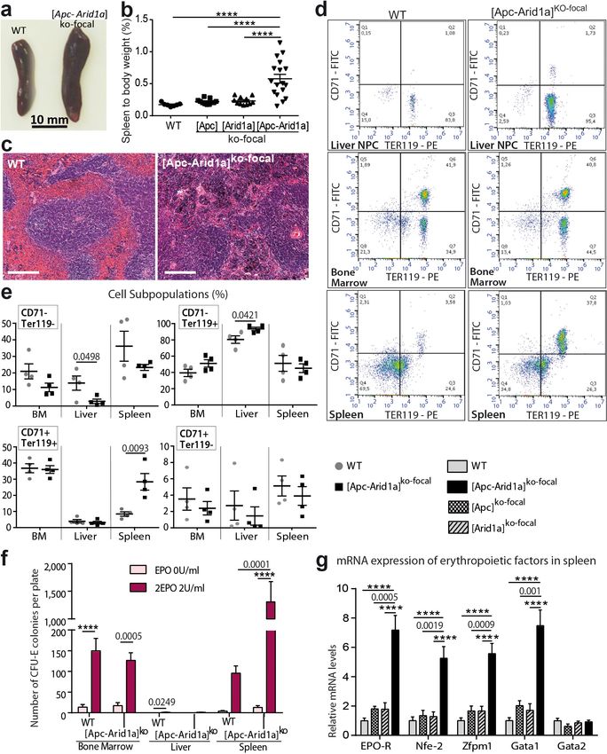
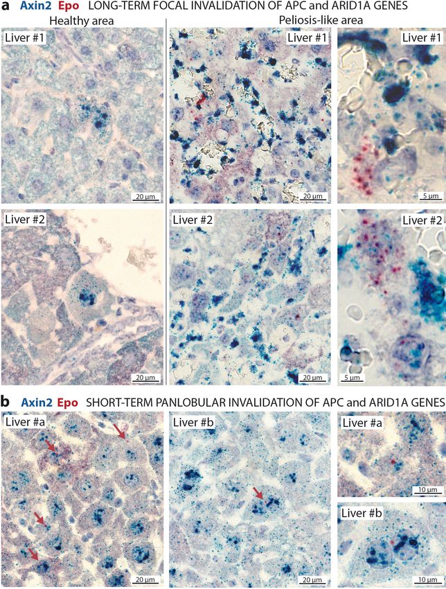
Research article                                                                Chromosomes and Gene Expression Developmental Biology

Figure 1. Development of peliosis-like regions after hepato-specific and focal Arid1a and Apc inactivation. (a) Cre-loxP-generated hepatocyte-specific
and inducible inactivation of Apc and/or Arid1a in 20% of hepatocytes after retro-orbital injection of infectious viral particles (ivp) of adenovirus
encoding Cre recombinase (AdCre). The resulting mice are referred to as [Apc-Arid1a]ko-focal, [Apc]ko-focal, and [Arid1a]ko-focal. (b) Gross examination of
mouse livers, 7 months after AdCre injection. Livers from [Apc-Arid1a]ko-focal mice had an irregular shape and a rough surface, with multiple dark red
zones (indicated by arrows). (c) Incidence of hepatic lesions detected in WT (n = 10) and [Apc-Arid1a]ko-focal (n = 24) mice by ultrasonography. (d)
Kaplan-Meier estimated survival curves of WT and [Apc-Arid1a]ko-focal mice over 15 months. n = 6 for each group. Inset: Liver of one mouse at necropsy
(13 months after AdCre injection, representative of the three analyzed mice). (e) Hematoxylin Eosin (HE)-stained sections of mouse livers at 7 months
post-injection. Large vascular spaces filled with blood cells were observed only in [Apc-Arid1a]ko-focal livers. Related data are found in Figure 1—figure
supplements 1–4, and source data in ‘Figure 1—source data 1; Figure 1—figure supplement 1—source data 1; Figure 1—figure supplement 3—
source data 1’.
The online version of this article includes the following source data and figure supplement(s) for figure 1:
Figure 1 continued on next page

Riou et al. eLife 2020;9:e53550. DOI: https://doi.org/10.7554/eLife.53550                                                                           3 of 29
Research article Chromosomes and Gene Expression Developmental Biology
Figure 1 continued
Source data 1. Emergence of peliosis (Figure 1c) and survival curve (Figure 1d).
Figure supplement 1. Focal inactivation of Apc and/or Arid1a genes in mouse liver.
Figure supplement 1—source data 1. Liver to body weight ratios (Figure 1—figure supplements 1a) and expression of Glul and Axin2 mRNAs (Fig-
ure 1—figure supplements 1b).
Figure supplement 2. Ultrasound features of livers from seven-month-old [Apc-Arid1a]ko-focal mice.
Figure supplement 3. Blood vessel enrichment and angiogenesis in [Apc-Arid1a]ko-focal livers.
Figure supplement 3—source data 1. qPCR expression of angiogenic mRNAs (Figure 1—figure supplements 3c).
Figure supplement 4. Hepatocarcinogenesis in b-catenin-activated and Arid1a-null context.
Histologically, the diseased [Apc-Arid1a]ko-focal liver showed abnormal blood vessels that were
partially or completely full of red blood cells (RBCs) (Figure 1e, Figure 1—figure supplements 3a),
associated with sinusoidal dilatation and liver cell dropout. Additionally, using microbubble-assisted
ultrasound, we showed a decrease in hepatic vascular perfusion within echogenic areas, illustrating
hence a vascular liver disease (Figure 1—figure supplements 2a). We thus characterized these areas
with dramatic histological features as peliosis-like areas, similar to the human vascular disease,
peliosis.
In accordance with previous results (Colnot et al., 2004), b-catenin-activated liver tumors devel-
oped in 92% of [Apc]ko-focal mice (Figure 1—figure supplements 4a). Here, only 8% of [Apc-Ari-
d1a]ko-focal mice developed liver tumors which were both b-catenin-activated and Arid1a-invalidated
(Figure 1—figure supplements 4a-c), suggesting that Arid1a loss suppresses the tumorigenic effect
of activated Wnt/b-catenin signaling in the liver. However, this model was not appropriate for assess-
ing the effects of Arid1a loss on Wnt/b-catenin-dependent hepatocarcinogenesis in these mice,
given the emergence of peliosis and lethality at a stage preceding or overlapping the expected
tumor initiation phase (Figure 1c, Figure 1—figure supplements 4a-c).
We reveal here that b-catenin activation and Arid1a loss cooperate to induce a dramatic hepatic
peliosis and lethality in the mouse.
Hepatic loss of both Arid1a and Apc results in erythrocytosis linked to
de novo transcription of Epo
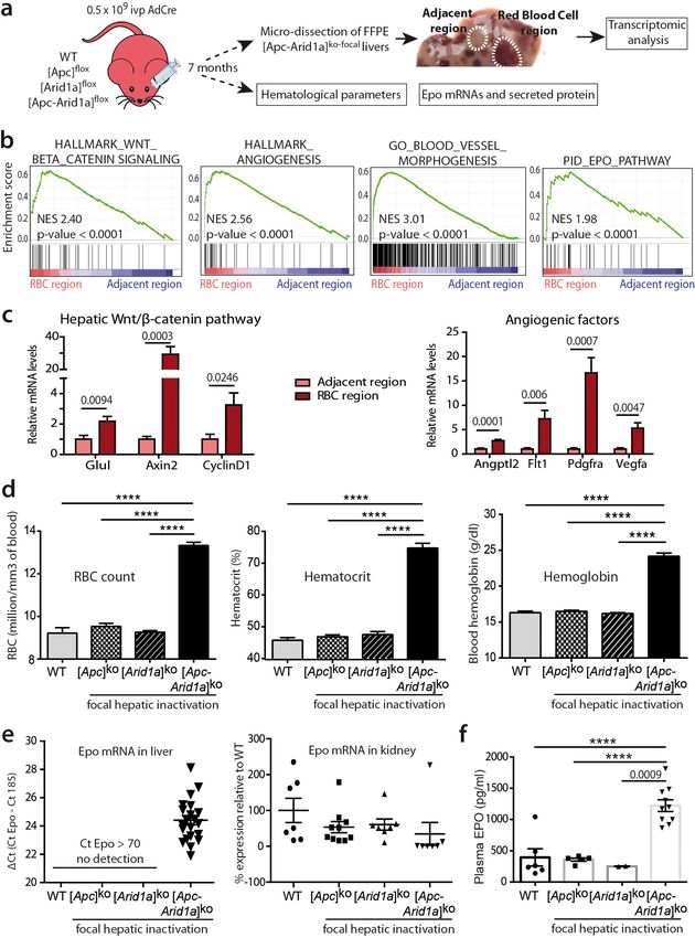
We performed transcriptomic microarray analysis of micro-dissected [Apc-Arid1a]ko-focal livers
(Figure 2a). Firstly, gene set enrichment analysis (GSEA) revealed transcriptional signatures linked to
angiogenesis and the Erythropoietin (EPO) pathway in peliosis-like areas relative to adjacent regions
(Figure 2b,c and Figure 2—figure supplements 1). Additionally, these peliosis-like regions showed
a Wnt/b-catenin transcriptional signature, revealing enrichment of b-catenin-activated cells within
these areas.
We then analyzed the hematological parameters and complete blood cell counts from peripheral
blood. RBC counts, as well as hematocrit and hemoglobin levels, were significantly higher in [Apc-
Arid1a]ko-focal mice than in control or single knockout mice (Figure 2d). This confirmed that blood
erythrocytosis corresponded to erythrocyte overload.
The production of RBCs, known as erythropoiesis, is a dynamic process requiring the orchestra-
tion of specific molecular mechanisms (Nogueira-Pedro et al., 2016). These include for example the
key EPO cytokine, a circulating glycoprotein hormone (Jelkmann, 2007). In mouse embryos, hepato-
blasts are the primary source of Epo. In adults, the site of production switches from the liver to the
kidney (Weidemann and Johnson, 2009), but the adult liver can still produce Epo (Suzuki, 2015).
To determine whether erythrocytosis in [Apc-Arid1a]ko-focal mice could be due to dysregulation of
this key hematological regulator, we examined Epo transcript and protein levels within the entire
liver and the plasma fraction, respectively. We detected a marked reactivation of Epo expression in
[Apc-Arid1a]ko-focal livers, whereas no Epo expression was detected in either single knockout or con-
trol livers (Figure 2e). This was associated with distinctly higher Epo protein levels in the plasma of
[Apc-Arid1a]ko-focal mice (Figure 2f). We confirmed that plasma Epo derived from the liver as we
observed no change in Epo transcription in the kidneys of [Apc-Arid1a]ko-focal mice (Figure 2e). Inter-
estingly, we saw no changes in Epo mRNA levels in human HCC harboring the compound CTNNB1/
ARID1A mutations (Figure 1—figure supplements 4d).
Riou et al. eLife 2020;9:e53550. DOI: https://doi.org/10.7554/eLife.53550 4 of 29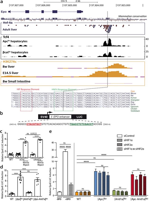
Research article                                                                  Chromosomes and Gene Expression Developmental Biology

Figure 2. Hepatic peliosis has ‘angiogenic’ and ‘erythropoietin’ transcriptional signatures, linked to a systemic erythrocytosis and to de novo hepatic
Epo expression in [Apc-Arid1a]ko-focal mice. (a) Experimental strategy; (b) Transcriptomic gene-set enrichment analysis (GSEA) of hepatic peliosis (n = 4)
relative to adjacent regions (n = 4) of [Apc-Arid1a]ko-focal mice. (c) Quantitative RT-PCR showing relative expression of mRNAs for positive targets of
hepatic Wnt/b-catenin pathway and angiogenic factors in hepatic peliosis (n = 10) compared to adjacent regions (n = 10) of [Apc-Arid1a]ko-focal mice
(unpaired t test analysis); (d) Hematological parameters from WT (n = 7), [Apc]ko-focal (n = 12), [Arid1a]ko-focal (n = 19), and [Apc-Arid1a]ko-focal (n = 20)
mice (One-way ANOVA analysis). (e) Evaluation of erythropoietin (Epo) mRNAs by quantitative RT-PCR in the livers analyzed by the DCt technique and
expressed relative to those for 18S RNA for the liver, and as relative levels in the kidney (One-way ANOVA analysis). (f) Plasma EPO concentrations at
sacrifice (WT (n = 6), [Apc]ko-focal (n = 5), [Arid1a]ko-focal (n = 2), and [Apc-Arid1a]ko-focal (n = 10)). Exact p-values are mentioned, ****p
Research article                                                                Chromosomes and Gene Expression Developmental Biology

Figure 2 continued
The online version of this article includes the following source data and figure supplement(s) for figure 2:
Source data 1. Gene expression (Figure 2c, e) and hematological parameters (Figure 2d).
Figure supplement 1. Peliosis-like regions from [Apc-Arid1a]ko-focal livers are enriched for ‘Endothelium development’ and ‘Erythrocyte homeostasis’
transcriptional signatures.

                                        Overall, our findings demonstrate that simultaneous Arid1a loss and b-catenin activation in single
                                      hepatocytes, occurring in a physiological but non-cancerous context, are responsible for a major
                                      hematological disorder that is linked to de novo expression and subsequent secretion of hepatic
                                      Epo.

                                      Erythropoiesis is induced in the spleens of [Apc-Arid1a] ko-focal mice
                                      To determine the site of pathological production of the RBCs observed in [Apc-Arid1a]ko-focal mice,
                                      we examined the liver, bone marrow (BM), and spleen; these are the three major organs responsible
                                      for erythropoiesis during embryogenesis (Suzuki et al., 2011), adult life (Suzuki, 2015), and stress
                                      responses in mice (Perry et al., 2009), respectively. Firstly, gross dissection of [Apc-Arid1a]ko-focal
                                      mice revealed a marked splenomegaly (Figure 3a,b). Histological sections from [Apc-Arid1a]ko-focal
                                      spleens showed prominent expansion of the red pulp with a predominance of erythroblasts relative
                                      to control spleens (Figure 3c).
                                          We additionally quantified erythroid precursors in the liver, BM, and spleen by flow cytometry
                                      (corresponding to the TER119+/CD71+ cell population). In [Apc-Arid1a]ko-focal liver non-parenchymal
                                      cells (NPCs) relative to controls, there was no difference in TER119+/CD71+ progenitors revealing no
                                      intra-hepatic erythropoiesis (Figure 3d,e). However, there was a striking increase in the RBC popula-
                                      tion (TER119+/CD71-). This liver erythrocytosis was confirmed by immunostaining of the hemoglobin
                                      subunit beta (HBB) in liver tissue sections, showing that RBCs, but not erythroblasts, accumulated in
                                      these livers (Figure 3—figure supplements 1). In addition, TER119+/CD71+ cell populations were
                                      similar in the BM of [Apc-Arid1a]ko-focal and control mice, whereas we found threefold more erythroid
                                      precursors in [Apc-Arid1a]ko-focal spleens than in control spleens (Figure 3d,e). This suggested that
                                      RBC overproduction came from splenic and not from medullary or hepatic erythroblasts. We then
                                      analyzed the ability of erythroid progenitors to expand by in vitro quantification of erythroid colony-
                                      forming units (CFU-E) from spleen cells, BM cells, and liver NPCs. We confirmed the presence of ery-
                                      throid progenitors in the BM and spleens of control mice after 3 days of culture in the presence of
                                      EPO, and their absence in control liver NPCs (Figure 3f). After EPO treatment, the spleens of [Apc-
                                      Arid1a]ko-focal mice contained 13-fold more CFU-E than control spleens (Figure 3f). This was not the
                                      case for the liver or BM. Finally, there were higher mRNA levels of erythropoiesis-related signaling
                                      components (Nogueira-Pedro et al., 2016) in the spleens of [Apc-Arid1a]ko-focal mice than those of
                                      control or single knockout mice (Figure 3g), including that of the Epo receptor.
                                          Overall, these data show a strong increase in erythropoiesis and erythrocyte progenitors in the
                                      spleens of [Apc-Arid1a]ko-focal mice.

                                      Blocking Epo signaling reverses erythrocytosis and splenic
                                      erythropoiesis, but maintains liver angiogenesis
                                      We analyzed the role of Epo in the dramatic phenotype of [Apc-Arid1a]ko-focal mice. We used an
                                      anti-EPO blocking serum which neutralizes soluble erythropoietin in mice (Mastrogiannaki et al.,
                                      2012). Anti-Epo treatment restored the hematocrit level of [Apc-Arid1a]ko-focal mice to that of
                                      untreated controls (Figure 4a, Figure 2d), showing a reversal of blood erythrocytosis. We quantified
                                      10-fold less erythroid precursors and a lower mRNA expression of erythropoiesis factors in the
                                      spleen of anti-Epo treated [Apc-Arid1a]ko-focal mice compared to untreated mice (Figure 4b–d).
                                         EPO is a pleiotropic growth factor which can stimulate vessel growth through an autocrine and/or
                                      paracrine loop (Kimáková et al., 2017). We tested the attractive possibility that hepatocyte-
                                      secreted Epo in [Apc-Arid1a]ko-focal mouse livers regulates RBC homing to the liver through
                                      increased angiogenesis. Liver tissue sections showed that blood vessels contained less RBCs in anti-
                                      EPO treated [Apc-Arid1a]ko-focal mice compared to untreated mice (Figure 4e, Figure 4—figure
                                      supplements 1), and these livers harbored less TER119+/CD71- mature RBCs (Figure 4f–g). Despite

Riou et al. eLife 2020;9:e53550. DOI: https://doi.org/10.7554/eLife.53550                                                                       6 of 29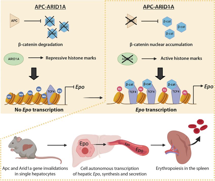
Research article                                                                  Chromosomes and Gene Expression Developmental Biology

Figure 3. Erythropoiesis occurs in the spleen of [Apc-Arid1a]ko-focal mice. (a) Gross morphology of spleens from representative control (WT) and [Apc-
Arid1a]ko-focal mice; (b) Spleen/body weight ratio of WT (n = 7), [Apc]ko-focal (n = 11), [Arid1a]ko-focal (n = 11), and [Apc-Arid1a]ko-focal (n = 17) mice (one-
way ANOVA). (c) Hematoxylin and Eosin staining of splenic sections. Scale bar is 200 mm. (d,e) FACS analysis of liver NPC, bone marrow, and spleens
from control (WT) or [Apc-Arid1a]ko-focal mice using the erythroid markers CD71 and Ter119. (e) FACS quantification from WT (n = 4) and [Apc-Arid1a]ko-
focal
      (n = 4) mice (multiple t-test). (f) Quantification of erythroid progenitors as erythroid colony-forming units (CFU-E) in the presence of EPO, using 2
105 cells from bone marrow or 2  106 cells from the liver and spleen of WT or [Apc-Arid1a]ko-focal mice (2-way ANOVA). (g) Q-PCR showing relative
expression of several factors, known to be involved in stress-induced erythropoiesis, in the spleens of WT (n = 9), [Apc]ko-focal (n = 5), [Arid1a]ko-focal
(n = 8), and [Apc-Arid1a]ko-focal (n = 8) mice (one-way ANOVA). ****p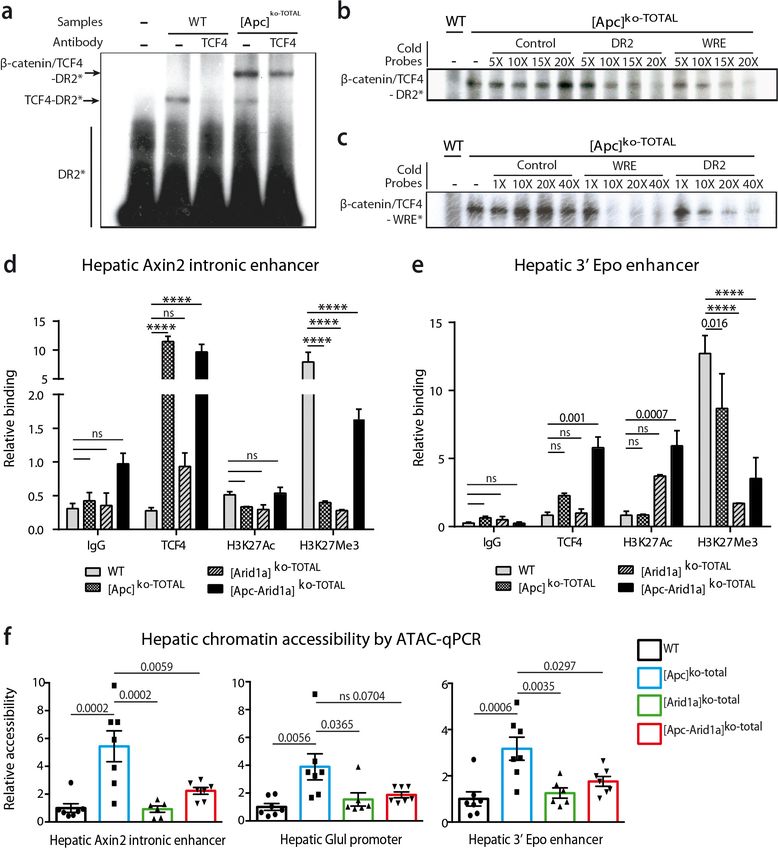
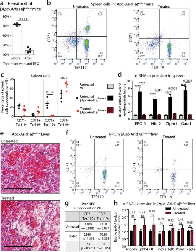
Research article                                                                Chromosomes and Gene Expression Developmental Biology

Figure 4. Blockade of Epo signaling with anti-EPO serum in [Apc-Arid1a]ko-focal mice eliminates aberrant erythropoiesis in the spleen, but maintains
angiogenesis in the liver. (a) Hematocrit before (n = 4) and after (n = 4) anti-EPO treatment (t-test). (b,c) FACS analysis (b) and quantification (c) of
spleens with/without anti-EPO (n = 4 for each group) (t-test). (d) RT-qPCR showing relative expression of erythropoiesis factors in the spleens of WT
(n = 9), treated [Apc-Arid1a]ko-focal (n = 4), untreated [Apc-Arid1a]ko-focal (n = 8) mice (one-way ANOVA). (e) Hematoxylin Eosin (HE)-stained sections of
livers from representative 7-month-old mice. (f,g) FACS analysis (f) and quantification (g) of liver NPC with/without anti-EPO. (h) RT-qPCR showing
relative expression of angiogenic factors in the livers with (n = 4) and without (n = 10) anti-EPO (t-test). ****p
Research article Chromosomes and Gene Expression Developmental Biology
this decrease in intrahepatic RBCs, we did not observe any change in the disruption of the liver vas-
cular architecture as shown by both histological (Figure 4e, Figure 4—figure supplements 1) and
gene expression analyses (Figure 4h).
We demonstrate here that high plasma Epo concentration is directly responsible for splenic eryth-
ropoiesis and erythrocytosis in [Apc-Arid1a]ko-focal mice. However, this cytokine alone is not responsi-
ble for alterations in liver angiogenesis.
Epo is cell-autonomously expressed by b-catenin-activated Arid1a-null
hepatocytes in both the mouse and in humans
We investigated whether Epo is expressed by hepatocytes after Apc and/or Arid1a hepato-specific
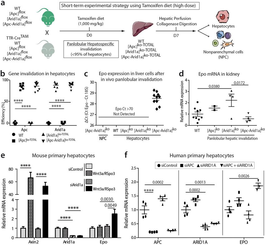
inactivations. We generated Tamoxifen-induced mouse models (Figure 5a) with short-term panlobu-
lar gene inactivations (Figure 5b) and Apc loss-induced hepatomegaly (Figure 5—figure supple-
ments 1a) as previously shown (Buenrostro et al., 2015). After diet-based Tamoxifen
administration, the Apc and/or Arid1a genes were invalidated in approximately 90% of hepatocytes
(Figure 5—figure supplements 1b). There was no gene invalidation in liver NPCs, thus highlighting
the high purity of the NPC fraction (Figure 5—figure supplements 2a). We detected Epo mRNA
expression only in the hepatocyte compartment and not in NPCs of [Apc-Arid1a]ko-TOTAL livers,
whereas a slight decrease of Epo expression was seen in the kidney of [Apc-Arid1a]ko-TOTAL mice
(Figure 5c,d).
To confirm the cell-autonomous expression of Epo in b-catenin-activated Arid1a-null hepatocytes,
we performed RNA in situ hybridization for Epo with Axin2 as a marker of b-catenin activation (Fig-
ure 6). Epo transcripts were not expressed in the livers, yet were abundant in rare interstitial renal
cells of control mice (Figure 6—figure supplements 1a); this localization of Epo in the kidney has
already been described (Lacombe et al., 1988). Conversely but as expected, we found Axin2 mRNA
transcripts in pericentral hepatocytes (Benhamouche et al., 2006). After Apc and Arid1a gene invali-
dation, we found a de novo expression of Epo in a subset of Axin2-expressing hepatocytes. In the
long-term focal model, this expression was restricted to the areas of peliosis (Figure 6a). In the
short-term panlobular model, rare Axin2-expressing hepatocytes also expressed single Epo mRNA
transcripts (Figure 6b). In both models, Epo expression was not found elsewhere in the liver.
We examined whether Epo expression is specific to the loss of Apc or can be initiated regardless
of how Wnt/b-catenin signaling is activated. We successfully activated b-catenin via its Wnt/Spondin
ligand in murine primary hepatocytes (Figure 5—figure supplements 2e). We consecutively per-
formed in vivo Arid1a knockout followed by in vitro Wnt/Spondin stimulation, or in vivo Apc loss fol-
lowed by efficient in vitro siRNA-mediated Arid1a knockdown (si-Arid1a) (Figure 5—figure
supplements 2f). Epo expression significantly increased in these conditions (Figure 5e, Figure 5—
figure supplements 2b, c). Mutational activation of b-catenin coupled with si-Arid1a also led to the
induction of Epo expression in the b-catenin-mutated HEPA1.6 murine hepatoma-derived cell line
(Figure 5—figure supplements 2d).
We assessed the conservation of EPO regulation from mouse to humans. We found that EPO
mRNA expression was also regulated by both the chromatin remodeler ARID1A and the Wnt/b-cate-
nin signaling pathway in primary human hepatocytes after siRNA-mediated ARID1A and APC down-
regulation (Figure 5f).
Overall, these in vivo and in vitro findings strongly demonstrate a conserved and cell-autonomous
role of Wnt/b-catenin activation and Arid1a loss in hepatic Epo expression. This occurs as a stochas-
tic transcriptional event in b-catenin-activated Arid1a-null hepatocytes.
Wnt/b-catenin pathway control of 3’ Epo enhancer activity is hypoxia-
and HIF-independent
We questioned if b-catenin directly controls Epo transcription through cis-regulatory sequences. We
previously performed ChIP-Seq experiments to assess Tcf4/b-catenin occupancy in the chromatin of
hepatocytes isolated from [Apc]ko-TOTALversus [b-catenin]ko-TOTAL murine models (Gougelet et al.,
2014). The only DNA region bound by Tcf4 in the vicinity of the Epo gene was its 3’enhancer (Epo-
3’E), known to be involved in Epo transcription in the embryonic liver, as well as the known Hif- (HIF-
REs) and Hnf4-containing responses elements (HREs) (Suzuki et al., 2011; Semenza et al., 1991;
Figure 7a). This Tcf4 binding was at the same location as HRE binding, and was stronger in activated
Riou et al. eLife 2020;9:e53550. DOI: https://doi.org/10.7554/eLife.53550 9 of 29Research article Chromosomes and Gene Expression Developmental Biology Figure 5. Cell-autonomous Epo expression after Arid1a inactivation and Wnt/b-catenin activation in murine and human hepatocytes. (a) In vivo and ex vivo strategy. WT (n = 8), [Apc]ko-TOTAL (n = 7), [Arid1a]ko-TOTAL (n = 8), and [Apc-Arid1a]ko-TOTAL (n = 10) mice. (b) Inactivation efficiency of Apc and Arid1a genes in isolated hepatocytes. (c,d) RT-qPCR assessment of erythropoietin (Epo) transcription (c) in the hepatocyte and NPC compartments of the livers, (d) in the kidney (1-way ANOVA). (e) In vitro analysis of Axin2, Arid1a (Arid1a floxed-exon detection), and Epo expression by RT-qPCR of mouse hepatocytes after Wnt3a and R-Spondin3 stimulation, and si-Arid1a/si-Control treatments, showing Arid1a knockdown efficiency and Wnt/b- catenin pathway activation, as the mRNA levels of Axin2, a canonical target gene of Wnt signaling, significantly increased (2-way ANOVA). (f) In vitro analysis of Apc, Arid1a, and Epo by RT-qPCR of cryopreserved human hepatocytes after siRNA transfection (one-way ANOVA analysis). Data are presented as the mean ± SEM. ****p
Research article                                                         Chromosomes and Gene Expression Developmental Biology

Figure 5 continued
Figure supplement 2—source data 1. Efficiency of gene invalidation (Figure 5—figure supplements 2a), mRNA expression (Figure 5—figure supple-
ments 2b-d), western blots (Figure 5—figure supplements 2e).

                                    b-catenin than in b-catenin-null hepatocytes (Figure 7a). We demonstrated from ENCODE data that
                                    H3K27Ac, a histone mark indicating active promoters or enhancers, also bound to this region; this
                                    binding was present in mouse liver chromatin at E14.5, an embryonic stage in which the Epo gene is
                                    actively transcribed (Figure 7a). However, Epo was only partially present in the livers of eight-week-
                                    old mice, with no Epo transcription, and completely absent in the adult small intestine, a tissue
                                    known not to transcribe the Epo gene (Figure 7a).
                                       We thus tested whether Wnt/b-catenin signaling directly activates hepatic Epo transcription
                                    through the Epo-3’E. We transfected a luciferase reporter (pEpoE-luc) containing the HIF and HNF4-
                                    binding sites into primary mouse hepatocytes (Figure 7b). After Wnt/Spondin stimulation, and
                                    regardless of si-Arid1a treatment, Epo enhancer activity was five- to eight-fold higher (Figure 7c,d).
                                    Hence, in this in vitro reporter assay context, b-catenin signaling increases Epo-3’E activity and it is
                                    independent of the chromatin landscape.
                                       Hypoxia-inducible factor (HIF) signaling is the master pathway regulating EPO transcription and
                                    Hif2a has a prominent role in hepatic Epo transcription (Mastrogiannaki et al., 2012). We investi-
                                    gated Hif2a involvement in b-catenin/Arid1a-dependent Epo expression. In vivo, we did not detect
                                    hypoxia or Hif1a/Hif2a accumulation in the absence of Apc and/or Arid1a in mouse livers (Figure 7—
                                    figure supplements 1a-c). A small subset of Hif1a/Hif2a targets, such as Eno2, Car9, and Rab42,
                                    was slightly overexpressed in both [Apc]ko and [Apc-Arid1a]ko livers, confirming that b-catenin and
                                    HIF signaling share some transcriptional targets (Figure 7—figure supplements 1d-e;
                                    Benhamouche et al., 2006). As expected, the hypoxia-mimetic agent desferrioxamine (DFO)
                                    markedly potentiated luciferase activity in pEpoE-luc-transfected hepatocytes, whereas efficient
                                    knockdown of both Hif1a or Hif2a (Figure 7—figure supplements 2) resulted in a significant
                                    decrease (Figure 7e). Interestingly, knockdown of HIFs, either alone or combined, did not reduce
                                    Epo-3’E induction by b-catenin signaling in hepatocytes, whether Apc be inactivated alone or in
                                    combination with Arid1a (Figure 7e).
                                       In all, the Wnt/b-catenin pathway controls erythropoietin expression in hepatocytes through the
                                    3’ Epo enhancer in a hypoxia- and HIF-independent manner.

                                    Both b-catenin signaling and Arid1a are key players in chromatin
                                    remodeling, histone recruitment, and Tcf4 binding on the hepatic Epo
                                    enhancer
                                    We previously showed similarities between HREs and WREs, and that Tcf4 can bind HREs and
                                    thereby participate in b-catenin-dependent transcription (Gougelet et al., 2014). Here, we found
                                    that Tcf4 bound DNA on the HRE region of the Epo-3’E in which there is no classical WRE. Indeed,
                                    by electrophoretic mobility shift assay (EMSA), we showed that Tcf4 weakly bound the Epo-3’E HRE
                                    (thereafter called DR2) in control liver nuclear extracts (Figure 8a). In [Apc]ko-TOTAL liver extracts, the
                                    nuclear translocation of b-catenin led to a stronger binding represented by a supershift (Figure 8a,
                                    b). This indicates that the Tcf4/b-catenin complex binds this DR2 motif, as well as a classical WRE
                                    shown by competitive EMSA (Figure 8b,c). These findings highlighted that Tcf4 binds to the HRE of
                                    the Epo enhancer and that activation of b-catenin increases this interaction.
                                        Endogenous hepatic Epo was expressed de novo after both Wnt/b-catenin activation and Arid1a
                                    knockout, but gene expression of classical b-catenin target genes (Glul, Axin2) was not affected by
                                    Arid1a status (Figure 8—figure supplements 1). We thus characterized Tcf4 binding, chromatin
                                    accessibility, and histone active (H3K27Ac) or repressive (H3K27Me3) marks of the Epo enhancer,
                                    the Axin2 intronic enhancer, and the Glul promoter in hepatocytes isolated from transgenic mouse
                                    livers.
                                        As previously described (Gougelet et al., 2014), Tcf4 efficiently bound to the Axin2 intronic
                                    enhancer in vivo and this increased when b-catenin signaling was activated (Figure 8d). This was

Riou et al. eLife 2020;9:e53550. DOI: https://doi.org/10.7554/eLife.53550                                                              11 of 29
Research article Chromosomes and Gene Expression Developmental Biology Figure 6. In situ hybridization of mRNAs showing a de novo expression of Epo in a subset of b-catenin-activated hepatocytes. (a) Seven months after Apc/Arid1a gene invalidation in single hepatocytes from two livers (#1 and #2); (b) 7 days after gene invalidation in more than 90% hepatocytes (two livers: #a and #b). Axin2 RNAScope probe stains b-catenin-activated hepatocytes (blue dots), and Epo RNAScope probe stains single Epo mRNAs as red dots. Related data are found in Figure 6—figure supplement 1. The online version of this article includes the following figure supplement(s) for figure 6: Figure supplement 1. Implementation of in situ Hybridization for Axin2 and Epo mRNAs using RNAScope, showing expressing mRNA as dots. Riou et al. eLife 2020;9:e53550. DOI: https://doi.org/10.7554/eLife.53550 12 of 29
Research article                                                           Chromosomes and Gene Expression Developmental Biology

Figure 7. Wnt/b-catenin directly controls EPO expression through 3’ Epo enhancer, in a HIF-independent manner. (a) Genomic environment of the Epo
gene (UCSC Genome Browser, mm9 database) and ChIP-seq peaks at the 3’ Epo enhancer. In blue/red: the crude reads of ChIP-Seq data performed in
adult livers against HNF-4a (54). In black: ChIP-Seq under Apcko or bcatko conditions with an antibody against TCF4 (16). In yellow: ENCODE data of
H3K27Ac marks in eight-week-old and E14.5 embryonic livers (Histone Mods by ChIP-Seq from ENCODE/LICR). (b) Schematic representation of the
Figure 7 continued on next page

Riou et al. eLife 2020;9:e53550. DOI: https://doi.org/10.7554/eLife.53550                                                                  13 of 29
Research article                                                                Chromosomes and Gene Expression Developmental Biology

Figure 7 continued
EpoE-Luc erythropoietin luciferase reporter, driven by the 3’ enhancer. (c–e) Luciferase reporter assays in mouse primary hepatocytes: (c) after in vitro
overactivation of Wnt/b-catenin signaling and Arid1a knockdown (d) after in vivo Cre-loxP-mediated gene inactivation; (e) Effect of hypoxic-mimic
conditions using desferrioxamine (DFO), and effect of knockdown of HIF factors (two separate experiments carried out in triplicate). Results are in
relative light units, and analyzed using 1-way (d) or 2-way ANOVA (c,e). ****p
Research article                                                              Chromosomes and Gene Expression Developmental Biology

Figure 8. b-catenin/Tcf4 complex binds to the HNF4-responsive element of Epo enhancer (Epo-HRE) after modifications of histone marks and
chromatin accessibility. (a) EMSA using nuclear proteic extracts from WT or [Apc]ko-TOTAL livers and 32P-labeled probes containing Epo-HRE (DR2). (b, c)
Competitive EMSA using 32P-labeled DR2 (b) and 32P-labeled WRE (c) probes and increasing concentrations of cold probes containing HNF4, WRE or
control-responsive element. WRE cold probes compete with radiolabeled DR2 motif for the Tcf4/b-catenin binding and vice versa. (d, e) Chromatin
ImmunoPrecipitation (ChIP) assays of hepatocytes from WT, [Apc]ko-TOTAL, [Arid1a]ko-TOTAL, and [Apc-Arid1a]ko-TOTAL livers. ChIP-qPCR against IgG,
Tcf4, Acetylation of Histone3 in Lysine27 (H3K27Ac), and Tri-methylation of Histone3 in Lysine27 (H3K27me3) for Axin2 (d) and Epo (e) enhancer regions.
WT (n = 3), [Apc]ko-TOTAL (n = 2), [Arid1a]ko-TOTAL (n = 2), and [Apc-Arid1a]ko-TOTAL (n = 3) mice. Enrichment by ChIP was assessed relative to the input
DNA and normalized to the level of negative controls. (f) ATAC-qPCR using frozen livers from WT (n = 7), [Apc]ko-TOTAL (n = 7), [Arid1a]ko-TOTAL (n = 6),
and [Apc-Arid1a]ko-TOTAL (n = 7) mice. Data are analyzed with one-way ANOVA. ****p
Research article                                                              Chromosomes and Gene Expression Developmental Biology

Figure 9. Schematic model of the role of Arid1a in hepatic Epo expression linked to overactivation of the Wnt/b-catenin pathway. Under physiological
conditions, the presence of Arid1a is associated with histone repressive marks at the Epo enhancer and b-catenin is constantly degraded; thus, Epo is
not produced. In the absence of Apc, b-catenin/Tcf4 complex binds the Epo enhancer, and enhances chromatin accessibility, but the histone marks
remain repressive. The loss of Arid1a increases active histone marks, which is insufficient to induce Epo transcription. After both Wnt/b-catenin
activation and Arid1a inactivation, active histone marks and binding of b-catenin/Tcf4 to the Epo enhancer drive Epo liver transcription, and subsequent
secretion of Epo into the bloodstream, resulting in splenic erythropoiesis and in substantial blood and liver erythrocytosis.

                                        The consequences of this HIF signaling-independent Epo regulation is significant for the genetic
                                     engineering of EPO for therapeutic purposes. In anemia, a major complication of chronic kidney dis-
                                     ease, HIF stabilizers are currently used to restore circulating EPO levels. The long-term safety of this
                                     strategy is hindered by the lack of targeting specificity (Kular and Macdougall, 2019). The use of
                                     cell transcriptional machinery to produce therapeutic levels of EPO has been put forward to over-
                                     come the side effects associated with HIF stabilizers. The EPO-producing cells of the adult kidney
                                     are potential candidates, but anemic patients have damaged kidneys. Based on our results, here we

Riou et al. eLife 2020;9:e53550. DOI: https://doi.org/10.7554/eLife.53550                                                                      16 of 29
Research article Chromosomes and Gene Expression Developmental Biology
can propose an alternative involving the restoration of the ability of hepatocytes to synthetize EPO,
independently of hypoxia, by targeting Wnt/b-catenin and ARID1A signaling in the liver.
Our demonstration that Arid1a inactivation is required in Epo transcription opposes previously
described roles of chromatin remodeling complexes in hepatic regulation of Epo (Wang et al.,
2004; Sena et al., 2013). However, firstly these studies analyzed hypoxia-dependent Epo regulation
which is distinct from our study; we firmly established that the b-catenin-dependent control of Epo
transcription depends on Arid1a loss, is Hif-independent, and occurs in a normoxic adult liver. Sec-
ondly, these studies focused on BRG1/BRM ATPases, essential core subunits of both the BAF and
pBAF complexes. The loss of Arid1a, a facultative component of the BAF complex, does not disrupt
BAF complex functionality as Arid1b is known to compensate for Arid1a loss. This highlights a spe-
cific role for Arid1a in transcriptional repression through the modulation of chromatin accessibility to
transcription factors at their target DNA sequences (Sun et al., 2016; Nagl et al., 2005). We show
increased binding of the Tcf4/b-catenin complex to Epo-3’E Hnf4-RE is Arid1a-dependent and
Arid1a loss decreases the H3K27me3 repressive mark. That could be due to the intricate balance
existing between the Polycomb complex PRC2 and the SWI/SNF complex (Kadoch et al., 2016).
Accordingly, the inhibition of the Polycomb EZH2 subunit is synthetically lethal in ARID1A-mutated
tumors (Bitler et al., 2015; Alldredge and Eskander, 2017). Therefore, Arid1a and the Polycomb
complex could act in concert to modulate Epo gene expression in the liver.
We illustrate that Arid1a loss renders the liver Epo-inducible element more accessible to Tcf4,
and even more so to b-catenin. Contrary to the paradigm that Tcf4 binds its DNA targets regardless
of b-catenin activation status, we previously reported that Tcf4 DNA-binding was stronger in the
presence of nuclear b-catenin in the liver (Gougelet et al., 2014). More broadly, numerous interac-
tions between chromatin remodeling and Wnt/b-catenin signaling have already been described
(Barker et al., 2001; Eckey et al., 2012; Mathur et al., 2017; Song et al., 2009; Yan et al., 2014;
Zhai et al., 2016) and can explain the impact of b-catenin signaling on chromatin accessibility at the
Epo enhancer. Single-RNA in situ hybridization revealed that Epo gene expression only occurs in
rare hepatocytes, emphasizing the complexity of Epo liver transcription in the liver. This contributes
to previous studies using single-RNA in situ hybridization, showing that transcription in the liver is
gene-dependent, and is either bursty and dynamic or stable (Bahar Halpern et al., 2015).
We found here that the loss of Arid1a does not change the transcription of hepatic canonical
Wnt/b-catenin target genes. As for Epo, it could potentially unmask new chromatin-dependent b-
catenin target genes. Among these new Arid1a/b-catenin target genes are those involved in liver
angiogenesis. In the near future, genome-wide studies will be required to firmly identify these genes,
combining the analysis of transcriptome, chromatin accessibility (ATAC-Seq), histone mark, b-catenin
and Arid1a cistromes (ChIP-Seq) in liver chromatin from Arid1a-null and b-catenin-activated
hepatocytes.
The initial aim of our study was to better elucidate oncogenic cooperation in liver carcinogenesis.
In our in vivo experimental models reported here, the loss of Arid1a protects against b-catenin-
dependent carcinogenesis. However, these results were not fully exploitable due to the deleterious
effect of the dramatic hematological disorder developed by the mice. New mouse models are there-
fore required for further investigation of the oncogenic role of Arid1a in liver carcinogenesis. In turn,
confirmation of such a role would corroborate a recent study showing that hepatic Arid1a can harbor
either a tumor suppressor or oncogenic role depending on the cellular context (Sun et al., 2018).
An additional study also demonstrated that Arid1a is protumoral rather than a tumor suppressor in
colorectal cancer with Apc mutations (Mathur et al., 2017).
Lastly, some liver cancer studies have identified pathological erythrocytosis and/or hepatic vascu-
lar lesions, potentially with EPO production and peliosis. However, the molecular mechanisms under-
lying these pathological observations are still poorly understood (Matsuyama et al., 2000;
Bunn, 2013; Ke et al., 2017; Hoshimoto et al., 2009; Tsuchiya et al., 2009; Vik et al., 2009). Our
study contributes molecular clues by indicating that this is not linked to CTNNB1/ARID1A mutations,
but more likely attributed to the hypoxia frequently found in cancers. Future studies should use
mouse models and data from patients with HCC to address the specific transcriptional output of
CTNNB1/ARID1A-mutated liver tumors.
Riou et al. eLife 2020;9:e53550. DOI: https://doi.org/10.7554/eLife.53550 17 of 29Research article Chromosomes and Gene Expression Developmental Biology
Materials and methods
Key resources table
Reagent
type
(species) or Source or Additional
resource Designation reference Identifiers information
Gene Epo GenBank NM_007942.2 Erythropoietin
(Mus musculus)
Gene Arid1a GenBank NM_001080819.2 Arid1a
(Mus musculus)
Gene Ctnnb1 GenBank NM_007614.3 Beta-catenin
(Mus musculus)
Gene Apc GenBank NM_001360980.1 Adenomatous
(Mus musculus) polyposis coli
Strain, strain Arid1a-lox From Arid1atm1.1Zhwa/J https://www.jax.
background Z. Wang’s lab org/strain/027717
(Mus musculus)
Strain, strain Apc-lox From Perret- Apctm2.1Cip https://www.
background Colnot’s lab infrafrontier.
(Mus musculus) eu/search?
keyword=
EM:05566
Strain, strain Ttr-Cre-Tam From Perret- Tg(Ttr-cre/ https://www.
background Colnot’s lab Esr1*)1Vco infrafrontier.
(Mus musculus) eu/search?
keyword=
EM:01713
Genetic Ad-Cre Université de Ad5-CAG-Cre https://umr1089.univ-
reagent Nantes, France nantes.fr/facilities-
(Adenovirus 5) cores/cpv/translational-
vector-core-2201753.
kjsp?RH=1519296751975
Cell line Mouse From Christine Hepa 1-6 [Hepa1- For transfection
(Mus musculus) hepatoma Perret’s lab 6] (ATCC experiments
CRL-1830)
Antibody anti-Arid1a Abcam Cat# 182560 IHC(1:1000),
(Rabbit [EPR13501] WB (1:2000)
monoclonal)
Antibody anti-Glul (GS) BD Biosciences Cat# 610518, IHC(1:400),
(Mouse RRID:AB_397880 WB (1:5000)
monoclonal)
Antibody anti-HBB Proteintech Cat# 16216–1-AP, IHC(1:200),
(Mouse RRID:AB_10598329 WB (1:2000)
monoclonal)
Antibody anti-HIF1a Novus Cat# NB100-449, WB nuclear
(Rabbit RRID:AB_10001045 extract (1:500)
polyclonal)
Antibody anti-HIF2a Novus Cat# NB100-122, WB nuclear
(Rabbit RRID:AB_10002593 extract (1:500)
polyclonal)
Antibody Anti-Tcf4 Millipore Cat# 05–511, ChIP: 3 mg
(Tcf7l2) (Mouse RRID:AB_309772
monoclonal)
Antibody Anti-H3K27Ac Active Motif Cat# 39133, ChIP: 3 mg
(Rabbit RRID:AB_2561016
polyclonal)
Antibody Anti-H3K27me3 Active Motif Cat# 39155, ChIP: 3 mg
(Rabbit RRID:AB_2561020
polyclonal)
Continued on next page
Riou et al. eLife 2020;9:e53550. DOI: https://doi.org/10.7554/eLife.53550 18 of 29Research article Chromosomes and Gene Expression Developmental Biology
Continued
Reagent
type
(species) or Source or Additional
resource Designation reference Identifiers information
Antibody IgG (Mouse) Thermo Cat# 10400C, ChIP: 3 mg
Fisher Scientific RRID:AB_2532980
Antibody Anti-CD71- BD Biosciences Cat# 553266, FACS (1:100)
FITC (Rat RRID:AB_394743
monoclonal)
Antibody Anti-Ter119- BD Biosciences Cat# 553673, FACS (1:100)
PE (rat RRID:AB_394986
monoclonal)
Antibody Anti-b-actin Sigma-Aldrich Cat# A5441, WB (1:10000)
(mouse RRID:AB_476744
monoclonal)
Antibody Anti-lamin A/C Cell Signaling Cat# 2032, WB nuclear
(rabbit polyclonal) Technology RRID:AB_2136278 extract (1:500)
Antibody IgG, HRP- Cell Signaling Cat# 7076, WB (1:2000)
conjugated Technology RRID:AB_330924
(horse,
anti-mouse)
Antibody IgG, Cell Signaling Cat# 7074, WB (1:2000)
HRP-conjugated Technology RRID:AB_2099233
(goat, anti-rabbit)
Antibody IgG, biotinylated Vector lab Cat# BA-1000, IHC (1:200)
(goat, anti-rabbit) RRID:AB_2313606
Commercial MOM mouse Vector Cat# BMK-2202, Kit
assay or kit on mouse Laboratories RRID:AB_2336833
Sequence- 18S Thermo Taqman qPCR primers
based reagent Fisher Assay 4308329
Scientific
Sequence- Glul Thermo Taqman Assay qPCR primers
based reagent Fisher Mm00725701_si Mus musculus
Scientific
Sequence- Axin2 Thermo Taqman Assay qPCR primers
based reagent Fisher Scientific Mm00443610_m1 Mus musculus
Sequence- Arid1a (total) Thermo Taqman Assay qPCR primers
based reagent Fisher Scientific Mm00473838_m1 Mus musculus
Sequence- Arid1a (not Thermo Taqman Assay qPCR primers
based reagent excised by Cre) Fisher Scientific Mm00473841_m1 Mus musculus
Sequence- Apc (total) Thermo Taqman Assay qPCR primers
based reagent Fisher Scientific Mm00545877_m1 Mus musculus
Sequence- Apc (not Thermo Taqman Assay qPCR primers
based reagent excised by Cre) Fisher Scientific Mm01130462_m1 Mus musculus
Sequence- Epo Thermo Taqman Assay qPCR primers
based reagent Fisher Mm01202755_m1 Mus musculus
Scientific
Sequence- 18 s Eurogentec F_GTAACCCGT SybrGreen
based reagent TGAACCCCATT qPCR primers
R_CCATCCAA
TCGGTAGCG
Sequence- Angiopoietin- Eurogentec F_CCGCAACAT SybrGreen qPCR
based reagent like 2 (Angptl2) GAACTCGAGAG primers Mus musculus
R_GTGCTCCAGG
TCCTTGTACT
Sequence- Carbonic Eurogentec F_GACCTCGTG SybrGreen qPCR
based reagent anhydrase 9 (Car9) ATTCTCGGCTA primers Mus musculus
R_GAGAAGGC
CAAACACCAAGG
Continued on next page
Riou et al. eLife 2020;9:e53550. DOI: https://doi.org/10.7554/eLife.53550 19 of 29Research article Chromosomes and Gene Expression Developmental Biology
Continued
Reagent
type
(species) or Source or Additional
resource Designation reference Identifiers information
Sequence- Cyclin D1 Eurogentec F_AGAAGTGCG SybrGreen qPCR
based reagent (Ccnd1) AAGAGGAGGTC primers Mus musculus
R_TTCTCGGC
AGTCAAGGGAAT
Sequence- Enolase 2, Eurogentec F_TGGATTTCA SybrGreen qPCR
based reagent gamma neuronal AGTCTCCCGCT primers Mus musculus
(Eno2) R_TCAGGTCAT
CGCCCACTATC
Sequence- Erythropoietin Eurogentec F_ATGACTTTCG SybrGreen qPCR
based reagent receptor (Epo-r) TGACTCACCCT primers Mus musculus
R_GGGCTCCG
AAGAACTTCTGTG
Sequence- FMS-like Eurogentec F_AGAGGAGGA SybrGreen qPCR
based reagent tyrosine TGAGGGTGTCT primers Mus musculus
kinase 1 (Flt1) R_GGGAACTT
CATCTGGGTCCA
Sequence- GATA binding Eurogentec F_TTCCCACTA SybrGreen qPCR
based reagent protein 1 (Gata1) CTGCTGCTACC primers Mus musculus
R_GCGGCCTC
TATTTCAAGCTC
Sequence- GATA binding Eurogentec F_GCCGGTTCT SybrGreen qPCR
based reagent protein 2 (Gata2) GTCCATTCATC primers Mus musculus
R_ATGGCAGCA
GTCTCTTCCAT
Sequence- Inhibin beta-B (Inhbb) Eurogentec F_GTACCTGAAA SybrGreen qPCR
based reagent CTGCTCCCCT primers Mus musculus
R_ATGGCCTC
TGTGATGGGAAA
Sequence- Potassium Eurogentec F_TGACTTCTAC SybrGreen qPCR
based reagent channel CAGATCCGGC primers Mus musculus
tetramer domain R_TCAGGGTCAG
contain. 11 (Kctd11) TGCAGAAGAG
Sequence- Kinase insert Eurogentec F_AGAAGATGC SybrGreen qPCR
based reagent domain protein CCATGACCCAA primers Mus musculus
receptor (Kdr) R_TCACCCATC
CTCAACACACA
Sequence- Nuclear factor, Eurogentec F_GATGTCCCGA SybrGreen qPCR
based reagent erythroid derived ACTAGAGCCA primers Mus musculus
2 (Nfe2) R_ACACCCTTG
GCCTTAGAGTC
Sequence- Platelet derived Eurogentec F_ACAGCTCAC SybrGreen qPCR
based reagent growth factor AGACTTCGGAA primers Mus musculus
receptor, alpha R_AGAAGATGA
polypeptide TACCCGGAGCG
(Pdgfra)
Sequence- Phosphoglycerate Eurogentec F_TGGCACCAG SybrGreen qPCR
based reagent kinase 1 (Pgk1) GAACCCTTAAA primers Mus musculus
R_AGCTCAGCC
TTTACAGCTCA
Sequence- Placenta- Eurogentec F_TGATTGCTT SybrGreen qPCR
based reagent specific 8 (Plac8) CAGTGACTGCG primers Mus musculus
R_GTTCATGGC
TCTCCTCCTGT
Sequence- Protein tyrosine Eurogentec F_TGGACCCTG SybrGreen qPCR
based reagent phosphatase, GGATCTAAGGA primers Mus musculus
receptor type, R_GTGGTCACT
B (Ptprb) GCAAGCTTCAA
Continued on next page
Riou et al. eLife 2020;9:e53550. DOI: https://doi.org/10.7554/eLife.53550 20 of 29Research article Chromosomes and Gene Expression Developmental Biology
Continued
Reagent
type
(species) or Source or Additional
resource Designation reference Identifiers information
Sequence- Member RAS Eurogentec F_GGCGTTCTG SybrGreen qPCR
based reagent oncogene family TTGGTCTTTGA primers
(Rab42) R_GCAAGTTCCT Mus musculus
CTGCTTCCTG
Sequence- Vascular Eurogentec F_GCTGTAACGAT SybrGreen qPCR
based reagent endothelial GAAGCCCTG primers Mus musculus
growth factor R_CGCTCCAGG
A (Vegfa) ATTTAAACCGG
Sequence- Zinc finger Eurogentec F_CCTTGAGATG SybrGreen qPCR
based reagent protein, GCGTTCACAG primers Mus musculus
multitype 1 (Zfpm1) R_CCTGCTCTA
CTACTGTGCCA
Sequence- AT-rich interaction Eurogentec F_AAGCCACCAA SybrGreen qPCR
based reagent domain 1A (ARID1A) CTCCAGCATCCA primers (Homo sapiens)
R_CGCTTCTGG
AATGTGGAGTCAC
Sequence- Adenomatous Eurogentec F_CACACTTCCAA SybrGreen qPCR
based reagent polyposis coli CTTCTCGCAACG primers (Homo sapiens)
(APC) R_AGGCTGCAT
GAGAGCACTTGTG
Sequence- Erythropoietin Eurogentec F_GCATGTGGAT SybrGreen qPCR
based reagent (EPO) AAAGCCGTCAGTG primers
R_GAGTTTGCGGA (Homo sapiens)
AAGTGTCAGCAG
Sequence- DOS7-binding Eurogentec F_GGGGTAGG EMSA probe
based reagent site (Control) AACCAATGAAA Mus musculus
R_TTTCATTGG
TTCCTACCCC
Sequence- HNF4-responsive Eurogentec F_GCCCGGCTGACC EMSA probe
based reagent element (DR2) TCTTGACCCCTCT Mus musculus
GGGCTTGAG
R_CTCAAGCCCAGA
GGGGTCAAGAG
GTCAGCCGGGC
Sequence- Wnt-reponsive Eurogentec F_CATCCCCCT EMSA probe
based reagent element TTGATCTTACC
R_GGTAAGATC
AAAGGGGGATG
Sequence- Negative Eurogentec F_ACACACCTT qPCR primers for
based reagent control region GAATCCCGT ChIP and ATAC
R_CCCAGCTA
GAATGAACAAG
Sequence- Hepatic Eurogentec F_CTGTACCTCA qPCR primers for
based reagent Epo 3’ enhancer CCCCATCTGGTC ChIP and ATAC
R_CCCAGCTCA
CTCAGCACTTGTCC
Sequence- EPO-enh-5’ (1) Eurogentec F_GGCAACAGC qPCR primers
based reagent TGAAATCACCAA for ATAC
R_TCCCAGATC
TGATGCCTTGC
Sequence- EPO-enhHIF (2) Eurogentec F_CTGTACCTC qPCR primers for
based reagent ACCCCATCTGG ChIP and ATAC
R_CAGAGGG
GTCAAGAGGTCAG
Sequence- EPO-enhHnf4 (3) Eurogentec F_GCAAGGCAT qPCR primers for
based reagent CAGATCTGGGA ChIP and ATAC
R_AGACAGCCT
TGAATGGAGCC
Riou et al. eLife 2020;9:e53550. DOI: https://doi.org/10.7554/eLife.53550 21 of 29Research article Chromosomes and Gene Expression Developmental Biology
Animals
Mice carrying two floxed alleles in the 14th exon of the Apc gene (generated in our laboratory
[Colnot et al., 2004]) or the 8th exon of the Arid1a gene (created by the Zhong Wang laboratory
[Gao et al., 2008]),were interbred with TTR-CreTam mice (Tannour-Louet et al., 2002), resulting in
Apcflox/+/TTR-CreTam or Arid1aflox/+/TTR-CreTam mice. For focal genetic inactivation, 8-week-old
Apcflox/flox and Arid1aflox/flox male mice were injected intravenously with 0.5 109 infectious particles
of Ad5-CAG-cre (AdCre) adenovirus as described (Colnot et al., 2004). Mice with hepato-specific
and AdCre-mediated inactivation of Apc and/or Arid1a in single hepatocytes are referred to as
[Apc-Arid1a]ko-focal, [Apc]ko-focal, and [Arid1a]ko-focal mice. The development of tumors and peliosis
were followed monthly by 2D-ultrasound (Vevo 770, Visualsonics). For panlobular genetic inactiva-
tion, 8-week-old Apcflox/flox/Ttr-CreTam and Arid1aflox/flox/Ttr-CreTammale mice were given a tamoxi-
fen diet (M-Z, low phytoestrogen +1000 mg/kg TAM citrate, SSNIFF, Soest, Germany) for 4 days.
These mice are referred to as [Apc-Arid1a]ko-TOTAL, [Apc]ko-TOTAL, and [Arid1a]ko-TOTAL mice.
Mice were housed under conventional conditions and all reported animal procedures were car-
ried out according to French government regulations (Ethics Committee of Descartes University,
Paris). The animal welfare assurance number is APAFIS#14472.
Immunohistochemistry and in situ hybridization experiments
After sacrifice, livers were harvested, fixed overnight in 4% formalin buffer, and embedded in paraf-
fin. FFPE liver sections were treated as previously described for immunocytochemistry and HE stain-
ings (de La Coste et al., 1998). Antibodies used are listed in the Key Resources Table.
RNA in situ hybridization was done on freshly cut 7 mm FFPE liver or kidney sections using the
RNAScope 2.5 HD Duplex Kit, with HybEZ II hybridization system, following the manufacturer’s
instructions (Advanced Cell Diagnostics). The following RNAscope probes were used: Epo (Mm-Epo-
C2, Cat. 315501-C2, NM_007942.2, region 39–685), Axin2 (Mm-Axin2, Cat. 400331, NM_015732.4,
region 330–1287), DapB (negative control, Cat. 320751, CP015375.1, region 2252107–2252555),
Polr2a (positive control, Mm-Polr2a, Cat. 320761, NM_001291068.1, region 3212–4088).
Hematological analysis and red blood cell counts
Hematological parameters were measured using a CoulterMAXM automatic analyzer (Beckman
Coulter) as previously described (Mastrogiannaki et al., 2009).
Plasma collection and ELISA for erythropoietin
At sacrifice, peripheral blood was collected from the inferior vena cava with a heparinized needle
(Sigma Aldrich – H3393-50KU). Plasma samples were stored at 80˚C. Plasma EPO protein levels
were determined using a Quantikine mouse EPO enzyme-linked immunosorbent assay kit (R and D
systems – MEP00B), according to the manufacturer’s instructions.
Treatment with anti-erythropoietin blocking serum
One-year-old [Apc-Arid1a]ko-focal and control mice were injected with anti-erythropoietin rabbit
serum, as previously described (Mastrogiannaki et al., 2012), with minor modifications: injections
were performed for 7 consecutive days and mice were sacrificed 18 hr after the last injection. The
dose injected was described as able to neutralize a 10-fold excess of circulating erythropoietin
(Mastrogiannaki et al., 2012). At sacrifice, liver and spleen were collected for immunochemistry and
cytometry analysis.
Hepatocyte isolation and cell culture
Livers from 3-month-old mice were perfused 7 days after the beginning of the tamoxifen diet (1000
mg/kg) with collagenase. The liver cell suspension was collected, and hepatocytes were separated
from NPCs by centrifugation for 2 min at 48 g as previously described (Anson et al., 2012). The
supernatant containing the NPCs was collected and centrifuged for 10 min at 440 g. Hepatocytes
were plated as previously described (Gougelet et al., 2014; Torre et al., 2011; Guidotti et al.,
2003). Hepa1-6 hepatoma cell line was a gift from C. Perret’s lab, authenticated by its CTNNB1
mutation, assessed by Sanger sequencing. It was tested negative for mycoplasma contamination.
Riou et al. eLife 2020;9:e53550. DOI: https://doi.org/10.7554/eLife.53550 22 of 29You can also read