Autoimmune and Rheumatic Manifestations Associated With COVID-19 in Adults: An Updated Systematic Review
←
→
Page content transcription
If your browser does not render page correctly, please read the page content below
SYSTEMATIC REVIEW
                                                                                                                                             published: 12 March 2021
                                                                                                                                     doi: 10.3389/fimmu.2021.645013

                                            Autoimmune and Rheumatic
                                            Manifestations Associated With
                                            COVID-19 in Adults: An Updated
                                            Systematic Review
                                            Kuo-Tung Tang 1,2,3 , Bo-Chueh Hsu 4 and Der-Yuan Chen 5,6,7*
                                            1
                                             Division of Allergy, Immunology, and Rheumatology, Taichung Veterans General Hospital, Taichung, Taiwan, 2 Faculty of
                                            Medicine, National Yang-Ming University, Taipei, Taiwan, 3 Ph.D. Program in Translational Medicine and Rong Hsing Research
                                            Center for Translational Medicine, National Chung Hsing University, Taichung, Taiwan, 4 Division of Allergy, Immunology and
                                            Rheumatology, Taichung Veterans General Hospital Puli Branch, Nantou, Taiwan, 5 Translational Medicine Laboratory, China
                                            Medical University Hospital, Taichung, Taiwan, 6 Rheumatology and Immunology Center, China Medical University Hospital,
                                            Taichung, Taiwan, 7 College of Medicine, China Medical University, Taichung, Taiwan

                                            Background: Numerous cases of the coronavirus disease 2019 (COVID-19) with
                                            autoimmune and rheumatic manifestations have been reported. Despite the available
                                            reviews that summarized its autoimmune/rheumatic manifestations, a systematic
                                            approach is still lacking. Therefore, we conducted a comprehensive systematic review in
                             Edited by:
                      Pier Luigi Meroni,    order to give an overview upon these rare but clinically significant manifestations.
            Istituto Auxologico Italiano
                          (IRCCS), Italy
                                            Methods: We performed a literature search of PubMed and EMBASE as of October
                       Reviewed by:
                                            9, 2020. All articles relevant to either systemic or organ-specific autoimmune and
                          Tadej Avcin,      rheumatic manifestations potentially associated with COVID-19 were collected. The
           University Medical Centre        reviewed literature were limited to adults ≥18 years.
                  Ljubljana, Slovenia
                 Janine Adele Lamb,         Results: Although most of the existing evidence was based on case reports or case
        The University of Manchester,
                                            series without a long-term follow-up, a variety of autoimmune/rheumatic manifestations
                     United Kingdom
                                            were associated with COVID-19. The manifestations that have a consistent association
                 *Correspondence:
                     Der-Yuan Chen          with COVID-19 include autoimmune cytopenia, cutaneous vasculitis, encephalitis, and
             dychen1957@gmail.com           Guillain-Barre syndrome. Such association is conflicting as regards to antiphospholipid
                                            syndrome, hemophagocytic lymphohistiocytosis, and myasthenia gravis.
                   Specialty section:
         This article was submitted to      Conclusion: Our systematic review indicated the potential of the COVID-19 virus
   Autoimmune and Autoinflammatory
                             Disorders,
                                            to trigger a myriad of autoimmune and rheumatic manifestations, which should be
               a section of the journal     considered amid global efforts to combat COVID-19.
              Frontiers in Immunology
                                            Keywords: autoimmune disease, rheumatic disease, COVID-19, SARS-CoV-2, treatment
       Received: 22 December 2020
        Accepted: 17 February 2021
         Published: 12 March 2021
                                            INTRODUCTION
                            Citation:
    Tang K-T, Hsu B-C and Chen D-Y          Since the initial outbreak at Wuhan in December 2019, the coronavirus disease 2019 (COVID-19)
  (2021) Autoimmune and Rheumatic
                                            pandemic has brought about a tremendous burden to the healthcare systems, and is still a huge
      Manifestations Associated With
     COVID-19 in Adults: An Updated
                                            threat to all human beings. As of 25th November 2020, nearly 60 million cases had been diagnosed,
                   Systematic Review.       unfortunately with 1.4 million fatalities globally (1). Its manifestations ranged from asymptomatic
          Front. Immunol. 12:645013.        infection, mild respiratory illness, acute respiratory distress syndrome (ARDS), and multiple organs
    doi: 10.3389/fimmu.2021.645013          failure. More and more reports regarding its associated autoimmune and rheumatic manifestations

Frontiers in Immunology | www.frontiersin.org                                     1                                           March 2021 | Volume 12 | Article 645013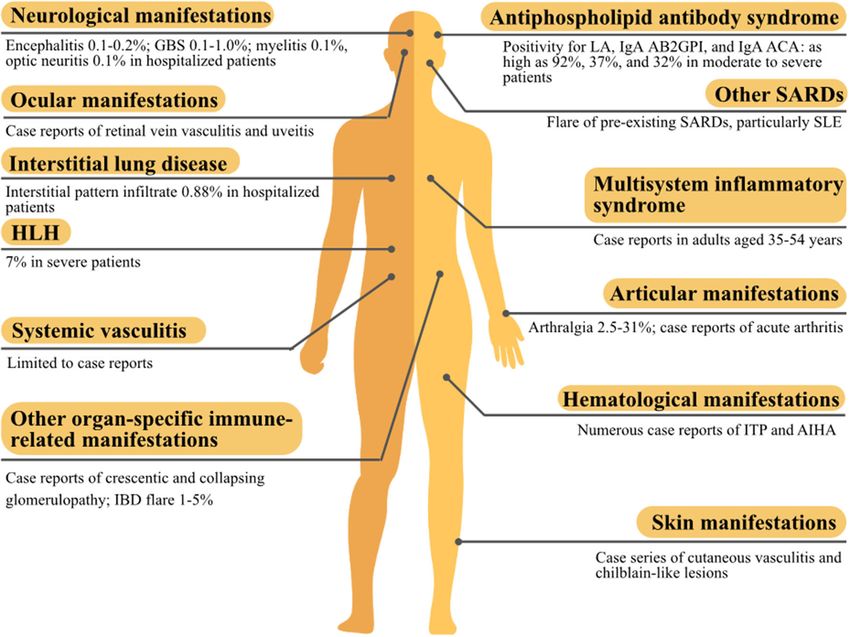
Tang et al.                                                                                                                    Autoimmunity in COVID-19

appeared as COVID-19 cases surged. These manifestations are                     (HLH), etc.; those for organ-specific immune-related diseases
noteworthy since they were either associated with increased                     included Guillain-Barré syndrome (GBS), uveitis, and interstitial
morbidity, e.g., antiphospholipid antibody syndrome (APS),                      lung disease (ILD), etc.
or life-threatening, e.g., multisystem inflammatory syndrome
(MIS), as summarized in previous reviews (2, 3). Furthermore,                   Study Selection
one of the concerns about vaccination is its potential to cause                 Three authors (KT Tang, BC Hsu, and DY Chen) independently
similar autoimmune and rheumatic complications. With new                        assessed the titles and abstracts identified by the aforementioned
reports accumulating at a rapid speed, we have undertaken                       search, and the relevant full-text articles were retrieved. Two
a comprehensive systematic review and hoped it would                            authors (KT Tang and DY Chen) independently assessed the
help delineate the landscape of autoimmune and rheumatic                        full-text articles for eligibility, resolved discrepancies through
manifestations associated with COVID-19.                                        discussion or consultation with the third author (BC Hsu).
                                                                                The references for the selected articles were also examined for
                                                                                relevance. Articles were selected if they: (1) involved adults
MATERIALS AND METHODS
                                                                                ≥18 years; (2) were potentially relevant to autoimmune or
Literature Search                                                               rheumatic manifestations concurrent with or following COVID-
This systematic review focused on the autoimmune and                            19 infection; (3) were potentially relevant to an exacerbation
rheumatic manifestations associated with COVID-19 infection.                    of pre-existing autoimmune or rheumatic diseases concurrent
The algorithm of the systematic review follows the Preferred                    with or following COVID-19 infection. There was no language
Reporting Items for Systematic Reviews and Meta-Analyses                        restriction. Studies describing manifestations that were less likely
(PRISMA) checklist, as shown in Figure 1. Firstly, we searched                  immune-mediated were excluded, such as manifestations with
the PubMed and EMBASE on October 9, 2020. The search                            onset apparently before the COVID-19 symptoms or those very
strategy is illustrated in detail in Appendix 1 in Supplementary                likely medication-related. Two authors (KT Tang and DY Chen)
Material. The search keywords for systemic autoimmune                           independently extracted data from these studies electronically.
diseases included systemic lupus erythematosus (SLE),                           Our emphasis was on the temporal relationship between these
spondyloarthropathy, and hemophagocytic lymphohistiocytosis                     manifestations and COVID-19 infection, other clinical evidence

 FIGURE 1 | Selection of studies to be included in the systematic review.

Frontiers in Immunology | www.frontiersin.org                               2                                     March 2021 | Volume 12 | Article 645013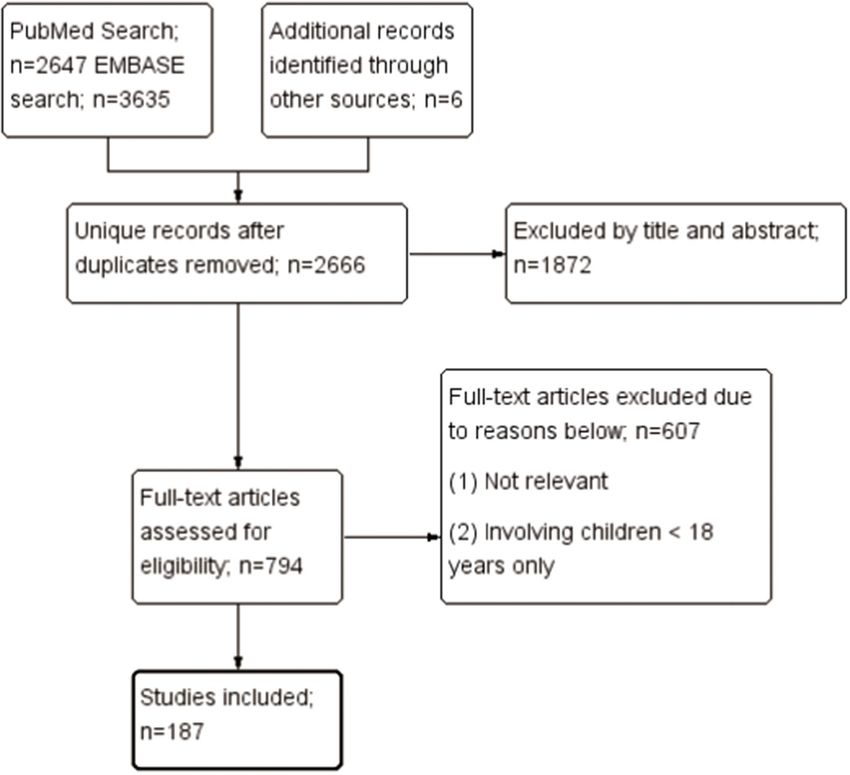
Tang et al.                                                                                                             Autoimmunity in COVID-19

supporting the immune-mediated mechanisms underlying these                nucleoprotein could cross-react with various tissue proteins, such
manifestations, the epidemiology of these manifestations, and             as nuclear antigens, extractable nuclear antigen, mitochondria,
therapeutic response to immunomodulating therapies.                       thyroglobulin, thyroid peroxidase, transglutaminases, myelin
                                                                          basic protein, actin, and α-myosin (17). In summary, molecular
RESULTS                                                                   similarities between viral and human proteins existed, but the
                                                                          clinical significance requires further verification.
Overview
Probably owning to the characteristics of SARS-CoV-2 and                  Autoantibodies
the elicited immune response, COVID-19 infection has been                 As shown in Table 1, five studies reported the screening results
reported to be associated with a variety of autoimmune                    of circulating autoantibodies in patients with COVID-19 (17–
and rheumatic manifestations. Most of these manifestations                21). Anti-nuclear antibodies (ANA) was found in 4–50% of
have been associated with other microbial infections and                  COVID-19 patients, most of whom were older people. This was
their underlying immune-mediated mechanisms are evident.                  consistent with previous reports of an increased prevalence of
However, the existing data were mostly case reports or case series,       autoantibodies in the elderly without autoimmune diseases (22).
and sometimes even conflicting; therefore a causal relationship           In accordance, Schiaffino et al. found an association between
could not be ascertained.                                                 older age and the presence of autoantibodies in COVID-19
                                                                          patients (17). Preliminary results of these studies indicated a
Potential Molecular Mechanisms                                            higher incidence of neurologic and thrombotic events, and
Contributing to Autoimmune and                                            poor outcome in the autoantibody-positive subgroup compared
Rheumatic Manifestations                                                  with the autoantibody-negative subgroup (17, 18). However, the
Severe Acute Respiratory Syndrome Coronavirus 2                           pathogenic potential of these autoantibodies, and whether these
(SARS-CoV-2)                                                              autoantibodies persist after resolution of COVID-19 infection,
Coronaviruses contain the largest single-stranded RNA in                  remains unclear. Interestingly, Bastard et al. demonstrated the
nature, and the SARS-CoV-2 genome is composed of around                   presence of anti-type I interferon (IFN) antibodies in 10.2%
30,000 nucleotides (4). The complex transcriptome, due to its             of 987 patients with life-threatening COVID-19 pneumonia,
discontinuous transcription and recombination activities, may             although they speculated that these antibodies might appear
further expand the ability to interact with the immune system             before COVID-19 infection (23). In summary, autoantibodies
(5). Additionally, the variability of protein sequences provides a        were prevalent in COVID-19 patients, albeit with unknown
rich source of epitopes to stimulate the immune system (6). These         clinical significance.
factors may contribute to the development of immune-mediated
                                                                          Cytokine Storm
manifestations associated with COVID-19 infection.
                                                                          COVID-19 triggers an exaggerated immune response in
Molecular Mimicry                                                         infected patients, and a variety of inflammatory cytokines,
It is well-known that microbial infection could lead to                   such as interleukin (IL)-1β, IL-6, IL-8, interferon (IFN)-
autoimmunity through three mechanisms: molecular mimicry,                 γ, and chemokines, such as granulocyte colony stimulating
bystander activation, and epitope spreading (7). The possibility          factor (G-CSF), interferon gamma-induced protein 10 (IP-10),
of molecular mimicry in COVID-19 disease has been proposed,               monocyte chemoattractant protein-1 (MCP-1), and macrophage
and peptide sharing analysis revealed massive hexapeptide and             inflammatory protein 1α (MIP-1α), were elevated in severe
heptapeptide sharing between SARS-CoV-2 spike glycoprotein                COVID-19 patients (24, 25). In particular, a meta-analysis has
and human proteins compared with other mammals and human                  demonstrated a nearly 3-fold higher serum levels of IL-6 in
coronaviruses (8). Another study also found hexapeptide sharing           patients with complicated COVID-19 when compared with those
between viral epitopes and 460 human proteins (9). Interestingly,         patients with non-complicated disease (26). Regulatory T cells
some of these proteins are associated with pulmonary, cardiac,            were also below normal levels in COVID-19 patients, which
vascular, coagulation, and immunological disorders. Other                 further aggravate the inflammatory response (27). Ultimately,
studies have demonstrated a similarity between SARS-CoV-2                 the resultant cytokine storm leads to tissue damage and multiple
and human proteins, including pulmonary surfactants (10),                 organ failure. Clinically, inflammatory markers, such as C-
brainstem neuronal proteins (11), chaperons (12), heat shock              reactive protein, procalcitonin, D-dimer, and ferritin, were
proteins 60 and 90 (13), ankyrin 1 (an erythrocyte membrane               increased in COVID-19 patients and associated with a poor
protein) (14), odorant receptor 7D4, poly (ADP-ribose)                    prognosis (28). Taken together, the uncontrolled inflammatory
polymerase family member 9 (PARP9), and solute carrier family             milieu triggered by COVID-19 infection may lead to organ
12 member 6 (SLC12A6) (15), which have been hypothesized                  damage and the generation of autoimmunity, too.
to contribute to lung disease, respiratory failure, endothelitis,
neuroimmune diseases, autoimmune hemolytic anemia (AIHA),                 Systemic Autoimmune and Rheumatic
leukopenia, and vascular damage. Also, three immunogenic                  Manifestations
epitopes with high sequence identity to viral proteins were               Arthritis
found in patients with dermatomyositis (16). Of note, the                 Articular symptoms are often observed in virus infection,
monoclonal antibodies against SARS-CoV-2 spike protein and                with the severity ranging from arthralgia, acute arthritis, to

Frontiers in Immunology | www.frontiersin.org                         3                                    March 2021 | Volume 12 | Article 645013
Tang et al. Autoimmunity in COVID-19
TABLE 1 | Prevalence of circulating autoantibodies in patients with COVID-19 disease.
Study Reference Country Patients Mean/median Proportion Autoantibodies
number age (years) of males
Pascolini et al. 18 Italy 33 referred 70 (range 22–90) 52% ANA detected by IFA on HEp-2 cells (33%),
patients anti-histone antibody detected by immunoblot (3%),
but negative for autoantibodies against Sm and
RNP/Sm, RNP70, A, and C, SSA-Ro52, SSA-Ro60,
SSB, Scl-70, PM-Scl, Jo-1, CENP-B, PCNA,
dsDNA, nucleosomes, ribosomal P protein, and M2
detected by immunoblot, ANCA detected by IFA,
and anti-PR3 and anti-MPO antibodies detected by
FEIA
Schiaffino et al. 19 Spain 53 hospitalized 64 (IQR 24–91) 58% ANA detected by unknown method (3.8%), IgG/M
patients autoantibodies against hepatocytes and gastric
glandular cells detected by IFA on rat
kidney/stomach/liver (23%)
Vlachoyiannopoulos 20 Greece 29 ICU patients 64 (range 43–85) 72% ANA detected by unknown method (34.5%),
et al. anti-CCP detected by ELISA (3.5%), c-ANCA
detected by immunofluorescence (6.9%), and
p-ANCA detected by immunofluorescence (6.9%),
but negative for anti-ENA detected by immunoblot,
and anti-dsDNA, anti-PR3 and anti-MPO antibodies
detected by ELISA
Vojdani et al. 17 USA 5 patients N.A. N.A. ANA, anti-ENA, anti-actin and anti-mitochondrial
antibodies detected by unknown methods (60%),
but negative for anti-dsDNA antibody and RF
detected by unknown methods
Zhou et al. 21 China 21 ICU patients 66 (SD 13) 62% ANA (50%), anti–Ro52 (20%), anti–Ro60 (25%),
anti-Scl-70 (5%), and anti-U1-RNP antibodies (5%),
but negative for autoantibodies against Jo-1,
centromere B, SmD1, SSB and dsDNA (all detected
by chemiluminescence immunoassay)
ANA, antinuclear antibody; CCP, cyclic citrullinated peptide antibody; COVID-19, the coronavirus disease 2019; c-ANCA, cytoplasmic anti-neutrophil cytoplasmic antibody; dsDNA,
double-stranded DNA; ELISA, enzyme-linked immunosorbent assay; ENA, extractable nuclear antigen; FEIA, fluorescent-enzyme immuno-assay; ICU, intensive care unit; IFA, indirect
immunofluorescence assay; IQR, interquartile range; MPO, myeloperoxidase; N.A., not available; p-ANCA, perinuclear anti-neutrophil cytoplasmic antibody; PCNA, proliferating cell
nuclear antigen; PR3, proteinase 3; RF, rheumatoid factor; RNP, ribonucleoprotein; SD, standard deviation.
chronic arthritis (29). Only two studies reported the prevalence Another 50-year-old female also demonstrated worsening of
of arthralgia alone, instead of myalgia/arthralgia, in COVID- pre-existing rheumatoid arthritis (RA), which improved after
19 patients, which were 31% of 417 patients with COVID- sarilumab treatment (32). In summary, SARS-CoV-2 infection
19 from 12 European hospitals and 2.5% of 40 patients in was associated with the development of arthralgia, acute arthritis,
Thailand, respectively (30, 31). Five cases of acute mono-, oligo- and possibly, chronic arthritis.
, or polyarthritis as an initial presentation (32) or a delayed
phenomenon 3–29 days after COVID-19 symptoms onset (33– Antiphospholipid Antibody Syndrome (APS)
36) have been reported. Two cases had accompanying features As demonstrated in Table 2, the presence of antiphospholipid
of reactive arthritis, such as enthesitis and urethritis (35, 36). antibodies (aPL) has been observed in COVID-19 patients
Using RT-PCR, negative SARS-CoV-2 RNA in joint fluid was around the world (18–20, 39–55). The association between aPL
demonstrated in two cases (33, 36), implying that the arthritis and disease severity was shown in three studies (39, 54, 56), but
was mediated by immune mechanisms rather than direct viral not in another study (46). Lupus anticoagulant and non-criteria
invasion. Acute arthritis either resolved spontaneously (33) or IgA anti-β2glycoprotein-I/anticardiolipin antibodies (57) were
responded to treatment with non-steroidal anti-inflammatory the most prevalent aPL, with the prevalence of 3–92, 0–37, and
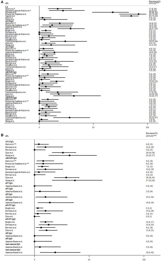
drugs (NSAIDs), corticosteroids, and even baricitinib (32, 34– 0–32% in patients with moderate to severe disease (Figure 2).
36). Nevertheless, two acute arthritis patients did not enter However, the lupus anticoagulant testing might be interfered by
remission at follow-up despite treatment (32, 35). Furthermore, heparin use or elevated C-reactive protein in COVID-19 patients.
insufficient follow-up time in these reports might miss out on IgG anticardiolipin and anti-β2glycoprotein-I antibodies were
the opportunity to observe arthritis recurrences after arthritis also prevalent, but often in low titers. Notably, one study revealed
remission and being drug-free, since infection-related reactive 3 (5%) of 58 COVID-19 patients had a highly thrombogenic
arthritis may persist for years (37). Development of chronic anti-β2glycoprotein-I domain I IgG antibody, although not
arthritis concurrent with SARS-CoV-2 infection was found in correlated with thrombosis (41). The strong association of aPL
a 45-year-old male, which responded to corticosteroids (38). with thrombotic events was not observed in most studies, even
Frontiers in Immunology | www.frontiersin.org 4 March 2021 | Volume 12 | Article 645013Frontiers in Immunology | www.frontiersin.org
Tang et al.
TABLE 2 | Prevalence of antiphospholipid autoantibodies in COVID-19 disease.
Study Reference Country Patients Mean/median age Proportion of Prevalence of aPL Findings
number (years) males
Amezcua-Guerra 39 Mexico 21 ICU patients 62 (IQR 54–67) 43% Anti-annexin V IgG (5%), anti-annexin V IgM (19%), ACA IgG Elevated levels of interleukin-6/ferritin/C-reactive
et al. (10%), ACA IgM (14%), AB2GPI IgG (5%), AB2GPI IgM (0%), aPT protein only in patients with aPL; pulmonary embolism
IgG (0%), aPT IgM (5%), aPS IgG (10%), aPS IgM (14%), aPI IgG in two aPL+ patients but in no aPL- patients
(0%), and aPI IgM (0%)*
Bertin et al. 40 France 56 patients with 67 59% ACA IgG (29%), ACA IgM (5%), AB2GPI IgG (2%), and AB2GPI ACA IgG was associated with severe disease
moderate and severe IgM (7%)*
disease
Borghi et al. 41 Italy 122 patients with 69 (SD 16) 63% ACA IgG (6%), ACA IgM (7%), AB2GPI IgG (16%), AB2GPI IgG No association between aPL and thrombotic events,
severe disease domain I (5%), AB2GPI IgM (9%), AB2GPI IgA (7%), aPS/PT IgG even for AB2GPI domain I IgG
(3%), and aPS/PT IgM (10%)*
Bowles et al. 42 UK 35 patients with a 57 (95%CI 19–83) 69% LA (53%)**
prolonged aPTT
Cuenca Saez et al. 43 Spain 11 patients with (range 2–40) N.A. LA (0%), ACA IgG (0%), ACA IgM (0%), and low titer ACA IgA
perniosis (100%)
Devreese et al. 44 Belgium 31 ICU patients 63 (range 38–82) 90% LA (68%), ACA IgG (0%), ACA IgM (3%), ACA IgA (10%), AB2GPI No association between aPL and thrombotic events
IgG (3%), AB2GPI IgM (3%), AB2GPI IgA (10%), aPS/PT IgG
(10%), and aPS/PT IgM (13%)
Galeano-Valle et al. 45 Spain 24 patients with 64 (SD 14) 58% ACA IgG (0%), low titer ACA IgM (8.3%), AB2GPI IgG (0%), and
venous low titer AB2GPI IgM (8.3%)
thromboembolism
Gatto et al. 46 Italy 122 patients with mild 54 (SD 19) 49% LA (22%)**, ACA IgG (13%), ACA IgM (3%), ACA IgA (2%), A trend toward an association between aPL and
to severe disease AB2GPI IgG (6%), AB2GPI IgM (7%), and AB2GPI IgA (3%) thrombotic events
Gutierrez López de 47 Spain 27 hospitalized 58 (range 20–90) 44% LA (22%)**, ACA IgG (0%), ACA IgM (0%), AB2GPI IgG (0%), No association between aPL and thrombotic events
Ocáriz et al. patients AB2GPI IgM (0%) and AB2GPI IgA (4%)*
5
Harzallah et al. 48 France 56 patients N.A. N.A. LA (45%)** and ACA IgG/M/AB2GPI IgG/M (10%)
Pascolini et al. 18 Italy 33 hospitalized 70 (range 22–90) 52% ACA IgG (9%), ACA IgM (15%), AB2GPI IgG (6%), and AB2GPI None of the patients had thrombotic events
patients IgM (6%)*
Pineton de Chambrun 49 France 25 ICU patients 48 (range 35–64), 68% LA (92%), ACA IgG (12%), ACA IgM (0%), ACA IgA (8%), AB2GPI Massive pulmonary embolism in 6 patients, all aPL+
et al. IgG (0%), AB2GPI IgM (0%), and AB2GPI IgA (8%)
Previtali et al. 50 Italy 35 deceased patients 73 (range 52–82) 74% Low titer ACA IgG (3%), low titer ACA IgM (6%), ACA IgA (0%), Catastrophic APS was less likely despite multiple
AB2GPI IgG (0%), AB2GPI IgM (0%), low titer aPS/PT IgG (3%), thrombosis at autopsies
and low titer aPS/PT IgM (6%)
Reyes Gil et al. 51 USA 68 patients 57 50% LA (60%)**, ACA IgG (0%), ACA IgM (1%), AB2GPI IgG (0%), and LA associated with thrombotic events
AB2GPI IgM (1%)*
Schiaffino et al. 19 Spain 53 hospitalized 64 (range 24–91) 58% ACA IgG (2%), ACA IgM (9%), AB2GPI IgG (2%), and AB2GPI IgM No association between aPL and thrombotic events
patients (6%)*
Siguret et al. 52 France 74 mechanically 64 N.A. LA (85%) and ACA IgG/IgM/AB2GPI IgG (12%)* No association between aPL and thrombotic events
ventilated patients
Tvito et al. 53 Israel 43 patients with mild N.A. 63% LA (37%)**, ACA IgG (0%), ACA IgM (0%), AB2GPI IgG (0%), and No association between aPL and thrombotic events
March 2021 | Volume 12 | Article 645013
to severe disease AB2GPI IgM (0%)
Vlachoyiannopoulos 20 Greece 29 ICU patients 64 (range 43–85) 72% ACA IgG (24%), ACA IgM (10%), ABGPI IgG (17%), and ABGPI
et al. IgM (28%)*
Xiao et al. 54 China 66 ICU patients 65 59% LA (3%), ACA IgG (6%), ACA IgM (3%), ACA IgA (26%), AB2GPI Patients with multiple aPLs had a significantly higher
IgG (18%), AB2GPI IgM (2%), AB2GPI IgA (29%), aPS/PT IgG incidence of cerebral infarction
Autoimmunity in COVID-19
(0%), and aPS/PT IgM (11%)*
Zhang et al. 55 China 19 ICU patients 65 (IQR 60–70) 53% LA (5%), ACA IgG (11%), ACA IgM (5%), ACA IgA (32%), AB2GPI All 4 patients with cerebral infarction had aPL with
IgG (32%), AB2GPI IgM (0%), and AB2GPI IgA (37%)* multiple isotypes whereas no thrombotic events
developed in aPL-patients.
*Probably including low titer aPL as positive.
**Determined by two tests based on different principles per the International Society of Thrombosis and Haemostasis criteria.
AB2GPI, anti-β2glycoprotein I; ACA, anticardiolipin antibody; aPI, antiphosphatidylinositol antibody; aPL, antiphospholipid antibodies; aPS, anti-phosphotidylserine antibody; aPT, antiprothrombin antibody; aPTT, activated
partial-thromboplastin time; CI, confidence interval; COVID-19, the coronavirus disease 2019; ICU, intensive care unit; IQR, interquartile range; LA, lupus anticoagulant; N.A., not available; SD, standard deviation.Tang et al. Autoimmunity in COVID-19 FIGURE 2 | The prevalence of (A) criteria and (B) non-criteria antiphospholipid antibodies (aPL) based on the revised Sapporo criteria for antiphospholipid antibody syndrome in COVID-19 patients with moderate to severe disease. *Determined by two tests based on different principles per the International Society of Thrombosis and Haemostasis criteria. **Moderate-to-high titer aPL. ***95% exact confidence intervals. AB2GPI, anti-β2glycoprotein I antibody; ACA, anticardiolipin antibody; aPI, antiphosphatidylinositol antibody; aPL, antiphospholipid antibodies; aPS, antiphosphotidylserine antibody; aPT, antiprothrombin antibody; COVID-19, the coronavirus disease 2019; LA, lupus anticoagulant. Frontiers in Immunology | www.frontiersin.org 6 March 2021 | Volume 12 | Article 645013
Tang et al.                                                                                                               Autoimmunity in COVID-19

for patients with double/triple positivity. Besides, repeated testing       who then only received supportive treatment (72). Notably,
showed that the titer of aPL fluctuated during the disease course           virus particles have been found in the cytoplasm of vascular
(52, 54), and the aPL turned to be negative 1 month later in most           endothelial cells in the biopsy specimen of one patient
of the aPL-positive patients (44).                                          (72), whereas SARS-CoV-2 RT-PCR was negative in another
    COVID-19 was associated with the flares of pre-existing APS.            patient’s biopsy (71). A 37-year-old woman suffered from
The complications include bilateral adrenal glands hemorrhage               anti-proteinase 3 (PR3)-positive diffuse alveolar hemorrhage
in a 66-year-old female and limb ischemia in another 48-year-               concurrent with SARS-CoV-2 infection and later received
old male, both of which were controlled by anticoagulants                   treatments of intravenous methylprednisolone pulse therapy,
(58, 59). Interestingly, one study revealed 5 (63%) of eight                plasmapheresis, and IVIG. Her hemoptysis improved after IVIG,
pregnant women with COVID-19 infection fulfilled diagnostic                 but finally she expired while on ventilator (73). Two cases were
criteria for pre-eclampsia/HELLP syndrome (hemolysis; elevated              reported on anti-neutrophilic cytoplasmic antibodies (ANCA)-
liver enzymes; low platelet count), perhaps due to overlapping              associated vasculitis with necrotizing nephritis concomitant with
features. Only one of them was more likely to have pre-eclampsia,           COVID-19 infection. They responded to methylprednisolone
an obstetric complication of APS (60). However, aPL were not                pulse therapy plus rituximab (74). Henoch-Schönlein purpura
examined in these pregnant women. In summary, low titer and                 with nephritis was found in a 78-year-old man 3 weeks
transient aPL were prevalent in COVID-19 patients, but like aPL             after COVID-19 infection. His condition improved after
generated in other infections, most of them were not pathogenic.            methylprednisolone pulse therapy and rituximab (75). Henoch-
                                                                            Schönlein purpura with suspected gastrointestinal involvement
Multisystem Inflammatory Syndrome in Adults                                 occurred in another 24-year-old man, and improved after
(MIS-A)                                                                     corticosteroids treatment (76). In summary, case reports of large,
Pediatric cases of Kawasaki-like multisystem inflammatory                   medium, and small vessel vasculitis involving multiple organs
syndrome (MIS-C) are accumulating mainly in Western                         have been reported in COVID-19 patients. However, we could
countries. With a favorable prognosis, the disease was                      not ascertain whether these vasculitis resulted from direct virus-
characterized      by    hyper-inflammation,     gastrointestinal           induced endothelitis or immune-mediated mechanisms.
symptoms, and cardiac dysfunction, such as myocarditis, and
shock, which were somewhat different from classical Kawasaki                Other Systemic Autoimmune Rheumatic Diseases
disease. Multisystem inflammatory syndrome associated with                  (SARDs)
COVID-19 infection has also been found in adults (MIS-A) aged               Three cases of new onset SLE concurrent with COVID-19
35–54 years (61–64). Like children, these adult patients were               infection have been published, and two of them eventually
reported in Western countries and recovered after treatment                 deceased (77–79). In other studies, myositis of proximal limbs
with corticosteroids, intravenous immunoglobulin (IVIG),                    and paraspinal myositis have been demonstrated on magnetic
and tocilizumab, an IL-6 receptor inhibitor. An IL-1 receptor               resonance imaging (MRI) along with elevated creatine kinase
antagonist, anakinra, may also be a therapeutic option based on             in one and seven COVID-19 patients, respectively (80, 81).
the experiences in children (65). Although some did not undergo             However, a complete workup, such as electromyography and
complete coronary evaluation, none of these adult patients                  muscle biopsy, was lacking in these cases. None of them
had dilatation or aneurysm. Interestingly, isolated myocarditis             received treatment due to being either in critical conditions or
has also been reported in two adult COVID-19 patients: one                  asymptomatic. In a Chinese cohort of 21 COVID-19 patients
28-year-old woman presented with myocarditis shortly after                  with pre-existing SARDs, a disease flare was demonstrated in one
COVID-19 infection, which improved after methylprednisolone                 SLE patient (skin rashes and hemolytic anemia), one ankylosing
pulse therapy (66); the other 53-year-old woman developed                   spondylitis patient (back and ankle pain), and one patient with
myocarditis a week after COVID-19 symptoms onset, which was                 polymyalgia rheumatica (muscle pain), whose symptoms were
stabilized by corticosteroids (67).                                         attenuated after treatment with hydroxychloroquine, NSAID,
                                                                            corticosteroids, or mycophenolate mofetil (82). Another Italian
Systemic Vasculitis                                                         multicenter cohort reported 40 (17%) of 232 SARD patients with
An autopsy study in Italy revealed vasculitis in the lung,                  moderate to severe disease activity upon COVID-19 infection
brain, and other organs in individuals who succumbed to                     but provided no further details (83). The Asia Pacific Lupus
COVID-19 (68). Clinical evidence for systemic vasculitis was                Collaboration (APLC) cohort reported three cases of SARS-CoV-
limited to case reports. A 69-year-old woman and a 71-                      2-infected SLE patients, two of whom developed a concurrent
year-old man presented with asymptomatic aortitis and had                   lupus flare (thrombocytopenia and nephritis) and improved after
concurrent SARS-CoV-2 infection (69, 70). A 73-year-man                     corticosteroids and IVIG treatment (84). Another two SARS-
presented with arterial vasculitis at splenic hilum resulting               CoV-2-infected SLE patients presented with an exacerbation
in splenic infarction concomitantly with COVID-19 infection.                of thrombocytopenia, which were successfully treated with
The condition improved after splenectomy (71). Another 71-                  corticosteroids and IVIG (85, 86). In summary, SARDs, especially
year-old man developed abdominal and bilateral common iliac                 SLE, might flare upon COVID-19 infection. Nevertheless, the
arteritis concurrent with COVID-19 infection, with subsequent               overlapping features between the wide spectrum of COVID-
spontaneous remission (70). Concurrent ileocecal vasculitic                 19 manifestations and SARDs made the distinction ambiguous.
ulcers was found in a 40-year-old female COVID-19 patient,                  Furthermore, medication adherence in these patients was

Frontiers in Immunology | www.frontiersin.org                           7                                    March 2021 | Volume 12 | Article 645013
Tang et al. Autoimmunity in COVID-19
questionable during the pandemic, which might also contribute found to have concomitant Evans syndrome and COVID-
to a flare of SARDs. 19 infection (123). Autoimmune thrombotic thrombocytopenic
purpura has been demonstrated in two COVID-19 patients
Hemophagocytic Lymphohistiocytosis (HLH) and improved after plasmapheresis (124, 125). Another 66-
Infection is a well-known trigger factor of HLH. In fact, year-old man whose acquired hemophilia flared concomitantly
severe COVID-19 patients presented with hyperferritinemia with COVID-19 infection, responded to corticosteroids and
and cytokine storm, reminiscent of HLH. Autopsy studies cyclophosphamide (126). In summary, many cases of ITP or
demonstrated a high percentage of hemophagocytosis in AIHA associated with COVID-19 have been reported, implying
COVID-19 patients’ bone marrow, pulmonary lymph nodes, a possible link between them.
or spleen, ranging from 75 to 94% (87, 88). Using a validated
hemophagocytic syndrome diagnostic score (HScore) cut-off Skin Manifestations
point of 168 (89), we identified a total of 15 cases of COVID- Case series had been reported in Western countries on Raynaud’s
19-associated HLH (Table 3) (88, 90–94). However, the reported phenomenon and chilblains-like lesions in patients with recent
prevalence of HLH in severe COVID-19 patients was as low as 7% COVID-19 infection (positive anti-SARS-CoV-2 IgG) or in
(93, 94). Notably, routine examinations for serum triglycerides close contact with confirmed COVID-19 cases (127–130). Some
and fibrinogen, hepatosplenomegaly, or tissue hemophagocytosis biopsies revealed positive findings for vasculitis or vascular
were not always performed in these studies. In summary, microthrombi, but negative findings for SARS-CoV-2 based on
COVID-19 infection was associated with HLH in a few cases as RT-PCR. Biopsy-proven cases had been reported on cutaneous
defined by the HScore. vasculitis, manifesting as purpuric papules (131) and plaques
(132, 133), hemorrhagic bullae (134), and urticarial (135,
Organ-Specific Immune-Related 136), and targetoid lesions (137). Most of these conditions
Manifestations occurred 5–35 days after onset of COVID-19 symptoms and
Hematological Manifestations they responded well to topical or oral corticosteroids. Besides,
Thrombocytopenia is common but usually mild in COVID- livedo reticularis developed without aPL in a 57-year-old
19 infection. Chen et al. reviewed 271 hospitalized COVID-19 man concomitant with COVID-19 infection (138). Flares of
patients and found the prevalence of delayed thrombocytopenia psoriasis or newly-developed psoriatic arthritis concurrent with
14 days after COVID symptoms to be 12%. The authors or following COVID-19 infection had been reported. These
speculated that it was partly immune-mediated (95). Table 4 conditions had resolved either spontaneously or after NSAID
demonstrated 38 cases of immune thrombocytopenic purpura and topical corticosteroids treatment (139–141). In summary,
(ITP) associated with COVID-19 infection, and some of them cutaneous vasculitis were potentially associated with COVID-19
only had mild or asymptomatic COVID-19 (90, 96–113). Most infection, mainly in the Western population.
of these patients with thrombocytopenia were diagnosed by
exclusion and had a favorable response to corticosteroids, IVIG, Neurological Manifestations
and even thrombopoietin receptor agonists. Positive direct A spectrum of neurological manifestations, including
Coomb’s test was shown in 13% of 267 anemic COVID-19 neuroimmune manifestations were presented in COVID-19
patients and 46% of the other 113 COVID-19 patients (114, 115). patients. The frequencies of these manifestations in hospitalized
Furthermore, both warm and cold AIHA have been reported in patients are as follows: encephalitis 0.1–0.2%, GBS 0.1–1%,
COVID-19 patients and most of them recovered spontaneously myelitis 0.1%, and optic neuritis 0.1% in Western countries
(116–122). In line with these findings, a 39-year-old man was (142–144). In Singapore, two cases of acute disseminated
TABLE 3 | Hemophagocytic lymphohistiocytosis (HLH) in COVID-19 disease.
Study Reference Country Patient Age (years), Sex HSscore, Treatment response
number number median (range) median (range)
Debliquis et al. 90 France 1 63 1M 207 Deceased without specific treatment
Dimopoulos et al. 91 Greece and the 8 68 (51, 84) 7M1F 175 (171, 188) Decreased HScore after anakinra but
Netherlands 3 (38%) of them eventually deceased
Faguer et al. 92 France 1 51 1M 253 Decreased HScore after tocilizumab
Hakim et al. 93 USA 1 37 1M 204 Decreased HScore after tocilizumab
but eventually deceased on ventilator
Prilutskiy et al. 88 USA 1 72 1M 217 Hemophagocytosis found
post-mortem despite anakinra
Wood et al. 94 UK 3 N.A. N.A. N.A. Decreased HScore after tocilizumab
but then contracting a bacterial
pneumonia
COVID-19, the coronavirus disease 2019; N.A., not available.
Frontiers in Immunology | www.frontiersin.org 8 March 2021 | Volume 12 | Article 645013Tang et al. Autoimmunity in COVID-19
TABLE 4 | Cases of immune thrombocytopenic purpura (ITP) in COVID-19 disease.
Study Reference Age (years) Patient number Findings Treatment response
number and sex
Artru et al. 96 38 1M Responsive to corticosteroids and IVIG
Bennett et al. 97 73 1F Responsive to methylprednisolone pulse
therapy and IVIG
Bomhof et al. 98 59, 66, 67 2M1F Two responsive to corticosteroids and IVIG,
and one died of intracerebral bleeding despite
platelet transfusion
Debliquis et al. 90 78 1M Increased megakaryocytes at bone Responsive to IVIG
marrow biopsy, positive antiplatelet
antibodies
Deruelle et al. 99 41 1M Increased megakaryocytes at bone Responsive to IVIG
marrow biopsy
Hindilerden et al. 100 86 1M Increased megakaryocytes at bone Responsive to corticosteroids
marrow biopsy
Hu et al. 101 72 1F History of ITP Responsive to corticosteroids
Humbert et al. 102 84 1M Responsive to corticosteroids and IVIG
Lévesque et al. 103 53 1M Responsive to romiplostim, vincristine, and
methylprednisolone pulse therapy
Mahevas et al. 104 Median 64 (range 7M7F All responsive to corticosteroids and IVIG
53–79)
Malik et al. 105 29 1F Increased megakaryocytes at bone Responsive to corticosteroids
marrow biopsy
Martincic et al. 106 48 1M Responsive to corticosteroids and IVIG
Murt et al. 107 41 1M Responsive to IVIG
Nesr et al. 108 34 1F History of ITP, pregnant Responsive to corticosteroids and IVIG
Pascolini et al. 109 31, 69, and 88 2M1F Positive IgM antiplatelet antibodies Recovery after resolution of COVID-19 infection
Patel et al. 110 67 1M Responsive to romiplostim
Revuz et al. 111 39, 57, and 76 2M1F All responsive to IVIG
Sadr et al. 112 57 1F Recovery after resolution of COVID-19 infection
Zulfiqar et al. 113 65 1F Increased megakaryocytes at bone Responsive to corticosteroids and eltrombopag
marrow biopsy
COVID-19, the coronavirus disease 2019; IVIG, intravenous immunoglobulin.
encephalomyelitis (ADEM), two encephalitis, and one GBS were 5–7 days after COVID-19 symptoms onset, which responded to
reported among 47,572 COVID-19 cases (145). The development pyridostigmine, corticosteroids, IVIG and plasmapheresis (151).
of encephalitis and GBS were generally delayed after COVID-19 In a cohort of 15 MG patients with concomitant SARS-CoV-2
symptoms onset. MRI-proven central nervous system vasculitis infection, a high percentage of them experienced MG worsening
occurred in two patients 11–29 days after COVID-19 symptoms (87%) and even mechanical ventilation (73%), although it was
onset and responded to either corticosteroids or tocilizumab difficult to ascertain exacerbated respiratory muscle weakness
(146, 147). Using RT-PCR or antibody testing, if available, in patients with concomitant pneumonia and respiratory failure
cerebrospinal fluid SARS-CoV-2 were positive in only some (152). However, 8 (62%) of them recovered after treated with
of these cases (144). Most neuroimmune diseases patients corticosteroids, IVIG or plasmapheresis. In another cohort,
responded to standard immunomodulating therapies. one (20%) of 5 SARS-CoV-2-infected MG patients experienced
A 29-year-old woman developed multiple sclerosis with MG worsening (ptosis and dysphagia) but responded to
right optic neuritis 2–3 weeks after COVID-19 infection corticosteroids plus IVIG (153). In summary, a myriad of
(148). Another 26-year-old man had myelin oligodendrocyte neuroimmune manifestations might develop concurrently with
glycoprotein antibody-positive neuromyelitis optica, presenting or following SARS-CoV-2 infection. However, it was difficult
as bilateral optic neuritis and longitudinal extensive transverse to differentiate between direct neuronal infection and immune-
myelitis a few days after COVID-19 symptoms onset and mediated mechanisms underlying these manifestations.
responsive to methylprednisolone pulse therapy (149). Also,
a study of 76 patients with multiple sclerosis demonstrated Interstitial Lung Disease (ILD)
disease recrudescence preceding or concurrent with COVID- Lung involvement is critical in COVID-19 infection. Chest
19 in 16 (21%) (150). There were reports of newly-onset computed tomography (CT) demonstrated interlobular septal
acetylcholine receptor antibody-positive myasthenia gravis (MG) and interstitial thickening in 0.88% of 130 infected inpatients at
Frontiers in Immunology | www.frontiersin.org 9 March 2021 | Volume 12 | Article 645013Tang et al. Autoimmunity in COVID-19
Wuhan (154). In accordance, autopsy of COVID-19 patients with Other Organ-Specific Immune-Related
critical illness showed interstitial mononuclear inflammatory Manifestations
infiltrates, organizing pneumonia, or fibrosis in the lung (155, Crescentic glomerulonephritis had been found in two COVID-
156). Long-term follow-ups of SARS survivors had residual 19 patients, which stabilized after methylprednisolone pulse
fibrosis on chest radiographs and accompanying respiratory therapy, plasmapheresis, IVIG and cyclophophomide (162).
dysfunction 6 months later (157). Similarly, a subset of COVID- Collapsing glomerulopathy concomitant with COVID-19
19 patients displayed interstitial change 2 weeks after disease infection was reported in two cases, with virus-like particles
onset (158) or upon discharge (159), as well as decreased found in their renal biopsies (163, 164). In the North West
diffusing capacity for carbon monoxide (DLCO) (160). However, London, eight unexpected new cases of Goodpasture syndrome
a longer follow-up is required to monitor the progression were diagnosed, representing a 5-fold increase of the background
of pulmonary interstitial change after COVID-19 infection to rate, and four of them were positive for IgM and/or IgG
determine if these changes are merely post-ARDS change or antibodies to SARS-CoV-2 (165). However, the clinical
progressive ILD. significance required further research. A 19-year-old woman
developed ulcerative colitis 9 days after the resolution of
Ocular Manifestations COVID-19 infection (166). A cross-sectional study of 82 patients
Retinal vein vasculitic occlusion was found in a 52-year-old with inflammatory bowel disease (IBD) revealed an IBD flare in
man 10 days after COVID-19 symptoms appeared, but his one (1%) patient during SARS-CoV-2 infection (167). In another
visual acuity improved after treatment with corticosteroids and study of 79 IBD patients infected with SARS-CoV-2 revealed 4
intravitreal anti-vascular endothelial growth factor (anti-VEGF) (5%) patients had severe concomitant IBD flare (168). Finally,
injection (161). Another 54-year-old woman developed bilateral subacute thyroiditis has been reported in an 18-year-old woman
anterior uveitis 14 days after COVID-19-associated MIS-A but 15 days after diagnosis of COVID-19, which improved after
responded to topical corticosteroids (61). corticosteroids therapy (169).
FIGURE 3 | The summary of autoimmune and rheumatic manifestations associated with the coronavirus disease 2019 (COVID-19). AB2GPI, anti-β2glycoprotein I
antibody; ACA, anticardiolipin antibody; AIHA, autoimmune hemolytic anemia; GBS, Guillain-Barré syndrome; HLH, hemophagocytic lymphohistiocytosis; IBD,
inflammatory bowel disease; ITP, immune thrombocytopenic purpura; LA, lupus anticoagulant; SARD, systemic autoimmune rheumatic disease; SLE, systemic
lupus erythematosus.
Frontiers in Immunology | www.frontiersin.org 10 March 2021 | Volume 12 | Article 645013Tang et al. Autoimmunity in COVID-19
DISCUSSION SARS-CoV-2. In general, these manifestations were effectively
treated in a strategy similarly used for patients without
Limitations concomitant infection. Spontaneous recovery could happen
Some limitations should be addressed. First, most of the but was uncommon, although expectant management
clinical evidence was not systematic and based on case reports was rarely undertaken in these patients. Based on the
or case series without a long-term follow-up. Second, the temporal relationship (sometimes delayed after COVID-
sensitivity and specificity of different testing kits for SARS- 19 infection resolves), well-known immune-mediated
CoV-2 infection were not well-validated, and the false positive mechanisms, and treatment response to immunomodulators,
or negative results could undermine our appraisal of the these manifestations were probably consequences of the
literature. Third, most of the reports were from patients immune dysregulation caused by COVID-19 infection,
with moderate to severe COVID-19 disease. It was impossible particularly autoimmune cytopenia, cutaneous vasculitis,
to determine the epidemiology of these manifestations in encephalitis, and GBS. But the evidence was still conflicting
asymptomatic and mild COVID-19 cases. Fourth, overlapping as regards to manifestations, such as APS, HLH, and MG.
COVID-19 features and concomitant medications could make Herein, we provided a comprehensive overview of the
it difficult to determine if these manifestations were immune- evidence and literature concerning these rare but clinically
mediated. Fifth, it was difficult to assure whether direct significant manifestations; vaccine developers should take
cytopathic effect consequent to viral invasion or immune- these findings into account in their vaccine design and
mediated mechanisms were responsible for these manifestations. post-marketing surveillance.
Lastly, the immunomodulators often used to treat these
manifestations are also potential therapies for COVID-19.
It was hard to know whether the immunomodulators exert DATA AVAILABILITY STATEMENT
their beneficial effect directly upon the immune mechanisms
underlying these manifestations or indirectly through the The raw data supporting the conclusions of this article will be
alleviation of COVID-19 infection. However, there are no made available by the authors, without undue reservation, upon
immunomodulating therapies that have consistently shown request to the corresponding author.
therapeutic efficacy toward SARS-CoV-2.
AUTHOR CONTRIBUTIONS
Conclusions
SARS-CoV-2 has a complex transcriptome and shares molecular K-TT, B-CH, and D-YC performed the literature search and
similarities with human proteins, and its infection could retrieved relevant articles. K-TT and D-YC appraised the selected
generate various autoantibodies and cytokine storm, which articles and drafted the manuscript. All authors made substantive
form the basis for developing autoimmune and rheumatic intellectual contributions to the present study and approved the
manifestations. Accordingly, a variety of systemic or organ- final manuscript.
specific manifestations have been reported to be associated
with COVID-19 (summarized in Figure 3). Most of these SUPPLEMENTARY MATERIAL
manifestations have been reported in other microbial infections
except for MIS-A. MIS-A shared some similarities with The Supplementary Material for this article can be found
Kawasaki disease, but the distinct differences between online at: https://www.frontiersin.org/articles/10.3389/fimmu.
the two entities made MIS-A more likely to be specific to 2021.645013/full#supplementary-material
REFERENCES 6. V’kovski P, Kratzel A, Steiner S, Stalder H, Thiel V. Coronavirus biology
and replication: implications for SARS-CoV-2. Nat Rev Microbiol. (2021)
1. Johns Hopkins University. Coronavirus Resource Center. Available online at: 19:155–70. doi: 10.1038/s41579-020-00468-6
https://coronavirus.jhu.edu/map.html (accessed December 25, 2020). 7. Fujinami RS, Von Herrath MG, Christen U, Whitton JL.
2. Rodriguez Y, Novelli L, Rojas M, De Santis M, Acosta-Ampudia Molecular mimicry, bystander activation, or viral persistence:
Y, Monsalve DM, et al. Autoinflammatory and autoimmune infections and autoimmune disease. Clin Microbiol Rev. (2006)
conditions at the crossroad of COVID-19. J Autoimmun. (2020) 19:80–94. doi: 10.1128/CMR.19.1.80-94.2006
114:102506. doi: 10.1016/j.jaut.2020.102506 8. Kanduc D, Shoenfeld Y. Molecular mimicry between SARS-CoV-2 spike
3. Ehrenfeld M, Tincani A, Andreoli L, Cattalini M, Greenbaum A, glycoprotein and mammalian proteomes: implications for the vaccine.
Kanduc D, et al. Covid-19 and autoimmunity. Autoimmun Rev. (2020) Immunol Res. (2020) 68:310–3. doi: 10.1007/s12026-020-09152-6
19:102597. doi: 10.1016/j.autrev.2020.102597 9. Kanduc D. From anti-SARS-CoV-2 immune responses to
4. Naqvi AaT, Fatima K, Mohammad T, Fatima U, Singh IK, Singh A, et al. COVID-19 via molecular mimicry. Antibodies (Basel). (2020)
Insights into SARS-CoV-2 genome, structure, evolution, pathogenesis and 9:33. doi: 10.3390/antib9030033
therapies: Structural genomics approach. Biochim Biophys Acta. (2020) 10. Kanduc D, Shoenfeld Y. On the molecular determinants of the SARS-CoV-2
1866:165878. doi: 10.1016/j.bbadis.2020.165878 attack. Clin Immunol. (2020) 215:108426. doi: 10.1016/j.clim.2020.108426
5. Kim D, Lee JY, Yang JS, Kim JW, Kim VN, Chang H. The 11. Lucchese G, Floel A. Molecular mimicry between SARS-CoV-
architecture of SARS-CoV-2 transcriptome. Cell. (2020) 181:914–21 2 and respiratory pacemaker neurons. Autoimmun Rev. (2020)
e910. doi: 10.1016/j.cell.2020.04.011 19:102556. doi: 10.1016/j.autrev.2020.102556
Frontiers in Immunology | www.frontiersin.org 11 March 2021 | Volume 12 | Article 645013Tang et al. Autoimmunity in COVID-19
12. Marino Gammazza A, Legare S, Lo Bosco G, Fucarino A, Angileri F, 32. Alivernini S, Cingolani A, Gessi M, Paglionico A, Pasciuto G, Tolusso B,
Conway De Macario E, et al. Human molecular chaperones share with SARS- et al. Comparative analysis of synovial inflammation after SARS-CoV-2
CoV-2 antigenic epitopes potentially capable of eliciting autoimmunity infection. Ann Rheum Dis. (2020). doi: 10.1136/annrheumdis-2020-218315.
against endothelial cells: possible role of molecular mimicry in COVID- [Epub ahead of print].
19. Cell Stress Chaperones. (2020) 25:737–41. doi: 10.1007/s12192-020-0 33. Yokogawa N, Minematsu N, Katano H, Suzuki T. Case of
1148-3 acute arthritis following SARS-CoV-2 infection. Ann Rheum Dis.
13. Lucchese G, Floel A. SARS-CoV-2 and Guillain-Barre syndrome: (2020). doi: 10.1136/annrheumdis-2020-218281. [Epub ahead of print].
molecular mimicry with human heat shock proteins as potential 34. Saricaoglu EM, Hasanoglu I, Guner R. The first reactive
pathogenic mechanism. Cell Stress Chaperones. (2020) 25:731– arthritis case associated with COVID-19. J Med Virol. (2021)
5. doi: 10.1007/s12192-020-01145-6 93:192–3. doi: 10.1002/jmv.26296
14. Angileri F, Legare S, Marino Gammazza A, Conway De Macario E, Macario 35. Ono K, Kishimoto M, Shimasaki T, Uchida H, Kurai D, Deshpande GA,
AJL, Cappello F. Is molecular mimicry the culprit in the autoimmune et al. Reactive arthritis after COVID-19 infection. RMD Open. (2020)
haemolytic anaemia affecting patients with COVID-19? Br J Haematol. 6:e001350. doi: 10.1136/rmdopen-2020-001350
(2020) 190:e92–3. doi: 10.1111/bjh.16883 36. Liew IY, Mak TM, Cui L, Vasoo S, Lim XR. A case of reactive arthritis
15. Angileri F, Legare S, Marino Gammazza A, Conway De Macario secondary to coronavirus disease 2019 infection. J Clin Rheumatol. (2020)
E, Jl Macario A, Cappello F. Molecular mimicry may explain 26:233. doi: 10.1097/RHU.0000000000001560
multi-organ damage in COVID-19. Autoimmun Rev. (2020) 37. Garcia Ferrer HR, Azan A, Iraheta I, Von Feldt J, Espinoza LR, Manasson J,
19:102591. doi: 10.1016/j.autrev.2020.102591 et al. Potential risk factors for reactive arthritis and persistence of symptoms
16. Megremis S, Walker TDJ, He X, Ollier WER, Chinoy H, Hampson L, et al. at 2 years: a case-control study with longitudinal follow-up. Clin Rheumatol.
Antibodies against immunogenic epitopes with high sequence identity to (2018) 37:415–22. doi: 10.1007/s10067-017-3911-3
SARS-CoV-2 in patients with autoimmune dermatomyositis. Ann Rheum 38. Talarico R, Stagnaro C, Ferro F, Carli L, Mosca M. Symmetric peripheral
Dis. (2020) 79:1383–6. doi: 10.1136/annrheumdis-2020-217522 polyarthritis developed during SARS-CoV-2 infection. The Lancet
17. Vojdani A, Kharrazian D. Potential antigenic cross-reactivity between SARS- Rheumatology. (2020) 2:e518–9. doi: 10.1016/s2665-9913(20)30216-2
CoV-2 and human tissue with a possible link to an increase in autoimmune 39. Amezcua-Guerra LM, Rojas-Velasco G, Brianza-Padilla M, Vazquez-
diseases. Clin Immunol. (2020) 217:108480. doi: 10.1016/j.clim.2020.108480 Rangel A, Marquez-Velasco R, Baranda-Tovar F, et al. Presence of
18. Pascolini S, Vannini A, Deleonardi G, Ciordinik M, Sensoli A, Carletti I, et al. antiphospholipid antibodies in COVID-19: case series study. Ann Rheum
COVID-19 and immunological dysregulation: can autoantibodies be useful? Dis. (2020). doi: 10.1136/annrheumdis-2020-218100. [Epub ahead of print].
Clin Transl Sci. (2020). doi: 10.1111/cts.12908. [Epub ahead of print]. 40. Bertin D, Brodovitch A, Beziane A, Hug S, Bouamri A, Mege JL,
19. Schiaffino MT, Di Natale M, Garcia-Martinez E, Navarro J, Munoz-Blanco et al. Anticardiolipin IgG autoantibody level is an independent risk
JL, Demelo-Rodriguez P, et al. Immunoserologic detection and diagnostic factor for COVID-19 severity. Arthritis Rheumatol. (2020) 72:1953–
relevance of cross-reactive autoantibodies in coronavirus disease 2019 55. doi: 10.1002/art.41409
patients. J Infect Dis. (2020) 222:1439–43. doi: 10.1093/infdis/jiaa485 41. Borghi MO, Beltagy A, Garrafa E, Curreli D, Cecchini G, Bodio C,
20. Vlachoyiannopoulos PG, Magira E, Alexopoulos H, Jahaj E, et al. Anti-phospholipid antibodies in COVID-19 are different from those
Theophilopoulou K, Kotanidou A, et al. Autoantibodies related to systemic detectable in the anti-phospholipid syndrome. Front Immunol. (2020)
autoimmune rheumatic diseases in severely ill patients with COVID-19. 11:584241. doi: 10.3389/fimmu.2020.584241
Ann Rheum Dis. (2020) 79:1661–3. doi: 10.1136/annrheumdis-2020-218009 42. Bowles L, Platton S, Yartey N, Dave M, Lee K, Hart DP, et al. Lupus
21. Zhou Y, Han T, Chen J, Hou C, Hua L, He S, et al. Clinical and autoimmune anticoagulant and abnormal coagulation tests in patients with Covid-19. N
characteristics of severe and critical cases of COVID-19. Clin Transl Sci. Engl J Med. (2020) 383:288–90. doi: 10.1056/NEJMc2013656
(2020) 13:1077–86. doi: 10.1111/cts.12805 43. Cuenca Saez MA, Gomez-Biezna SL. Immunoglobulin A antiphospholipid
22. Vadasz Z, Haj T, Kessel A, Toubi E. Age-related autoimmunity. BMC Med. antibodies in patients with chilblain-like lesions during the COVID-19
(2013) 11:94. doi: 10.1186/1741-7015-11-94 pandemic. Actas Dermosifiliogr. (2020). doi: 10.1016/j.ad.2020.08.006. [Epub
23. Bastard P, Rosen LB, Zhang Q, Michailidis E, Hoffmann HH, Zhang Y, ahead of print].
et al. Autoantibodies against type I IFNs in patients with life-threatening 44. Devreese KMJ, Linskens EA, Benoit D, Peperstraete H. Antiphospholipid
COVID-19. Science. (2020) 370:eabd4585. doi: 10.1126/science.abd4585 antibodies in patients with COVID-19: a relevant observation? J Thromb
24. Tang Y, Liu J, Zhang D, Xu Z, Ji J, Wen C. Cytokine storm in COVID- Haemost. (2020) 18:2191–201. doi: 10.1111/jth.14994
19: the current evidence and treatment strategies. Front Immunol. (2020) 45. Galeano-Valle F, Oblitas CM, Ferreiro-Mazon MM, Alonso-Munoz
11:1708. doi: 10.3389/fimmu.2020.01708 J, Del Toro-Cervera J, Di Natale M, et al. Antiphospholipid
25. Huang C, Wang Y, Li X, Ren L, Zhao J, Hu Y, et al. Clinical features of antibodies are not elevated in patients with severe COVID-19
patients infected with 2019 novel coronavirus in Wuhan, China. Lancet. pneumonia and venous thromboembolism. Thromb Res. (2020)
(2020) 395:497–506. doi: 10.1016/s0140-6736(20)30183-5 192:113–5. doi: 10.1016/j.thromres.2020.05.017
26. Coomes EA, Haghbayan H. Interleukin-6 in Covid-19: a systematic review 46. Gatto M, Perricone C, Tonello M, Bistoni O, Cattelan AM, Bursi R, et al.
and meta-analysis. Rev Med Virol. (2020) 30:1–9. doi: 10.1002/rmv.2141 Frequency and clinical correlates of antiphospholipid antibodies arising in
27. Qin C, Zhou L, Hu Z, Zhang S, Yang S, Tao Y, et al. Dysregulation of immune patients with SARS-CoV-2 infection: findings from a multicentre study on
response in patients with coronavirus 2019 (COVID-19) in Wuhan, China. 122 cases. Clin Exp Rheumatol. (2020) 38:754–9.
Clin Infect Dis. (2020) 71:762–8. doi: 10.1093/cid/ciaa248 47. Gutierrez Lopez De Ocariz X, Castro Quismondo N, Vera Guerrero E,
28. Zeng F, Huang Y, Guo Y, Yin M, Chen X, Xiao L, et al. Association of Rodriguez Rodriguez M, Ayala Diaz R, Martinez Lopez J. Thrombosis
inflammatory markers with the severity of COVID-19: a meta-analysis. Int and antiphospholipid antibodies in patients with SARS-COV-2 infection
J Infect Dis. (2020) 96:467–74. doi: 10.1016/j.ijid.2020.05.055 (COVID-19). Int J Lab Hematol. (2020) 42:e280–2. doi: 10.1111/ijlh.13320
29. Marks M, Marks JL. Viral arthritis. Clin Med (Lond). (2016) 16:129– 48. Harzallah I, Debliquis A, Drenou B. Lupus anticoagulant is
34. doi: 10.7861/clinmedicine.16-2-129 frequent in patients with Covid-19. J Thromb Haemost. (2020)
30. Lechien JR, Chiesa-Estomba CM, De Siati DR, Horoi M, Le Bon SD, 18:2064–5. doi: 10.1111/jth.14867
Rodriguez A, et al. Olfactory and gustatory dysfunctions as a clinical 49. Pineton De Chambrun M, Frere C, Miyara M, Amoura Z, Martin-Toutain
presentation of mild-to-moderate forms of the coronavirus disease (COVID- I, Mathian A, et al. High frequency of antiphospholipid antibodies in
19): a multicenter European study. Eur Arch Otorhinolaryngol. (2020) critically ill COVID-19 patients: a link with hypercoagulability? J Intern Med.
277:2251–61. doi: 10.1007/s00405-020-05965-1 (2020). doi: 10.1111/joim.13126. [Epub ahead of print].
31. Joob B, Wiwanitkit V. Arthralgia as an initial presentation of COVID-19: 50. Previtali G, Seghezzi M, Moioli V, Sonzogni A, Cerutti L, Marozzi R, et al.
observation. Rheumatol Int. (2020) 40:823. doi: 10.1007/s00296-020-04561-0 The pathogenesis of thromboembolic disease in covid-19 patients: could be
Frontiers in Immunology | www.frontiersin.org 12 March 2021 | Volume 12 | Article 645013You can also read