HHS Public Access Author manuscript Neurobiol Dis. Author manuscript; available in PMC 2017 January 05.
←
→
Page content transcription
If your browser does not render page correctly, please read the page content below
HHS Public Access
                                 Author manuscript
                                 Neurobiol Dis. Author manuscript; available in PMC 2017 January 05.
Author Manuscript

                    Published in final edited form as:
                     Neurobiol Dis. 2017 January ; 97(Pt A): 46–59. doi:10.1016/j.nbd.2016.10.006.

                    The sigma-1 receptor mediates the beneficial effects of
                    pridopidine in a mouse model of Huntington disease
                    Daniel Ryskampa, Jun Wua, Michal Gevab, Rebecca Kuskoc, Iris Grossmanb, Michael
                    Haydenb,*, and Ilya Bezprozvannya,**
                    aDepartment        of Physiology, University of Texas Southwestern Medical Center, Dallas, TX 75390,
                    USA
Author Manuscript

                    bTeva   Pharmaceutical Industries, 5 Basel St., Petach Tikva 49131, Israel
                    cImmuneering         Corporation, Cambridge, MA 02142, USA

                    Abstract
                         The tri-nucleotide repeat expansion underlying Huntington disease (HD) results in corticostriatal
                         synaptic dysfunction and subsequent neurodegeneration of striatal medium spiny neurons (MSNs).
                         HD is a devastating autosomal dominant disease with no disease-modifying treatments.
                         Pridopidine, a postulated “dopamine stabilizer”, has been shown to improve motor symptoms in
                         clinical trials of HD. However, the target(s) and mechanism of action of pridopidine remain to be
                         fully elucidated. As binding studies identified sigma-1 receptor (S1R) as a high-affinity receptor
                         for pridopidine, we evaluated the relevance of S1R as a therapeutic target of pridopidine in HD.
Author Manuscript

                         S1R is an endoplasmic reticulum - (ER) resident transmembrane protein and is regulated by ER
                         calcium homeostasis, which is perturbed in HD. Consistent with ER calcium dysregulation, we
                         observed striatal upregulation of S1R in aged YAC128 transgenic HD mice and HD patients. We
                         previously demonstrated that dendritic MSN spines are lost in aged corticostriatal co-cultures from
                         YAC128 mice. We report here that pridopidine and the chemically similar S1R agonist 3-PPP
                         prevent MSN spine loss in aging YAC128 co-cultures. Spine protection was blocked by neuronal
                         deletion of S1R. Pridopidine treatment suppressed supranormal ER Ca2+ release, restored ER
                         calcium levels and reduced excessive store-operated calcium (SOC) entry in spines, which may
                         account for its synaptoprotective effects. Normalization of ER Ca2+ levels by pridopidine was
                         prevented by S1R deletion. To evaluate long-term effects of pridopidine, we analyzed expression
                         profiles of calcium signaling genes. Pridopidine elevated striatal expression of calbindin and
                         homer1a, whereas their striatal expression was reduced in aged Q175KI and YAC128 HD mouse
Author Manuscript

                         models compared to WT. Pridopidine and 3-PPP are proposed to prevent calcium dysregulation
                         and synaptic loss in a YAC128 corticostriatal co-culture model of HD. The actions of pridopidine

                    *
                     Correspondence to: M. Hayden, 5 Basel St., Petach Tikva 49131, Israel. **Correspondence to: I. Bezprozvanny, 5323 Harry Hines
                    Blvd., ND12.200, Dallas, TX 75390, USA.
                    Competing interests
                    The remaining authors declare no competing financial interests.
                    No non-financial conflicts of interest exist for any of the authors.
                    Authors' contributions
                    DR and JW carried out studies with primary neuronal cultures and Western blotting analysis. MG, RK, IG analyzed gene expression
                    data. MH, IB – designed and conceived the study and participated in its design and coordination. DR, IB – drafted the manuscript,
                    MG, RK, IG, MH – helped to revise the manuscript. All authors read and approved the final manuscript.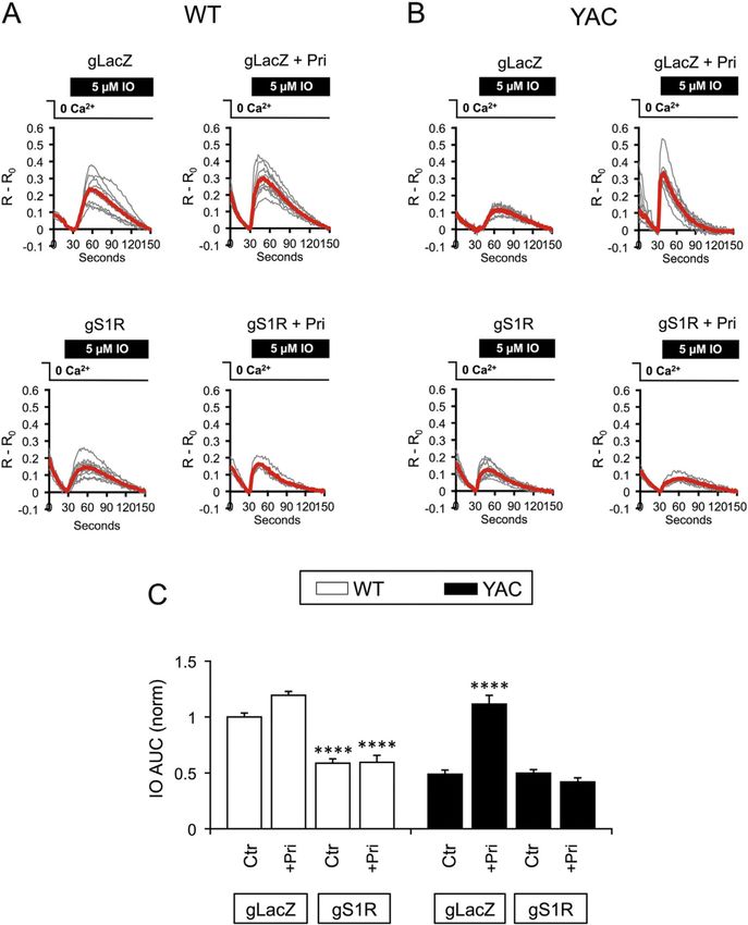
Ryskamp et al.                                                                                             Page 2

                            were mediated by S1R and led to normalization of ER Ca2+ release, ER Ca2+ levels and spine
Author Manuscript

                            SOC entry in YAC128 MSNs. This is a new potential mechanism of action for pridopidine,
                            highlighting S1R as a potential target for HD therapy. Upregulation of striatal proteins that
                            regulate calcium, including calbindin and homer1a, upon chronic therapy with pridopidine, may
                            further contribute to long-term beneficial effects of pridopidine in HD.

                       Keywords
                            Huntington disease; Pridopidine; 3-PPP; Sigma-1 receptor; Medium spiny neurons; YAC128 mice;
                            Corticostriatal co-culture; Synaptic instability; Store-operated calcium entry

                       1. Introduction
                                      Huntington disease (HD) is a progressive neurodegenerative disease resulting from a
Author Manuscript

                                      dominantly-inherited trinucleotide (CAG) repeat expansion in the huntingtin gene, encoding
                                      polyglutamine-expanded mutant Huntingtin (mHtt) protein (MacDonald et al., 1993). HD
                                      symptoms, which include motor, cognitive and psychiatric disturbances, typically present
                                      around 40 years of age and progressively worsen until death approximately 20 years after
                                      diagnosis (Foroud et al., 1999). HD management is limited to supportive care and
                                      symptomatic treatment (Bates et al., 2015). Pridopidine (ACR16) is emerging in clinical
                                      trials as a potential therapeutic to mitigate motor symptoms (e.g., total motor score
                                      improvement was observed when tested as a secondary endpoint in two independent clinical
                                      trials) in HD patients (de Yebenes et al., 2011; Esmaeilzadeh et al., 2011; Huntington Study
                                      Group, 2013; Lundin et al., 2010). Pridopidine was initially identified as a stabilizer of the
                                      dopamine system, normalizing hyper- and hypodopaminergic behaviors, with the proposed
                                      mode of action of a D2 receptor (D2R) antagonist, a partial weak agonist, or both a positive
Author Manuscript

                                      allosteric modulator and an orthosteric antagonist (Dyhring et al., 2010; Nilsson et al., 2004;
                                      Rung et al., 2008). However, the affinity of pridopidine for D2R is low (IC50 and Ki ~10
                                      µM) (Dyhring et al., 2010) compared to its affinity for the sigma-1 receptor (S1R; Ki ~80
                                      nM) (Sahlholm et al., 2013). Indeed, pridopidine exhibits efficient S1R binding, but not D2R
                                      binding, at behaviorally relevant doses in vivo (Sahlholm et al., 2015), indicating that the
                                      therapeutic mechanism of action for pridopidine may primarily involve the S1R.

                                      S1R is a brain-enriched, transmembrane protein of 223 amino acids in the endoplasmic
                                      reticulum (ER) (Kourrich et al., 2012). S1R is evolutionarily conserved and lacks sequence
                                      homology with other mammalian proteins. Computational modeling and NMR studies
                                      indicate that S1R contains 2 transmembrane domains in ER membrane (Brune et al., 2014;
                                      Ortega-Roldan et al., 2015), although a recent crystal structure indicated a single
Author Manuscript

                                      transmembrane domain topology (Schmidt et al., 2016). S1R is often referred to as a
                                      “chaperone” (Su et al., 2010), but its primary function appears to involve modulation of ion
                                      channels (Kourrich et al., 2012). S1R is normally restricted to mitochondrial-associated
                                      membrane (MAM) domains where it regulates calcium (Ca2+) signaling between the ER and
                                      mitochondria, as well as lipid transport (Hayashi and Su, 2003; Hayashi and Su, 2007).
                                      However, high concentrations of S1R agonists, or alternatively ER stress, lead to dislocation
                                      of S1R beyond the MAM domain (Su et al., 2010) so as to regulate ion channels on the

                                          Neurobiol Dis. Author manuscript; available in PMC 2017 January 05.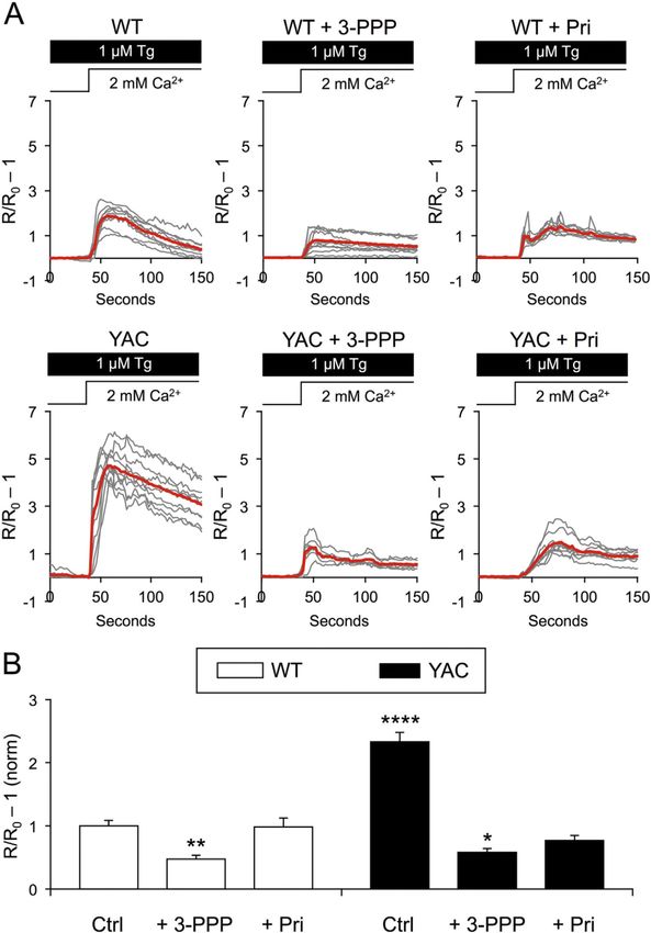
Ryskamp et al.                                                                                            Page 3

                                     plasma membrane (Kourrich et al., 2012). Other roles have been reported for S1R in brain
Author Manuscript

                                     function, including neuromodulation (Maurice et al., 2006) and neuroplasticity (Kourrich et
                                     al., 2012; Takebayashi et al., 2004; Tang et al., 2009; Tsai et al., 2009).

                                     S1R was first identified as a target for treating neuropsychiatric disorders, including drug
                                     addiction, depression and schizophrenia (Maurice and Su, 2009). Additional indications are
                                     now emerging from genetic data pertaining to neurodegenerative diseases, such as
                                     Alzheimer's disease (Fehér et al., 2012; Mishina et al., 2008; Uchida et al., 2005),
                                     amyotrophic lateral sclerosis (Al-Saif et al., 2011), hereditary motor neuropathy (Li et al.,
                                     2015) and frontotemporal lobar degeneration (Luty et al., 2010). Several studies have
                                     identified neuroprotective properties of S1R modulators (Fisher et al., 2016; Marrazzo et al.,
                                     2005; Ruscher et al., 2011; Schetz et al., 2007; Smith et al., 2008). In previous studies, the
                                     S1R agonist PRE-084 displayed neuroprotective properties in PC6.3 cells expressing N-
                                     terminal mHtt (Hyrskyluoto et al., 2013). Similarly, pridopidine improved motor
Author Manuscript

                                     performance and prolonged survival of R6/2 HD mice and exerted neuroprotective effects in
                                     a mouse striatal knock-in cellular model of HD (STHdh111/111) (Squitieri et al., 2015).
                                     These data suggest that pridopidine might act as a disease-modifying therapeutic in HD by
                                     stimulating S1R activity.

                                     Early neuropathological features of HD include perturbed corticostriatal synaptic function
                                     and connectivity (Miller and Bezprozvanny, 2010; Milnerwood and Raymond, 2007;
                                     Milnerwood and Raymond, 2010; Murmu et al., 2013; Orth et al., 2010; Schippling et al.,
                                     2009), eventually leading to overt neurodegeneration of medium spiny neurons (MSNs) in
                                     the striatum (Myers et al., 1988; Vonsattel and DiFiglia, 1998). Perturbed stability of
                                     synaptic spines has been suggested to underlie the development of HD symptoms
                                     (Bezprozvanny and Hiesinger, 2013; Murmu et al., 2013; Ryskamp et al., 2016). In recent
Author Manuscript

                                     studies, we demonstrated that post-synaptic dendritic spines of MSNs are lost in aged
                                     corticostriatal co-cultures established from YAC128 mice (Wu et al., 2016). In the present
                                     study, we used this in vitro HD MSN spine loss model to investigate the potential
                                     mechanism of action of pridopidine in HD and to assess S1R as a therapeutic target of
                                     pridopidine.

                       2. Materials and methods
                       2.1. Mice
                                     Experiments involving mice were approved by the Institutional Animal Care and Use
                                     Committee of the University of Texas Southwestern Medical Center at Dallas and followed
                                     the National Institutes of Health Guidelines for the Care and Use of Experimental Animals.
Author Manuscript

                                     Wild type (WT; FVB/NJ) and YAC128 transgenic (FVB-Tg(YAC128)53Hay/J; Jackson
                                     Labs: stock # 004938) mice (Slow et al., 2003) were maintained at UT Southwestern
                                     Medical Center in barrier facility (12 h light/dark cycle) and genotyped as in (Wu et al.,
                                     2011).

                                         Neurobiol Dis. Author manuscript; available in PMC 2017 January 05.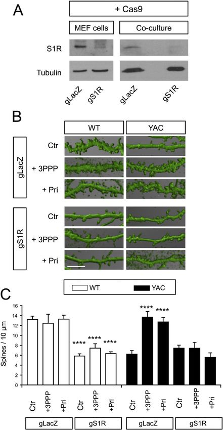
Ryskamp et al.                                                                                           Page 4

                       2.2. Statistical analysis of the Q175 HD mouse model gene expression (CHDI allelic series
Author Manuscript

                       six month dataset)
                                     CAG knockin mouse RNAseq data were downloaded from the Cure Huntington's Disease
                                     Initiative (CHDI) website http://chdifoundation.org/datasets/ and were normalized using the
                                     voom transform from R package limma v3.18.13 in R v3.1.3. Using lmFit, genes were tested
                                     for differential expression in striata comparing between 6 months old Q20 (normal
                                     phenotype) and 6 months old Q175 heterozygotes. Q175 mice express an allele encoding the
                                     human HTT exon 1 sequence with a ~190 CAG repeat tract that replaces mouse Htt exon 1
                                     and results in an HD phenotype.

                       2.3. Chronic treatment animal studies of gene expression profiles
                                     Gene expression analysis of pridopidine-treated rats was recently described (Geva et al.,
                                     2016). Briefly, Sprague Dawley rats (n = 6) were treated daily by oral gavage with
Author Manuscript

                                     pridopidine (60 mg/kg) over 10 days. Six control Sprague Dawley rats were vehicle-treated.
                                     On the 10th day, 90 min following the last drug administration, brains were removed and
                                     RNA was isolated from the striatum of each rat and was analyzed using Affymetrix Rat
                                     230_2 arrays. The gene expression data from 12 striatum samples was RMA normalized
                                     with affy package v1.42.3 in R v3.1.2. Probesets were annotated according to the Affymetrix
                                     Rat230_2 Release 22 annotation file. Processing of the Affymetrix data is detailed in the
                                     previous publication (Geva et al., 2016). The limma package v3.18.13 in R v3.1.3 was used
                                     to test if relevant calcium-related genes were differentially expressed between the two
                                     groups of biological replicates and multiple hypothesis testing was corrected for using the
                                     Bonferroni correction. Limma employs an empirical Bayes method to moderate standard
                                     error (Ritchie et al., 2015). When a gene had multiple probesets, the probeset with the
                                     highest absolute value of fold change was reported.
Author Manuscript

                       2.4. Western blot analysis
                                     Striata from WT and YAC128 mice at 2, 6, and 12 months of age were isolated by dissection
                                     in PBS following euthanasia by euthasol injection and cervical dislocation. Cortices were
                                     similarly isolated for WT and YAC128 mice at 12 months of age. Human samples from
                                     caudate/nucleus accumbens/putamen and globus pallidus were obtained from the Harvard
                                     Brain Tissue Resource Center (http://www.brainbank.mclean.org/) and were processed as in
                                     (Sun et al., 2014). Isolated tissue was weighed and for each 100 mg of tissue, 200 µl of cold
                                     lysis buffer (1% CHAPS, 137 mM NaCl, 2.7 mM KCl, 4.3 mM Na2HPO4, 1.4 mM
                                     KH2PO4, pH 7.2, 5 mM EDTA, 5 mM EGTA, 1 mM PMSF, 50 mM NaF, 1 mM Na3VO4
                                     and protease inhibitors) was used for homogenizing tissue and solubilizing protein (4 °C for
                                     1 h). Samples were centrifuged at 10,000g for 10 min at 4 °C and the supernatant was
Author Manuscript

                                     transferred to a new tube. For human samples, the protein concentration was measured with
                                     a NanoDrop. Based on the volume of supernatant, an appropriate amount of 6 × SDS buffer
                                     was added to each sample. Human samples were diluted with 1 × SDS buffer based on the
                                     measured concentration of protein to standardize protein loading into the gel. For Western
                                     blot analysis of primary neuron cultures, WT and YAC128 striatal, cortical and
                                     corticostriatal cultures were aged to DIV21. Culture media was replaced with 1 × SDS lysis
                                     buffer. A pipette tip was used to scrape the bottoms of wells. Lysates were transferred to 1.5

                                        Neurobiol Dis. Author manuscript; available in PMC 2017 January 05.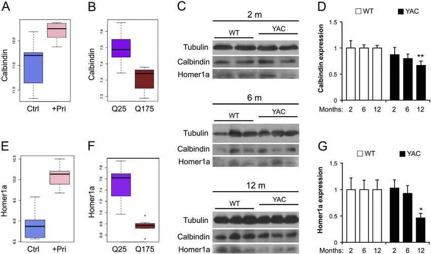
Ryskamp et al.                                                                                            Page 5

                                     ml tubes on ice and then sonicated. Samples were temporarily transferred from ice to a
Author Manuscript

                                     90 °C tube rack for 3 min. The protein lysates were separated by SDS-PAGE and analyzed
                                     by Western blotting with mouse anti-S1R pAb (1:200, Santa Cruz, sc-137,075), mouse anti-
                                     calbindin-D-28K (1:500, clone CB-955, Sigma), rabbit anti-homer1a (1:500, Synaptic
                                     Systems, Cat. No. 160 013), and mouse anti-tubulin (1:5000, DSHB, E7-c). The homer1a
                                     antibody was validated by immunoblotting brain lysates from homer1a KO mice (i.e., the
                                     ~28 kDa band was absent for homer1a KO brain samples). The HRP-conjugated anti-mouse
                                     secondary antibody (111–035-144) and anti-rabbit secondary antibody (115-035-146) were
                                     from Jackson ImmunoResearch. Data were densitometrically analyzed using ImageJ by
                                     normalizing the density of each band to tubulin signal of the same sample after subtracting
                                     the background.

                       2.5. In vitro spine loss assay
Author Manuscript

                                     To study MSN spine loss in vitro, corticostriatal co-cultures were prepared from WT and
                                     heterozygous YAC128 littermates as in (Wu et al., 2016). Striata and cortices were dissected
                                     from pups on postnatal day 0–1 (in 1 × Hank's Balanced Salt Solution, 16.36 mM HEPES,
                                     10 mM NaHCO3, 1 × penicillin-streptomycin). Brain tissue was cut into small pieces (~500
                                     µm chunks), centrifuged (800 rpm for 4 min), digested with papain dissolved in Neurobasal-
                                     A medium (30 min at 37 °C in 500 µl of 114 U papain/10 ml NBA; Worthington), rinsed and
                                     centrifuged (2000 rpm for 4 min; Neurobasal-A medium with 10% FBS and 25 µg/ml
                                     DNAse I), mechanically dissociated (in 500 µl of dissection media with 5 mg/ml DNAse I),
                                     rinsed and centrifuged (2000 rpm for 4 min; in dissection media then again in plating
                                     media), plated on poly-D-lysine coated 12 mm coverslips (0.5 ml/well of 0.1 mg/ml poly-D-
                                     lysine in PBS for 30 min at 37 °C, rinsed with dissection media then plating media and dried
                                     with the coverslips centered in each well) in 80 µl of Neurobasal-A medium supplemented
Author Manuscript

                                     with 5% FBS, 2% B27 and 0.5 mM L-glutamine (Invitrogen) for 7 min. Then an additional
                                     920 µl of plating media was added to each well and cells were maintained at 37 °C in a 5%
                                     CO2 incubator, feeding weekly by addition of 400 µl of NBA, 2% B27 and 0.5 mM L-
                                     glutamine. The anterior half of bilateral cortices from one brain and striata from three brains
                                     were used to plate 24 wells of a 24-well plate. The average plating densities were ~350
                                     cells/mm2 for cortical neurons (Ctx) and ~1060 cells/mm2 for MSNs (a 3:1 MSN:Ctx ratio).

                                     Drugs were dissolved in Neurobasal A media (NBA) from 10 mM stock solutions (DMSO)
                                     immediately prior to experiments. Starting on DIV21 co-cultures were treated with
                                     pridopidine or 3-PPP (100 nM or 1 µM). The compounds or the vehicle control (NBA +
                                     DMSO) were added directly to culture media from 10 µM NBA stocks. Cultures were fixed
                                     after 16 h of drug treatment and processed for immunohistochemistry. Co-cultures were
                                     fixed for 20 min in 4% formaldehyde plus 4% sucrose in PBS (pH 7.4; 4 °C), rinsed twice
Author Manuscript

                                     with cold PBS, permeabilized for 5 min in 0.25% Triton-X-100 (RT) and rinsed again with
                                     PBS (RT). Cultures were blocked with 5% BSA in PBS and immunostained with a rabbit
                                     anti-DARPP-32 antibody (1:500, Cell Signaling, 2306 s) and, after rinsing 3 × with PBS, a
                                     goat anti-rabbit Alexa488 secondary antibody (1:1000). DARPP32 is highly expressed in
                                     MSNs and can be immunolabeled to reliability visualize MSN spine morphology in vitro
                                     (Wu et al., 2016). Z-stacks were captured using a 63 × glycerol objective (N.A. 1.3) on a
                                     confocal microscope (Leica SP5) with the pinhole set to one airy unit. The density of

                                         Neurobiol Dis. Author manuscript; available in PMC 2017 January 05.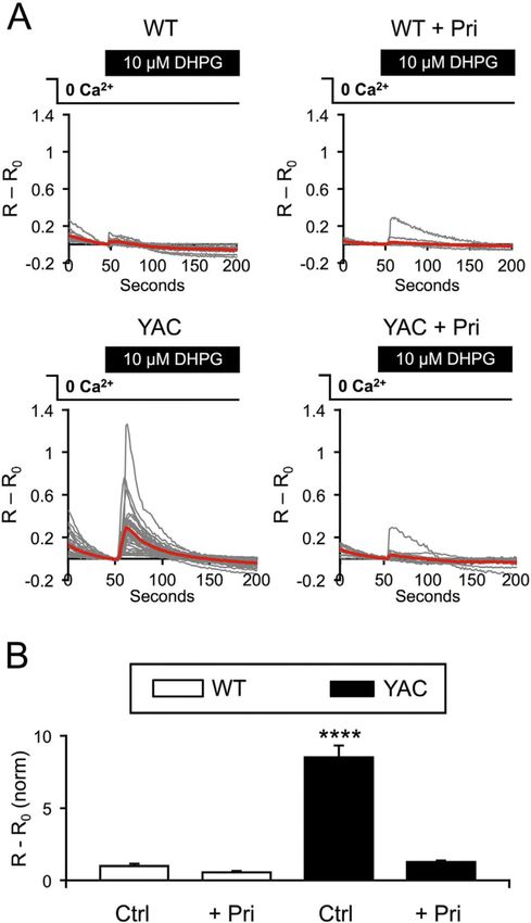
Ryskamp et al.                                                                                          Page 6

                                     dendritic spines of DARPP32-positive MSNs was automatically quantified by using the
Author Manuscript

                                     NeuronStudio software package (Rodriguez et al., 2008) with manual correction. Spine
                                     shapes were categorized as described in (Wu et al., 2016). Data were analyzed from at least
                                     three batches of cultures for each experiment.

                       2.6. Lenti-virus preparation
                                     We used a lenti-expression vector (FUGW; addgene.org/14883/) to drive expression of GFP
                                     or Cherry, lenti-Cas9-Blast (addgene.org/52962/) or lenti-GuidePuro (addgene.org/52963/).
                                     To generate lenti-viruses, plasmids of interest were mixed with lenti-viral production
                                     plasmids (Δ8.9 and VSVG) in 1 ml DMEM and 60 µl polyethylenimine (PEI) for 20 min at
                                     RT. Culture media (DMEM + 10% FBS) was replaced with 11.5 ml of NBA and the plasmid
                                     mixture was dripped into the fresh NBA media to transfect the HEK293T cells for virus
                                     production and packaging. This media was collected 48 h later, centrifuged (2000 RPM for 5
Author Manuscript

                                     min), filtered (0.45 µm pore size) and aliquoted into cryotubes, which were flash-frozen in
                                     liquid nitrogen and stored at −80 °C until use. 100 µl of lenti-virus media was added to each
                                     well of neuron cultures on DIV7. Lenti-viruses produced through this approach exhibited
                                     selective neuronal tropism as revealed by GFP expression and MAP2 immunostaining, with
                                     a ~90% neuronal transfection rate (Wu et al., 2016).

                       2.7. CRISPR/Cas9-mediated deletion of S1R
                                     To delete neuronal S1R in corticostriatal co-cultures we used the CRISPR/Cas9 system.
                                     GuideRNA sequences targeting mouse S1R were designed using bioinformatics tools
                                     (crispr.mit.edu for maximizing specificity and http://www.broadinstitute.org/rnai/public/
                                     analysis-tools/sgrna-design for selecting guide sequences with predicted efficacy) and
                                     sgRNA plasmids targeting S1R (gS1R) were generated. A sgRNA sequence targeting exon 1
Author Manuscript

                                     of S1R (GCAGCTTGCTCGACAGTATG) was subcloned into the lentiGuide-Puro plasmid
                                     (addgene.org/52963/) as in (Sanjana et al., 2014) following their protocol (addgene.org/
                                     static/data/plasmids/52/52963/52963-attachment_IPB7ZL_hJcbm.pdf). The lenti-Cas9-Blast
                                     plasmid (addgene.org/52962/) was used to express Cas9. To validate these plasmids, MEF
                                     cells were co-transfected with Cas9-Blast and gS1R-Puro plasmids using FuGENE6 (1:4
                                     DNA to charge ratio) and cells transfected with both plasmids were selected with 5 µg/ml
                                     blasticidin and 10 µg/ml puromycin in the culture media. Western blotting analysis
                                     confirmed efficient deletion of S1R in MEF cells. Western blotting and functional analysis in
                                     the spine loss and calcium imaging assays confirmed efficient deletion by Cas9 and gS1R in
                                     co-cultures. As in (Platt et al., 2014), a guideRNA sequence
                                     (GTGCGAATACGCCCACGCGAT) targeting the bacterial gene β-galactosidase (LacZ) was
                                     used as a negative control (gLacZ).
Author Manuscript

                       2.8. Fura-2 calcium imaging experiments
                                     Fura-2 Ca2+ imaging experiments with corticostriatal co-cultures were performed as
                                     described previously (Wu et al., 2016) on DIV14–20. To distinguish cortical neurons from
                                     MSNs, in preparation of the co-culture, cortical neurons were plated on DIV0 and infected
                                     with lenti-GFP. 24 h later, media was replaced and striatal neurons were plated. GFP-
                                     negative striatal neurons were identified for analysis as in (Wu et al., 2016). The neurons
                                     were loaded with Fura-2-AM (5 µM; Molecular Probes) for 45 min at 37 °C and transferred

                                        Neurobiol Dis. Author manuscript; available in PMC 2017 January 05.
Ryskamp et al. Page 7
to a recording chamber filled with artificial cerebrospinal fluid (ACSF) (140 mM NaCl, 5
Author Manuscript
mM KCl, 1 mM MgCl2, 2 mM CaCl2, 10 mM HEPES [pH 7.3]). When Fura-2 fluorescence
was evoked with 340 and 380 nm excitation produced from a DeltaRAM-X illuminator, 510
nm emissions were captured with an Evolve camera and EasyRatioPro software (Photon
Technology International, Inc.) to measure the cytosolic calcium concentration (340F/380F
ratio). To measure InsP3R1-dependent Ca2+ release as described (Wu et al., 2016), external
Ca2+ was removed and the basal calcium level (R0 = 340F/380F at baseline) was recorded
for 60 s prior to addition of 10 µM (S)-3,5-DHPG (Tocris). R = 340F/380F at peak response
within 1 min of stimulus application. To measure the size of the ionomycin-sensitive Ca2+
pool, the neurons were washed with Ca2+-free ACSF (100 µM EGTA) for 30 s prior to the
application of 5 µM IO and the Ca2+ signals were recorded as R = 340F/380F. The ER Ca2+
pool size was calculated by integrating the area under the response curve as described (Wu
et al., 2016).
Author Manuscript
2.9. GCaMP5G Ca2+ imaging experiments
GCaMP5-based Ca2+ imaging of MSN spines was performed as described (Wu et al., 2016).
To distinguish cortical neurons from MSNs, corticostriatal co-cultures were prepared as in
Fura-2 Ca2+ imaging experiments except that lenti-Cherry was used to infect cortical
neurons. Co-cultures were transfected on DIV7 with a GCaMP5G expression plasmid (Jiang
and Chen, 2006) using a CalPhos Transfection Kit (Clontech). MSNs in the co-cultures were
identified as in (Wu et al., 2016) by GCaMP5 expression, morphology and lack of Cherry
expression. GCaMP5 was imaged with an Olympus IX70 inverted epifluorescence
microscope equipped with a 60 × lens, Cascade 650 digital camera (Roper Scientific) and
Prior Lumen 200 illuminator (488 nm excitation). Images were collected at 0.5 Hz with
MetaFluor (Universal Imaging). To measure neuronal store-operated Ca2+ entry (nSOC), the
co-cultures were incubated in Ca2+-free media containing 1 µM thapsigargin (Tg) and 400
Author Manuscript
µM EGTA for 5 min before returning to the ACSF containing 2 mM Ca2+, 1 µM Tg and a
Ca2+ channel inhibitor cocktail (1 µM TTX, 50 µM AP5, 10 µM CNQX and 50 µM
nifedipine). The basal calcium level (F0) in Ca2+-free media was recorded for 40 s prior to
Ca2+ add back. F = peak response from Ca2+ re-addition. Data analysis was performed using
ImageJ (NIH).
2.10. Statistical analyses
Data are presented as mean ± SE and were statistically analyzed with GraphPad Prism 6
using two-way ANOVAs followed by a multiple comparisons test. Sidak's post hoc test was
used when only comparing WT vs. YAC128 for each condition. Tukey's post hoc test was
used to make all comparisons. Dunnett's post hoc test was used when comparing against the
Author Manuscript
control condition. The multiplicity adjusted p value is reported. p > 0.05 = n.s, p < 0.05 = *,
p < 0.01 = **, p < 0.001 = *** and p < 0.0001 = ****.
3. Results
3.1. Upregulation of striatal S1R in both mice and patients with advanced but not early HD
In order to evaluate striatal S1R expression in association with ER calcium dysregulation,
we evaluated its protein levels in YAC128 HD mice by Western blotting. At 2 months,
Neurobiol Dis. Author manuscript; available in PMC 2017 January 05.Ryskamp et al. Page 8
normalized S1R expression in YAC128 striata (1.13 ± 0.2; n = 10 mice) was similar to WT
Author Manuscript
striata (Fig. 1A, B; n = 11). As normalized to WT (n = 12), at 6 months S1R expression was
slightly (not significantly) elevated in YAC128 striata (1.26 ± 0.19; n = 10) (Fig. 1A, B). By
12 months S1R expression was significantly upregulated in YAC128 striata (1.42 ± 0.1; n =
12) compared to WT striata (p < 0.05; n = 12; Sidak test) (Fig. 1A, B). By contrast, cortical
S1R expression was similar in WT and YAC128 mice at 12 months of age (WT = 1 ± 0.18, n
= 6; YAC128 = 0.93 ± 0.072, n = 6; p > 0.05; unpaired t-test). We also examined lysates
from primary striatal cultures, cortical cultures and corticostriatal co-cultures from WT and
YAC128 mice on DIV21. We observed no significant changes in S1R expression in WT vs.
YAC128 cultures (WT striatal culture = 1 ± 0.15, n = 3; YAC128 striatal culture = 0.71
±0.16, n = 3; WT cortical culture = 1 ± 0.12, n = 3; YAC128 cortical culture = 0.84 ± 0.2, n
= 3; WT corticostriatal co-culture = 1 ± 0.16, n = 4; YAC128 corticostriatal co-culture =
0.985 ± 0.103 n = 4; p > 0.05; unpaired t-tests), indicating that HD-related changes in striatal
Author Manuscript
S1R expression exclusively occur in vivo. We also evaluated S1R expression levels in
samples from the basal ganglia of HD patients (HD3 = moderate HD; HD4 = advanced HD)
compared to healthy controls (HC). S1R expression was moderately (not significantly)
elevated in striatal samples from HD3 patients (1.46 ± 0.2; n = 7) compared to HC (1 ± 0.11;
n = 5) (Fig. 1C, D). S1R expression was significantly upregulated in striatal samples from
HD4 patients (2.21 ± 0.6; n = 3; p < 0.05; Sidak test) compared to HC (Fig. 1C, D). We also
examined S1R expression in the human globus pallidus, which receives inputs from the
caudate nucleus and putamen. S1Rwas slightly, but non-significantly upregulated in globus
pallidus samples from HD3 and HD4 patients compared to healthy controls (HC = 1
± 0.096, n = 6; HD3 = 1.4 ± 0.19, n = 7; HD4 = 1.65 ± 0.34, n = 8; p > 0.05; Dunnett's test).
These data are consistent with a compensatory increase in striatal S1R expression in late HD
not seen in early HD.
Author Manuscript
3.2. Pridopidine stabilizes MSN spines in aging YAC128 corticostriatal co-cultures via S1R
In previous studies, we developed an in vitro model of synaptic pathology in HD that is
based on analysis of MSN spine density in corticostriatal co-cultures established from
YAC128 mice. We demonstrated that YAC128 MSNs in these cultures display age-
dependent spine loss when compared to WT MSNs (Wu et al., 2016). 3-PPP is a selective
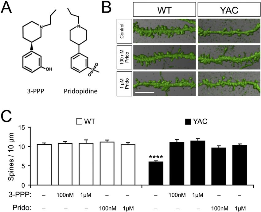
S1R agonist with a very similar chemical structure to pridopidine (Fig. 2A) and an identical
affinity for S1R (Sahlholm et al., 2013). To evaluate whether S1R agonists can stabilize
synaptic connections in HD, we administered 3-PPP to corticostriatal co-cultures from WT
and YAC128 mice. Exposure to 100 nM and 1 µM 3-PPP had no effect on the density of WT
MSN spines (WT = 10.5 ± 0.35 spines/10 µm of dendritic length, n = 23; WT 100 nM 3-PPP
= 10.7 ± 0.54 spines/10 µm, n = 8; WT 1 µM 3-PPP = 10.8 ± 0.86 spines/10 µm, n = 8) (Fig.
Author Manuscript
2B, C). Compared to WT, the spine density in co-cultured MSNs from YAC128 mice was
substantially reduced (YAC128 = 5.99 ± 0.26 spines/10 µm, n = 23; p < 0.0001) (Fig. 2B,
C), which is consistent with our previous observations (Wu et al., 2016). 100 nM and 1 µM
3-PPP elevated the density of YAC128 MSN spines to WT levels (YAC128 100 nM 3-PPP =
11.0 ± 0.78 spines/10 µm, n = 8; YAC128 1 µM 3-PPP = 11.4 ± 0.61 spines/10 µm, n = 8)
(Fig. 2B, C). Likewise, treatment with pridopidine had no effect on WT MSN spines (WT
100 nM pridopidine = 11.1 ± 0.52 spines/10 µm, n = 15; WT 1 µM pridopidine = 10.5
± 0.47 spines/10 µm, n = 15), but rescued MSN spines in YAC128 co-cultures (YAC128 100
Neurobiol Dis. Author manuscript; available in PMC 2017 January 05.Ryskamp et al. Page 9
nM pridopidine = 9.6 ± 0.54 spines/10 µm, n = 15; YAC128 1 µM pridopidine = 10.3 ± 0.36
Author Manuscript
spines/10 µm, n = 15) (Fig. 2B, C). In this model, all spine types (mushroom, thin, stubby)
are lost uniformly (Wu et al., 2016). Treatment with pridopidine uniformly rescued all spine
types (data not shown). We also tested other S1R agonists, specifically PRE-084 and (−)-
OSU6162, both of which also effectively rescued YAC128 MSN spines at 100 nM and 1 µM
concentrations (data not shown).
To evaluate whether S1R is required for the synapto-protective effects of 3-PPP and
pridopidine, we used the CRISPR/Cas9 system to delete S1R. Co-expression of Cas9 and
guideRNA targeting the mouse S1R gene (gS1R) effectively deleted S1R in MEF cells,
whereas co-expression of Cas9 and guideRNA targeting the bacterial β-galactosidase gene
(gLacZ) preserved S1R expression (Fig. 3A).
To determine the molecular mechanism of pridopidine's synapto-protective effect, MSN
Author Manuscript
spine analysis with corticostriatal co-cultures from YAC128 mice was performed following
deletion of S1R by coinfecting cultured neurons on DIV7 with lenti-Cas9 and lenti-gS1R or
lenti-gLacZ as a control (Fig. 3B, C). In our previous studies we used a similar approach to
delete STIM2 in corticostriatal co-cultures (Wu et al., 2016).
In gLacZ cultures, the WT MSN spine density was 13.2 ± 0.68 spines/10 µm (n = 7) (Fig.
3B, C) and the YAC128 MSN spine density was reduced to 6.2 ± 0.73 spines/10 µm (n = 7)
(Fig. 3B, C). Consistent with the previous results (Fig. 2B, C), 16-h treatment of DIV21 co-
cultures with 100 nM 3-PPP or pridopidine had no effect on the density of MSN spines in
WT gLacZ co-cultures (WT control = 13.2 ± 0.7 spines/10 µm, WT 3-PPP = 12.5 ± 1.8
spines/10 µm, WT pridopidine = 13.3 ± 0.8 spines/10 µm, n = 3–7, p > 0.05) and rescued
MSN spine loss in YAC128 gLacZ co-cultures (YAC128 control = 6.2 ± 0.7 spines/10 µm,
Author Manuscript
YAC128 3-PPP = 13.7 ± 1.1 spines/10 µm, YAC128 pridopidine = 12.7 ± 0.9 spines/10 µm,
n = 3–7, p < 0.0001) (Fig. 3B, C). The spine density of MSNs in WT gS1R co-cultures was
diminished to 5.83 ± 0.47 spines/10 µm (n = 7, p < 0.0001 compared to WT gLacZ),
indicating that S1R has a physiological role in maintaining MSN spine stability.
Although 3-PPP and pridopidine rescue YAC128 MSN spines (Figs. 2B, C and 3B, C), 3-
PPP and pridopidine were unable to prevent WT MSN spine loss in the absence of S1R (WT
gS1R + 3-PPP = 7.42 ± 0.89 spines/10 µm, n = 3, p < 0.0001 compared to WT gLacZ; WT
gS1R + pridopidine = 6.34 ± 0.35 spines/10 µm, n = 4, p < 0.0001 compared to WT gLacZ).
This is consistent with a crucial role for S1R in mediating the synapto-protective effects of
3-PPP and pridopidine.
Although co-expression of Cas9 and gS1R in WT co-cultures caused MSN spine loss, their
Author Manuscript
co-expression in YAC128 co-cultures did not cause additional spine loss beyond that caused
by mHtt (YAC128 gS1R = 7.42 ± 0.61 spines/10 µm, n = 7, p > 0.05 compared to YAC128
gLacZ). Even though 3-PPP and pridopidine restored the density of MSN spines to WT
levels in YAC128 gLacZ co-cultures (n = 3–7, p < 0.0001 compared to YAC128 gLacZ),
they were unable to rescue MSN spines in YAC128 gS1R co-cultures (YAC128 gS1R + 3-
PPP = 7.41 ± 1.19 spines/10 µm, n = 3, p > 0.05; YAC128 gS1R + pridopidine = 5.57 ± 0.90
spines/10 µm, n = 4, p > 0.05) (Fig. 3B, C).
Neurobiol Dis. Author manuscript; available in PMC 2017 January 05.Ryskamp et al. Page 10
This indicates that S1R is required for the protective effects of both 3-PPP and pridopidine.
Author Manuscript
These results are consistent with the hypothesis that pridopidine rescues spines in HD
neurons by activating S1R.
3.3. Pridopidine normalizes ER Ca2+ homeostasis in MSNs from YAC128 corticostriatal co-
cultures
Our recent data demonstrated that synaptic store-operated calcium entry (nSOC) plays an
important role in control of stability of hippocampal (Popugaeva et al., 2015; Sun et al.,
2014; Zhang et al., 2015) and striatal (Wu et al., 2016) synaptic spines. In HD, mHtt binds to
and sensitizes InsP3R1 to basal levels of InsP3 (Tang et al., 2003), increasing Ca2+ leakage
from the ER (Tang et al., 2005; Tang et al., 2003). This leads to excessively elevated SOC in
MSNs (Wu et al., 2011), an effect that is more pronounced in MSN spines (Wu et al., 2016).
This tonic Ca2+ signal is synaptotoxic, contributing to MSN spine loss in the YAC128 HD
Author Manuscript
mouse model in vitro and in vivo (Wu et al., 2016). S1R is known to modulate intracellular
Ca2+ signaling (Hayashi and Su, 2007; Srivats et al., 2016). In the next series of experiments
we evaluated effects of pridopidine on MSN Ca2+ homeostasis.
We previously demonstrated that DHPG-evoked Ca2+ release from the ER in MSNs is
mediated by InsP3R1 and that DHPG-evoked Ca2+ release is enhanced in YAC128 MSNs in
corticostriatal co-cultures (Wu et al., 2016). After removing external Ca2+, the application of
10 µM DHPG to corticostriatal co-cultures evoked a cytosolic Ca2+ elevation in the somata
of Fura-2-loaded MSNs (Fig. 4A, B). The amplitude of Ca2+ release in these experiments
was very low in WT MSNs but was elevated significantly in YAC128 MSNs (Fig. 4A). On
average, the amplitude of DHPG-induced response was 1 ± 0.139 (n = 153 MSNs) in WT
MSNs and 8.52 ± 0.804 (n = 185) in YAC128 MSNs (p < 0.0001), consistent with our
previous report (Wu et al., 2016). 16–24 h pre-incubation of co-cultures with 1 µM
Author Manuscript
Pridopidine had no effect on Ca2+ release in WT MSNs (0.551 ± 0.079, n = 175, p > 0.05),
but suppressed Ca2+ release in YAC128 MSNs to WT levels (1.27 ± 0.101, n = 398, p >
0.05) (Fig. 4A, B). These results indicated that incubation with pridopidine suppresses
supranormal activity of the InsP3R1 in mHtt-expressing MSNs.
Because pridopidine normalizes InsP3R1 hyperactivity, it would be expected to reduce the
leakage of Ca2+ from the ER in YAC128 MSNs and therefore increase ER Ca2+ content. To
measure ER Ca2+ content in the somata of Fura-2-loaded MSNs in corticostriatal co-
cultures, extracellular Ca2+ was removed and then ionomycin (IO) was added to release
Ca2+ from the internal stores. ER Ca2+ content was quantified by measuring the area under
the IO-evoked Ca2+ response curve. As we previously reported (Wu et al., 2016), the ER
Ca2+ levels were substantially depleted in YAC128 control MSNs (YAC128 gLacZ 0.49
Author Manuscript
± 0.038, n = 116, p > 0.05) compared to WT control MSNs (WT gLacZ 1 ± 0.036, n = 173,
p < 0.01) (Fig. 5A–C). Pre-incubation of co-cultures with 1 µM pridopidine for 16–24 h
non-significantly increased ER Ca2+ content in WT co-cultures (WT gLacZ + pridopidine
1.19 ± 0.036, n = 225), but significantly repleted ER Ca2+ in YAC128 MSNs (YAC128
gLacZ + pridopidine 1.12 ± 0.078, n = 94, p < 0.0001) (Fig. 5A–C).
To evaluate the potential role of S1R in mediating effects of pridopidine on MSN Ca2+
homeostasis, we used the CRISPR/Cas9 system to delete neuronal S1R and measured the
Neurobiol Dis. Author manuscript; available in PMC 2017 January 05.Ryskamp et al. Page 11
effect of pridopidine on ER Ca2+ levels. Co-infection of WT corticostriatal co-cultures with
lenti-Cas9 and lenti-gS1R suppressed ER Ca2+ levels (WT gS1R 0.59 ± 0.04, n = 103, p <
Author Manuscript
0.001) (Fig. 5A–C), indicating that basal S1R activity may partially attenuate tonic Ca2+
leakage in MSNs. Even though pridopidine treatment slightly elevated ER Ca2+ levels in
WT gLacZ MSNs, this effect was absent when S1R was deleted (WT gS1R + pridopidine,
0.60 ± 0.063, n = 76, p > 0.05) (Fig. 5A–C). Deletion of S1R had no additional effect on ER
Ca2+ levels in YAC128 MSNs (YAC128 gS1R, 0.50 ± 0.03, n = 92, p > 0.05 compared to
YAC128 gLacZ) (Fig. 5A–C), indicating that endogenous S1R is unable to adequately
regulate ER Ca2+ levels in YAC128 MSNs without stabilization from S1R ligands.
Additionally, the ability of pridopidine to elevate YAC128 ER calcium to WT levels was lost
in the absence of S1R (YAC128 gS1R + pridopidine, 0.419 ± 0.038, n = 95, p > 0.05
compared to YAC128 gLacZ) (Fig. 5A–C). This indicates that the ability of pridopidine to
restore ER Ca2+ homeostasis in YAC128 MSNs requires S1R.
Author Manuscript
3.4. Pridopidine normalizes spine nSOC in YAC128 MSNs from corticostriatal co-cultures
To compensate for constantly depleted ER calcium levels, YAC128 MSNs upregulate nSOC
to replenish ER calcium stores (Wu et al., 2011). The nSOC pathway is particularly
enhanced in synaptic spines of YAC128 MSNs, leading to spine loss (Wu et al., 2016). We
therefore examined whether incubating co-cultures with 1 µM 3-PPP or pridopidine for 16 h,
an amount of time sufficient to rescue spines, would suppress the hyperactive nSOC
pathway in the spines of YAC128 MSNs.
As previously reported (Wu et al., 2016), MSN spine nSOC was enhanced in corticostriatal
co-cultures from YAC128 mice (2.33 ± 0.15, n = 110 spines) when compared to that from
WT mice (1 ± 0.089, n = 110, p < 0.0001) (Fig. 6A, B). 3-PPP, but not pridopidine, slightly
reduced spine nSOC in WT MSNs (WT + 3-PPP = 0.47 ± 0.06, n = 97, p < 0.01; WT +
Author Manuscript
pridopidine = 0.98 ± 0.14, n = 70, p > 0.05) (Fig. 6A, B). Both 3-PPP and pridopidine
suppressed supranormal spine nSOC in YAC128 MSNs approximately to WT levels
(YAC128 + 3-PPP = 0.59 ± 0.058, n = 109, p < 0.05; YAC128 + pridopidine = 0.77 ± 0.079,
n = 110, p > 0.05) (Fig. 6A, B). From these results, we concluded that suppression of
supranormal nSOC may contribute to synaptoprotective effects of 3-PPP and pridopidine in
the YAC128 corticostriatal co-culture model of HD (Fig. 2).
3.5. Effects of pridopidine on striatal Ca2+ gene expression
To gain further insight into the role of pridopidine in regulation of calcium signaling, we
used previously published data (Geva et al., 2016) to analyze changes in Ca2+ signaling gene
expression profiles in rat striatum induced by chronic pridopidine therapy. In addition, we
leveraged publically available striatum RNAseq data from the Q175KI mouse model for HD
Author Manuscript
(Q175), and examined whether any calcium regulatory genes were downregulated in this
model as compared to the control Q25KI mouse (Q25).
Notably, calbindin (Calb1) mRNA was upregulated in rat striatum following 10 days of oral
pridopidine treatment (p < 0.01; adj. p = 0.4) (Fig. 7A), and it was downregulated in Q175
mice compared with Q25 (p < 0.001; adj. p < 0.01) (Fig. 7B). Calbindin functions as a buffer
Neurobiol Dis. Author manuscript; available in PMC 2017 January 05.Ryskamp et al. Page 12
for excessive intracellular calcium, and its downregulation is expected to predispose HD
MSNs to Ca2+ overload (Kiyama et al., 1990).
Author Manuscript
To substantiate observations of mRNA, we examined calbindin protein expression in the
striatum of untreated YAC128 mice by Western blotting. At 2 months of age, calbindin
expression was similar in WT and YAC128 striata (WT = 1.0 ± 0.14, n = 3 mice; YAC128 =
0.87 ± 0.14, n = 3 mice; Fig. 7C–D). Likewise, calbindin expression was similar in WT and
YAC128 striata at 6 months of age (WT = 1.0 ± 0.061, n = 12 mice; YAC128 = 0.79 ± 0.091,
n = 11 mice; Fig. 7C–D). Similar to findings with Q175 mice, striatal calbindin was
significantly downregulated in YAC128 mice at 12 months of age (WT = 1.0 ± 0.05, n = 12
mice; YAC128 = 0.67 ± 0.08, n = 11 mice; p < 0.01, Fig. 7C–D).
We also found that homer1a mRNA was upregulated in rat striatum post-pridopidine
treatment (p < 0.0001; adj. p < 0.05, Fig. 7E), and it was downregulated in Q175 vs. Q25
Author Manuscript
(WT) mice (p < 0.0001; adj. p < 0.001, Fig. 7F). When analyzed by Western blotting, in
striatal samples from untreated YAC128 mice, homer1a protein expression was similar at 2
months (WT = 1 ± 0.22, n = 4; YAC128 = 1.03 ± 0.16, n = 7) and 6 months (WT = 1 ± 0.18,
n = 12; YAC128 = 0.93 ± 0.14, n = 11) of age, but was significantly downregulated at 12
months of age (WT = 1 ± 0.18, n = 13; YAC128 = 0.46 ± 0.09, n = 14; p < 0.05; Fig. 7C, G).
Homer proteins (Homer 1–3) are postsynaptic scaffolding proteins that are known to play a
central role in calcium signaling by forming multi-protein complexes with calcium
regulators including InsP3R1, mGluR1/5 and Shank (Jardin et al., 2013; Shiraishi-
Yamaguchi and Furuichi, 2007; Worley et al., 2007). Homer1a is a truncated splice-variant
that acts as a dominant negative regulator of the full length homer proteins by preventing
physical coupling and juxtaposition of mGluR1/5-Homer1-InsP3R1 complex components,
thereby attenuating Ca2+ release from the ER (Jardin et al., 2013; Shiraishi-Yamaguchi and
Author Manuscript
Furuichi, 2007; Worley et al., 2007). Thus, the pridopidine-induced increase in Homer1a
expression level may help to stabilize Ca2+ dysregulation in HD MSNs by reducing
InsP3R1-mediated Ca2+ release.
4. Discussion
We report here that S1R is upregulated in the striatum of both mice and patients with
advanced but not early HD. This may reflect a compensatory response to mitigate the effects
of ER Ca2+ dysregulation and synaptic instability. Consistent with this, treatment of
corticostriatal co-cultures prepared from YAC128 mice with the S1R agonist 3-PPP
prevented age-dependent MSN spine loss. Likewise, pridopidine improved synaptic
connectivity in this in vitro model of HD, whereas S1R deletion decreased the density of
Author Manuscript
WT MSN spines to YAC128 MSN levels without causing additional spine loss in YAC128
MSNs. The synapto-protective property of these compounds was eliminated by neuronal
deletion of S1R, suggesting that 3-PPP and pridopidine bolster synaptic connections by
stimulating S1R activity.
Given the role of S1R in the regulation of ER Ca2+ signaling (Hayashi and Su, 2007; Srivats
et al., 2016), we evaluated whether 3-PPP and pridopidine stabilize calcium homeostasis in
Neurobiol Dis. Author manuscript; available in PMC 2017 January 05.Ryskamp et al. Page 13
co-cultured YAC128 MSNs. 16–24 hour treatment with pridopidine suppressed DHPG-
induced Ca2+ release in YAC128 MSNs to WT levels. Pridopidine also elevated ER Ca2+
Author Manuscript
levels in YAC128 MSNs to WT levels, a S1R-dependent effect. Overnight treatment with 3-
PPP or pridopidine reduced nSOC signaling in YAC128 MSN spines below synaptotoxic
levels. These results suggest that pridopidine may prevent spine loss in YAC128 MSNs by
modulating ER Ca2+ homeostasis. Moreover, the Ca2+ regulatory proteins calbindin and
homer1a were upregulated in the striatum of rats following chronic pridopidine treatment,
whereas their expression was downregulated in the striatum of aged Q175 and YAC128
mice. These transcriptional/translational changes may further fine tune Ca2+ signaling and
synaptic stability.
4.1. S1R is necessary for formation and stability of MSN spines
Previous studies suggested that S1R plays an important role in the development and stability
Author Manuscript
of dendritic spines in hippocampal neurons, as well as in the control of neuronal oxidative
stress and Rac GTP signaling (Tsai et al., 2009). More recently, a microarray approach was
used to evaluate transcriptional changes following RNAi knockdown of S1R in cultured
hippocampal neurons (Tsai et al., 2012). S1R knockdown primarily affected pathways
involved in sterol biosynthesis, protein ubiquitination, the actin cytoskeleton network, and
oxidative stress (Tsai et al., 2012).
The potential role of S1R in the response to oxidative stress was corroborated by analysis of
liver tissues and retinal glial cells from S1R knockout mice (Pal et al., 2012; Wang et al.,
2015a) and may be important for dendritic spine stability in the hippocampus given that
spine loss from S1R knockdown was rescued by reducing oxidative stress (Tsai et al., 2009).
Consistent with important neuronal functions of S1R, S1R knockout mice are viable (Langa
et al., 2003), but exhibit late-onset neurodegeneration (Bernard-Marissal et al., 2015; Ha et
Author Manuscript
al., 2011; Mavlyutov et al., 2010). We demonstrate here that genetic ablation of S1R using
the CRISPR/Cas9 system resulted in MSN spine loss in WT corticostriatal co-cultures,
identifying an essential role of S1R in the development and/or maintenance of synaptic
connections between cortical and striatal neurons.
4.2. Role of S1R in neurodegenerative disorders
Considering the important role of S1R in synaptic biology, it is not surprising that an
important role of S1R is emerging in neurodegenerative diseases, such as Alzheimer's
disease (Fehér et al., 2012; Maruszak et al., 2007; Mishina et al., 2008; Uchida et al., 2005),
amyotrophic lateral sclerosis (Al-Saif et al., 2011; Belzil et al., 2013) and frontotemporal
lobar degeneration (Luty et al., 2010). The mechanistic basis for these connections is
starting to be elucidated (Nguyen et al., 2015). For example, it was recently demonstrated
Author Manuscript
that S1R promotes turnover of p35, an activator of cyclin-dependent kinase 5 (cdk5) (Tsai et
al., 2015). This function of S1R is connected with its ability to control phosphorylation of
tau protein (Tsai et al., 2015) and may be responsible for the connection between reduced
S1R function and Alzheimer's disease.
We observed herein upregulation of S1R in striatal samples from aged YAC128 HD mice
and patients with severe HD. Striatal S1R upregulation may occur as a compensatory
Neurobiol Dis. Author manuscript; available in PMC 2017 January 05.Ryskamp et al. Page 14
response to chronic ER Ca2+ depletion (Hayashi and Su, 2007) in HD MSNs and, possibly,
Author Manuscript
to synaptic dysfunction. Thus, changes in striatal S1R expression could represent a marker
of HD progression. This was evident with the non-significant elevation of S1R protein in the
basal ganglia of patients with HD3 (defined in (Shoulson and Fahn, 1979)), and would
become significant with patients featuring more advanced pathology and atrophy (HD4).
Given that S1R levels can be assessed in human patients using PET (Mishina et al., 2008),
this may have implications for defining the optimal therapeutic window for pridopidine
therapy and/or personalizing treatment. Interestingly, striatal S1R mRNA expression was
unchanged in Q175 mice compared to Q20 controls at 6 months of age (p = 0.653; CHDI
RNAseq dataset). Thus, the upregulation of striatal S1R in HD may result from changes in
S1R translation and/or degradation. Either way, the trend toward a compensatory increase of
S1R protein with progression of disease may also highlight the timing for treatment with
pridopidine, with earlier treatment potentially of greater and more beneficial effect.
Author Manuscript
4.3. S1R as a target for pridopidine in HD
S1R agonists enhance cognitive performance, synaptic plasticity and neuronal survival in
conditions of neuronal stress (Antonini et al., 2011; Antonini et al., 2009; Hindmarch and
Hashimoto, 2010; Ruscher et al., 2011). Pharmacological activation of S1R was beneficial in
experimental models of Parkinson's disease (Francardo et al., 2014) and Alzheimer's disease
(Fisher et al., 2016; Marrazzo et al., 2005; Meunier et al., 2006; Villard et al., 2009). The
S1R agonist PRE-084 exerted neuroprotective effects in PC6.3 cells expressing N-terminal
mHtt (Hyrskyluoto et al., 2013). Pridopidine improved motor performance and prolonged
survival of R6/2 HD mice and exerted neuroprotective effects in a mouse striatal knock-in
cellular HD model (STHdh111/111) (Squitieri et al., 2015). In our experiments, pridopidine
and the chemically similar S1R agonist 3-PPP rescued MSN spine loss in YAC128 cortico-
Author Manuscript
striatal co-cultures. Genetic ablation of S1R prevented the rescue of YAC128 MSN spines by
3-PPP and pridopidine, suggesting that S1R is a target for these compounds. This is
consistent with the high affinity of 3-PPP and pridopidine for S1R (Sahlholm et al., 2013;
Sahlholm et al., 2015). Our results suggest that the beneficial actions of pridopidine are
likely to be mediated via activation of S1R, resulting in bolstering of HD MSN spines. These
data suggest that pridopidine might act as a disease-modifying therapeutic in HD by
stimulating S1R activity.
4.4. Effects on Ca2+ signaling from S1R activation by pridopidine
As discussed above, stimulation of S1R activity by pridopidine may lead to remodeling of
the actin cytoskeleton in spines and/or a reduction in oxidative stress (Tsai et al., 2012).
Another well-established function of S1R is to control ER Ca2+ signaling. It was
Author Manuscript
demonstrated that S1R regulates InsP3R3-mediated ER-mitochondrial Ca2+ transfer
(Hayashi and Su, 2007) and removes the inhibitory actions of ankyrin on InsP3R3 (Wu and
Bowen, 2008). However, these actions of S1R appear to be specific for InsP3R3 isoform,
whereas InsP3R1 is a predominant neuronal isoform (Taylor et al., 1999). S1R directly binds
to InsP3R1, but no direct functional effects on InsP3R1 activity were observed in
experiments with hepatocytes (Abou-Lovergne et al., 2011). It was concluded that activation
of S1R leads to stimulation of PKC activity and suppression of InsP3 synthesis in
hepatocytes (Abou-Lovergne et al., 2011).
Neurobiol Dis. Author manuscript; available in PMC 2017 January 05.Ryskamp et al. Page 15
We previously discovered that mHtt enhances the sensitivity of InsP3R1 to InsP3, resulting
in a tonic calcium leakage that decreases ER Ca2+ levels (Tang et al., 2009; Tsai et al., 2009;
Author Manuscript
Tang et al., 2005; Tang et al., 2003). This overactivates STIM2-depedendent store-operated
Ca2+ entry (SOC) in YAC128 MSN spines, causing spine loss (Wu et al., 2016). In our
experiments herein, we discovered that incubation with pridopidine normalized ER Ca2+
release, ER Ca2+ levels and spine SOC in corticostriatal co-cultures from YAC128 mice.
Moreover, incubation with pridopidine was sufficient to restore ER Ca2+ levels in YAC128
MSNs in a S1R-dependent manner.
Actions of pridopidine in these experiments could be mediated by stabilizing effects on
InsP3R1 function, by stimulation of PKC activity or by a different mechanism. Overnight
treatment with pridopidine was sufficient to normalize ER Ca2+ levels and the enhanced
nSOC pathway in YAC128 MSN spines. Of potential relevance to these findings,
knockdown of S1R by RNAi was reported to affect expression levels of the SOC channel
Author Manuscript
subunit Orai1 (Tsai et al., 2012). It has been reported that S1R overexpression or incubation
with S1R agonists suppresses SOC in various cell lines (Brailoiu et al., 2016; Srivats et al.,
2016). To explain these results it has been proposed that S1R may suppress SOC by binding
to STIM1 and obstructing the interaction of STIM1 and Orai1 (Srivats et al., 2016).
However, in our experiments pridopidine elevated ER Ca2+ levels in YAC128 MSNs, which
would decouple STIMs from SOC channels. Thus, the attenuation of supranormal nSOC in
YAC128 MSN spines by pridopidine is more likely to be explained by its effects on ER Ca2+
handling, although direct association of S1R with STIM1 and/or STIM2 may also contribute
to the observed effects. Collectively, our findings suggest that the ability of pridopidine to
stabilize corticostriatal synaptic connections involves normalization of synaptic Ca2+
signaling.
Author Manuscript
Analysis of genes that regulate Ca2+ revealed that calbindin and homer1a are upregulated in
the striatum of rats following chronic pridopidine treatment and reciprocally downregulated
in the striatum of Q175 mice. In Western blotting experiments we confirmed that these
proteins are downregulated in the striatum of aged YAC128 mice. Calbindin, which chelates
cytosolic Ca2+, is absent in HD postmortem brain neurons (Kiyama et al., 1990), suggesting
that loss of Ca2+ buffering is associated with disease progression and severity. Increased
calbindin expression from pridopidine treatment should increase the cytosolic Ca2+
buffering capacity in HD MSNs and exert protective effects, as has been demonstrated in
variety of neuronal insult models (Hugon et al., 1996; Yamada et al., 1990; Yenari et al.,
2001).
Homer1a is a neuronal, activity-dependent, immediate early gene that suppresses mGluR1/5-
induced ER Ca2+ release via InsP3Rs by preventing full length Homer1-mediated coupling
Author Manuscript
between mGluR1/5 and InsP3Rs (Ango et al., 2001; Mao et al., 2005; Tu et al., 1998).
Upregulation of Homer1a is expected to suppress excessive ER Ca2+ release in HD MSNs,
leading to restoration of ER Ca2+ levels and reduced SOC. In support of this hypothesis,
Homer1a overexpression suppressed mGluR activity and clustering in various cell culture
assays (Ango et al., 2000; Ciruela et al., 1999; Roche et al., 1999; Tadokoro et al., 1999).
Increased Homer1a expression attenuated neuronal injury induced by mGluR1 activation in
a model of traumatic brain injury (Luo et al., 2014) and reduced Ca2+ overload and neuronal
Neurobiol Dis. Author manuscript; available in PMC 2017 January 05.Ryskamp et al. Page 16
injury in a MPP(+)-toxicity model with cultured rat mesencephalic cells (Zeng et al., 2013).
Author Manuscript
Increased Homer1a expression was also recently demonstrated to exert neuroprotective
effects against NMDA-induced neuronal injury (Wang et al., 2015b). Interestingly,
expression of homer1a has been shown to be elevated following inhibition of neuronal SOC
in PC12 cells, suggesting potential cross-talk between SOC and Homer1a expression in
neuronal cells (Li et al., 2013). From these results we conclude that observed changes in
expression of calbindin1 and homer1a may further contribute to the long-term beneficial
effects of pridopidine in HD.
5. Conclusions
Pridopidine and 3-PPP are proposed to prevent Ca2+ dysregulation and synaptic loss in a
YAC128 corticostriatal co-culture model of HD. The actions of pridopidine in striatal
neurons are mediated by S1R and involve stabilization of ER Ca2+ levels, reduction of
Author Manuscript
synaptic store-operated Ca2+ entry, and increased expression of the Ca2+ regulating proteins
calbindin1 and homer1a. These results reveal a new potential mechanism of action for
pridopidine and highlight S1R as a potential target for HD therapy.
Acknowledgments
TEVA Pharmaceuticals provided funding for the study. M.G., I.G. and M.H are employees of TEVA
Pharmaceuticals and I.B. is a paid consultant to TEVA Pharmaceuticals.
We thank Leah Taylor for administrative assistance and Pippa Loupe for editorial assistance. We thank Drs. Paul
Worley and Tao Xu (John Hopkins University) for providing samples of Homer 1a knockout brains. The research
described herein was supported by TEVA Pharmaceuticals (SRA #109415) and by the National Institute of
Neurological Disorders and Stroke of the National Institutes of Health (R01NS074376 and R01NS056224, IB;
F32NS093786, DAR). IB holds the Carl J. and Hortense M. Thomsen Chair in Alzheimer's Disease Research.
Author Manuscript
Abbreviations
HD Huntington disease
Htt Huntingtin
MSN medium spiny neuron
S1R sigma-1 receptor
ER endoplasmic reticulum
YAC128 yeast artificial chromosome 128Q expansion mice
3-PPP R(+)-3-(3-Hydroxyphenyl)-N-propylpiperidine hydrochloride
Author Manuscript
SOC store-operated calcium current
MAM mitochondrial-associated membrane
gLacZ guideRNA targeting the bacterial β-galactosidase gene
gS1R guideRNA targeting the mouse S1R gene
Neurobiol Dis. Author manuscript; available in PMC 2017 January 05.Ryskamp et al. Page 17
MEF mouse embryonic fibroblasts
Author Manuscript
DHPG (S)-3,5-dihydroxyphenylglycine
IO ionomycin
References
Abou-Lovergne A, Collado-Hilly M, Monnet FP, Koukoui O, Prigent S, Coquil JF, Dupont G,
Combettes L. Investigation of the role of sigma1-receptors in inositol 1,4,5-trisphosphate dependent
calcium signaling in hepatocytes. Cell Calcium. 2011; 50:62–72. [PubMed: 21641033]
Al-Saif A, Al-Mohanna F, Bohlega S. A mutation in sigma-1 receptor causes juvenile amyotrophic
lateral sclerosis. Ann. Neurol. 2011; 70:913–919. [PubMed: 21842496]
Ango F, Pin JP, Tu JC, Xiao B, Worley PF, Bockaert J, Fagni L. Dendritic and axonal targeting of type
5 metabotropic glutamate receptor is regulated by homer1 proteins and neuronal excitation. J.
Neurosci. 2000; 20:8710–8716. [PubMed: 11102477]
Author Manuscript
Ango F, Prézeau L, Muller T, Tu JC, Xiao B, Worley PF, Pin JP, Bockaert J, Fagni L. Agonist-
independent activation of metabotropic glutamate receptors by the intracellular protein Homer.
Nature. 2001; 411:962–965. [PubMed: 11418862]
Antonini V, Marrazzo A, Kleiner G, Coradazzi M, Ronsisvalle S, Prezzavento O, Ronsisvalle G,
Leanza G. Anti-amnesic and neuroprotective actions of the sigma-1 receptor agonist (−)-MR22 in
rats with selective cholinergic lesion and amyloid infusion. J. Alzheimers Dis. 2011; 24:569–586.
[PubMed: 21297260]
Antonini V, Prezzavento O, Coradazzi M, Marrazzo A, Ronsisvalle S, Arena E, Leanza G. Anti-
amnesic properties of (±)-PPCC, a novel sigma receptor ligand, on cognitive dysfunction induced
by selective cholinergic lesion in rats. J. Neurochem. 2009; 109:744–754. [PubMed: 19245662]
Bates GP, Dorsey R, Gusella JF, Hayden MR, Kay C, Leavitt BR, Nance M, Ross CA, Scahill RI,
Wetzel R, Wild EJ, Tabrizi SJ. Huntington disease. Nat. Rev. Dis. Primers. 2015; 1:15005.
[PubMed: 27188817]
Belzil VV, Daoud H, Camu W, Strong MJ, Dion PA, Rouleau GA. Genetic analysis of SIGMAR1 as a
cause of familial ALS with dementia. Eur. J. Hum. Genet. 2013; 21:237–239. [PubMed: 22739338]
Author Manuscript
Bernard-Marissal N, Médard JJ, Azzedine H, Chrast R. Dysfunction in endoplasmic reticulum-
mitochondria crosstalk underlies SIGMAR1 loss of function mediated motor neuron degeneration.
Brain. 2015; 138:875–890. [PubMed: 25678561]
Bezprozvanny I, Hiesinger PR. The synaptic maintenance problem: membrane recycling, Ca2+
homeostasis and late onset degeneration. Mol. Neurodegener. 2013; 8:23. [PubMed: 23829673]
Brailoiu GC, Deliu E, Console-Bram LM, Soboloff J, Abood ME, Unterwald EM, Brailoiu E. Cocaine
inhibits store-operated Ca2+ entry in brain microvascular endothelial cells: critical role for sigma-1
receptors. Biochem. J. 2016; 473:1–5. [PubMed: 26467159]
Brune S, Schepmann D, Klempnauer KH, Marson D, Dal Col V, Laurini E, Fermeglia M, Wunsch B,
Pricl S. The sigma enigma: in vitro/in silico site-directed mutagenesis studies unveil sigma1
receptor ligand binding. Biochemistry. 2014; 53:2993–3003. [PubMed: 24766040]
Ciruela F, Soloviev M, McIlhinney R. Co-expression of metabotropic glutamate receptor type 1α with
Homer-1a/Vesl-1S increases the cell surface expression of the receptor. Biochem. J. 1999;
341:795–803. [PubMed: 10417346]
Author Manuscript
Dyhring T, Nielsen EØ, Sonesson C, Pettersson F, Karlsson J, Svensson P, Christophersen P, Waters N.
The dopaminergic stabilizers pridopidine (ACR16) and (−)-OSU6162 display dopamine D(2)
receptor antagonism and fast receptor dissociation properties. Eur. J. Pharmacol. 2010; 628:19–26.
[PubMed: 19919834]
Esmaeilzadeh M, Kullingsjö J, Ullman H, Varrone A, Tedroff J. Regional cerebral glucose metabolism
after pridopidine (ACR16) treatment in patients with Huntington disease. Clin. Neuropharmacol.
2011; 34:95–100. [PubMed: 21586914]
Neurobiol Dis. Author manuscript; available in PMC 2017 January 05.You can also read